
Dynamic Hydrogels with Viscoelasticity and Tunable Stiffness for the Regulation of Cell Behavior and Fate
 and
and 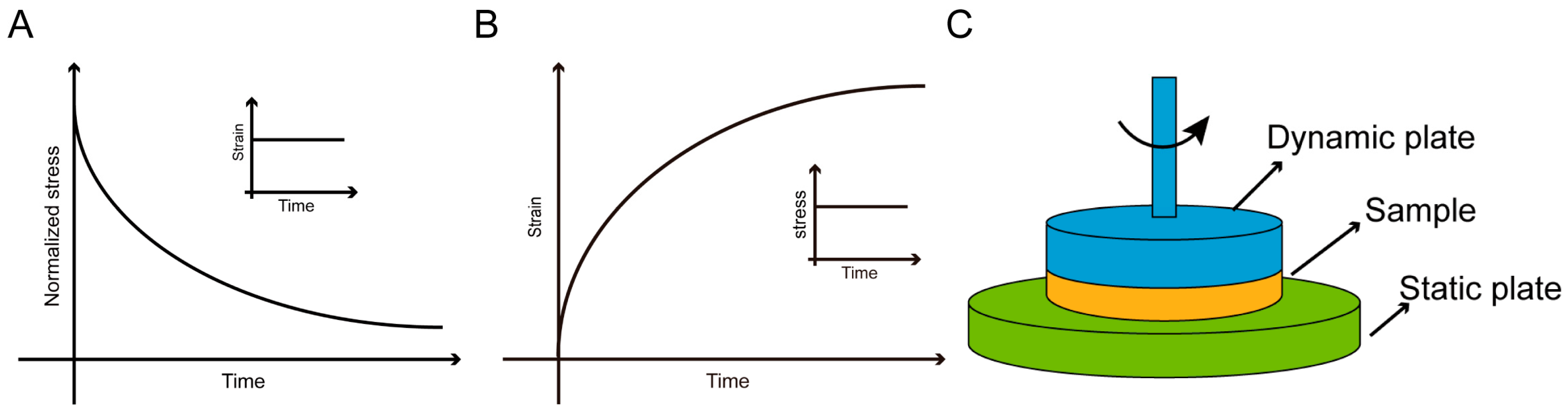
Abstract
:1. Introduction
2. Dynamic Mechanical Properties of Tissue
2.1. Viscoelasticity of Biological Tissue
2.2. Dynamic Stiffness of Biological Tissue
3. Multiscale Characterization of Dynamic Mechanics
3.1. Macroscopic Characterization
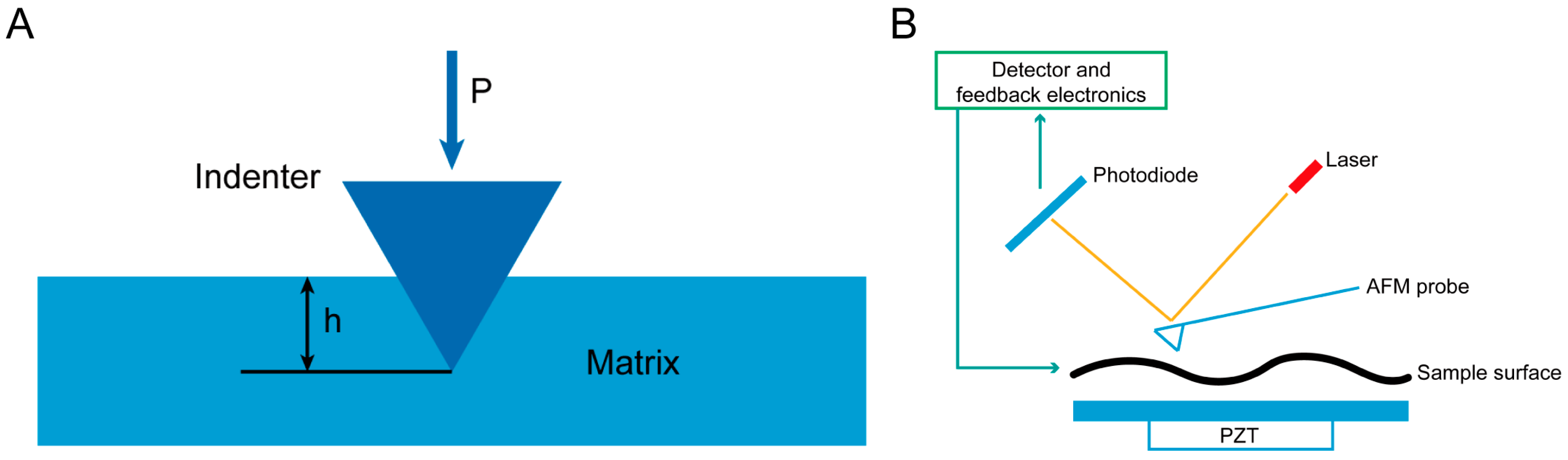
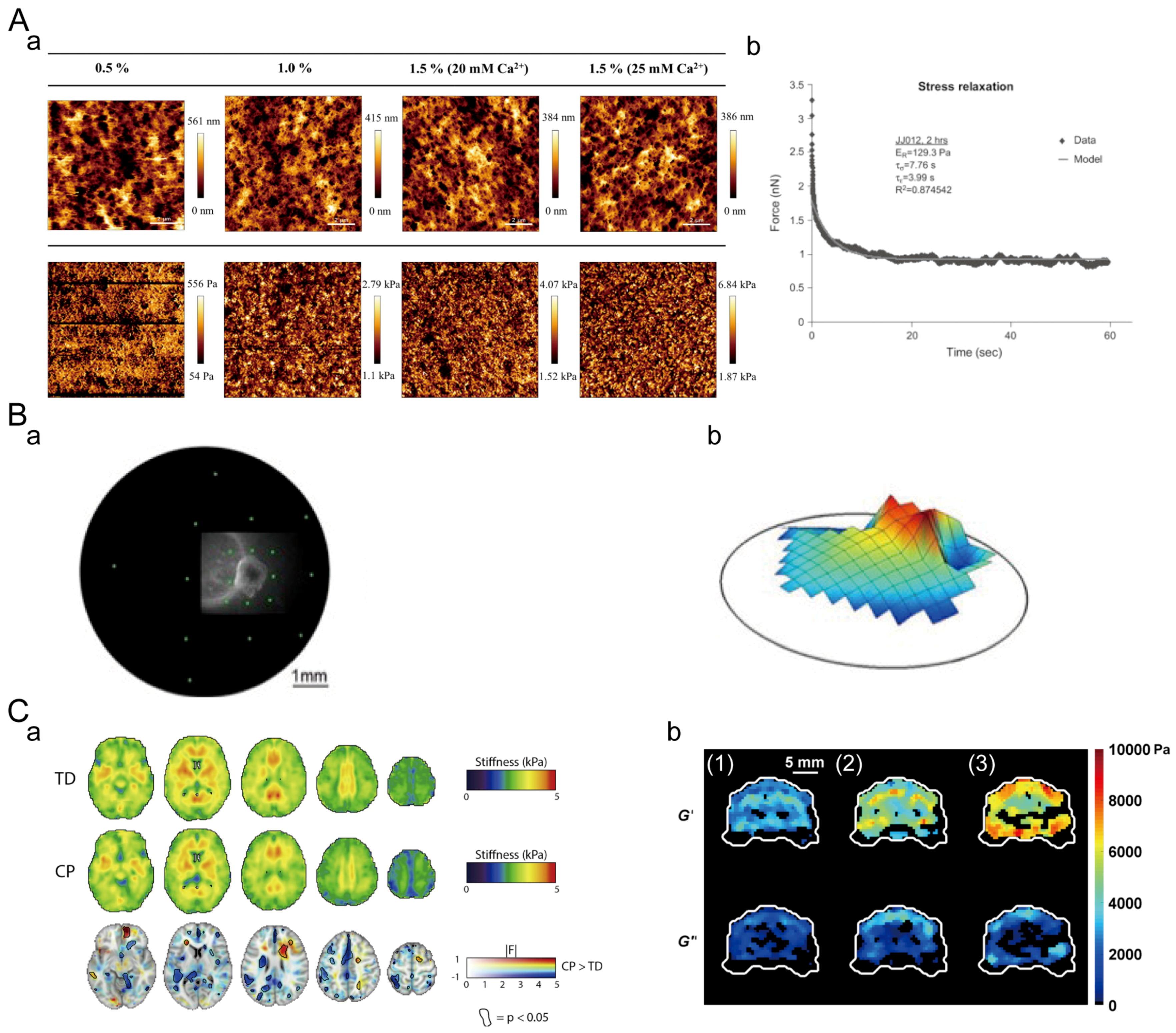
3.2. Microscopic Characterization

4. Construction of Hydrogels with Dynamic Mechanical Properties
4.1. Crosslinking Strategies for Viscoelastic Hydrogels
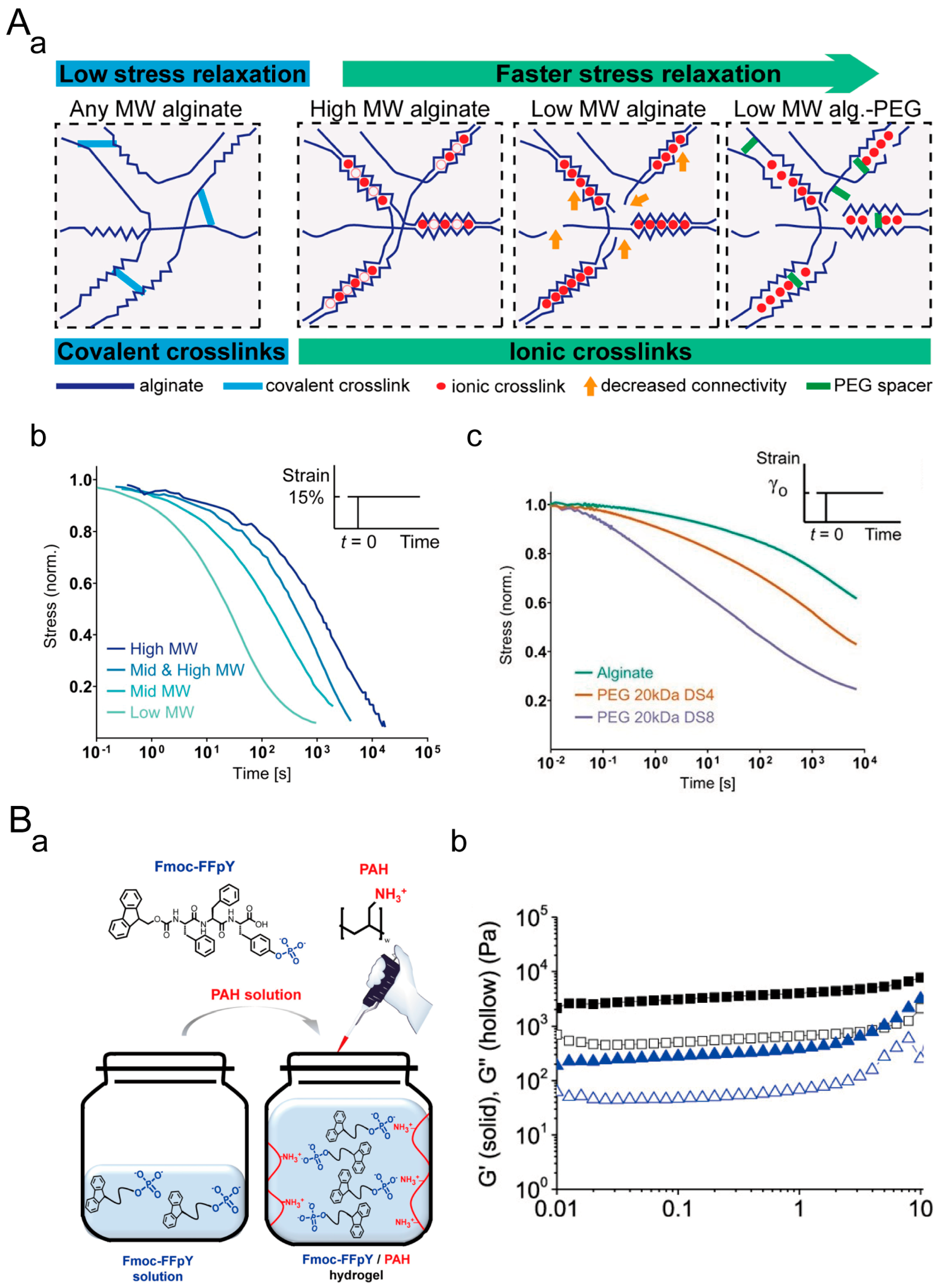
4.1.1. Ionically Crosslinked Hydrogels
4.1.2. Hydrogen-Bonded Crosslinked Hydrogels
4.1.3. Hydrophobic-Interaction-Crosslinked Hydrogels
4.1.4. Supramolecular Hydrogels
4.1.5. Dynamic Covalent Hydrogels
4.2. Crosslinking Strategies for Hydrogels with Dynamic Stiffness
4.2.1. Dynamic Softening Hydrogel
4.2.2. Dynamic Stiffening Hydrogel
4.2.3. Hydrogel with Dynamic Reversible Stiffness
5. The Influence of Dynamic Mechanical Microenvironments on Cell Behavior and Fate
5.1. Viscoelastic Microenvironment Affects Cell Behavior and Fate
5.1.1. Cell Spreading
5.1.2. Cell Proliferation
5.1.3. Cell Migration
5.1.4. Cell Differentiation
5.2. The Dynamic Stiffness Affects Cell Behavior and Fate
5.2.1. Cell Spreading
5.2.2. Cell Proliferation
5.2.3. Cell Differentiation
6. Conclusions and Future Perspectives
Author Contributions
Funding
Conflicts of Interest
References
- Chandra, P.; Lee, S.J. Synthetic Extracellular Microenvironment for Modulating Stem Cell Behaviors. Biomark. Insights 2015, 10, 105–116. [Google Scholar] [CrossRef] [Green Version]
- Singh, A.; Yadav, C.B.; Tabassum, N.; Bajpeyee, A.K.; Verma, V. Stem Cell Niche: Dynamic Neighbor of Stem Cells. Eur. J. Cell Biol. 2019, 98, 65–73. [Google Scholar] [CrossRef]
- Shi, J.; Zhao, Y.-C.; Niu, Z.-F.; Fan, H.-J.; Hou, S.-K.; Guo, X.-Q.; Sang, L.; Lv, Q. Mesenchymal Stem Cell-Derived Small Extracellular Vesicles in the Treatment of Human Diseases: Progress and Prospect. World J. Stem Cells 2021, 13, 49–63. [Google Scholar] [CrossRef] [PubMed]
- Witwer, K.W.; Van Balkom, B.W.M.; Bruno, S.; Choo, A.; Dominici, M.; Gimona, M.; Hill, A.F.; De Kleijn, D.; Koh, M.; Lai, R.C.; et al. Defining Mesenchymal Stromal Cell (MSC)-Derived Small Extracellular Vesicles for Therapeutic Applications. J. Extracell. Vesicles 2019, 8, 1609206. [Google Scholar] [CrossRef] [Green Version]
- Karamanos, N.K.; Theocharis, A.D.; Piperigkou, Z.; Manou, D.; Passi, A.; Skandalis, S.S.; Vynios, D.H.; Orian-Rousseau, V.; Ricard-Blum, S.; Schmelzer, C.E.H.; et al. A Guide to the Composition and Functions of the Extracellular Matrix. FEBS J. 2021, 288, 6850–6912. [Google Scholar] [CrossRef] [PubMed]
- Yue, B. Biology of the Extracellular Matrix: An Overview. J. Glaucoma 2014, 23, S20–S23. [Google Scholar] [CrossRef] [Green Version]
- Theocharis, A.D.; Skandalis, S.S.; Gialeli, C.; Karamanos, N.K. Extracellular Matrix Structure. Adv. Drug Deliv. Rev. 2016, 97, 4–27. [Google Scholar] [CrossRef] [PubMed]
- Walker, C.; Mojares, E.; Del Río Hernández, A. Role of Extracellular Matrix in Development and Cancer Progression. Int. J. Mol. Sci. 2018, 19, 3028. [Google Scholar] [CrossRef] [Green Version]
- Walma, D.A.C.; Yamada, K.M. The Extracellular Matrix in Development. Development 2020, 147, dev175596. [Google Scholar] [CrossRef]
- Harvey, A.E.; Lashinger, L.M.; Hays, D.; Harrison, L.M.; Lewis, K.; Fischer, S.M.; Hursting, S.D. Calorie Restriction Decreases Murine and Human Pancreatic Tumor Cell Growth, Nuclear Factor-ΚB Activation, and Inflammation-Related Gene Expression in an Insulin-like Growth Factor-1-Dependent Manner. PLoS ONE 2014, 9, e94151. [Google Scholar] [CrossRef] [Green Version]
- Cahill, E.F.; Kennelly, H.; Carty, F.; Mahon, B.P.; English, K. Hepatocyte Growth Factor Is Required for Mesenchymal Stromal Cell Protection Against Bleomycin-Induced Pulmonary Fibrosis. Stem Cells Transl. Med. 2016, 5, 1307–1318. [Google Scholar] [CrossRef]
- Floren, M.; Bonani, W.; Dharmarajan, A.; Motta, A.; Migliaresi, C.; Tan, W. Human Mesenchymal Stem Cells Cultured on Silk Hydrogels with Variable Stiffness and Growth Factor Differentiate into Mature Smooth Muscle Cell Phenotype. Acta Biomater. 2016, 31, 156–166. [Google Scholar] [CrossRef] [Green Version]
- Qian, Y.; Han, Q.; Chen, W.; Song, J.; Zhao, X.; Ouyang, Y.; Yuan, W.; Fan, C. Platelet-Rich Plasma Derived Growth Factors Contribute to Stem Cell Differentiation in Musculoskeletal Regeneration. Front. Chem. 2017, 5, 89. [Google Scholar] [CrossRef] [Green Version]
- Poveda-Reyes, S.; Moulisova, V.; Sanmartín-Masiá, E.; Quintanilla-Sierra, L.; Salmerón-Sánchez, M.; Ferrer, G.G. Gelatin-Hyaluronic Acid Hydrogels with Tuned Stiffness to Counterbalance Cellular Forces and Promote Cell Differentiation. Macromol. Biosci. 2016, 16, 1311–1324. [Google Scholar] [CrossRef]
- Zhao, W.; Li, X.; Liu, X.; Zhang, N.; Wen, X. Effects of Substrate Stiffness on Adipogenic and Osteogenic Differentiation of Human Mesenchymal Stem Cells. Mater. Sci. Eng. C 2014, 40, 316–323. [Google Scholar] [CrossRef]
- Wu, S.; Xu, R.; Duan, B.; Jiang, P. Three-Dimensional Hyaluronic Acid Hydrogel-Based Models for in Vitro Human IPSC-Derived NPC Culture and Differentiation. J. Mater. Chem. B 2017, 5, 3870–3878. [Google Scholar] [CrossRef] [PubMed]
- Hou, Y.; Yu, L.; Xie, W.; Camacho, L.C.; Zhang, M.; Chu, Z.; Wei, Q.; Haag, R. Surface Roughness and Substrate Stiffness Synergize To Drive Cellular Mechanoresponse. Nano Lett. 2020, 20, 748–757. [Google Scholar] [CrossRef] [PubMed]
- Yuan, W.; Wang, H.; Fang, C.; Yang, Y.; Xia, X.; Yang, B.; Lin, Y.; Li, G.; Bian, L. Microscopic Local Stiffening in a Supramolecular Hydrogel Network Expedites Stem Cell Mechanosensing in 3D and Bone Regeneration. Mater. Horiz. 2021, 8, 1722–1734. [Google Scholar] [CrossRef] [PubMed]
- Wang, C.; Tong, X.; Yang, F. Bioengineered 3D Brain Tumor Model To Elucidate the Effects of Matrix Stiffness on Glioblastoma Cell Behavior Using PEG-Based Hydrogels. Mol. Pharm. 2014, 11, 2115–2125. [Google Scholar] [CrossRef]
- Wang, C.; Sinha, S.; Jiang, X.; Murphy, L.; Fitch, S.; Wilson, C.; Grant, G.; Yang, F. Matrix Stiffness Modulates Patient-Derived Glioblastoma Cell Fates in Three-Dimensional Hydrogels. Tissue Eng. Part A 2021, 27, 390–401. [Google Scholar] [CrossRef] [PubMed]
- Narkhede, A.A.; Crenshaw, J.H.; Manning, R.M.; Rao, S.S. The Influence of Matrix Stiffness on the Behavior of Brain Metastatic Breast Cancer Cells in a Biomimetic Hyaluronic Acid Hydrogel Platform. J. Biomed. Mater. Res. Part A 2018, 106, 1832–1841. [Google Scholar] [CrossRef]
- Goldshmid, R.; Seliktar, D. Hydrogel Modulus Affects Proliferation Rate and Pluripotency of Human Mesenchymal Stem Cells Grown in Three-Dimensional Culture. ACS Biomater. Sci. Eng. 2017, 3, 3433–3446. [Google Scholar] [CrossRef]
- Shkumatov, A.; Thompson, M.; Choi, K.M.; Sicard, D.; Baek, K.; Kim, D.H.; Tschumperlin, D.J.; Prakash, Y.S.; Kong, H. Matrix Stiffness-Modulated Proliferation and Secretory Function of the Airway Smooth Muscle Cells. Am. J. Physiol. Lung Cell Mol. Physiol. 2015, 308, L1125–L1135. [Google Scholar] [CrossRef] [PubMed] [Green Version]
- Li, Y.; Zhang, Y.; Shi, F.; Tao, L.; Wei, Y.; Wang, X. Modulus-Regulated 3D-Cell Proliferation in an Injectable Self-Healing Hydrogel. Colloids Surf. B Biointerfaces 2017, 149, 168–173. [Google Scholar] [CrossRef]
- Pogoda, K.; Bucki, R.; Byfield, F.J.; Cruz, K.; Lee, T.; Marcinkiewicz, C.; Janmey, P.A. Soft Substrates Containing Hyaluronan Mimic the Effects of Increased Stiffness on Morphology, Motility, and Proliferation of Glioma Cells. Biomacromolecules 2017, 18, 3040–3051. [Google Scholar] [CrossRef] [PubMed]
- Missirlis, D.; Spatz, J.P. Combined Effects of PEG Hydrogel Elasticity and Cell-Adhesive Coating on Fibroblast Adhesion and Persistent Migration. Biomacromolecules 2014, 15, 195–205. [Google Scholar] [CrossRef] [PubMed]
- Hadden, W.J.; Young, J.L.; Holle, A.W.; McFetridge, M.L.; Kim, D.Y.; Wijesinghe, P.; Taylor-Weiner, H.; Wen, J.H.; Lee, A.R.; Bieback, K.; et al. Stem Cell Migration and Mechanotransduction on Linear Stiffness Gradient Hydrogels. Proc. Natl. Acad. Sci. USA 2017, 114, 5647–5652. [Google Scholar] [CrossRef]
- Liu, Q.-P.; Luo, Q.; Deng, B.; Ju, Y.; Song, G.-B. Stiffer Matrix Accelerates Migration of Hepatocellular Carcinoma Cells through Enhanced Aerobic Glycolysis Via the MAPK-YAP Signaling. Cancers 2020, 12, 490. [Google Scholar] [CrossRef] [Green Version]
- Kim, T.H.; An, D.B.; Oh, S.H.; Kang, M.K.; Song, H.H.; Lee, J.H. Creating Stiffness Gradient Polyvinyl Alcohol Hydrogel Using a Simple Gradual Freezing–Thawing Method to Investigate Stem Cell Differentiation Behaviors. Biomaterials 2015, 40, 51–60. [Google Scholar] [CrossRef]
- Žigon-Branc, S.; Markovic, M.; Van Hoorick, J.; Van Vlierberghe, S.; Dubruel, P.; Zerobin, E.; Baudis, S.; Ovsianikov, A. Impact of Hydrogel Stiffness on Differentiation of Human Adipose-Derived Stem Cell Microspheroids. Tissue Eng. Part A 2019, 25, 1369–1380. [Google Scholar] [CrossRef]
- Oh, S.H.; An, D.B.; Kim, T.H.; Lee, J.H. Wide-Range Stiffness Gradient PVA/HA Hydrogel to Investigate Stem Cell Differentiation Behavior. Acta Biomater. 2016, 35, 23–31. [Google Scholar] [CrossRef] [PubMed]
- Martinez-Garcia, F.D.; de Hilster, R.H.J.; Sharma, P.K.; Borghuis, T.; Hylkema, M.N.; Burgess, J.K.; Harmsen, M.C. Architecture and Composition Dictate Viscoelastic Properties of Organ-Derived Extracellular Matrix Hydrogels. Polymers 2021, 13, 3113. [Google Scholar] [CrossRef]
- Elosegui-Artola, A. The Extracellular Matrix Viscoelasticity as a Regulator of Cell and Tissue Dynamics. Curr. Opin. Cell Biol. 2021, 72, 10–18. [Google Scholar] [CrossRef] [PubMed]
- Budday, S.; Sommer, G.; Holzapfel, G.A.; Steinmann, P.; Kuhl, E. Viscoelastic Parameter Identification of Human Brain Tissue. J. Mech. Behav. Biomed. Mater. 2017, 74, 463–476. [Google Scholar] [CrossRef] [Green Version]
- Xie, R.; Yu, X.; Cao, T.; Yang, C.; Zhang, Y.; Wang, X.; Liu, Y.-J.; Duan, S.; Ye, F.; Fan, Q. Fibrous Viscoelastic Extracellular Matrix Assists Precise Neuronal Connectivity. Adv. Funct. Mater. 2023, 2301926. [Google Scholar] [CrossRef]
- Baker, L.X.; Chen, F.; Ssempijja, Y.; Byrne, M.; Kim, T.K.; Vain, A.; Jagasia, M.; Tkaczyk, E.R. Longitudinal Tracking of Skin Dynamic Stiffness to Quantify Evolution of Sclerosis in Chronic Graft-versus-Host Disease. Bone Marrow Transplant. 2021, 56, 989–991. [Google Scholar] [CrossRef]
- Morgan, G.; Martin, R.; Welch, H.; Williams, L.; Morris, K. Objective Assessment of Stiffness in the Gastrocnemius Muscle in Patients with Symptomatic Achilles Tendons. BMJ Open Sport Exerc. Med. 2019, 5, e000622. [Google Scholar] [CrossRef] [PubMed] [Green Version]
- Lim, K.T.; Choi, W.J. Soft Tissue Stiffness over the Hip Increases with Age and Its Implication in Hip Fracture Risk in Older Adults. J. Biomech. 2019, 93, 28–33. [Google Scholar] [CrossRef]
- Handorf, A.M.; Zhou, Y.; Halanski, M.A.; Li, W.-J. Tissue Stiffness Dictates Development, Homeostasis, and Disease Progression. Organogenesis 2015, 11, 1–15. [Google Scholar] [CrossRef] [Green Version]
- Fitzgerald, M.M.; Bootsma, K.; Berberich, J.A.; Sparks, J.L. Tunable Stress Relaxation Behavior of an Alginate-Polyacrylamide Hydrogel: Comparison with Muscle Tissue. Biomacromolecules 2015, 16, 1497–1505. [Google Scholar] [CrossRef]
- Darnell, M.; Young, S.; Gu, L.; Shah, N.; Lippens, E.; Weaver, J.; Duda, G.; Mooney, D. Substrate Stress-Relaxation Regulates Scaffold Remodeling and Bone Formation In Vivo. Adv. Healthc. Mater. 2017, 6, 1601185. [Google Scholar] [CrossRef] [PubMed] [Green Version]
- Hazur, J.; Endrizzi, N.; Schubert, D.W.; Boccaccini, A.R.; Fabry, B. Stress Relaxation Amplitude of Hydrogels Determines Migration, Proliferation, and Morphology of Cells in 3-D Culture. Biomater. Sci. 2022, 10, 270–280. [Google Scholar] [CrossRef] [PubMed]
- Song, G.; Zhao, Z.; Peng, X.; He, C.; Weiss, R.A.; Wang, H. Rheological Behavior of Tough PVP-in Situ-PAAm Hydrogels Physically Cross-Linked by Cooperative Hydrogen Bonding. Macromolecules 2016, 49, 8265–8273. [Google Scholar] [CrossRef]
- Zhang, S.; Xu, B.; Lu, X.; Wang, L.; Li, Y.; Ma, N.; Wei, H.; Zhang, X.; Wang, G. Readily Producing a Silly Putty-like Hydrogel with Good Self-Healing, Conductive and Photothermal Conversion Properties Based on Dynamic Coordinate Bonds and Hydrogen Bonds. J. Mater. Chem. C 2020, 8, 6763–6770. [Google Scholar] [CrossRef]
- Zhao, D.; Feng, M.; Zhang, L.; He, B.; Chen, X.; Sun, J. Facile Synthesis of Self-Healing and Layered Sodium Alginate/Polyacrylamide Hydrogel Promoted by Dynamic Hydrogen Bond. Carbohydr. Polym. 2021, 256, 117580. [Google Scholar] [CrossRef]
- Su, E.; Okay, O. Polyampholyte Hydrogels Formed via Electrostatic and Hydrophobic Interactions. Eur. Polym. J. 2017, 88, 191–204. [Google Scholar] [CrossRef]
- Hafeez, S.; Aldana, A.A.; Duimel, H.; Ruiter, F.A.A.; Decarli, M.C.; Lapointe, V.; van Blitterswijk, C.; Moroni, L.; Baker, M.B. Molecular Tuning of a Benzene-1,3,5-Tricarboxamide Supramolecular Fibrous Hydrogel Enables Control over Viscoelasticity and Creates Tunable ECM-Mimetic Hydrogels and Bioinks. Adv. Mater. 2022, 35, 2207053. [Google Scholar] [CrossRef]
- Mihajlovic, M.; Wyss, H.M.; Sijbesma, R.P. Effects of Surfactant and Urea on Dynamics and Viscoelastic Properties of Hydrophobically Assembled Supramolecular Hydrogel. Macromolecules 2018, 51, 4813–4820. [Google Scholar] [CrossRef] [Green Version]
- Fumagalli, M.; Belal, K.; Guo, H.; Stoffelbach, F.; Cooke, G.; Marcellan, A.; Woisel, P.; Hourdet, D. Supramolecular Polymer Hydrogels Induced by Host–Guest Interactions with Di-[Cyclobis(Paraquat-p-Phenylene)] Cross-Linkers: From Molecular Complexation to Viscoelastic Properties. Soft Matter 2017, 13, 5269–5282. [Google Scholar] [CrossRef] [Green Version]
- Tan, C.S.Y.; Liu, J.; Groombridge, A.S.; Barrow, S.J.; Dreiss, C.A.; Scherman, O.A. Controlling Spatiotemporal Mechanics of Supramolecular Hydrogel Networks with Highly Branched Cucurbit[8]Uril Polyrotaxanes. Adv. Funct. Mater. 2018, 28, 1702994. [Google Scholar] [CrossRef]
- Zhou, Y.; Zhang, Y.; Dai, Z.; Jiang, F.; Tian, J.; Zhang, W. A Super-Stretchable, Self-Healing and Injectable Supramolecular Hydrogel Constructed by a Host–Guest Crosslinker. Biomater. Sci. 2020, 8, 3359–3369. [Google Scholar] [CrossRef]
- Hafeez, S.; Ooi, H.W.; Morgan, F.L.C.; Mota, C.; Dettin, M.; Van Blitterswijk, C.; Moroni, L.; Baker, M.B. Viscoelastic Oxidized Alginates with Reversible Imine Type Crosslinks: Self-Healing, Injectable, and Bioprintable Hydrogels. Gels 2018, 4, 85. [Google Scholar] [CrossRef] [PubMed] [Green Version]
- Arefkhalily, M.; Goktas, M.; Guler, M.O. Tuning Viscoelastic Properties of Supramolecular Peptide Gels via Dynamic Covalent Crosslinking. Org. Biomol. Chem. 2014, 13, 1983–1987. [Google Scholar] [CrossRef] [Green Version]
- Richardson, B.M.; Wilcox, D.G.; Randolph, M.A.; Anseth, K.S. Hydrazone Covalent Adaptable Networks Modulate Extracellular Matrix Deposition for Cartilage Tissue Engineering. Acta Biomater. 2019, 83, 71–82. [Google Scholar] [CrossRef]
- Shou, Y.; Xin, Y.T.; Li, X.; Le, Z.; Liu, L.; Sun, X.; Jonhson, W.; Ding, J.; Lim, C.T.; Tay, A. Dynamic Magneto-Softening of 3D Hydrogel Reverses Malignant Transformation of Cancer Cells and Enhances Drug Efficacy. ACS Nano 2023, 17, 2851–2867. [Google Scholar] [CrossRef] [PubMed]
- Liu, H.-Y.; Lin, C.-C. A Diffusion-Reaction Model for Predicting Enzyme-Mediated Dynamic Hydrogel Stiffening. Gels 2019, 5, 17. [Google Scholar] [CrossRef] [Green Version]
- Rombouts, W.H.; de Kort, D.W.; Pham, T.T.H.; van Mierlo, C.P.M.; Werten, M.W.T.; de Wolf, F.A.; van der Gucht, J. Reversible Temperature-Switching of Hydrogel Stiffness of Coassembled, Silk-Collagen-Like Hydrogels. Biomacromolecules 2015, 16, 2506–2513. [Google Scholar] [CrossRef]
- Rosales, A.M.; Mabry, K.M.; Nehls, E.M.; Anseth, K.S. Photoresponsive Elastic Properties of Azobenzene-Containing Poly(Ethylene-Glycol)-Based Hydrogels. Biomacromolecules 2015, 16, 798–806. [Google Scholar] [CrossRef] [Green Version]
- Kollarigowda, R.H.; Mathews, A.S.; Abraham, S. Super Mechanical Stimuli Responsive Hydrogel: Dynamic Cues for Cell Applications. ACS Appl. Bio. Mater. 2019, 2, 277–283. [Google Scholar] [CrossRef]
- Whitehead, J.; Griffin, K.H.; Gionet-Gonzales, M.; Vorwald, C.E.; Cinque, S.E.; Leach, J.K. Hydrogel Mechanics Are a Key Driver of Bone Formation by Mesenchymal Stromal Cell Spheroids. Biomaterials 2021, 269, 120607. [Google Scholar] [CrossRef]
- Chaudhuri, O.; Gu, L.; Darnell, M.; Klumpers, D.; Bencherif, S.A.; Weaver, J.C.; Huebsch, N.; Mooney, D.J. Substrate Stress Relaxation Regulates Cell Spreading. Nat. Commun. 2015, 6, 6365. [Google Scholar] [CrossRef] [Green Version]
- Engler, A.J.; Sen, S.; Sweeney, H.L.; Discher, D.E. Matrix Elasticity Directs Stem Cell Lineage Specification. Cell 2006, 126, 677–689. [Google Scholar] [CrossRef] [PubMed] [Green Version]
- Chaudhuri, O. Viscoelastic Hydrogels for 3D Cell Culture. Biomater. Sci. 2017, 5, 1480–1490. [Google Scholar] [CrossRef]
- Tang, S.; Richardson, B.M.; Anseth, K.S. Dynamic Covalent Hydrogels as Biomaterials to Mimic the Viscoelasticity of Soft Tissues. Prog. Mater. Sci. 2021, 120, 100738. [Google Scholar] [CrossRef]
- Elosegui-Artola, A.; Gupta, A.; Najibi, A.J.; Seo, B.R.; Garry, R.; Tringides, C.M.; de Lázaro, I.; Darnell, M.; Gu, W.; Zhou, Q.; et al. Matrix Viscoelasticity Controls Spatiotemporal Tissue Organization. Nat. Mater. 2023, 22, 117–127. [Google Scholar] [CrossRef]
- Chaudhuri, O.; Cooper-White, J.; Janmey, P.A.; Mooney, D.J.; Shenoy, V.B. Effects of Extracellular Matrix Viscoelasticity on Cellular Behaviour. Nature 2020, 584, 535–546. [Google Scholar] [CrossRef] [PubMed]
- Samadi-Dooki, A.; Voyiadjis, G.Z.; Stout, R.W. An Indirect Indentation Method for Evaluating the Linear Viscoelastic Properties of the Brain Tissue. J. Biomech. Eng. 2017, 139, 061007. [Google Scholar] [CrossRef]
- Vappou, J.; Breton, E.; Choquet, P.; Goetz, C.; Willinger, R.; Constantinesco, A. Magnetic Resonance Elastography Compared with Rotational Rheometry for in Vitro Brain Tissue Viscoelasticity Measurement. Magn. Reson. Mater. Phy. 2007, 20, 273–278. [Google Scholar] [CrossRef] [PubMed]
- Chaze, C.A.; McIlvain, G.; Smith, D.R.; Villermaux, G.M.; Delgorio, P.L.; Wright, H.G.; Rogers, K.J.; Miller, F.; Crenshaw, J.R.; Johnson, C.L. Altered Brain Tissue Viscoelasticity in Pediatric Cerebral Palsy Measured by Magnetic Resonance Elastography. NeuroImage Clin. 2019, 22, 101750. [Google Scholar] [CrossRef]
- Navajas, D.; Maksym, G.N.; Bates, J.H. Dynamic Viscoelastic Nonlinearity of Lung Parenchymal Tissue. J. Appl. Physiol. 1995, 79, 348–356. [Google Scholar] [CrossRef]
- Clancy, N.T.; Nilsson, G.E.; Anderson, C.D.; Leahy, M.J. A New Device for Assessing Changes in Skin Viscoelasticity Using Indentation and Optical Measurement. Ski. Res. Technol. 2010, 16, 210–228. [Google Scholar] [CrossRef] [PubMed]
- Crichton, M.L.; Chen, X.; Huang, H.; Kendall, M.A.F. Elastic Modulus and Viscoelastic Properties of Full Thickness Skin Characterised at Micro Scales. Biomaterials 2013, 34, 2087–2097. [Google Scholar] [CrossRef]
- Maccabi, A.; Shin, A.; Namiri, N.K.; Bajwa, N.; John, M.S.; Taylor, Z.D.; Grundfest, W.; Saddik, G.N. Quantitative Characterization of Viscoelastic Behavior in Tissue-Mimicking Phantoms and Ex Vivo Animal Tissues. PLoS ONE 2018, 13, e0191919. [Google Scholar] [CrossRef] [Green Version]
- Ramo, N.L.; Troyer, K.L.; Puttlitz, C.M. Viscoelasticity of Spinal Cord and Meningeal Tissues. Acta Biomater. 2018, 75, 253–262. [Google Scholar] [CrossRef] [PubMed]
- Hoyt, K.; Kneezel, T.; Castaneda, B.; Parker, K.J. Quantitative Sonoelastography for the in Vivo Assessment of Skeletal Muscle Viscoelasticity. Phys. Med. Biol. 2008, 53, 4063–4080. [Google Scholar] [CrossRef] [PubMed] [Green Version]
- Van Loocke, M.; Lyons, C.G.; Simms, C.K. Viscoelastic Properties of Passive Skeletal Muscle in Compression: Stress-Relaxation Behaviour and Constitutive Modelling. J. Biomech. 2008, 41, 1555–1566. [Google Scholar] [CrossRef] [PubMed]
- Wu, Z.; Ovaert, T.C.; Niebur, G.L. Viscoelastic Properties of Human Cortical Bone Tissue Depend on Gender and Elastic Modulus. J. Orthop. Res. 2012, 30, 693–699. [Google Scholar] [CrossRef] [PubMed] [Green Version]
- Fell, N.L.A.; Lawless, B.M.; Cox, S.C.; Cooke, M.E.; Eisenstein, N.M.; Shepherd, D.E.T.; Espino, D.M. The Role of Subchondral Bone, and Its Histomorphology, on the Dynamic Viscoelasticity of Cartilage, Bone and Osteochondral Cores. Osteoarthr. Cartil. 2019, 27, 535–543. [Google Scholar] [CrossRef] [PubMed] [Green Version]
- Cohen, R.E.; Hooley, C.J.; McCrum, N.G. Viscoelastic Creep of Collagenous Tissue. J. Biomech. 1976, 9, 175–184. [Google Scholar] [CrossRef]
- Troyer, K.L.; Shetye, S.S.; Puttlitz, C.M. Experimental Characterization and Finite Element Implementation of Soft Tissue Nonlinear Viscoelasticity. J. Biomech. Eng. 2012, 134, 114501. [Google Scholar] [CrossRef]
- Safshekan, F.; Tafazzoli-Shadpour, M.; Abdouss, M.; Shadmehr, M.B. Viscoelastic Properties of Human Tracheal Tissues. J. Biomech. Eng. 2016, 139, 011007. [Google Scholar] [CrossRef] [PubMed]
- Kasza, K.E.; Rowat, A.C.; Liu, J.; Angelini, T.E.; Brangwynne, C.P.; Koenderink, G.H.; Weitz, D.A. The Cell as a Material. Curr. Opin. Cell Biol. 2007, 19, 101–107. [Google Scholar] [CrossRef] [PubMed]
- Pravincumar, P.; Bader, D.L.; Knight, M.M. Viscoelastic Cell Mechanics and Actin Remodelling Are Dependent on the Rate of Applied Pressure. PLoS ONE 2012, 7, e43938. [Google Scholar] [CrossRef] [PubMed] [Green Version]
- Wahlquist, J.A.; DelRio, F.W.; Randolph, M.A.; Aziz, A.H.; Heveran, C.M.; Bryant, S.J.; Neu, C.P.; Ferguson, V.L. Indentation Mapping Revealed Poroelastic, but Not Viscoelastic, Properties Spanning Native Zonal Articular Cartilage. Acta Biomater. 2017, 64, 41–49. [Google Scholar] [CrossRef]
- Zheng, Y.; Jiang, Y.; Cao, Y. A Porohyperviscoelastic Model for the Shear Wave Elastography of the Liver. J. Mech. Phys. Solids 2021, 150, 104339. [Google Scholar] [CrossRef]
- Islam, M.R.; Virag, J.; Oyen, M.L. Micromechanical Poroelastic and Viscoelastic Properties of Ex-Vivo Soft Tissues. J. Biomech. 2020, 113, 110090. [Google Scholar] [CrossRef]
- Usgu, S.; Yakut, Y.; Çınar, M.A. The Comparison of Achilles Tendon Viscoelastic Properties in Elite Runners and Soccer Players. Spor Hekim. Derg. 2020, 55, 276–283. [Google Scholar] [CrossRef]
- Arampatzis, A.; Karamanidis, K.; Albracht, K. Adaptational Responses of the Human Achilles Tendon by Modulation of the Applied Cyclic Strain Magnitude. J. Exp. Biol. 2007, 210, 2743–2753. [Google Scholar] [CrossRef] [Green Version]
- Mountcastle, S.E.; Allen, P.; Mellors, B.O.L.; Lawless, B.M.; Cooke, M.E.; Lavecchia, C.E.; Fell, N.L.A.; Espino, D.M.; Jones, S.W.; Cox, S.C. Dynamic Viscoelastic Characterisation of Human Osteochondral Tissue: Understanding the Effect of the Cartilage-Bone Interface. BMC Musculoskelet. Disord. 2019, 20, 575. [Google Scholar] [CrossRef]
- Yin, Z.; Romano, A.J.; Manduca, A.; Ehman, R.L.; Huston, J. Stiffness and Beyond: What MR Elastography Can Tell Us About Brain Structure and Function Under Physiologic and Pathologic Conditions. Top. Magn. Reson. Imaging 2018, 27, 305–318. [Google Scholar] [CrossRef]
- Park, K.; Lonsberry, G.E.; Gearing, M.; Levey, A.I.; Desai, J.P. Viscoelastic Properties of Human Autopsy Brain Tissues as Biomarkers for Alzheimer’s Diseases. IEEE Trans. Biomed. Eng. 2019, 66, 1705–1713. [Google Scholar] [CrossRef]
- Munder, T.; Pfeffer, A.; Schreyer, S.; Guo, J.; Braun, J.; Sack, I.; Steiner, B.; Klein, C. MR Elastography Detection of Early Viscoelastic Response of the Murine Hippocampus to Amyloid β Accumulation and Neuronal Cell Loss Due to Alzheimer’s Disease. J. Magn. Reson. Imaging 2018, 47, 105–114. [Google Scholar] [CrossRef]
- Lipp, A.; Skowronek, C.; Fehlner, A.; Streitberger, K.-J.; Braun, J.; Sack, I. Progressive Supranuclear Palsy and Idiopathic Parkinson’s Disease Are Associated with Local Reduction of in Vivo Brain Viscoelasticity. Eur. Radiol. 2018, 28, 3347–3354. [Google Scholar] [CrossRef]
- Murphy, M.C.; Huston, J.; Ehman, R.L. MR Elastography of the Brain and Its Application in Neurological Diseases. Neuroimage 2019, 187, 176–183. [Google Scholar] [CrossRef]
- Gerischer, L.M.; Fehlner, A.; Köbe, T.; Prehn, K.; Antonenko, D.; Grittner, U.; Braun, J.; Sack, I.; Flöel, A. Combining Viscoelasticity, Diffusivity and Volume of the Hippocampus for the Diagnosis of Alzheimer’s Disease Based on Magnetic Resonance Imaging. Neuroimage Clin. 2018, 18, 485–493. [Google Scholar] [CrossRef]
- Deffieux, T.; Gennisson, J.-L.; Bousquet, L.; Corouge, M.; Cosconea, S.; Amroun, D.; Tripon, S.; Terris, B.; Mallet, V.; Sogni, P.; et al. Investigating Liver Stiffness and Viscosity for Fibrosis, Steatosis and Activity Staging Using Shear Wave Elastography. J. Hepatol. 2015, 62, 317–324. [Google Scholar] [CrossRef] [PubMed]
- Salameh, N.; Peeters, F.; Sinkus, R.; Abarca-Quinones, J.; Annet, L.; ter Beek, L.C.; Leclercq, I.; Van Beers, B.E. Hepatic Viscoelastic Parameters Measured with MR Elastography: Correlations with Quantitative Analysis of Liver Fibrosis in the Rat. J. Magn. Reson. Imaging 2007, 26, 956–962. [Google Scholar] [CrossRef] [PubMed]
- Wei, H.; Child, L.J. Clinical Utility of Viscoelastic Testing in Chronic Liver Disease: A Systematic Review. World J. Hepatol. 2020, 12, 1115–1127. [Google Scholar] [CrossRef] [PubMed]
- Cox, T.R.; Erler, J.T. Remodeling and Homeostasis of the Extracellular Matrix: Implications for Fibrotic Diseases and Cancer. Dis. Models Mech. 2011, 4, 165–178. [Google Scholar] [CrossRef] [Green Version]
- Isaksson, H.; Malkiewicz, M.; Nowak, R.; Helminen, H.J.; Jurvelin, J.S. Rabbit Cortical Bone Tissue Increases Its Elastic Stiffness but Becomes Less Viscoelastic with Age. Bone 2010, 47, 1030–1038. [Google Scholar] [CrossRef]
- John, D. Currey Role of Collagen and Other Organics in the Mechanical Properties of Bone. Osteoporos. Int. 2003, 14, 29–36. [Google Scholar] [CrossRef]
- Burr, D.B. Changes in Bone Matrix Properties with Aging. Bone 2019, 120, 85–93. [Google Scholar] [CrossRef]
- Iwashita, M.; Kataoka, N.; Toida, K.; Kosodo, Y. Systematic Profiling of Spatiotemporal Tissue and Cellular Stiffness in the Developing Brain. Development 2014, 141, 3793–3798. [Google Scholar] [CrossRef] [Green Version]
- Silver, J.S.; Günay, K.A.; Cutler, A.A.; Vogler, T.O.; Brown, T.E.; Pawlikowski, B.T.; Bednarski, O.J.; Bannister, K.L.; Rogowski, C.J.; Mckay, A.G.; et al. Injury-Mediated Stiffening Persistently Activates Muscle Stem Cells through YAP and TAZ Mechanotransduction. Sci. Adv. 2021, 7, eabe4501. [Google Scholar] [CrossRef]
- Liu, B.; McNally, S.; Kilpatrick, J.I.; Jarvis, S.P.; O’Brien, C.J. Aging and Ocular Tissue Stiffness in Glaucoma. Surv. Ophthalmol. 2018, 63, 56–74. [Google Scholar] [CrossRef]
- Liu, F.; Mih, J.D.; Shea, B.S.; Kho, A.T.; Sharif, A.S.; Tager, A.M.; Tschumperlin, D.J. Feedback Amplification of Fibrosis through Matrix Stiffening and COX-2 Suppression. J. Cell Biol. 2010, 190, 693–706. [Google Scholar] [CrossRef] [PubMed]
- Kondo, R.; Kage, M.; Iijima, H.; Fujimoto, J.; Nishimura, T.; Aizawa, N.; Akiba, J.; Naito, Y.; Kusano, H.; Nakayama, M.; et al. Pathological Findings That Contribute to Tissue Stiffness in the Spleen of Liver Cirrhosis Patients. Hepatol. Res. 2018, 48, 1000–1007. [Google Scholar] [CrossRef] [PubMed]
- Lung, C.-W.; Wu, F.-L.; Zhang, K.; Liau, B.-Y.; Townsend, R.; Jan, Y.-K. Using Elastographic Ultrasound to Assess Plantar Tissue Stiffness after Walking at Different Speeds and Durations. Appl. Sci. 2020, 10, 7498. [Google Scholar] [CrossRef]
- Bauer, A.; Gu, L.; Kwee, B.; Li, W.A.; Dellacherie, M.; Celiz, A.D.; Mooney, D.J. Hydrogel Substrate Stress-Relaxation Regulates the Spreading and Proliferation of Mouse Myoblasts. Acta Biomater. 2017, 62, 82–90. [Google Scholar] [CrossRef]
- Zhong, Y.; Chen, T.; Wang, X. Repairing Creep-Resistant and Kinetically Inert Hydrogels via Yeast Activity-Regulated Energy Dissipation. ACS Appl. Bio. Mater. 2020, 3, 4507–4513. [Google Scholar] [CrossRef]
- Zuidema, J.M.; Rivet, C.J.; Gilbert, R.J.; Morrison, F.A. A Protocol for Rheological Characterization of Hydrogels for Tissue Engineering Strategies. J. Biomed. Mater. Res. Part B Appl. Biomater. 2014, 102, 1063–1073. [Google Scholar] [CrossRef]
- Hajjarian, Z.; Nia, H.T.; Ahn, S.; Grodzinsky, A.J.; Jain, R.K.; Nadkarni, S.K. Laser Speckle Rheology for Evaluating the Viscoelastic Properties of Hydrogel Scaffolds. Sci. Rep. 2016, 6, 37949. [Google Scholar] [CrossRef] [Green Version]
- Kim, M.H.; Nguyen, H.; Chang, C.-Y.; Lin, C.-C. Dual Functionalization of Gelatin for Orthogonal and Dynamic Hydrogel Cross-Linking. ACS Biomater. Sci. Eng. 2021, 7, 4196–4208. [Google Scholar] [CrossRef] [PubMed]
- Jia, H.; Lin, X.; Wang, D.; Wang, J.; Shang, Q.; He, X.; Wu, K.; Zhao, B.; Peng, P.; Wang, H.; et al. Injectable Hydrogel with Nucleus Pulposus-Matched Viscoelastic Property Prevents Intervertebral Disc Degeneration. J. Orthop. Transl. 2022, 33, 162–173. [Google Scholar] [CrossRef] [PubMed]
- Kohn, J.C.; Lampi, M.C.; Reinhart-King, C.A. Age-Related Vascular Stiffening: Causes and Consequences. Front. Genet. 2015, 6, 112. [Google Scholar] [CrossRef] [PubMed] [Green Version]
- Xu, D.; Harvey, T.; Begiristain, E.; Domínguez, C.; Sánchez-Abella, L.; Browne, M.; Cook, R.B. Measuring the Elastic Modulus of Soft Biomaterials Using Nanoindentation. J. Mech. Behav. Biomed. Mater. 2022, 133, 105329. [Google Scholar] [CrossRef] [PubMed]
- Zhang, M.; Yan, S.; Xu, X.; Yu, T.; Guo, Z.; Ma, M.; Zhang, Y.; Gu, Z.; Feng, Y.; Du, C.; et al. Three-Dimensional Cell-Culture Platform Based on Hydrogel with Tunable Microenvironmental Properties to Improve Insulin-Secreting Function of MIN6 Cells. Biomaterials 2021, 270, 120687. [Google Scholar] [CrossRef]
- He, L.-H.; Swain, M.V. Nanoindentation Creep Behavior of Human Enamel. J. Biomed. Mater. Res. Part A 2009, 91A, 352–359. [Google Scholar] [CrossRef]
- Guglielmi, P.O.; Herbert, E.G.; Tartivel, L.; Behl, M.; Lendlein, A.; Huber, N.; Lilleodden, E.T. Mechanical Characterization of Oligo(Ethylene Glycol)-Based Hydrogels by Dynamic Nanoindentation Experiments. J. Mech. Behav. Biomed. Mater. 2015, 46, 1–10. [Google Scholar] [CrossRef] [Green Version]
- Akbarzadeh Solbu, A.; Koernig, A.; Kjesbu, J.S.; Zaytseva-Zotova, D.; Sletmoen, M.; Strand, B.L. High Resolution Imaging of Soft Alginate Hydrogels by Atomic Force Microscopy. Carbohydr. Polym. 2022, 276, 118804. [Google Scholar] [CrossRef]
- Darling, E.M.; Zauscher, S.; Block, J.A.; Guilak, F. A Thin-Layer Model for Viscoelastic, Stress-Relaxation Testing of Cells Using Atomic Force Microscopy: Do Cell Properties Reflect Metastatic Potential? Biophys. J. 2007, 92, 1784–1791. [Google Scholar] [CrossRef] [Green Version]
- Septiadi, D.; Crippa, F.; Moore, T.L.; Rothen-Rutishauser, B.; Petri-Fink, A. Nanoparticle–Cell Interaction: A Cell Mechanics Perspective. Adv. Mater. 2018, 30, 1704463. [Google Scholar] [CrossRef] [PubMed]
- Jones, D.P.; Hanna, W.; El-Hamidi, H.; Celli, J.P. Longitudinal Measurement of Extracellular Matrix Rigidity in 3D Tumor Models Using Particle-Tracking Microrheology. J. Vis. Exp. 2014, 88, e51302. [Google Scholar] [CrossRef] [Green Version]
- Lal, A.; Naranje, P.; Pavunesan, S.K. What’s New in Urologic Ultrasound? Indian J. Urol. 2015, 31, 176–184. [Google Scholar] [CrossRef]
- Feng, Y.; Clayton, E.H.; Chang, Y.; Okamoto, R.J.; Bayly, P.V. Viscoelastic Properties of the Ferret Brain Measured in Vivo at Multiple Frequencies by Magnetic Resonance Elastography. J. Biomech. 2013, 46, 863–870. [Google Scholar] [CrossRef] [Green Version]
- Charbonier, F.; Indana, D.; Chaudhuri, O. Tuning Viscoelasticity in Alginate Hydrogels for 3D Cell Culture Studies. Curr. Protoc. 2021, 1, e124. [Google Scholar] [CrossRef]
- Criado-Gonzalez, M.; Wagner, D.; Rodon Fores, J.; Blanck, C.; Schmutz, M.; Chaumont, A.; Rabineau, M.; Schlenoff, J.B.; Fleith, G.; Combet, J.; et al. Supramolecular Hydrogel Induced by Electrostatic Interactions between Polycation and Phosphorylated-Fmoc-Tripeptide. Chem. Mater. 2020, 32, 1946–1956. [Google Scholar] [CrossRef]
- Syed Azhar, S.N.A.; Ashari, S.E.; Zainuddin, N.; Hassan, M. Nanostructured Lipid Carriers-Hydrogels System for Drug Delivery: Nanohybrid Technology Perspective. Molecules 2022, 27, 289. [Google Scholar] [CrossRef] [PubMed]
- Wang, Y.J.; Zhang, X.N.; Song, Y.; Zhao, Y.; Chen, L.; Su, F.; Li, L.; Wu, Z.L.; Zheng, Q. Ultrastiff and Tough Supramolecular Hydrogels with a Dense and Robust Hydrogen Bond Network. Chem. Mater. 2019, 31, 1430–1440. [Google Scholar] [CrossRef]
- Wang, X.; Guo, Y.; Li, J.; You, M.; Yu, Y.; Yang, J.; Qin, G.; Chen, Q. Tough Wet Adhesion of Hydrogen-Bond-Based Hydrogel with On-Demand Debonding and Efficient Hemostasis. ACS Appl. Mater. Interfaces 2022, 14, 36166–36177. [Google Scholar] [CrossRef]
- Shao, C.; Chang, H.; Wang, M.; Xu, F.; Yang, J. High-Strength, Tough, and Self-Healing Nanocomposite Physical Hydrogels Based on the Synergistic Effects of Dynamic Hydrogen Bond and Dual Coordination Bonds. ACS Appl. Mater. Interfaces 2017, 9, 28305–28318. [Google Scholar] [CrossRef]
- Deng, Z.; He, Y.; Wang, Y.J.; Zhao, Y.; Chen, L. Chondroitin Sulfate Hydrogels Based on Electrostatic Interactions with Enhanced Adhesive Properties: Exploring the Bulk and Interfacial Contributions. Soft Matter 2020, 16, 6128–6137. [Google Scholar] [CrossRef]
- Parhi, R. Cross-Linked Hydrogel for Pharmaceutical Applications: A Review. Adv. Pharm. Bull. 2017, 7, 515–530. [Google Scholar] [CrossRef]
- Jiang, H.; Duan, L.; Ren, X.; Gao, G. Hydrophobic Association Hydrogels with Excellent Mechanical and Self-Healing Properties. Eur. Polym. J. 2019, 112, 660–669. [Google Scholar] [CrossRef]
- Yao, D.; Li, M.; Wang, T.; Sun, F.; Su, C.; Shi, T. Viscoelastic Silk Fibroin Hydrogels with Tunable Strength. ACS Biomater. Sci. Eng. 2021, 7, 636–647. [Google Scholar] [CrossRef]
- Huang, D.; Li, Y.; Ma, Z.; Lin, H.; Zhu, X.; Xiao, Y.; Zhang, X. Collagen Hydrogel Viscoelasticity Regulates MSC Chondrogenesis in a ROCK-Dependent Manner. Sci. Adv. 2023, 9, eade9497. [Google Scholar] [CrossRef] [PubMed]
- Mihajlovic, M.; Staropoli, M.; Appavou, M.-S.; Wyss, H.M.; Pyckhout-Hintzen, W.; Sijbesma, R.P. Tough Supramolecular Hydrogel Based on Strong Hydrophobic Interactions in a Multiblock Segmented Copolymer. Macromolecules 2017, 50, 3333–3346. [Google Scholar] [CrossRef]
- Sinawang, G.; Osaki, M.; Takashima, Y.; Yamaguchi, H.; Harada, A. Biofunctional Hydrogels Based on Host–Guest Interactions. Polym. J. 2020, 52, 839–859. [Google Scholar] [CrossRef]
- Yang, B.; Wei, K.; Loebel, C.; Zhang, K.; Feng, Q.; Li, R.; Wong, S.H.D.; Xu, X.; Lau, C.; Chen, X.; et al. Enhanced Mechanosensing of Cells in Synthetic 3D Matrix with Controlled Biophysical Dynamics. Nat. Commun. 2021, 12, 3514. [Google Scholar] [CrossRef]
- Xu, Y.; Cui, M.; Patsis, P.A.; Günther, M.; Yang, X.; Eckert, K.; Zhang, Y. Reversibly Assembled Electroconductive Hydrogel via a Host–Guest Interaction for 3D Cell Culture. ACS Appl. Mater. Interfaces 2019, 11, 7715–7724. [Google Scholar] [CrossRef]
- Fernandes-Cunha, G.M.; Jeong, S.H.; Logan, C.M.; Le, P.; Mundy, D.; Chen, F.; Chen, K.M.; Kim, M.; Lee, G.-H.; Na, K.-S.; et al. Supramolecular Host-Guest Hyaluronic Acid Hydrogels Enhance Corneal Wound Healing through Dynamic Spatiotemporal Effects. Ocul. Surf. 2022, 23, 148–161. [Google Scholar] [CrossRef]
- Konieczynska, M.D.; Villa-Camacho, J.C.; Ghobril, C.; Perez-Viloria, M.; Tevis, K.M.; Blessing, W.A.; Nazarian, A.; Rodriguez, E.K.; Grinstaff, M.W. On-Demand Dissolution of a Dendritic Hydrogel-Based Dressing for Second-Degree Burn Wounds through Thiol–Thioester Exchange Reaction. Angew. Chem. Int. Ed. 2016, 55, 9984–9987. [Google Scholar] [CrossRef] [PubMed] [Green Version]
- Ghobril, C.; Charoen, K.; Rodriguez, E.K.; Nazarian, A.; Grinstaff, M.W. A Dendritic Thioester Hydrogel Based on Thiol–Thioester Exchange as a Dissolvable Sealant System for Wound Closure. Angew. Chem. Int. Ed. 2013, 52, 14070–14074. [Google Scholar] [CrossRef]
- Brown, T.E.; Marozas, I.A.; Anseth, K.S. Amplified Photodegradation of Cell-Laden Hydrogels via an Addition–Fragmentation Chain Transfer Reaction. Adv. Mater. 2017, 29, 1605001. [Google Scholar] [CrossRef] [PubMed] [Green Version]
- Wu, H.-Y.; Yang, L.; Tu, J.-S.; Wang, J.; Li, J.-G.; Lv, H.-Y.; Yang, X.-N. Hydrogels with Dynamically Controllable Mechanics and Biochemistry for 3D Cell Culture Platforms. Chin. J. Polym. Sci. 2022, 40, 38–46. [Google Scholar] [CrossRef]
- Wu, L.; Di Cio, S.; Azevedo, H.S.; Gautrot, J.E. Photoconfigurable, Cell-Remodelable Disulfide Cross-Linked Hyaluronic Acid Hydrogels. Biomacromolecules 2020, 21, 4663–4672. [Google Scholar] [CrossRef]
- Wu, Z.M.; Zhang, X.G.; Zheng, C.; Li, C.X.; Zhang, S.M.; Dong, R.N.; Yu, D.M. Disulfide-Crosslinked Chitosan Hydrogel for Cell Viability and Controlled Protein Release. Eur. J. Pharm. Sci. 2009, 37, 198–206. [Google Scholar] [CrossRef]
- Lou, J.; Stowers, R.; Nam, S.; Xia, Y.; Chaudhuri, O. Stress Relaxing Hyaluronic Acid-Collagen Hydrogels Promote Cell Spreading, Fiber Remodeling, and Focal Adhesion Formation in 3D Cell Culture. Biomaterials 2018, 154, 213–222. [Google Scholar] [CrossRef]
- Marozas, I.A.; Anseth, K.S.; Cooper-White, J.J. Adaptable Boronate Ester Hydrogels with Tunable Viscoelastic Spectra to Probe Timescale Dependent Mechanotransduction. Biomaterials 2019, 223, 119430. [Google Scholar] [CrossRef]
- Tang, S.; Ma, H.; Tu, H.; Wang, H.; Lin, P.; Anseth, K.S. Adaptable Fast Relaxing Boronate-Based Hydrogels for Probing Cell–Matrix Interactions. Adv. Sci. 2018, 5, 1800638. [Google Scholar] [CrossRef] [Green Version]
- Sánchez-Morán, H.; Ahmadi, A.; Vogler, B.; Roh, K.-H. Oxime Cross-Linked Alginate Hydrogels with Tunable Stress Relaxation. Biomacromolecules 2019, 20, 4419–4429. [Google Scholar] [CrossRef]
- Morgan, F.L.C.; Fernández-Pérez, J.; Moroni, L.; Baker, M.B. Tuning Hydrogels by Mixing Dynamic Cross-Linkers: Enabling Cell-Instructive Hydrogels and Advanced Bioinks. Adv. Healthc. Mater. 2022, 11, 2101576. [Google Scholar] [CrossRef] [PubMed]
- Tang, L.; Chen, X.; Wang, L.; Qu, J. Metallo-Supramolecular Hydrogels Based on Amphiphilic Polymers Bearing a Hydrophobic Schiff Base Ligand with Rapid Self-Healing and Multi-Stimuli Responsive Properties. Polym. Chem. 2017, 8, 4680–4687. [Google Scholar] [CrossRef]
- Liao, S.; Tang, L.; Qu, J. Schiff-base-functionalized Polymeric Hydrogel with High Stretchability and Multifunction. Polym. Adv. Technol. 2021, 32, 1844–1852. [Google Scholar] [CrossRef]
- Montiel-Herrera, M.; Gandini, A.; Goycoolea, F.M.; Jacobsen, N.E.; Lizardi-Mendoza, J.; Recillas-Mota, M.; Argüelles-Monal, W.M. N-(Furfural) Chitosan Hydrogels Based on Diels–Alder Cycloadditions and Application as Microspheres for Controlled Drug Release. Carbohydr. Polym. 2015, 128, 220–227. [Google Scholar] [CrossRef]
- García-Astrain, C.; Gandini, A.; Peña, C.; Algar, I.; Eceiza, A.; Corcuera, M.; Gabilondo, N. Diels–Alder “Click” Chemistry for the Cross-Linking of Furfuryl-Gelatin-Polyetheramine Hydrogels. RSC Adv. 2014, 4, 35578–35587. [Google Scholar] [CrossRef]
- Su, H.; Zheng, R.; Jiang, L.; Zeng, N.; Yu, K.; Zhi, Y.; Shan, S. Dextran Hydrogels via Disulfide-Containing Schiff Base Formation: Synthesis, Stimuli-Sensitive Degradation and Release Behaviors. Carbohydr. Polym. 2021, 265, 118085. [Google Scholar] [CrossRef]
- Rodell, C.B.; MacArthur, J.W.; Dorsey, S.M.; Wade, R.J.; Wang, L.L.; Woo, Y.J.; Burdick, J.A. Shear-Thinning Supramolecular Hydrogels with Secondary Autonomous Covalent Crosslinking to Modulate Viscoelastic Properties In Vivo. Adv. Funct. Mater. 2015, 25, 636–644. [Google Scholar] [CrossRef] [PubMed]
- Wei, Z.; Schnellmann, R.; Pruitt, H.C.; Gerecht, S. Hydrogel Network Dynamics Regulate Vascular Morphogenesis. Cell Stem Cell 2020, 27, 798–812.e6. [Google Scholar] [CrossRef]
- Wiley, K.L.; Sutherland, B.P.; Ogunnaike, B.A.; Kloxin, A.M. Rational Design of Hydrogel Networks with Dynamic Mechanical Properties to Mimic Matrix Remodeling. Adv. Healthc. Mater. 2022, 11, 2101947. [Google Scholar] [CrossRef]
- Magin, C.M.; Alge, D.L.; Gould, S.T.; Anseth, K.S. Clickable, Photodegradable Hydrogels to Dynamically Modulate Valvular Interstitial Cell Phenotype. Adv. Healthc. Mater. 2014, 3, 1300288. [Google Scholar] [CrossRef] [PubMed]
- Mabry, K.M.; Lawrence, R.L.; Anseth, K.S. Dynamic Stiffening of Poly(Ethylene Glycol)-Based Hydrogels to Direct Valvular Interstitial Cell Phenotype in a Three-Dimensional Environment. Biomaterials 2015, 49, 47–56. [Google Scholar] [CrossRef] [PubMed] [Green Version]
- Chen, Z.; Lv, Y. Gelatin/Sodium Alginate Composite Hydrogel with Dynamic Matrix Stiffening Ability for Bone Regeneration. Compos. Part B Eng. 2022, 243, 110162. [Google Scholar] [CrossRef]
- Caliari, S.R.; Perepelyuk, M.; Cosgrove, B.D.; Tsai, S.J.; Lee, G.Y.; Mauck, R.L.; Wells, R.G.; Burdick, J.A. Stiffening Hydrogels for Investigating the Dynamics of Hepatic Stellate Cell Mechanotransduction during Myofibroblast Activation. Sci. Rep. 2016, 6, 21387. [Google Scholar] [CrossRef] [Green Version]
- Lee, I.-N.; Dobre, O.; Richards, D.; Ballestrem, C.; Curran, J.M.; Hunt, J.A.; Richardson, S.M.; Swift, J.; Wong, L.S. Photoresponsive Hydrogels with Photoswitchable Mechanical Properties Allow Time-Resolved Analysis of Cellular Responses to Matrix Stiffening. ACS Appl. Mater. Interfaces 2018, 10, 7765–7776. [Google Scholar] [CrossRef] [Green Version]
- Arkenberg, M.R.; Moore, D.M.; Lin, C.-C. Dynamic Control of Hydrogel Crosslinking via Sortase-Mediated Reversible Transpeptidation. Acta Biomater. 2019, 83, 83–95. [Google Scholar] [CrossRef]
- Rosales, A.M.; Vega, S.L.; DelRio, F.W.; Burdick, J.A.; Anseth, K.S. Hydrogels with Reversible Mechanics to Probe Dynamic Cell Microenvironments. Angew. Chem. Int. Ed. 2017, 56, 12132–12136. [Google Scholar] [CrossRef] [PubMed]
- Huerta-López, C.; Alegre-Cebollada, J. Protein Hydrogels: The Swiss Army Knife for Enhanced Mechanical and Bioactive Properties of Biomaterials. Nanomaterials 2021, 11, 1656. [Google Scholar] [CrossRef]
- Kong, N.; Peng, Q.; Li, H. Rationally Designed Dynamic Protein Hydrogels with Reversibly Tunable Mechanical Properties. Adv. Funct. Mater. 2014, 24, 7310–7317. [Google Scholar] [CrossRef]
- Phuagkhaopong, S.; Mendes, L.; Müller, K.; Wobus, M.; Bornhäuser, M.; Carswell, H.V.O.; Duarte, I.F.; Seib, F.P. Silk Hydrogel Substrate Stress Relaxation Primes Mesenchymal Stem Cell Behavior in 2D. ACS Appl. Mater. Interfaces 2021, 13, 30420–30433. [Google Scholar] [CrossRef]
- Nam, S.; Stowers, R.; Lou, J.; Xia, Y.; Chaudhuri, O. Varying PEG Density to Control Stress Relaxation in Alginate-PEG Hydrogels for 3D Cell Culture Studies. Biomaterials 2019, 200, 15–24. [Google Scholar] [CrossRef] [PubMed]
- Lin, C.; He, Y.; Feng, Q.; Xu, K.; Chen, Z.; Tao, B.; Li, X.; Xia, Z.; Jiang, H.; Cai, K. Self-Renewal or Quiescence? Orchestrating the Fate of Mesenchymal Stem Cells by Matrix Viscoelasticity via PI3K/Akt-CDK1 Pathway. Biomaterials 2021, 279, 121235. [Google Scholar] [CrossRef]
- Xu, X.; Feng, Q.; Ma, X.; Deng, Y.; Zhang, K.; Ooi, H.S.; Yang, B.; Zhang, Z.-Y.; Feng, B.; Bian, L. Dynamic Gelatin-Based Hydrogels Promote the Proliferation and Self-Renewal of Embryonic Stem Cells in Long-Term 3D Culture. Biomaterials 2022, 289, 121802. [Google Scholar] [CrossRef] [PubMed]
- Adebowale, K.; Gong, Z.; Hou, J.C.; Wisdom, K.M.; Garbett, D.; Lee, H.; Nam, S.; Meyer, T.; Odde, D.J.; Shenoy, V.B.; et al. Enhanced Substrate Stress Relaxation Promotes Filopodia-Mediated Cell Migration. Nat. Mater. 2021, 20, 1290–1299. [Google Scholar] [CrossRef] [PubMed]
- Sinha, S.; Ayushman, M.; Tong, X.; Yang, F. Dynamically Crosslinked Poly(Ethylene-Glycol) Hydrogels Reveal a Critical Role of Viscoelasticity in Modulating Glioblastoma Fates and Drug Responses in 3D. Adv. Healthc. Mater. 2023, 12, 2202147. [Google Scholar] [CrossRef]
- Wisdom, K.M.; Adebowale, K.; Chang, J.; Lee, J.Y.; Nam, S.; Desai, R.; Rossen, N.S.; Rafat, M.; West, R.B.; Hodgson, L.; et al. Matrix Mechanical Plasticity Regulates Cancer Cell Migration through Confining Microenvironments. Nat. Commun. 2018, 9, 4144. [Google Scholar] [CrossRef] [Green Version]
- Liu, C.; Yu, Q.; Yuan, Z.; Guo, Q.; Liao, X.; Han, F.; Feng, T.; Liu, G.; Zhao, R.; Zhu, Z.; et al. Engineering the Viscoelasticity of Gelatin Methacryloyl (GelMA) Hydrogels via Small “Dynamic Bridges” to Regulate BMSC Behaviors for Osteochondral Regeneration. Bioact. Mater. 2023, 25, 445–459. [Google Scholar] [CrossRef]
- Yang, R.; Xue, W.; Ma, X.; Ren, Y.; Xu, L.; Kong, W.; Zhang, W.; Wang, P.; Tan, X.; Chi, B. Engineering the Dynamics of Biophysical Cues in Supramolecular Hydrogels to Facile Control Stem Cell Chondrogenesis for Cartilage Regeneration. Compos. Part B Eng. 2023, 250, 110429. [Google Scholar] [CrossRef]
- Han, S.-B.; Kim, J.-K.; Lee, G.; Kim, D.-H. Mechanical Properties of Materials for Stem Cell Differentiation. Adv. Biosyst. 2020, 4, 2000247. [Google Scholar] [CrossRef]
- Chaudhuri, O.; Gu, L.; Klumpers, D.; Darnell, M.; Bencherif, S.A.; Weaver, J.C.; Huebsch, N.; Lee, H.-P.; Lippens, E.; Duda, G.N.; et al. Hydrogels with Tunable Stress Relaxation Regulate Stem Cell Fate and Activity. Nat. Mater. 2016, 15, 326–334. [Google Scholar] [CrossRef] [Green Version]
- Wei, D.; Liu, A.; Sun, J.; Chen, S.; Wu, C.; Zhu, H.; Chen, Y.; Luo, H.; Fan, H. Mechanics-Controlled Dynamic Cell Niches Guided Osteogenic Differentiation of Stem Cells via Preserved Cellular Mechanical Memory. ACS Appl. Mater. Interfaces 2020, 12, 260–274. [Google Scholar] [CrossRef]
- Higuchi, A.; Ling, Q.-D.; Chang, Y.; Hsu, S.-T.; Umezawa, A. Physical Cues of Biomaterials Guide Stem Cell Differentiation Fate. Chem. Rev. 2013, 113, 3297–3328. [Google Scholar] [CrossRef]
- Mahajan, A.; Singh, A.; Datta, D.; Katti, D.S. Bioinspired Injectable Hydrogels Dynamically Stiffen and Contract to Promote Mechanosensing-Mediated Chondrogenic Commitment of Stem Cells. ACS Appl. Mater. Interfaces 2022, 14, 7531–7550. [Google Scholar] [CrossRef]
- Zhang, J.; Yang, H.; Abali, B.E.; Li, M.; Xia, Y.; Haag, R. Dynamic Mechanics-Modulated Hydrogels to Regulate the Differentiation of Stem-Cell Spheroids in Soft Microniches and Modeling of the Nonlinear Behavior. Small 2019, 15, 1901920. [Google Scholar] [CrossRef]
- Chen, Z.; Zou, Y.; Lv, Y. Dynamic-Stiffening Collagen-Coated Substrate Enhances Osteogenic Differentiation of Mesenchymal Stem Cells through Integrin A2β1. Biomater. Sci. 2023, 11, 4700–4712. [Google Scholar] [CrossRef]
- Crocini, C.; Walker, C.J.; Anseth, K.S.; Leinwand, L.A. Three-Dimensional Encapsulation of Adult Mouse Cardiomyocytes in Hydrogels with Tunable Stiffness. Prog. Biophys. Mol. Biol. 2020, 154, 71–79. [Google Scholar] [CrossRef]
- Vappou, J.; Breton, E.; Choquet, P.; Willinger, R.; Constantinesco, A. Assessment of in Vivo and Post-Mortem Mechanical Behavior of Brain Tissue Using Magnetic Resonance Elastography. J. Biomech. 2008, 41, 2954–2959. [Google Scholar] [CrossRef] [PubMed]
- Wang, Z.; Lakes, R.S.; Golob, M.; Eickhoff, J.C.; Chesler, N.C. Changes in Large Pulmonary Arterial Viscoelasticity in Chronic Pulmonary Hypertension. PLoS ONE 2013, 8, e78569. [Google Scholar] [CrossRef] [PubMed] [Green Version]
- Tamate, R.; Ueki, T.; Kitazawa, Y.; Kuzunuki, M.; Watanabe, M.; Akimoto, A.M.; Yoshida, R. Photo-Dimerization Induced Dynamic Viscoelastic Changes in ABA Triblock Copolymer-Based Hydrogels for 3D Cell Culture. Chem. Mater. 2016, 28, 6401–6408. [Google Scholar] [CrossRef]
- Pang, L.; Dong, X.; Niu, C.; Qi, M. Dynamic Viscoelasticity and Magnetorheological Property of Magnetic Hydrogels. J. Magn. Magn. Mater. 2020, 498, 166140. [Google Scholar] [CrossRef]


Disclaimer/Publisher’s Note: The statements, opinions and data contained in all publications are solely those of the individual author(s) and contributor(s) and not of MDPI and/or the editor(s). MDPI and/or the editor(s) disclaim responsibility for any injury to people or property resulting from any ideas, methods, instructions or products referred to in the content. |
© 2023 by the authors. Licensee MDPI, Basel, Switzerland. This article is an open access article distributed under the terms and conditions of the Creative Commons Attribution (CC BY) license (https://creativecommons.org/licenses/by/4.0/).
Share and Cite
Zhang, Y.; Wang, Z.; Sun, Q.; Li, Q.; Li, S.; Li, X. Dynamic Hydrogels with Viscoelasticity and Tunable Stiffness for the Regulation of Cell Behavior and Fate. Materials 2023, 16, 5161. https://doi.org/10.3390/ma16145161
Zhang Y, Wang Z, Sun Q, Li Q, Li S, Li X. Dynamic Hydrogels with Viscoelasticity and Tunable Stiffness for the Regulation of Cell Behavior and Fate. Materials. 2023; 16(14):5161. https://doi.org/10.3390/ma16145161
Chicago/Turabian StyleZhang, Yuhang, Zhuofan Wang, Qingqing Sun, Qian Li, Shaohui Li, and Xiaomeng Li. 2023. "Dynamic Hydrogels with Viscoelasticity and Tunable Stiffness for the Regulation of Cell Behavior and Fate" Materials 16, no. 14: 5161. https://doi.org/10.3390/ma16145161