
Fibroblasts Mediate Ectopic Bone Formation of Calcium Phosphate Ceramics

 , and
, and 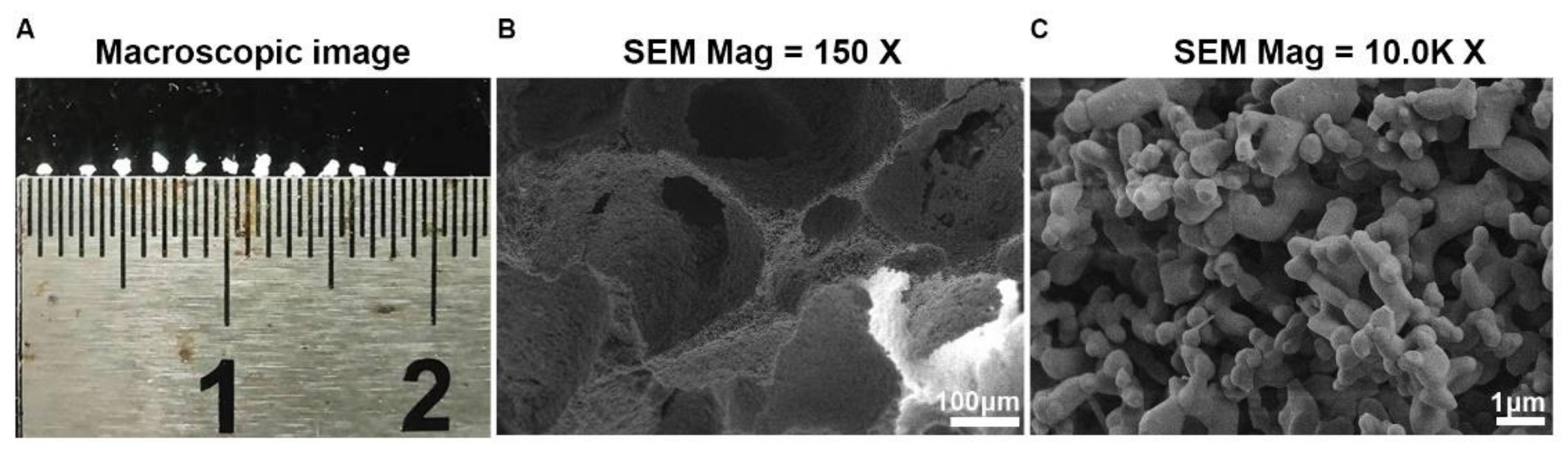
Abstract
:1. Introduction
2. Materials and Methods
2.1. Preparation of BCP Ceramics
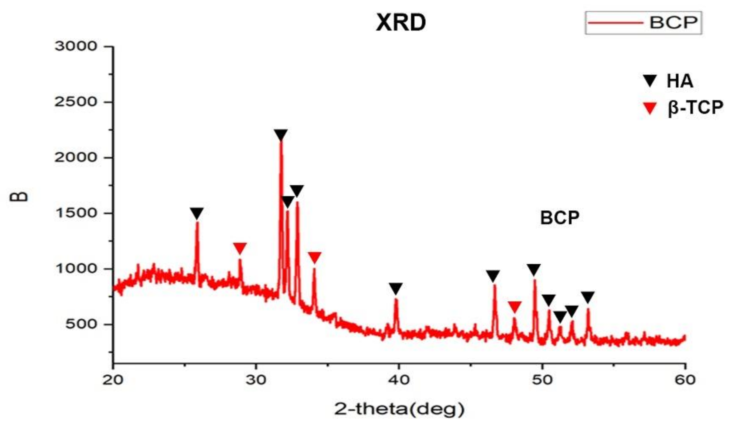
2.2. Morphological and Phase Analysis of BCP
2.3. Mice and Experimental Model
2.4. Histological Staining
2.5. Cell Culture
2.6. Cell Proliferation Assay
2.7. Transwell Assay
2.8. Quantitative Real—Time PCR
2.9. Alkaline Phosphatase (ALP) Staining
2.10. Statistical Analysis
3. Results
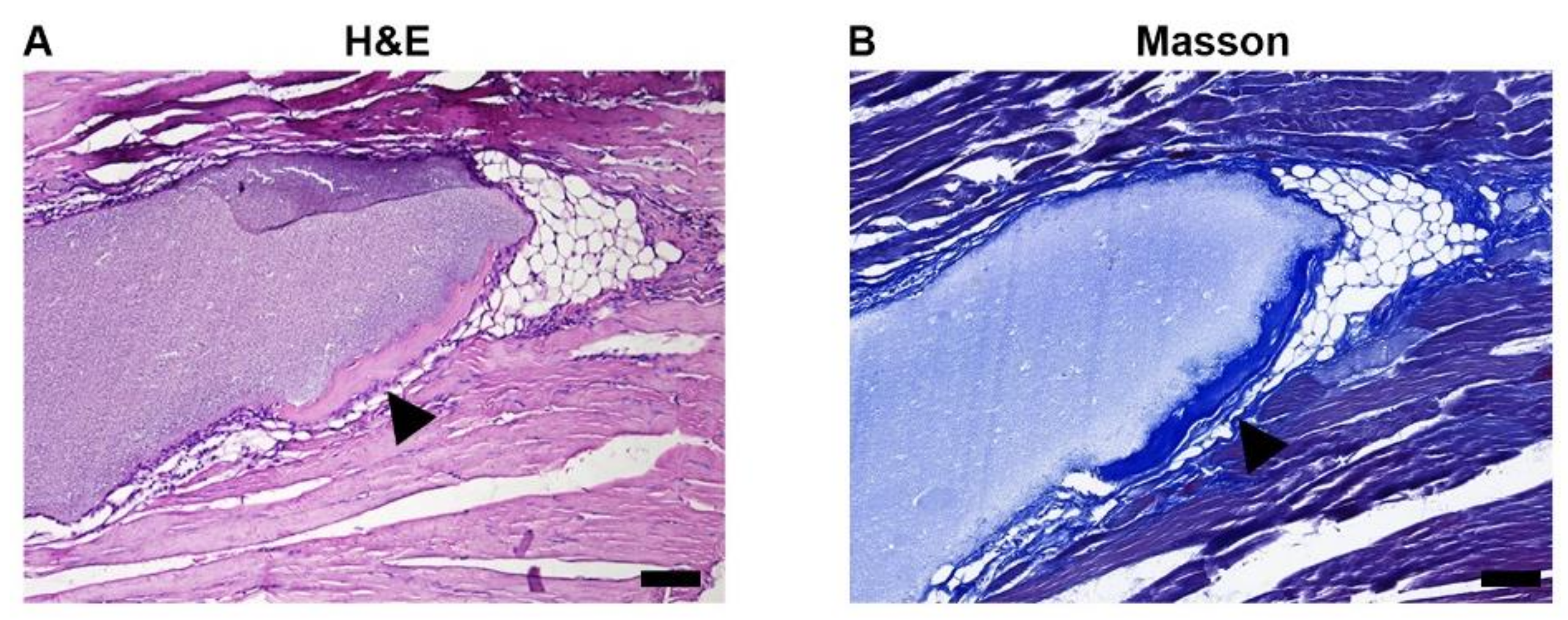
3.1. Bioactive BCP Implantation in Muscle Leads to Bone-like Formation
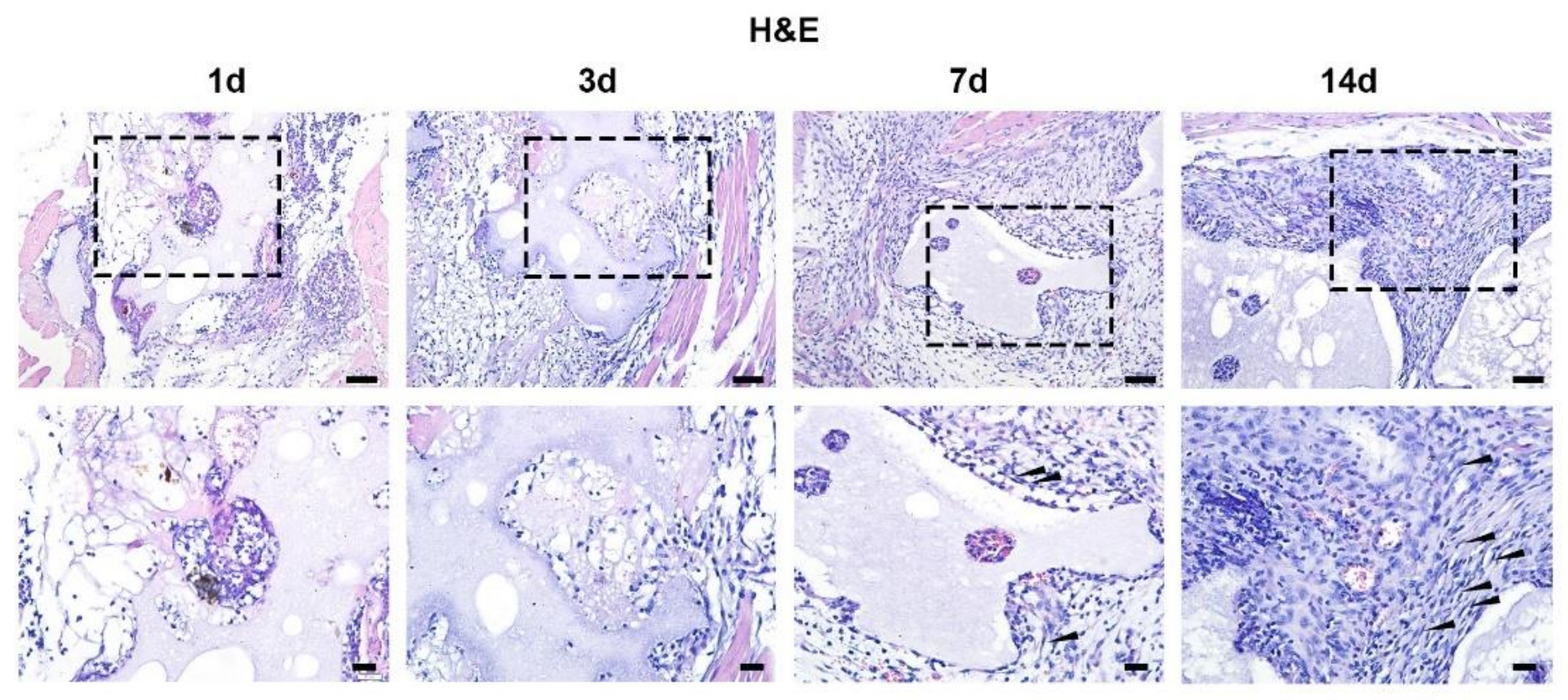
3.2. Temporal and Spatial Distribution of Fibroblasts around BCP during Bone-like Formation
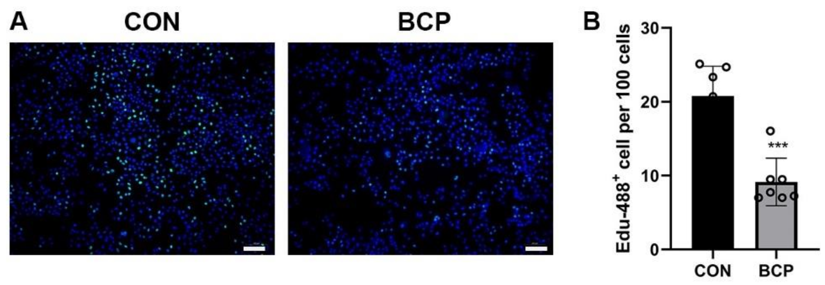
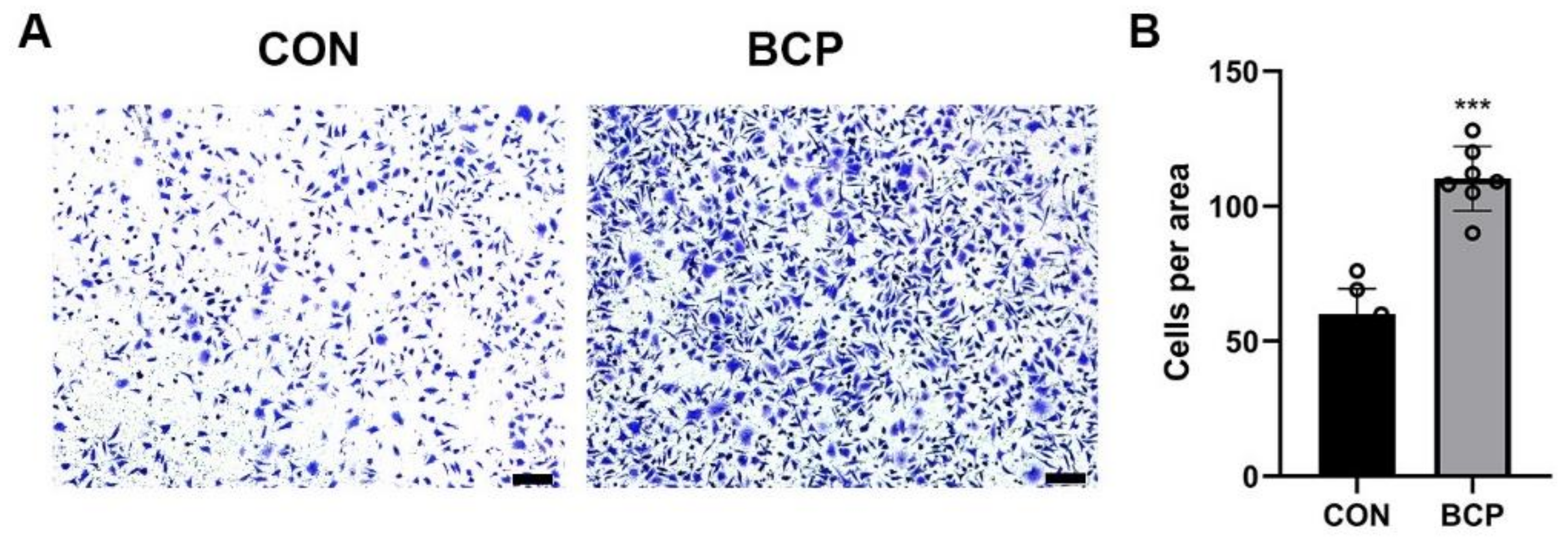
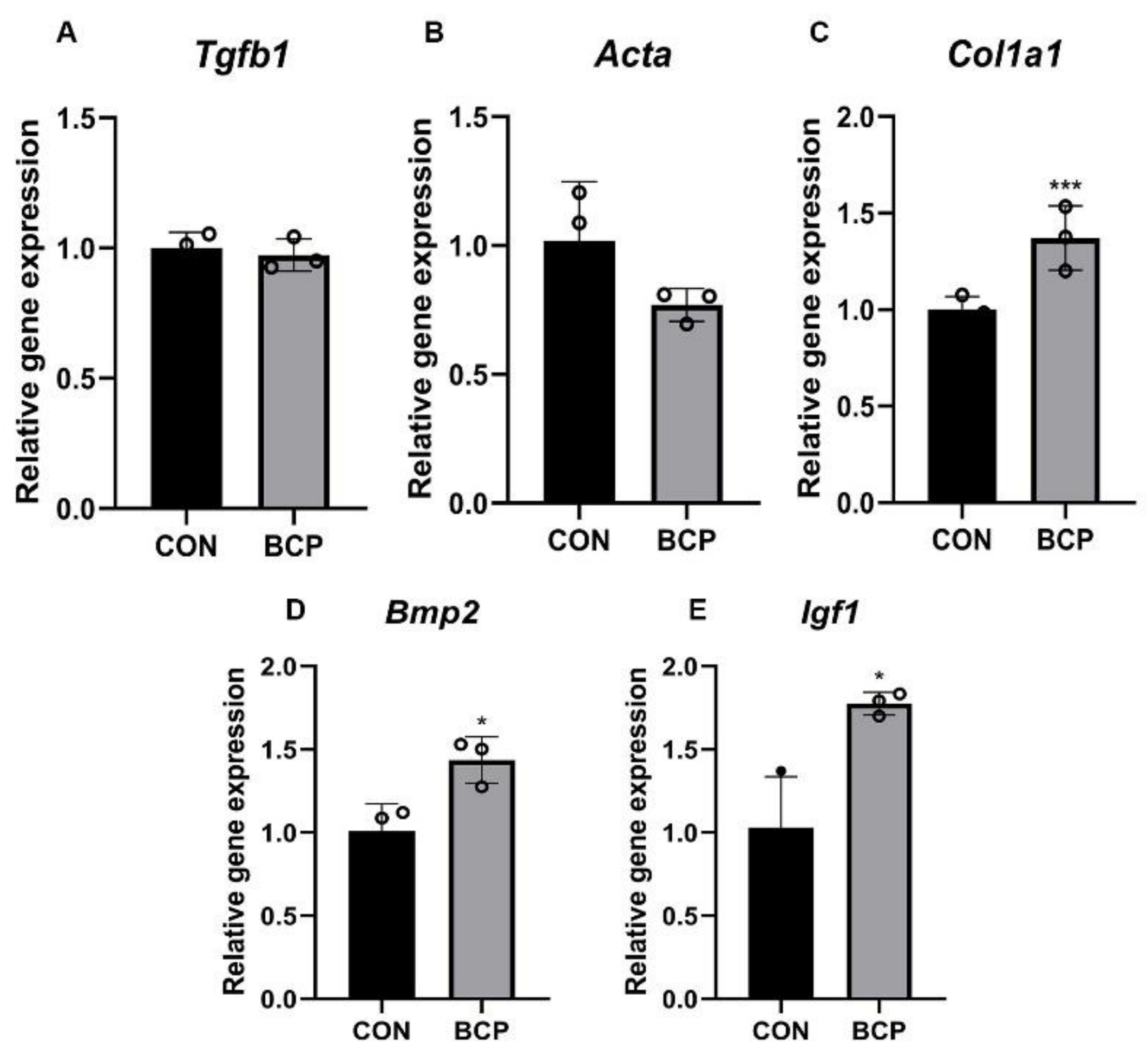
3.3. BCP Promoted Migration and Collagen Secretion of Fibroblasts
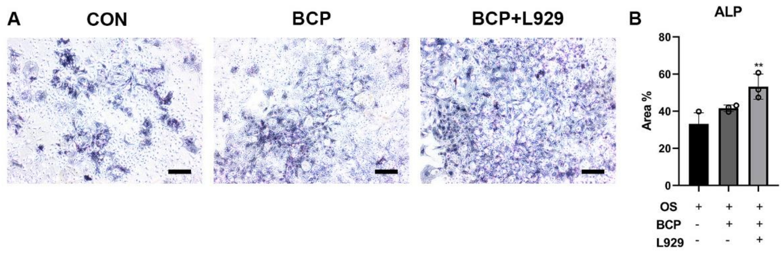
3.4. BCP Enhanced the Mineralization of the BMSCs through Fibroblasts
4. Discussion
5. Conclusions
Supplementary Materials
Author Contributions
Funding
Institutional Review Board Statement
Informed Consent Statement
Data Availability Statement
Conflicts of Interest
Abbreviations
| SEM | Scanning Electron Microscope |
| XRD | X-ray Diffraction |
| BCP | Biphasic Calcium Phosphate |
| BMSC | Bone Marrow-Derived Mesenchymal Stem Cells |
| CaP | Calcium Phosphates |
| ALP | Alkaline phosphatase |
| IHC | Immunohistochemistry |
| HE | Haematoxylin-eosin |
| HA | Hydroxyapatite |
| TCP | Tricalcium Phosphate |
References
- Wei, Y.; Zhu, G.; Zhao, Z.; Yin, C.; Zhao, Q.; Xu, H.; Wang, J.; Zhang, J.; Zhang, X.; Zhang, Y.; et al. Individualized plasticity autograft mimic with efficient bioactivity inducing osteogenesis. Int. J. Oral Sci. 2021, 13, 14. [Google Scholar] [CrossRef] [PubMed]
- Zhao, Z.; Zhao, Q.; Gu, B.; Yin, C.; Shen, K.; Tang, H.; Xia, H.; Zhang, X.; Zhao, Y.; Yang, X.; et al. Minimally invasive implantation and decreased inflammation reduce osteoinduction of biomaterial. Theranostics 2020, 10, 3533–3545. [Google Scholar] [CrossRef] [PubMed]
- Li, J.; Yuan, H.; Chandrakar, A.; Moroni, L.; Habibovic, P. 3D porous Ti6Al4V-beta-tricalcium phosphate scaffolds directly fabricated by additive manufacturing. Acta Biomater. 2021, 126, 496–510. [Google Scholar] [CrossRef] [PubMed]
- Wang, M.; Li, H.; Yang, Y.; Yuan, K.; Zhou, F.; Liu, H.; Zhou, Q.; Yang, S.; Tang, T. A 3D-bioprinted scaffold with doxycycline-controlled BMP2-expressing cells for inducing bone regeneration and inhibiting bacterial infection. Bioact. Mater. 2021, 6, 1318–1329. [Google Scholar] [CrossRef] [PubMed]
- Scott, M.A.; Levi, B.; Askarinam, A.; Nguyen, A.; Rackohn, T.; Ting, K.; Soo, C.; James, A.W. Brief review of models of ectopic bone formation. Stem. Cells Dev. 2012, 21, 655–667. [Google Scholar] [CrossRef] [Green Version]
- Mofakhami, S.; Salahinejad, E. Biphasic calcium phosphate microspheres in biomedical applications. J. Control. Release 2021, 338, 527–536. [Google Scholar] [CrossRef]
- Jacobs, A.; Renaudin, G.; Charbonnel, N.; Nedelec, J.M.; Forestier, C.; Descamps, S. Copper-Doped Biphasic Calcium Phosphate Powders: Dopant Release, Cytotoxicity and Antibacterial Properties. Materials 2021, 14, 2393. [Google Scholar] [CrossRef]
- Zhao, Q.; Shi, M.; Yin, C.; Zhao, Z.; Zhang, J.; Wang, J.; Shen, K.; Zhang, L.; Tang, H.; Xiao, Y.; et al. Dual-Wavelength Photosensitive Nano-in-Micro Scaffold Regulates Innate and Adaptive Immune Responses for Osteogenesis. Nano-Micro Lett. 2020, 13, 28. [Google Scholar] [CrossRef]
- Van Linthout, S.; Miteva, K.; Tschöpe, C. Crosstalk between fibroblasts and inflammatory cells. Cardiovasc. Res. 2014, 102, 258–269. [Google Scholar] [CrossRef] [Green Version]
- Bollyky, P.L.; Lord, J.D.; Masewicz, S.A.; Evanko, S.P.; Buckner, J.H.; Wight, T.N.; Nepom, G.T. Cutting edge: High molecular weight hyaluronan promotes the suppressive effects of CD4+CD25+ regulatory T cells. J. Immunol. 2007, 179, 744–747. [Google Scholar] [CrossRef] [PubMed] [Green Version]
- Cappato, S.; Gamberale, R.; Bocciardi, R.; Brunelli, S. Genetic and Acquired Heterotopic Ossification: A Translational Tale of Mice and Men. Biomedicines 2020, 8, 611. [Google Scholar] [CrossRef] [PubMed]
- Muhl, L.; Genové, G.; Leptidis, S.; Liu, J.; He, L.; Mocci, G.; Sun, Y.; Gustafsson, S.; Buyandelger, B.; Chivukula, I.V.; et al. Single-cell analysis uncovers fibroblast heterogeneity and criteria for fibroblast and mural cell identification and discrimination. Nat. Commun. 2020, 11, 3953. [Google Scholar] [CrossRef] [PubMed]
- Chen, K.; Kwon, S.H.; Henn, D.; Kuehlmann, B.A.; Tevlin, R.; Bonham, C.A.; Griffin, M.; Trotsyuk, A.A.; Borrelli, M.R.; Noishiki, C.; et al. Disrupting biological sensors of force promotes tissue regeneration in large organisms. Nat. Commun. 2021, 12, 5256. [Google Scholar] [CrossRef] [PubMed]
- Kim, S.W.; Im, G.B.; Jeong, G.J.; Baik, S.; Hyun, J.; Kim, Y.J.; Pang, C.; Jang, Y.C.; Bhang, S.H. Delivery of a spheroids-incorporated human dermal fibroblast sheet increases angiogenesis and M2 polarization for wound healing. Biomaterials 2021, 275, 120954. [Google Scholar] [CrossRef]
- Sun, L.; He, L.; Wu, W.; Luo, L.; Han, M.; Liu, Y.; Shi, S.; Zhong, K.; Yang, J.; Li, J. Fibroblast membrane-camouflaged nanoparticles for inflammation treatment in the early stage. Int. J. Oral Sci. 2021, 13, 39. [Google Scholar] [CrossRef]
- Yang, R.; Huang, J.; Zhang, W.; Xue, W.; Jiang, Y.; Li, S.; Wu, X.; Xu, H.; Ren, J.; Chi, B. Mechanoadaptive injectable hydrogel based on poly(γ-glutamic acid) and hyaluronic acid regulates fibroblast migration for wound healing. Carbohydr. Polym. 2021, 273, 118607. [Google Scholar] [CrossRef] [PubMed]
- Zhang, Q.; Chang, C.; Qian, C.; Xiao, W.; Zhu, H.; Guo, J.; Meng, Z.; Cui, W.; Ge, Z. Photo-crosslinkable amniotic membrane hydrogel for skin defect healing. Acta Biomater. 2021, 125, 197–207. [Google Scholar] [CrossRef] [PubMed]
- Wang, J.; Zhao, Q.; Zheng, S.; Wang, J.; Fu, L.; Wang, C.; Zhao, Z.; Zhang, Y. Break monopoly of polarization: CD301b+ macrophages play positive roles in osteoinduction of calcium phosphate ceramics. Appl. Mater. Today 2021, 24, 101111. [Google Scholar] [CrossRef]
- Zhao, Q.; Gong, Z.; Li, Z.; Wang, J.; Zhang, J.; Zhao, Z.; Zhang, P.; Zheng, S.; Miron, R.J.; Yuan, Q.; et al. Target Reprogramming Lysosomes of CD8+ T Cells by a Mineralized Metal-Organic Framework for Cancer Immunotherapy. Adv. Mater. 2021, 33, e2100616. [Google Scholar] [CrossRef] [PubMed]
- Zhao, Q.; Wang, J.; Yin, C.; Zhang, P.; Zhang, J.; Shi, M.; Shen, K.; Xiao, Y.; Zhao, Y.; Yang, X.; et al. Near-Infrared Light-Sensitive Nano Neuro-Immune Blocker Capsule Relieves Pain and Enhances the Innate Immune Response for Necrotizing Infection. Nano Lett. 2019, 19, 5904–5914. [Google Scholar] [CrossRef] [PubMed]
- Fu, L.; Wang, M.; Zhu, G.; Zhao, Z.; Sun, H.; Cao, Z.; Xia, H. REV-ERBs negatively regulate mineralization of the cementoblasts. Biochem. Biophys. Res. Commun. 2022, 587, 9–15. [Google Scholar] [CrossRef] [PubMed]
- Zhi, W.; Wang, X.; Sun, D.; Chen, T.; Yuan, B.; Li, X.; Chen, X.; Wang, J.; Xie, Z.; Zhu, X.; et al. Optimal regenerative repair of large segmental bone defect in a goat model with osteoinductive calcium phosphate bioceramic implants. Bioact. Mater. 2022, 11, 240–253. [Google Scholar] [CrossRef]
- Cecen, B.; Kalemtas, A.; Topates, G.; Kozaci, L.D. Chapter 17—Cellular response to calcium phosphate cements. In Handbook of Biomaterials Biocompatibility; Mozafari, M., Ed.; Woodhead Publishing: Sawston, UK, 2020; pp. 369–393. [Google Scholar]
- Guo, X.; Li, M.; Qi, W.; Bai, H.; Nie, Z.; Hu, Z.; Xiao, Y.; de Bruijn, J.D.; Bao, C.; Yuan, H. Serial cellular events in bone formation initiated by calcium phosphate ceramics. Acta Biomater. 2021, 134, 730–743. [Google Scholar] [CrossRef] [PubMed]
- Hulsart-Billström, G.; Hu, Q.; Bergman, K.; Jonsson, K.B.; Åberg, J.; Tang, R.; Larsson, S.; Hilborn, J. Calcium phosphates compounds in conjunction with hydrogel as carrier for BMP-2: A study on ectopic bone formation in rats. Acta Biomater. 2011, 7, 3042–3049. [Google Scholar] [CrossRef] [PubMed]
- Lee, J.H.; Lee, K.M.; Baek, H.R.; Jang, S.J.; Lee, J.H.; Ryu, H.S. Combined effects of porous hydroxyapatite and demineralized bone matrix on bone induction: In vitro and in vivo study using a nude rat model. Biomed. Mater. 2011, 6, 015008. [Google Scholar] [CrossRef] [PubMed] [Green Version]
- van Gaalen, S.M.; Dhert, W.J.; Kruyt, M.C.; Yuan, H.; Oner, F.C.; van Blitterswijk, C.A.; Verbout, A.J.; de Bruijn, J.D. Goat bone tissue engineering: Comparing an intramuscular with a posterolateral lumbar spine location. Tissue Eng. Part A 2010, 16, 685–693. [Google Scholar] [CrossRef] [PubMed]
- Barbieri, D.; Yuan, H.; de Groot, F.; Walsh, W.R.; de Bruijn, J.D. Influence of different polymeric gels on the ectopic bone forming ability of an osteoinductive biphasic calcium phosphate ceramic. Acta Biomater. 2011, 7, 2007–2014. [Google Scholar] [CrossRef] [PubMed]
- Muniz, F.T.; Miranda, M.A.; Morilla Dos Santos, C.; Sasaki, J.M. The Scherrer equation and the dynamical theory of X-ray diffraction. Acta Crystallogr. Sect. A Found. Adv. 2016, 72, 385–390. [Google Scholar] [CrossRef]
- von Doernberg, M.C.; von Rechenberg, B.; Bohner, M.; Grünenfelder, S.; van Lenthe, G.H.; Müller, R.; Gasser, B.; Mathys, R.; Baroud, G.; Auer, J. In vivo behavior of calcium phosphate scaffolds with four different pore sizes. Biomaterials 2006, 27, 5186–5198. [Google Scholar] [CrossRef] [PubMed]
- Zhang, J.; Wu, Q.; Yin, C.; Jia, X.; Zhao, Z.; Zhang, X.; Yuan, G.; Hu, H.; Zhao, Q. Sustained calcium ion release from bioceramics promotes CaSR-mediated M2 macrophage polarization for osteoinduction. J. Leukoc. Biol. 2021, 110, 485–496. [Google Scholar] [CrossRef]
- Contreras, O.; Rossi, F.M.V.; Theret, M. Origins, potency, and heterogeneity of skeletal muscle fibro-adipogenic progenitors-time for new definitions. Skelet. Muscle 2021, 11, 16. [Google Scholar] [CrossRef]
- Mascharak, S.; desJardins-Park, H.E.; Davitt, M.F.; Griffin, M.; Borrelli, M.R.; Moore, A.L.; Chen, K.; Duoto, B.; Chinta, M.; Foster, D.S.; et al. Preventing Engrailed-1 activation in fibroblasts yields wound regeneration without scarring. Science 2021, 372, eaba2374. [Google Scholar] [CrossRef] [PubMed]
- Rohani, M.G.; Parks, W.C. Matrix remodeling by MMPs during wound repair. Matrix Biol. 2015, 44–46, 113–121. [Google Scholar] [CrossRef]
- Silvestre, J.S.; Théry, C.; Hamard, G.; Boddaert, J.; Aguilar, B.; Delcayre, A.; Houbron, C.; Tamarat, R.; Blanc-Brude, O.; Heeneman, S.; et al. Lactadherin promotes VEGF-dependent neovascularization. Nat. Med. 2005, 11, 499–506. [Google Scholar] [CrossRef] [PubMed]
- Rivera-Gonzalez, G.C.; Shook, B.A.; Andrae, J.; Holtrup, B.; Bollag, K.; Betsholtz, C.; Rodeheffer, M.S.; Horsley, V. Skin Adipocyte Stem Cell Self-Renewal Is Regulated by a PDGFA/AKT-Signaling Axis. Cell Stem Cell 2016, 19, 738–751. [Google Scholar] [CrossRef] [PubMed] [Green Version]
- Plikus, M.V.; Guerrero-Juarez, C.F.; Ito, M.; Li, Y.R.; Dedhia, P.H.; Zheng, Y.; Shao, M.; Gay, D.L.; Ramos, R.; Hsi, T.C.; et al. Regeneration of fat cells from myofibroblasts during wound healing. Science 2017, 355, 748–752. [Google Scholar] [CrossRef] [PubMed] [Green Version]
- Descamps, E.; Molto, A.; Borderie, D.; Lories, R.; Richard, C.M.; Pons, M.; Roux, C.; Briot, K. Changes in bone formation regulator biomarkers in early axial spondyloarthritis. Rheumatology 2021, 60, 1185–1194. [Google Scholar] [CrossRef]
- Kusano, T.; Nakatani, M.; Ishiguro, N.; Ohno, K.; Yamamoto, N.; Morita, M.; Yamada, H.; Uezumi, A.; Tsuchida, K. Desloratadine inhibits heterotopic ossification by suppression of BMP2-Smad1/5/8 signaling. J. Orthop. Res. 2021, 39, 1297–1304. [Google Scholar] [CrossRef]
- Buechler, M.B.; Pradhan, R.N.; Krishnamurty, A.T.; Cox, C.; Calviello, A.K.; Wang, A.W.; Yang, Y.A.; Tam, L.; Caothien, R.; Roose-Girma, M.; et al. Cross-tissue organization of the fibroblast lineage. Nature 2021, 593, 575–579. [Google Scholar] [CrossRef]
- Uezumi, A.; Fukada, S.; Yamamoto, N.; Ikemoto-Uezumi, M.; Nakatani, M.; Morita, M.; Yamaguchi, A.; Yamada, H.; Nishino, I.; Hamada, Y.; et al. Identification and characterization of PDGFRα+ mesenchymal progenitors in human skeletal muscle. Cell Death Dis. 2014, 5, e1186. [Google Scholar] [CrossRef] [PubMed] [Green Version]
- Eisner, C.; Cummings, M.; Johnston, G.; Tung, L.W.; Groppa, E.; Chang, C.; Rossi, F.M. Murine Tissue-Resident PDGFRα+ Fibro-Adipogenic Progenitors Spontaneously Acquire Osteogenic Phenotype in an Altered Inflammatory Environment. J. Bone Miner. Res. 2020, 35, 1525–1534. [Google Scholar] [CrossRef] [PubMed]


Publisher’s Note: MDPI stays neutral with regard to jurisdictional claims in published maps and institutional affiliations. |
© 2022 by the authors. Licensee MDPI, Basel, Switzerland. This article is an open access article distributed under the terms and conditions of the Creative Commons Attribution (CC BY) license (https://creativecommons.org/licenses/by/4.0/).
Share and Cite
Fu, L.; Zhao, Q.; Li, J.; Zhao, Z.; Wang, M.; Sun, H.; Xia, H. Fibroblasts Mediate Ectopic Bone Formation of Calcium Phosphate Ceramics. Materials 2022, 15, 2569. https://doi.org/10.3390/ma15072569
Fu L, Zhao Q, Li J, Zhao Z, Wang M, Sun H, Xia H. Fibroblasts Mediate Ectopic Bone Formation of Calcium Phosphate Ceramics. Materials. 2022; 15(7):2569. https://doi.org/10.3390/ma15072569
Chicago/Turabian StyleFu, Liangliang, Qin Zhao, Jiaojiao Li, Zifan Zhao, Min Wang, Huifang Sun, and Haibin Xia. 2022. "Fibroblasts Mediate Ectopic Bone Formation of Calcium Phosphate Ceramics" Materials 15, no. 7: 2569. https://doi.org/10.3390/ma15072569