
Superrepellent Porous Polymer Surfaces by Replication from Wrinkled Polydimethylsiloxane/Parylene F
 , , ,
, , ,
Abstract
:1. Introduction
2. Materials and Methods
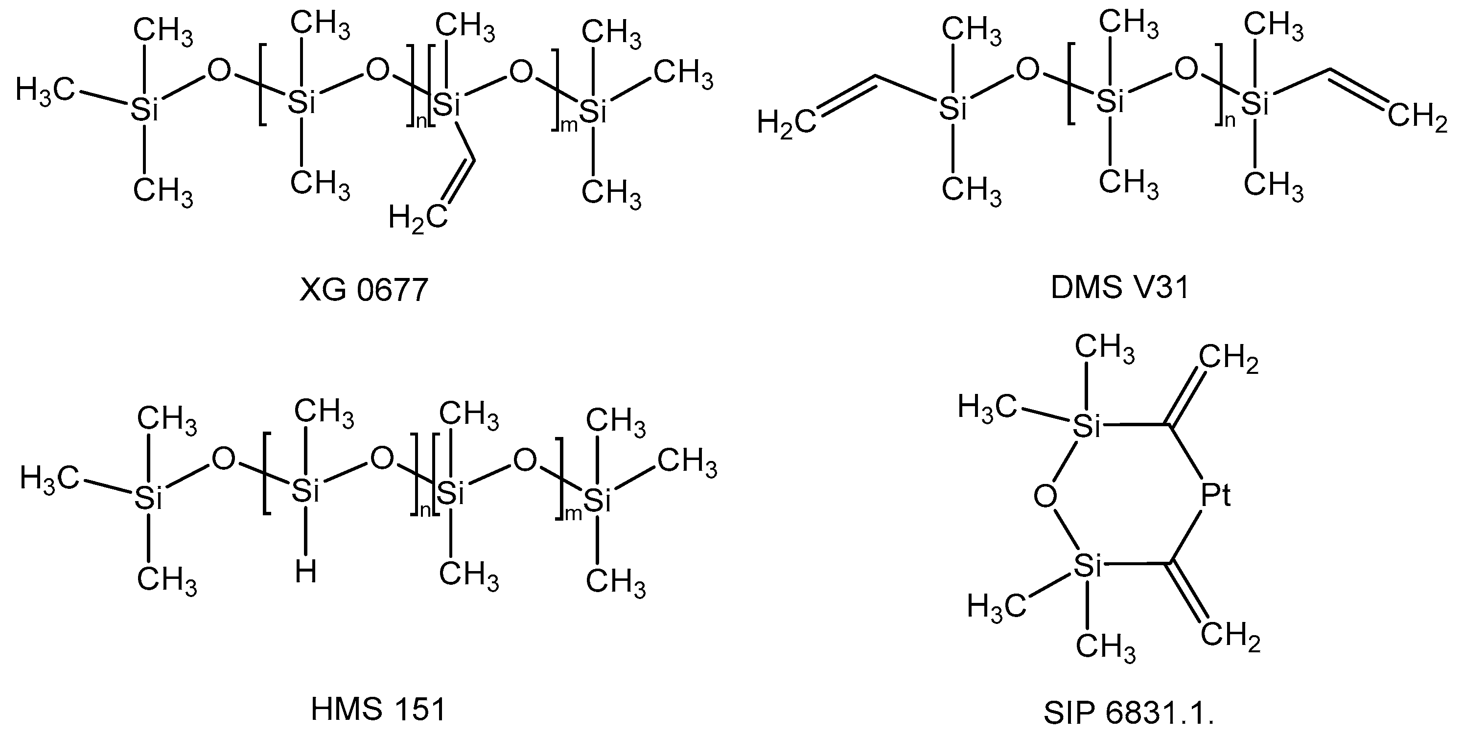
2.1. Materials
2.2. Methods
3. Results and Discussion
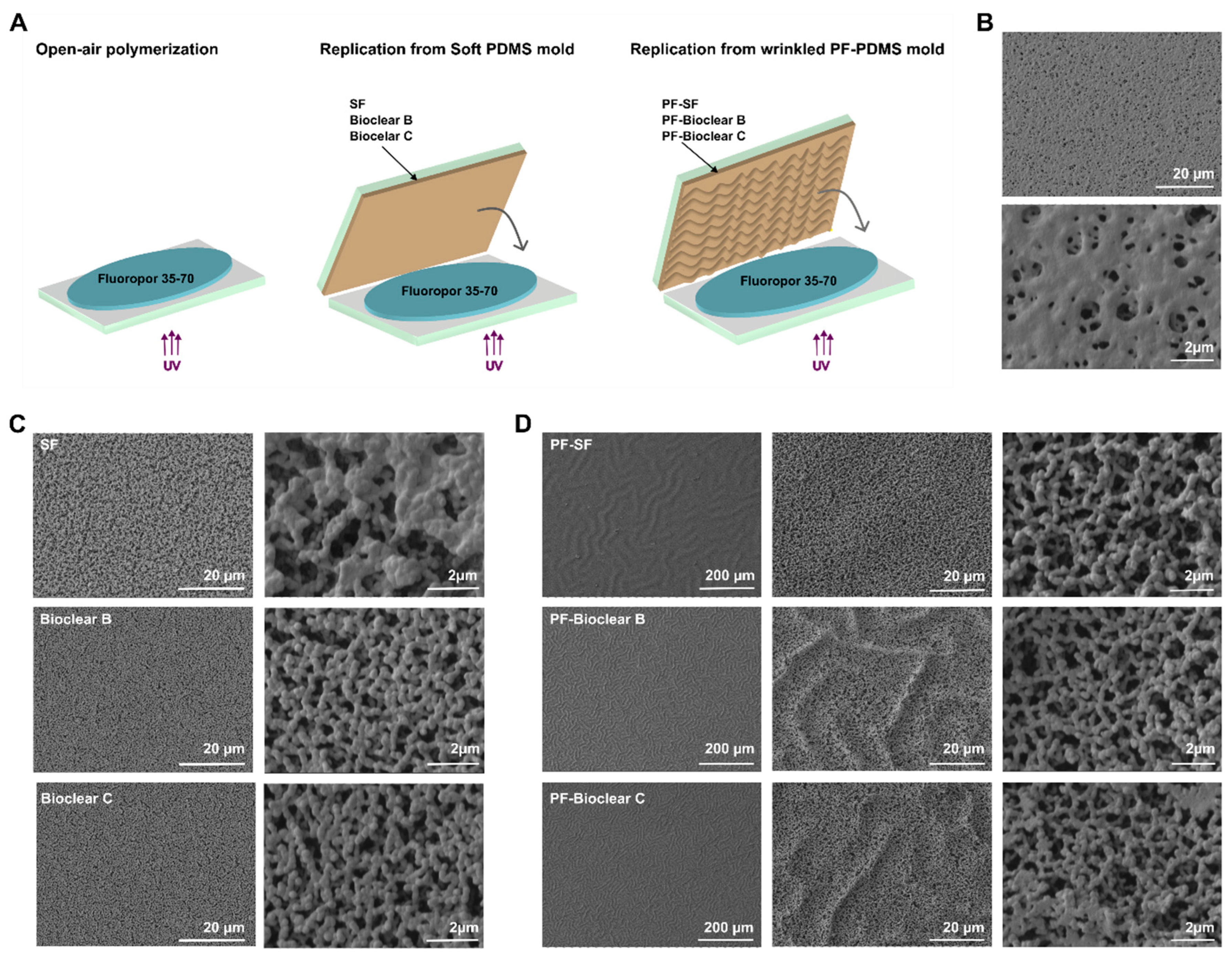
3.1. Soft PDMS Coatings
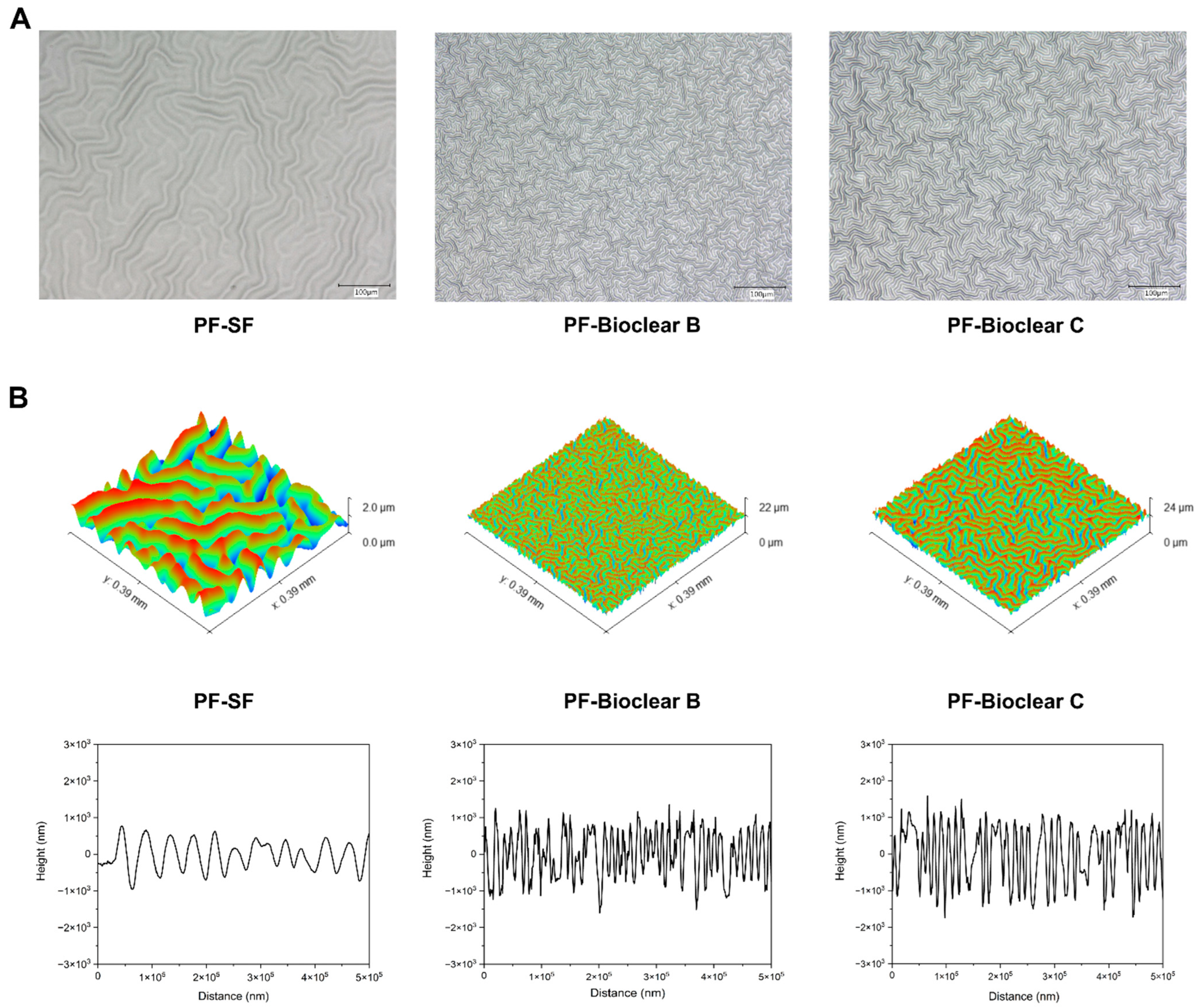
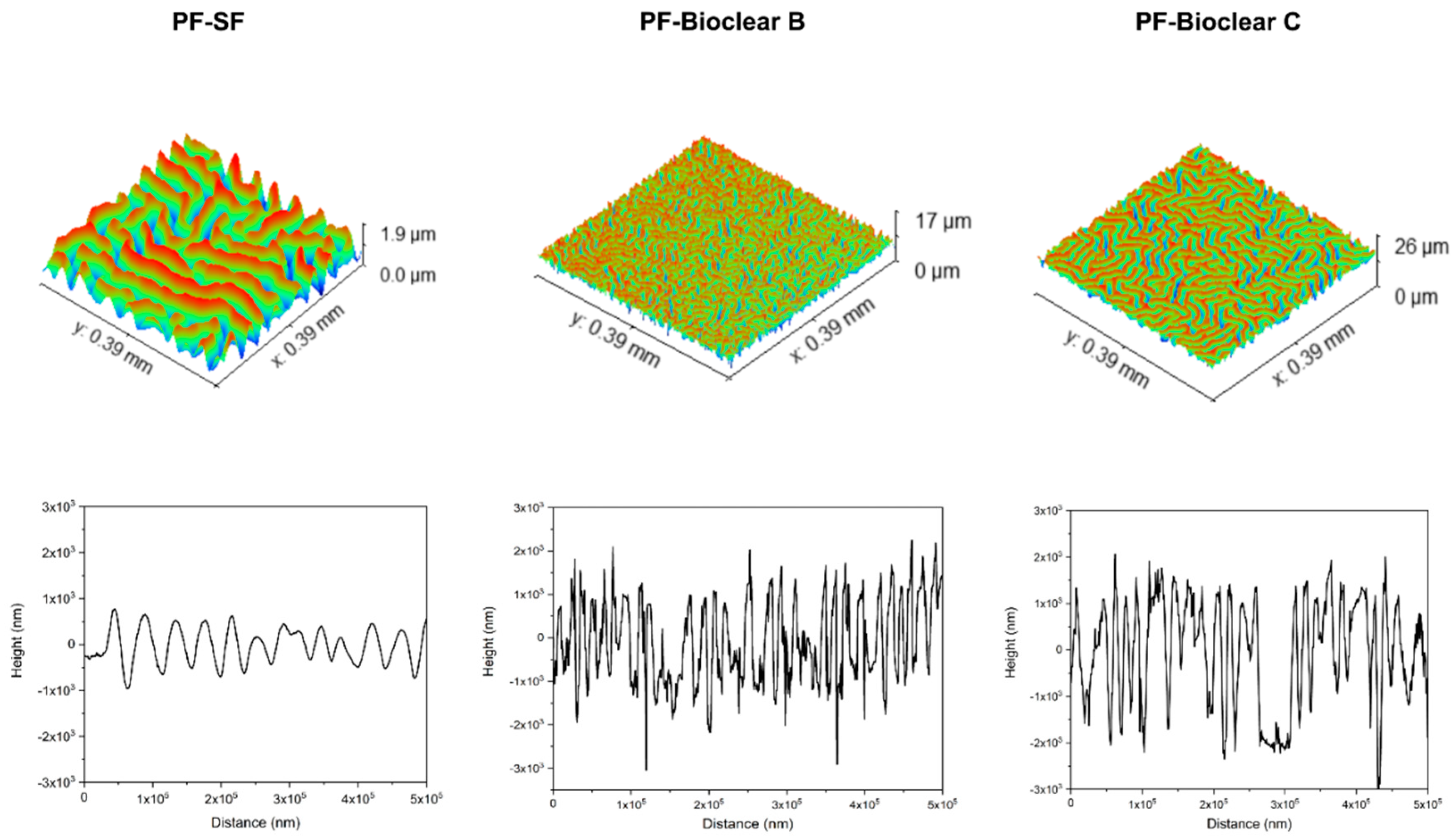
3.2. Properties of Parylene-F-Coated Wrinkled PDMS Thin Layers
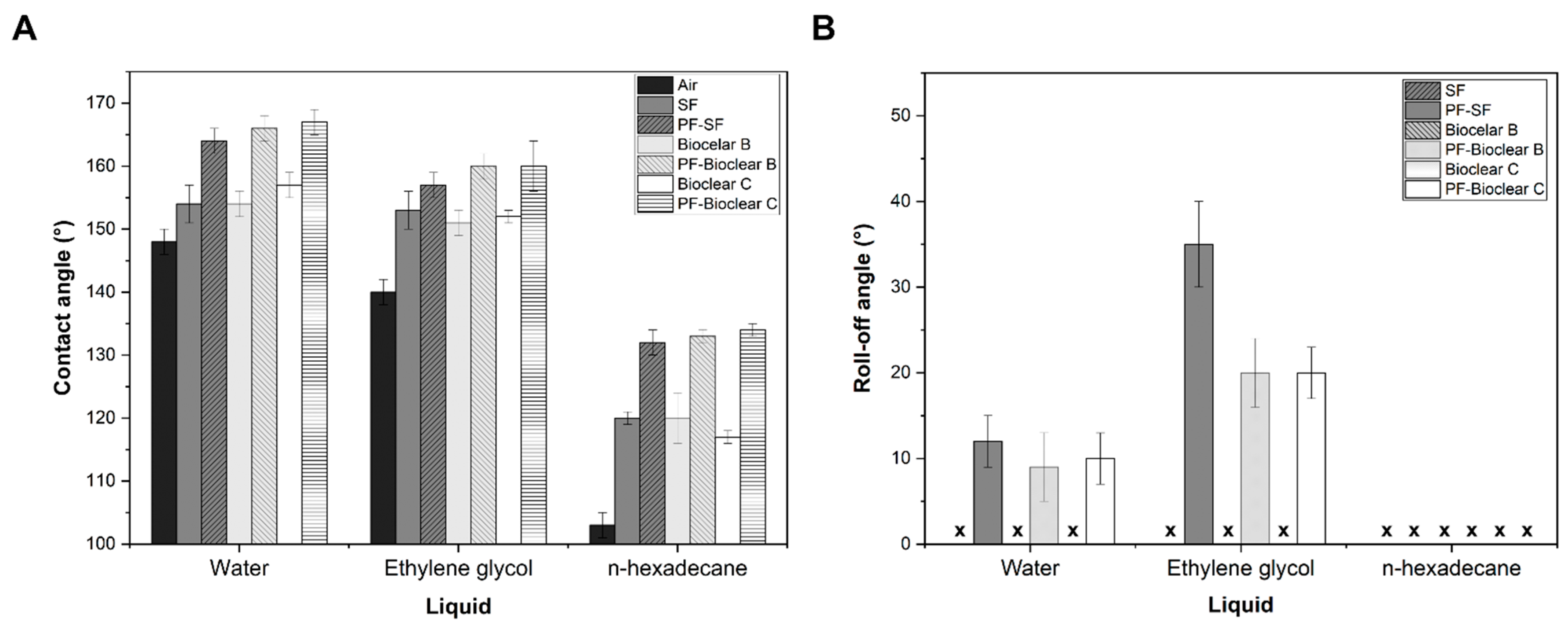
3.3. Superrepellent Wrinkled Porous Polymers
4. Conclusions
Author Contributions
Funding
Institutional Review Board Statement
Informed Consent Statement
Data Availability Statement
Conflicts of Interest
Appendix A


| Wrinkled Templates | |||
|---|---|---|---|
| PF-SF | PF-Bioclear B | PF-Bioclear C | |
| Surface free energy (mN/m) | 25 ± 1 | 29 ± 1 | 28 ± 1 |


| SF | PF-SF | Bioclear B | PF-Bioclear B | Bioclear C | PF-Bioclear C | Air | |
|---|---|---|---|---|---|---|---|
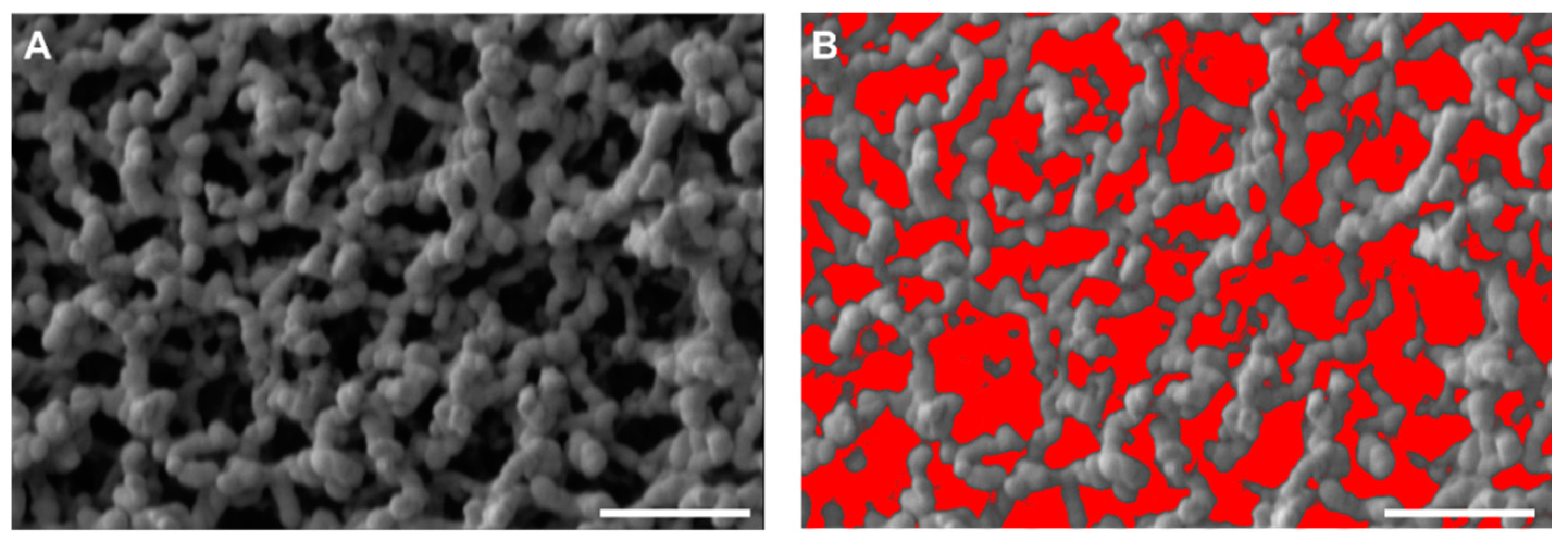
| Porosity % | 30 ± 1 | 47 ± 4 | 36 ± 2 | 46 ± 2 | 37 ± 3 | 48 ± 5 | 13 ± 1 |
References
- Blossey, R. Self-Cleaning Surfaces—Virtual Realities. Nat. Mater. 2003, 2, 301–306. [Google Scholar] [CrossRef]
- Farhadi, S.; Farzaneh, M.; Kulinich, S.A. Anti-Icing Performance of Superhydrophobic Surfaces. Appl. Surf. Sci. 2011, 257, 6264–6269. [Google Scholar] [CrossRef]
- Chapman, J.; Regan, F. Nanofunctionalized Superhydrophobic Antifouling Coatings for Environmental Sensor Applications—Advancing Deployment with Answers from Nature. Adv. Eng. Mater. 2012, 14, B175–B184. [Google Scholar] [CrossRef]
- Ishizaki, T.; Masuda, Y.; Sakamoto, M. Corrosion Resistance and Durability of Superhydrophobic Surface Formed on Magnesium Alloy Coated with Nanostructured Cerium Oxide Film and Fluoroalkylsilane Molecules in Corrosive NaCl Aqueous Solution. Langmuir 2011, 27, 4780–4788. [Google Scholar] [CrossRef] [PubMed]
- Mahalakshmi, P.V.; Vanithakumari, S.C.; Gopal, J.; Mudali, U.K.; Raj, B. Enhancing Corrosion and Biofouling Resistance through Superhydrophobic Surface Modification. Curr. Sci. 2011, 101, 1328–1336. [Google Scholar]
- Feng, L.; Zhang, Z.; Mai, Z.; Ma, Y.; Liu, B.; Jiang, L.; Zhu, D. A Super-Hydrophobic and Super-Oleophilic Coating Mesh Film for the Separation of Oil and Water. Angew. Chem. Int. Ed. 2004, 43, 2012–2014. [Google Scholar] [CrossRef]
- Cho, E.-C.; Chang-Jian, C.-W.; Chen, H.-C.; Chuang, K.-S.; Zheng, J.-H.; Hsiao, Y.-S.; Lee, K.-C.; Huang, J.-H. Robust Multifunctional Superhydrophobic Coatings with Enhanced Water/Oil Separation, Self-Cleaning, Anti-Corrosion, and Anti-Biological Adhesion. Chem. Eng. J. 2017, 314, 347–357. [Google Scholar] [CrossRef]
- Marmur, A. Wetting on Hydrophobic Rough Surfaces: To Be Heterogeneous or Not To Be? Langmuir 2003, 19, 8343–8348. [Google Scholar] [CrossRef]
- Cassie, A.B.D.; Baxter, S. Wettability of Porous Surfaces. Trans. Faraday Soc. 1944, 40, 546–551. [Google Scholar] [CrossRef]
- Di Mundo, R.; Palumbo, F.; d’Agostino, R. Nanotexturing of Polystyrene Surface in Fluorocarbon Plasmas: From Sticky to Slippery Superhydrophobicity. Langmuir 2008, 24, 5044–5051. [Google Scholar] [CrossRef]
- Han, J.T.; Zheng, Y.; Cho, J.H.; Xu, X.; Cho, K. Stable Superhydrophobic Organic−Inorganic Hybrid Films by Electrostatic Self-Assembly. J. Phys. Chem. B 2005, 109, 20773–20778. [Google Scholar] [CrossRef] [PubMed]
- Liu, H.; Feng, L.; Zhai, J.; Jiang, L.; Zhu, D. Reversible Wettability of a Chemical Vapor Deposition Prepared ZnO Film between Superhydrophobicity and Superhydrophilicity. Langmuir 2004, 20, 5659–5661. [Google Scholar] [CrossRef] [PubMed]
- Shirtcliffe, N.J.; McHale, G.; Newton, M.I.; Perry, C.C. Intrinsically Superhydrophobic Organosilica Sol−Gel Foams. Langmuir 2003, 19, 5626–5631. [Google Scholar] [CrossRef]
- Wu, L.-K.; Hu, J.-M.; Zhang, J.-Q. One Step Sol–Gel Electrochemistry for the Fabrication of Superhydrophobic Surfaces. J. Mater. Chem. A 2013, 1, 14471–14475. [Google Scholar] [CrossRef]
- Jeong, H.E.; Lee, S.H.; Kim, J.K.; Suh, K.Y. Nanoengineered Multiscale Hierarchical Structures with Tailored Wetting Properties. Langmuir 2006, 22, 1640–1645. [Google Scholar] [CrossRef]
- Zhu, X.; Zhang, Z.; Ren, G.; Yang, J.; Wang, K.; Xu, X.; Men, X.; Zhou, X. A Novel Superhydrophobic Bulk Material. J. Mater. Chem. 2012, 22, 20146–20148. [Google Scholar] [CrossRef]
- Jin, H.; Tian, X.; Ikkala, O.; Ras, R.H.A. Preservation of Superhydrophobic and Superoleophobic Properties upon Wear Damage. ACS Appl. Mater. Interfaces 2013, 5, 485–488. [Google Scholar] [CrossRef]
- Helmer, D.; Keller, N.; Kotz, F.; Stolz, F.; Greiner, C.; Nargang, T.M.; Sachsenheimer, K.; Rapp, B.E. Transparent, Abrasion-Insensitive Superhydrophobic Coatings for Real-World Applications. Sci. Rep. 2017, 7, 15078. [Google Scholar] [CrossRef] [Green Version]
- Keller, N.; Bruchmann, J.; Sollich, T.; Richter, C.; Thelen, R.; Kotz, F.; Schwartz, T.; Helmer, D.; Rapp, B.E. Study of Biofilm Growth on Slippery Liquid-Infused Porous Surfaces Made from Fluoropor. ACS Appl. Mater. Interfaces 2019, 11, 4480–4487. [Google Scholar] [CrossRef]
- Mayoussi, F.; Doeven, E.H.; Kick, A.; Goralczyk, A.; Thomann, Y.; Risch, P.; Guijt, R.M.; Kotz, F.; Helmer, D.; Rapp, B.E. Facile Fabrication of Micro-/Nanostructured, Superhydrophobic Membranes with Adjustable Porosity by 3D Printing. J. Mater. Chem. A 2021, 9, 21379–21386. [Google Scholar] [CrossRef]
- Goralczyk, A.; Zhu, M.; Mayoussi, F.; Lallemang, M.; Tschaikowsky, M.; Warmbold, A.; Caliaro, S.; Tauber, F.; Balzer, B.N.; Kotz-Helmer, F.; et al. Study of Repellence on Polymeric Surfaces with Two Individually Adjustable Pore Hierarchies. Chem. Eng. J. 2022, 437, 135287. [Google Scholar] [CrossRef]
- Goralczyk, A.; Bhagwat, S.; Mayoussi, F.; Nekoonam, N.; Sachsenheimer, K.; Hou, P.; Kotz-Helmer, F.; Helmer, D.; Rapp, B.E. Application of Micro/Nanoporous Fluoropolymers with Reduced Bioadhesion in Digital Microfluidics. Nanomaterials 2022, 12, 2201. [Google Scholar] [CrossRef] [PubMed]
- Job, N.; Théry, A.; Pirard, R.; Marien, J.; Kocon, L.; Rouzaud, J.-N.; Béguin, F.; Pirard, J.-P. Carbon Aerogels, Cryogels and Xerogels: Influence of the Drying Method on the Textural Properties of Porous Carbon Materials. Carbon 2005, 43, 2481–2494. [Google Scholar] [CrossRef]
- Sun, Q.; Ueno, K.; Misawa, H. In Situ Investigation of the Shrinkage of Photopolymerized Micro/Nanostructures: The Effect of the Drying Process. Opt. Lett. 2012, 37, 710–712. [Google Scholar] [CrossRef]
- García-González, C.A.; Camino-Rey, M.C.; Alnaief, M.; Zetzl, C.; Smirnova, I. Supercritical Drying of Aerogels Using CO2: Effect of Extraction Time on the End Material Textural Properties. J. Supercrit. Fluids 2012, 66, 297–306. [Google Scholar] [CrossRef]
- Levkin, P.A.; Svec, F.; Fréchet, J.M.J. Porous Polymer Coatings: A Versatile Approach to Superhydrophobic Surfaces. Adv. Funct. Mater. 2009, 19, 1993–1998. [Google Scholar] [CrossRef] [Green Version]
- Dong, Z.; Cui, H.; Zhang, H.; Wang, F.; Zhan, X.; Mayer, F.; Nestler, B.; Wegener, M.; Levkin, P.A. 3D Printing of Inherently Nanoporous Polymers via Polymerization-Induced Phase Separation. Nat. Commun. 2021, 12, 247. [Google Scholar] [CrossRef]
- Dong, Z.; Vuckovac, M.; Cui, W.; Zhou, Q.; Ras, R.H.A.; Levkin, P.A. 3D Printing of Superhydrophobic Objects with Bulk Nanostructure. Adv. Mater. 2021, 33, 2106068. [Google Scholar] [CrossRef]
- Mayer, F.; Ryklin, D.; Wacker, I.; Curticean, R.; Čalkovský, M.; Niemeyer, A.; Dong, Z.; Levkin, P.A.; Gerthsen, D.; Schröder, R.R.; et al. 3D Two-Photon Microprinting of Nanoporous Architectures. Adv. Mater. 2020, 32, 2002044. [Google Scholar] [CrossRef]
- Wenzel, R.N. Resistance of Solid Surfaces to Wetting by Water. Ind. Eng. Chem. 1936, 28, 988–994. [Google Scholar] [CrossRef]
- Neinhuis, C.; Barthlott, W. Characterization and Distribution of Water-Repellent, Self-Cleaning Plant Surfaces. Ann. Bot. 1997, 79, 667–677. [Google Scholar] [CrossRef]
- Lafuma, A.; Quéré, D. Superhydrophobic States. Nat. Mater 2003, 2, 457–460. [Google Scholar] [CrossRef] [PubMed]
- Yoshimitsu, Z.; Nakajima, A.; Watanabe, T.; Hashimoto, K. Effects of Surface Structure on the Hydrophobicity and Sliding Behavior of Water Droplets. Langmuir 2002, 18, 5818–5822. [Google Scholar] [CrossRef]
- Quéré, D. Wetting and Roughness. Annu. Rev. Mater. Res. 2008, 38, 71–99. [Google Scholar] [CrossRef]
- Martin, S.; Bhushan, B. Transparent, Wear-Resistant, Superhydrophobic and Superoleophobic Poly(Dimethylsiloxane) (PDMS) Surfaces. J. Colloid Interface Sci. 2017, 488, 118–126. [Google Scholar] [CrossRef]
- Wang, W.; Vahabi, H.; Movafaghi, S.; Kota, A.K. Superomniphobic Surfaces with Improved Mechanical Durability: Synergy of Hierarchical Texture and Mechanical Interlocking. Adv. Mater. Interfaces 2019, 6, 1900538. [Google Scholar] [CrossRef]
- Shieh, J.; Hou, F.J.; Chen, Y.C.; Chen, H.M.; Yang, S.P.; Cheng, C.C.; Chen, H.L. Robust Airlike Superhydrophobic Surfaces. Adv. Mater. 2010, 22, 597–601. [Google Scholar] [CrossRef]
- Nayak, B.K.; Caffrey, P.O.; Speck, C.R.; Gupta, M.C. Superhydrophobic Surfaces by Replication of Micro/Nano-Structures Fabricated by Ultrafast-Laser-Microtexturing. Appl. Surf. Sci. 2013, 266, 27–32. [Google Scholar] [CrossRef]
- Kang, S.M.; Kim, S.M.; Kim, H.N.; Kwak, M.K.; Tahk, D.H.; Suh, K.Y. Robust Superomniphobic Surfaces with Mushroom-like Micropillar Arrays. Soft Matter 2012, 8, 8563–8568. [Google Scholar] [CrossRef]
- Zhang, W.; Gao, J.; Deng, Y.; Peng, L.; Yi, P.; Lai, X.; Lin, Z. Tunable Superhydrophobicity from 3D Hierarchically Nano-Wrinkled Micro-Pyramidal Architectures. Adv. Funct. Mater. 2021, 31, 2101068. [Google Scholar] [CrossRef]
- Zeng, X.; Wang, Z.; Zhang, H.; Yang, W.; Xiang, L.; Zhao, Z.; Peng, L.-M.; Hu, Y. Tunable, Ultrasensitive, and Flexible Pressure Sensors Based on Wrinkled Microstructures for Electronic Skins. ACS Appl. Mater. Interfaces 2019, 11, 21218–21226. [Google Scholar] [CrossRef]
- Oh, J.-Y.; Kim, J.Y.; Park, C.W.; Jung, S.W.; Na, B.S.; Lee, K.; Park, N.-M.; Lee, S.S.; Koo, J.B.; Hwang, C.-S. Spontaneously Formed Wrinkled Substrates for Stretchable Electronics Using Intrinsically Rigid Materials. IEEE Electron Device Lett. 2016, 37, 588–590. [Google Scholar] [CrossRef]
- Epstein, A.K.; Hong, D.; Kim, P.; Aizenberg, J. Biofilm Attachment Reduction on Bioinspired, Dynamic, Micro-Wrinkling Surfaces. New J. Phys. 2013, 15, 095018. [Google Scholar] [CrossRef] [Green Version]
- Sabbah, A.; Youssef, A.; Damman, P. Superhydrophobic Surfaces Created by Elastic Instability of PDMS. Appl. Sci. 2016, 6, 152. [Google Scholar] [CrossRef] [Green Version]
- Xu, W.; Chen, S.; Yao, M.; Jiang, X.; Lu, Q. A Near-Infrared-Triggered Dynamic Wrinkling Biointerface for Noninvasive Harvesting of Practical Cell Sheets. ACS Appl. Mater. Interfaces 2021, 13, 32790–32798. [Google Scholar] [CrossRef]
- Bowden, N.; Brittain, S.; Evans, A.G.; Hutchinson, J.W.; Whitesides, G.M. Spontaneous Formation of Ordered Structures in Thin Films of Metals Supported on an Elastomeric Polymer. Nature 1998, 393, 146–149. [Google Scholar] [CrossRef]
- Chan, E.P.; Crosby, A.J. Spontaneous Formation of Stable Aligned Wrinkling Patterns. Soft Matter 2006, 2, 324–328. [Google Scholar] [CrossRef] [PubMed] [Green Version]
- Yu, S.; Guo, Y.; Li, H.; Lu, C.; Zhou, H.; Li, L. Tailoring Ordered Wrinkle Arrays for Tunable Surface Performances by Template-Modulated Gradient Films. ACS Appl. Mater. Interfaces 2022, 14, 11989–11998. [Google Scholar] [CrossRef]
- Bodö, P.; Sundgren, J.-E. Titanium Deposition onto Ion-Bombarded and Plasma-Treated Polydimethylsiloxane: Surface Modification, Interface and Adhesion. Thin Solid Film. 1986, 136, 147–159. [Google Scholar] [CrossRef]
- Yang, S.; Khare, K.; Lin, P.-C. Harnessing Surface Wrinkle Patterns in Soft Matter. Adv. Funct. Mater. 2010, 20, 2550–2564. [Google Scholar] [CrossRef]
- Park, S.K.; Kwark, Y.-J.; Nam, S.; Park, S.; Park, B.; Yun, S.; Moon, J.; Lee, J.-I.; Yu, B.; Kyung, K.-U. Wrinkle Structures Formed by Formulating UV-Crosslinkable Liquid Prepolymers. Polymer 2016, 99, 447–452. [Google Scholar] [CrossRef]
- Li, H.; Sheng, B.; Wu, H.; Huang, Y.; Zhang, D.; Zhuang, S. Ring Wrinkle Patterns with Continuously Changing Wavelength Produced Using a Controlled-Gradient Light Field. Materials 2018, 11, 1571. [Google Scholar] [CrossRef] [PubMed] [Green Version]
- Lee, S.; Byeon, E.; Jung, S.; Kim, D.-G. Heterogeneity of Hard Skin Layer in Wrinkled PDMS Surface Fabricated by Ar Ion-Beam Irradiation. Sci. Rep. 2018, 8, 14063. [Google Scholar] [CrossRef] [PubMed] [Green Version]
- Moon, M.-W.; Lee, S.H.; Sun, J.-Y.; Oh, K.H.; Vaziri, A.; Hutchinson, J.W. Wrinkled Hard Skins on Polymers Created by Focused Ion Beam. Proc. Natl. Acad. Sci. USA 2007, 104, 1130–1133. [Google Scholar] [CrossRef] [Green Version]
- Kim, E.S.; Kim, S.H.; Lee, S.-J.; Lee, J.H.; Byeon, M.; Suh, D.H.; Choi, W.J. Facile Fabrication of Micro/Nano-Structured Wrinkles by Controlling Elastic Properties of Polydimethylsiloxane Substrates. Polymer 2021, 212, 123087. [Google Scholar] [CrossRef]
- Han, B.; Wang, P.; Jin, H.; Hou, Z.; Bai, X. Wettability and Surface Energy of Parylene F Deposited on PDMS. Phys. Lett. A 2020, 384, 126628. [Google Scholar] [CrossRef]
- Tian, L.; Yin, Y.; Zhao, J.; Jin, H.; Shang, Y.; Yan, S.; Dong, S. Parylene F Coatings for Combating Marine Biofouling. Mater. Lett. 2021, 285, 129141. [Google Scholar] [CrossRef]
- Takei, A.; Tsukamoto, S.; Komazaki, Y.; Kusaka, Y.; Kuribara, K.; Yoshida, M. Stretchable and Durable Parylene/PEDOT:PSS/Parylene Multi-Layer Induced by Plastic Deformation for Stretchable Device Using Functionalized PDMS. AIP Adv. 2020, 10, 025205. [Google Scholar] [CrossRef] [Green Version]
- Lovchik, R.D.; Wolf, H.; Delamarche, E. High-Grade Optical Polydimethylsiloxane for Microfluidic Applications. Biomed. Microdevices 2011, 13, 1027–1032. [Google Scholar] [CrossRef]
- Liu, M.; Sun, J.; Sun, Y.; Bock, C.; Chen, Q. Thickness-Dependent Mechanical Properties of Polydimethylsiloxane Membranes. J. Micromech. Microeng. 2009, 19, 035028. [Google Scholar] [CrossRef]
- Wang, J.; Shi, F.G.; Nieh, T.G.; Zhao, B.; Brongo, M.R.; Qu, S.; Rosenmayer, T. Thickness Dependence of Elastic Modulus and Hardness of On-Wafer Low-k Ultrathin Polytetrafluoroethylene Films. Scr. Mater. 2000, 42, 687–694. [Google Scholar] [CrossRef]
- Owens, D.K.; Wendt, R.C. Estimation of the Surface Free Energy of Polymers. J. Appl. Polym. Sci. 1969, 13, 1741–1747. [Google Scholar] [CrossRef]


| PDMS Substrates | Weight (g) | ||
|---|---|---|---|
| XG 0677 | DMS V31 | HMS 151 | |
| Bioclear-B | 9.0 | 4.5 | 1.5 |
| Bioclear-C | 6.4 | - | 2.2 |
| Rq (nm) | |||
|---|---|---|---|
| PDMS | PF-PDMS (50 µm) | PF-PDMS (250 µm) | |
| SF (50:1) | 7 | 103 ± 7 | 110 ± 23 |
| Bioclear-B | 5 | 657 ± 34 | 487 ± 17 |
| Bioclear-C | 11 | 915 ± 25 | 815 ± 13 |
Publisher’s Note: MDPI stays neutral with regard to jurisdictional claims in published maps and institutional affiliations. |
© 2022 by the authors. Licensee MDPI, Basel, Switzerland. This article is an open access article distributed under the terms and conditions of the Creative Commons Attribution (CC BY) license (https://creativecommons.org/licenses/by/4.0/).
Share and Cite
Mayoussi, F.; Usama, A.; Karimi, K.; Nekoonam, N.; Goralczyk, A.; Zhu, P.; Helmer, D.; Rapp, B.E. Superrepellent Porous Polymer Surfaces by Replication from Wrinkled Polydimethylsiloxane/Parylene F. Materials 2022, 15, 7903. https://doi.org/10.3390/ma15227903
Mayoussi F, Usama A, Karimi K, Nekoonam N, Goralczyk A, Zhu P, Helmer D, Rapp BE. Superrepellent Porous Polymer Surfaces by Replication from Wrinkled Polydimethylsiloxane/Parylene F. Materials. 2022; 15(22):7903. https://doi.org/10.3390/ma15227903
Chicago/Turabian StyleMayoussi, Fadoua, Ali Usama, Kiana Karimi, Niloofar Nekoonam, Andreas Goralczyk, Pang Zhu, Dorothea Helmer, and Bastian E. Rapp. 2022. "Superrepellent Porous Polymer Surfaces by Replication from Wrinkled Polydimethylsiloxane/Parylene F" Materials 15, no. 22: 7903. https://doi.org/10.3390/ma15227903