

Numerical Analysis of Zirconium and Titanium Implants under the Effect of Critical Masticatory Load

 ,
,  ,
,  , and
, and
Abstract
:1. Introduction
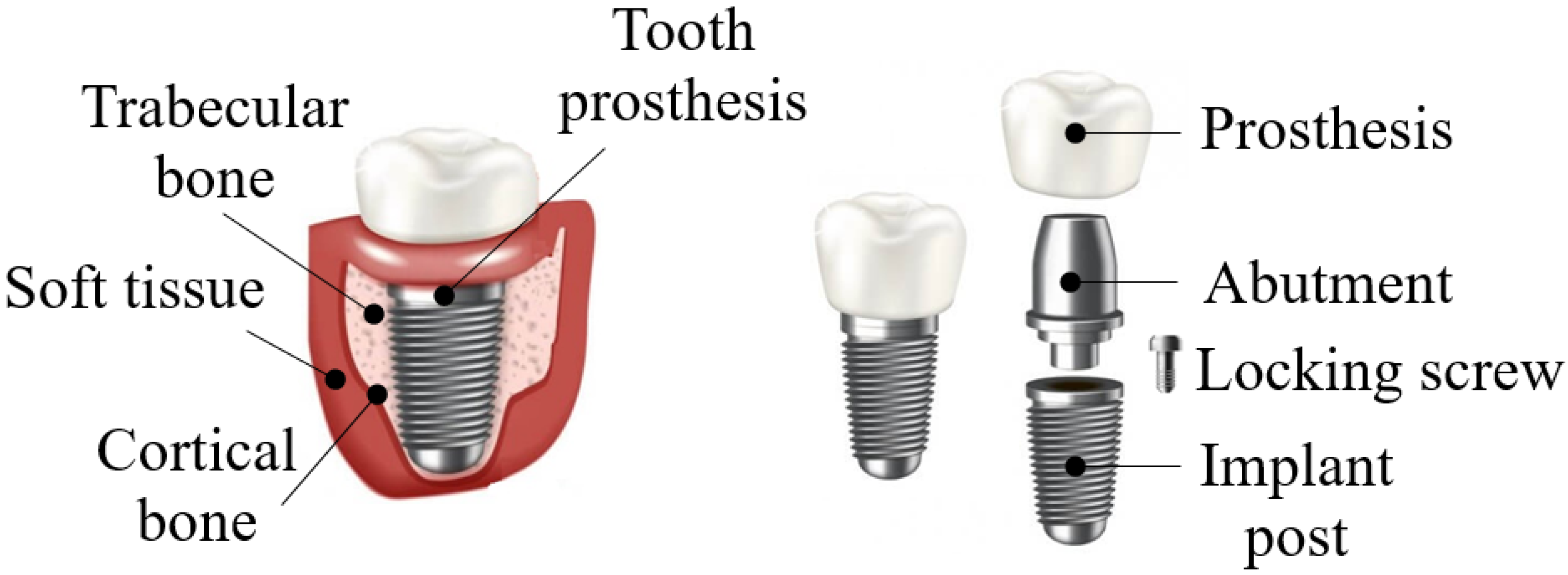
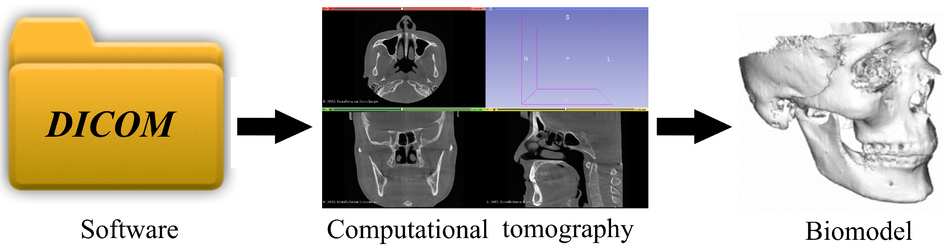
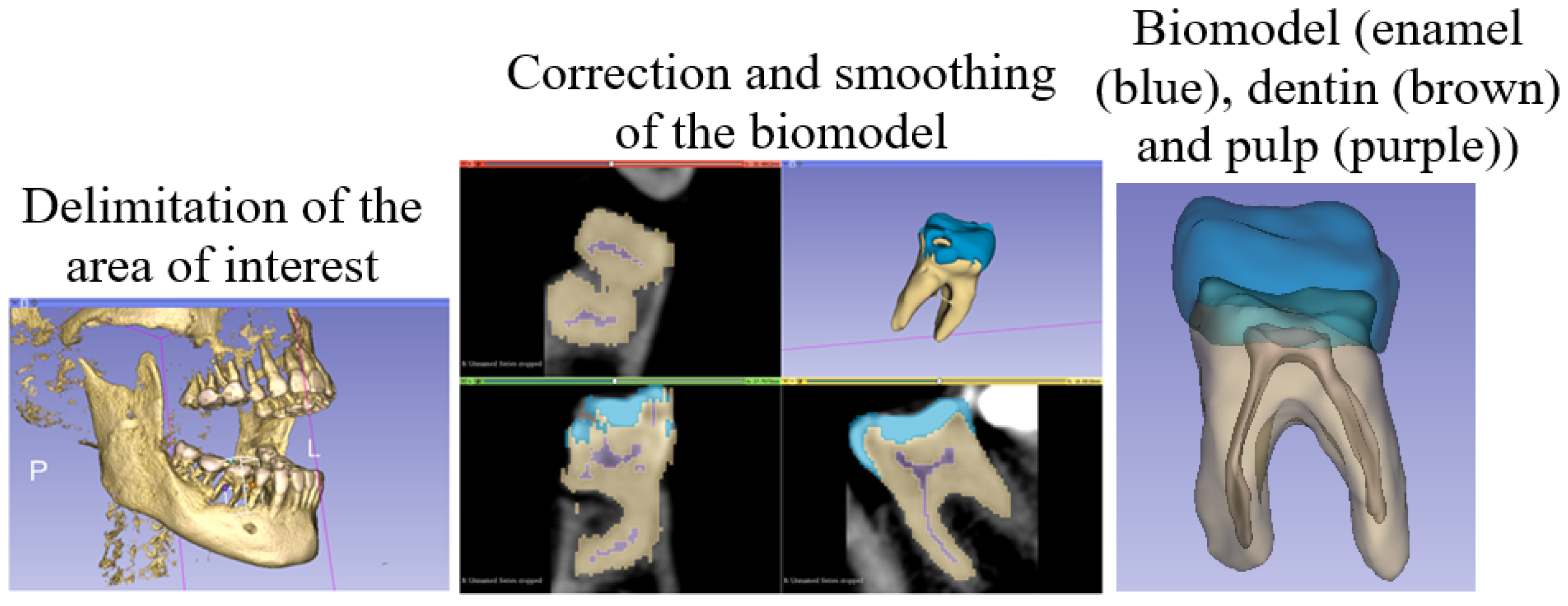
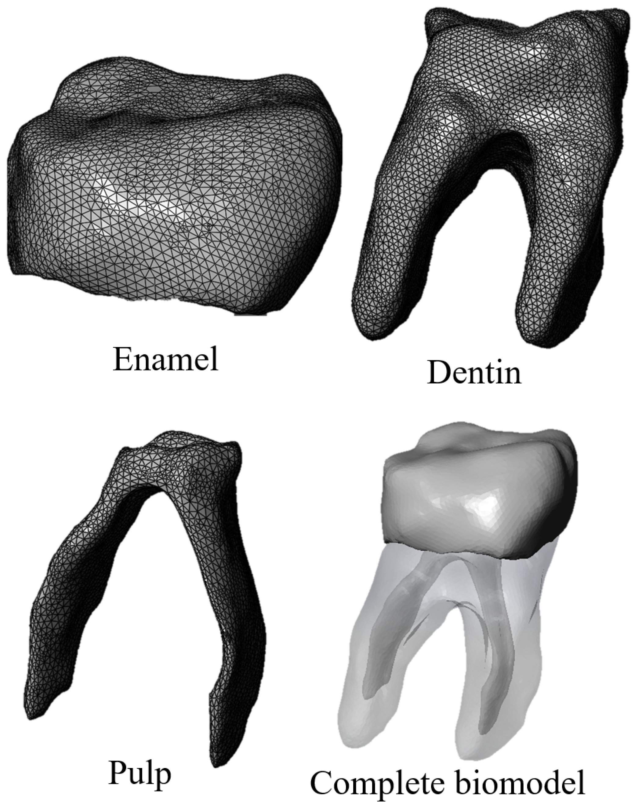
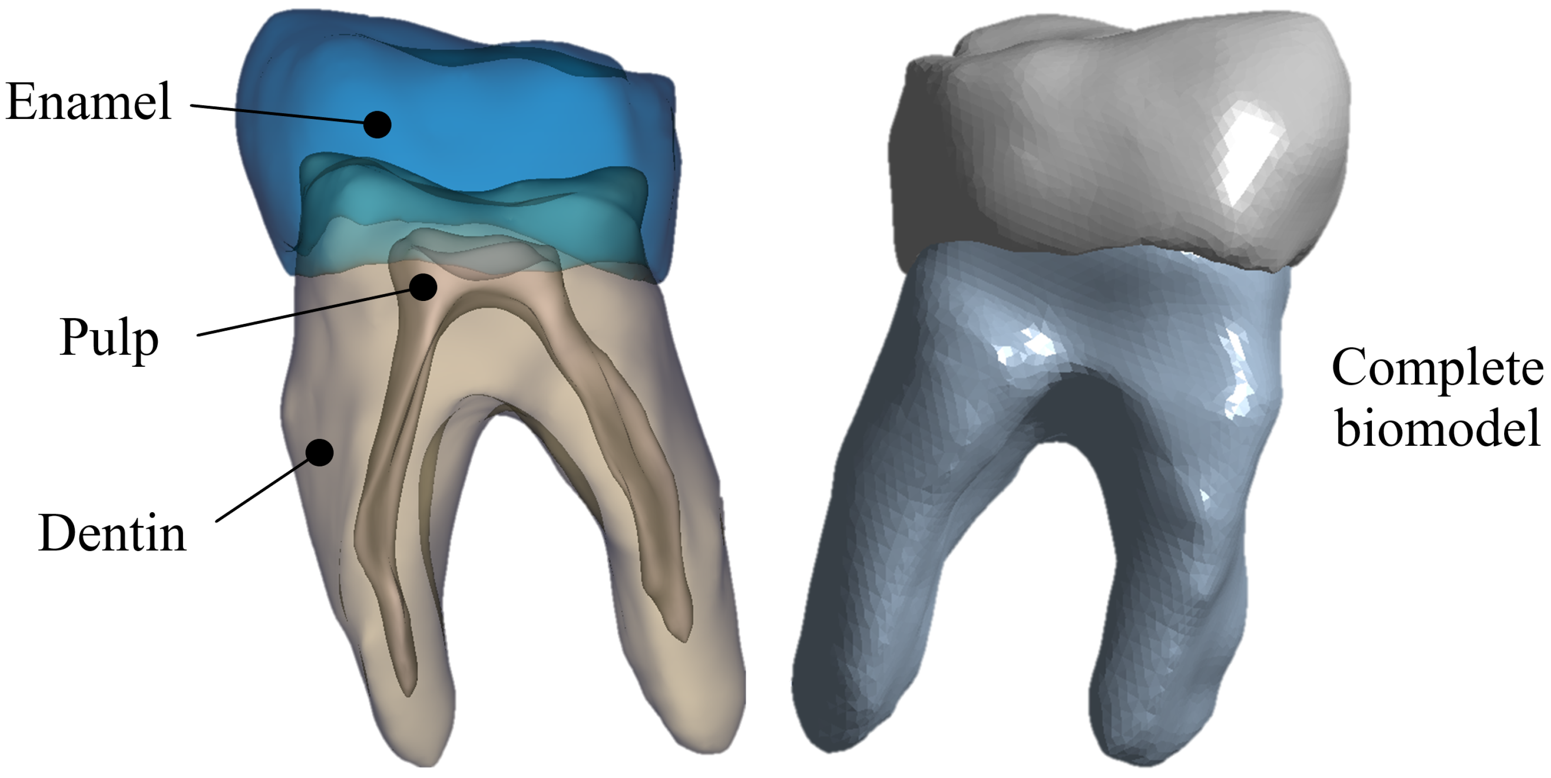
2. Biomodeling Methodology
- Obtaining tomographic images of the anatomical structure to be analyzed.
- Importing images in DICOM format and modeling Figure 3.
- Importing the STL model into a CAD program (student version) to convert it to a solid.
- Assembly of solidified models.
- Export of CAD model to a Finite Element program for the development of the analysis.
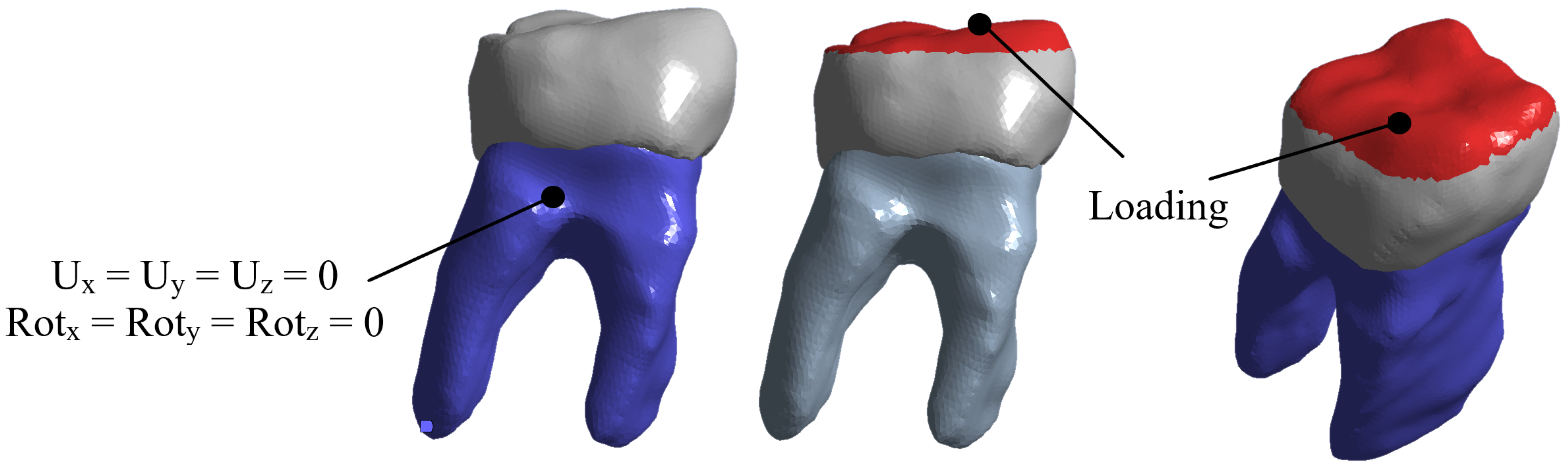
3. Materials and Methods
3.1. First Case of Study
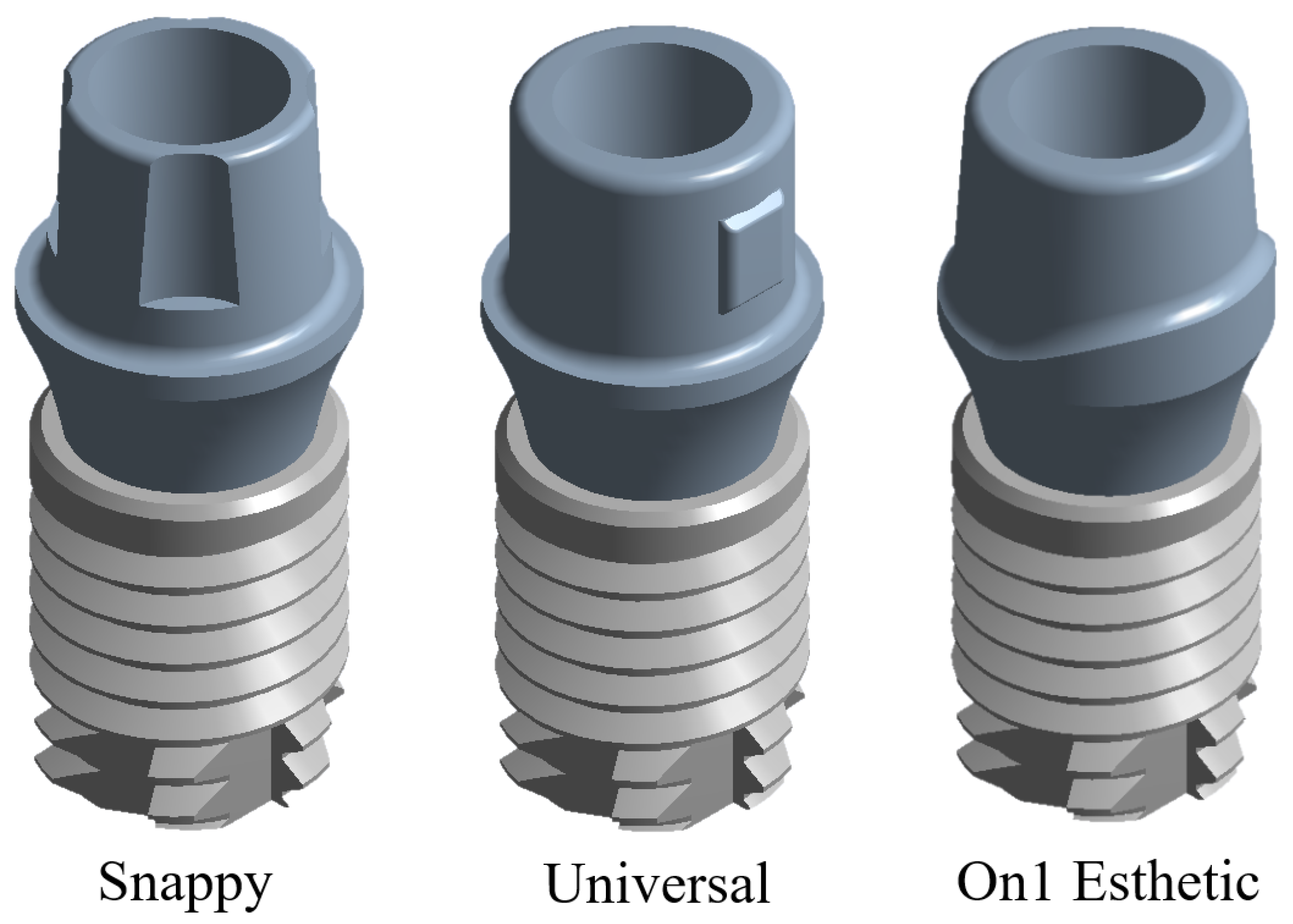
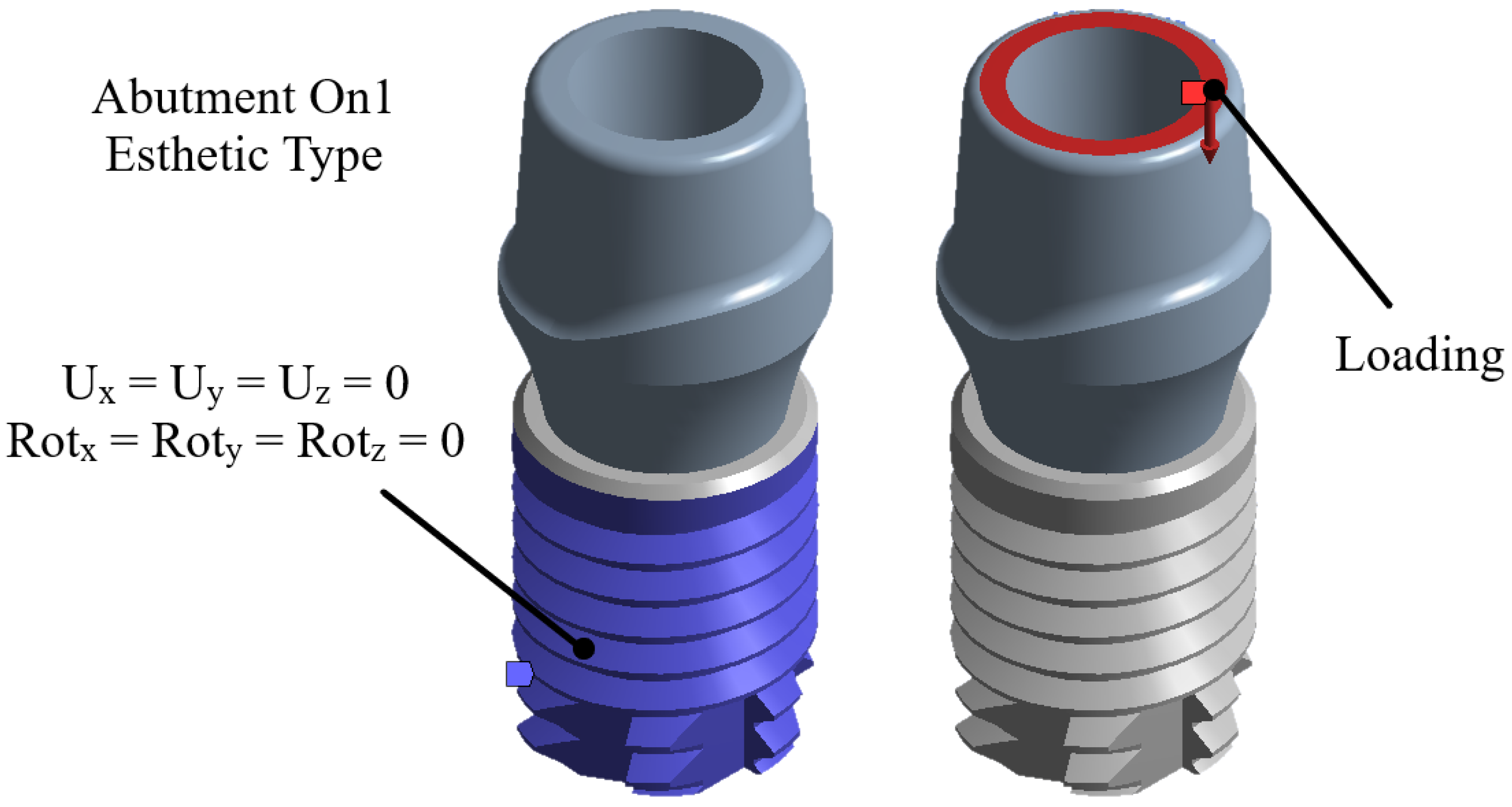
3.2. Second and Third Case Studies
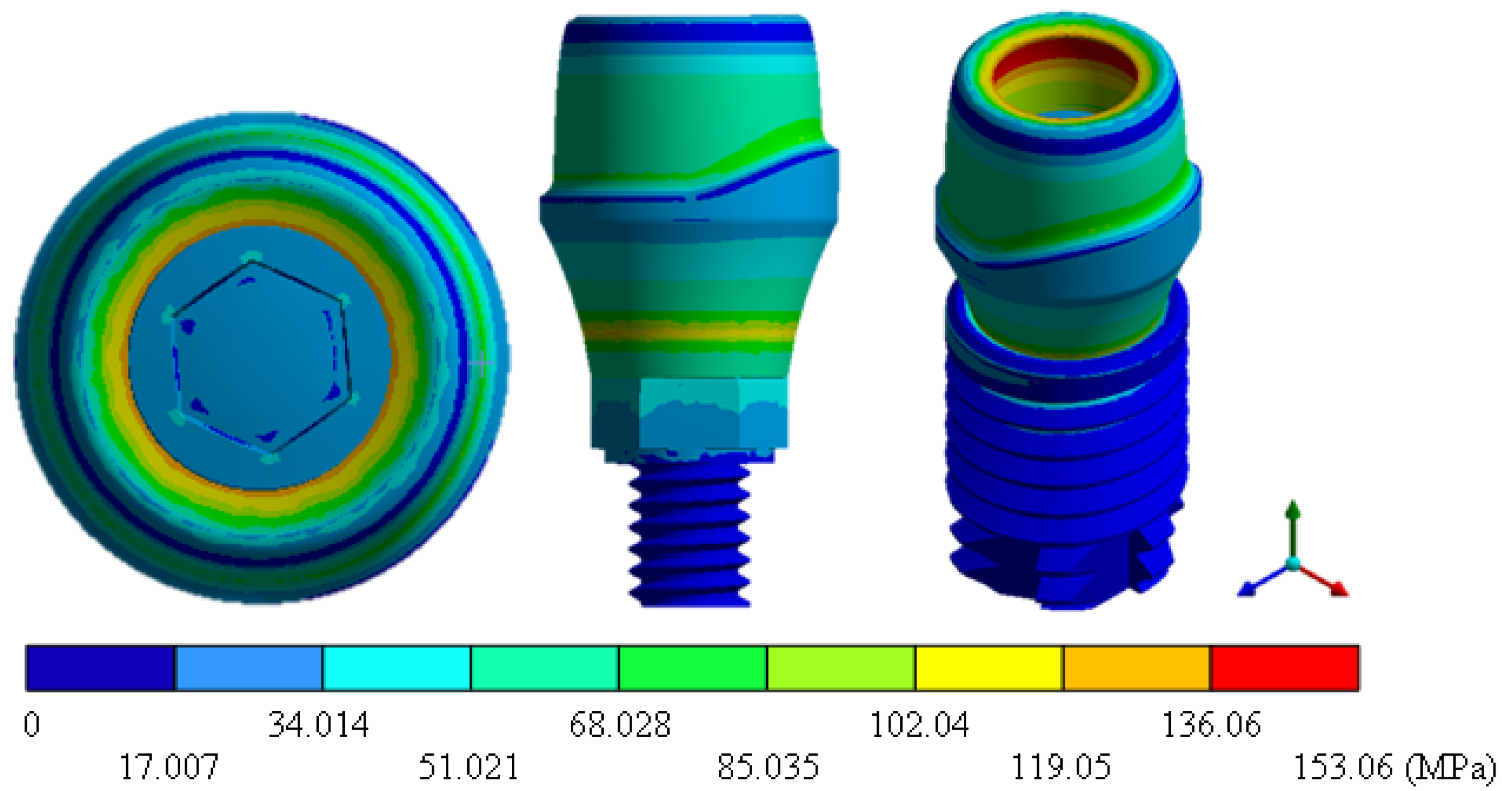
4. Results
5. Discussion
6. Conclusions
- The model developed of the healthy tooth structure has 95% similarity to the morphology of a tooth. In conclusion, a good simulation and model of any organ, tissue, or structure of the human body can be obtained from a computational CT scan.
- Ceramic implants are a great alternative for patients allergic to titanium; they prevent the formation of bacterial plaques and resist acid corrosion. In addition, their osseo-integration behavior and clinical survival rates are just as favorable as titanium implants.
- For both materials, the physical and mechanical properties (titanium and zirconium) allow replacing the tooth structure, fulfilling its 100% functionality.
- The general displacements of zirconium compared to titanium are less, because zirconium absorbs more impact energy and therefore is more tenacious.
- The zirconium implant showed lower resistance to failure compared to the titanium one. However, the difference is not as significant and meets the objective of using ceramic materials instead of metallic ones.
Author Contributions
Funding
Institutional Review Board Statement
Informed Consent Statement
Data Availability Statement
Acknowledgments
Conflicts of Interest
Abbreviations
| CAD | Computer-Aided Design |
| CBTC | Cone Beam Computed Tomography |
| CT | Computed Tomography |
| DICOM | Digital Imaging and Communication in Medicine |
| FEM | Finite Element Method |
| STL | STereoLithography |
Appendix A

| Value | Tooth | TiAlV | ZrO | ||||
|---|---|---|---|---|---|---|---|
| Snappy | Universal | On1 Esthetic | Snappy | Universal | On1 Esthetic | ||
| Total strain (mm/mm) | 0.0080 Max | 0.0023 Max | 0.0014 Max | 0.0013 Max | 0.0012 Max | 0.0009 Max | 0.0007 Max |
| 0 Min | 0 Min | 0 Min | 0 Min | 0 Min | 0 Min | 0 Min | |
| Strain nominal in X (mm/mm) | 0.0045 Max | 0.0010 Max | 0.0007 Max | 0.0007 Max | 0.0004 Max | 0.0003 Max | 0.0004 Max |
| −0.0022 Min | −0.0003 Min | −0.0004 Min | −0.0004 Min | −0.0002 Min | −0.0002 Min | −0.0003 Min | |
| Strain nominal in Y (mm/mm) | 0.0019 Max | 0.0004 Max | 0.0002 Max | 0.0002 Max | 0.0002 Max | 0.0002 Max | 0.0002 Max |
| −0.0072 Min | −0.0023 Min | −0.0014 Min | −0.0013 Min | −0.0012 Min | −0.0008 Min | −0.0007 Min | |
| Strain nominal in Z (mm/mm) | 0.0047 Max | 0.0010 Max | 0.0008 Max | 0.0007 Max | 0.0004 Max | 0.0003 Max | 0.0003 Max |
| −0.0035 Min | −0.0003 Min | −0.0003 Min | −0.0004 Min | −0.0002 Min | −0.0001 Min | −0.0003 Min | |
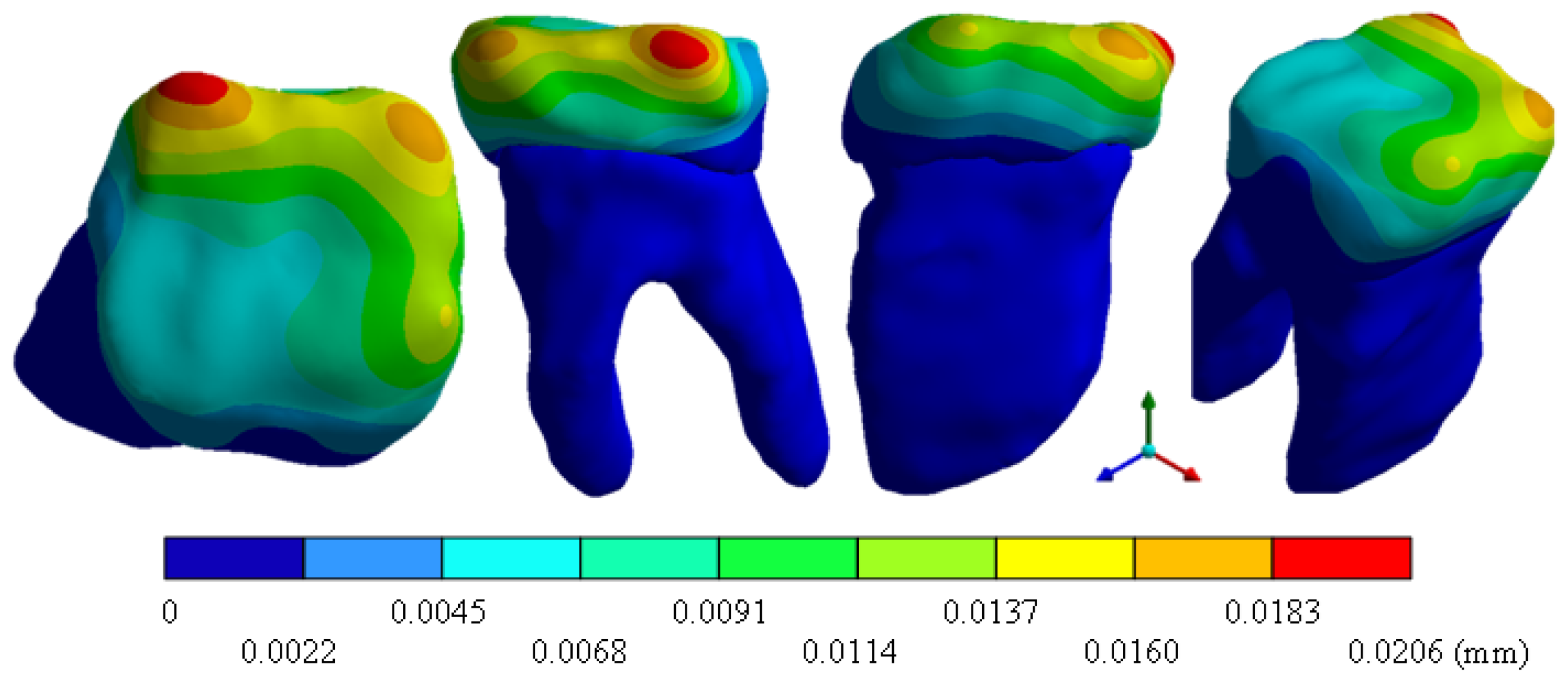
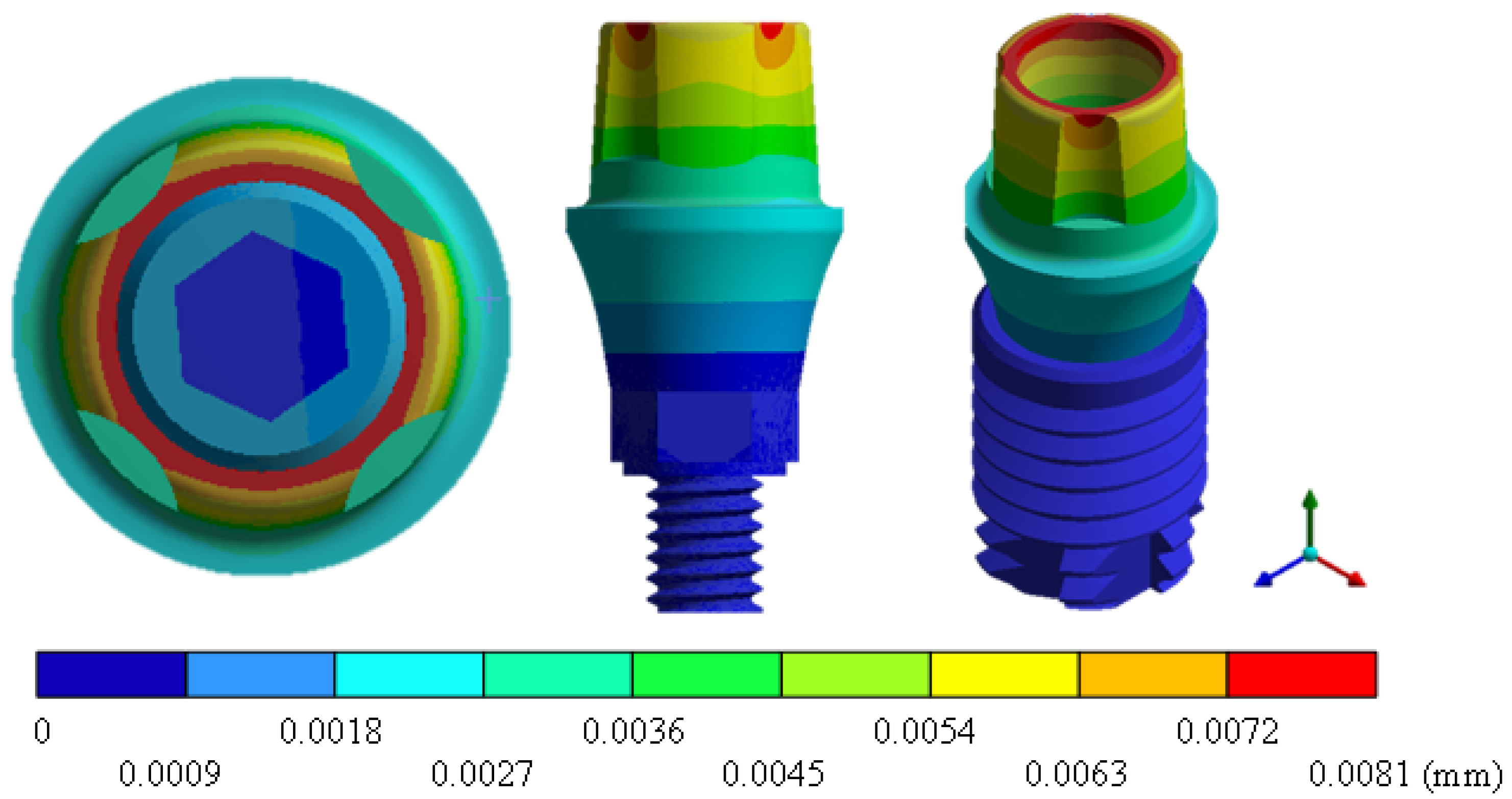
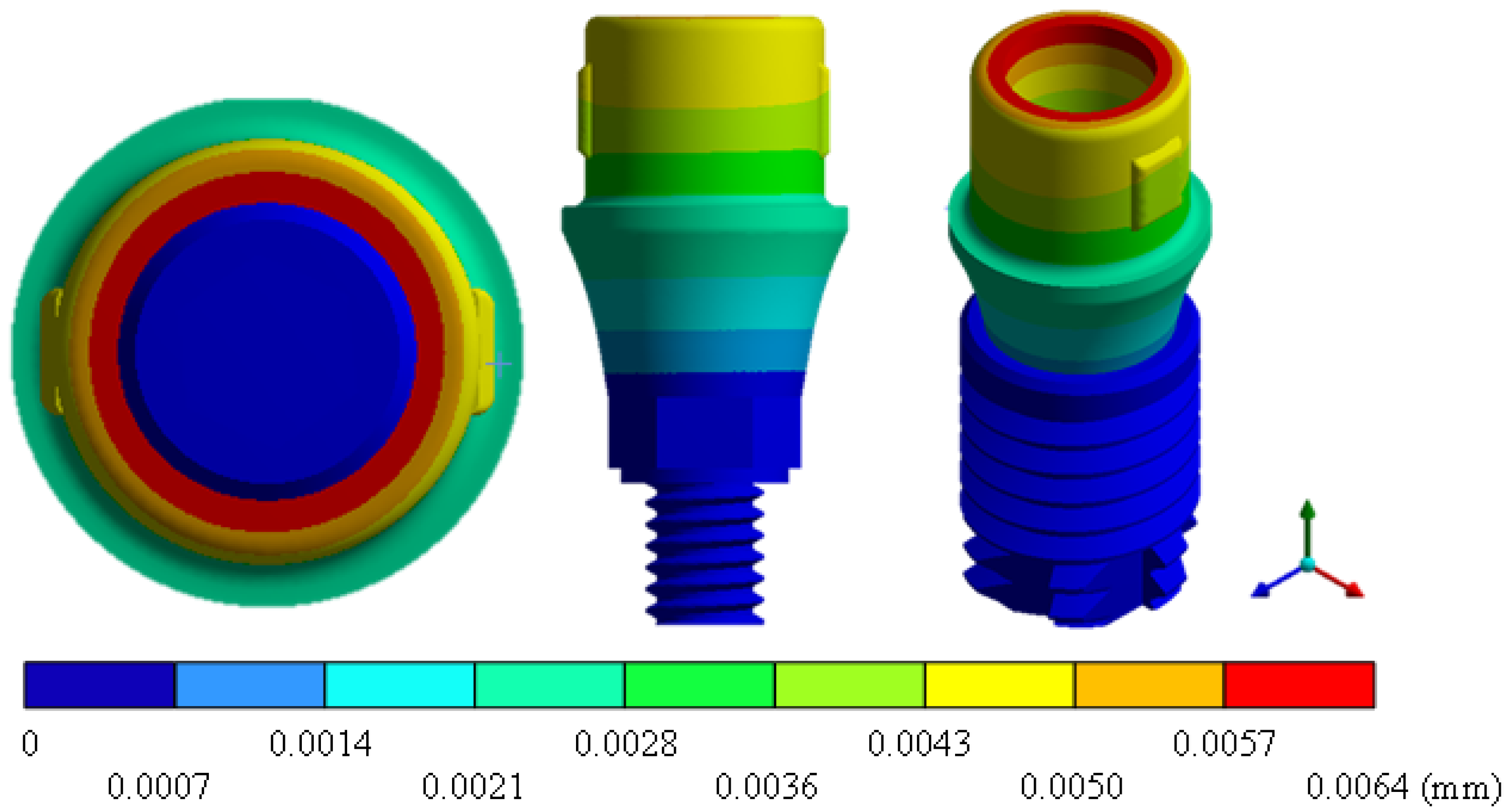
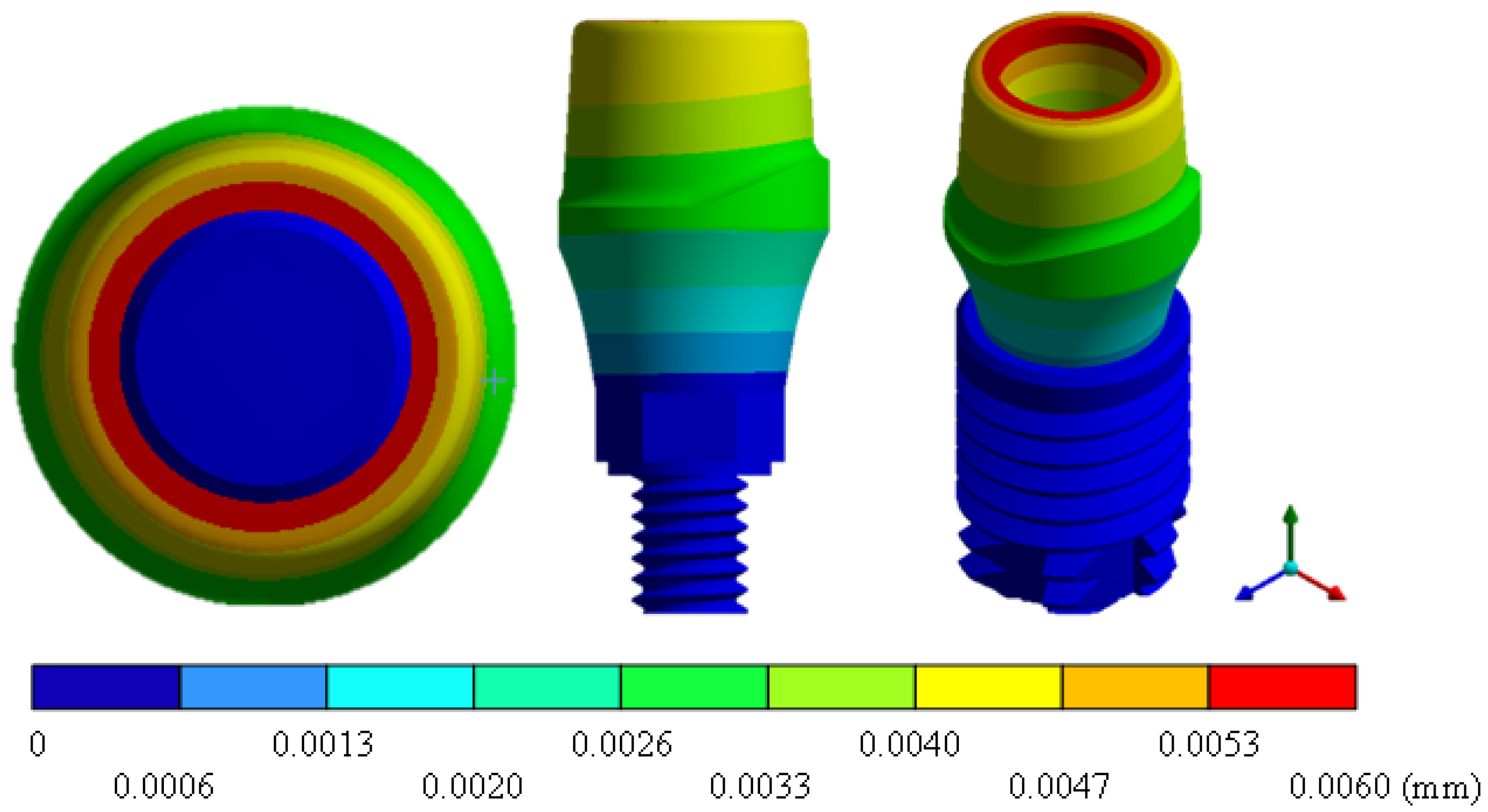
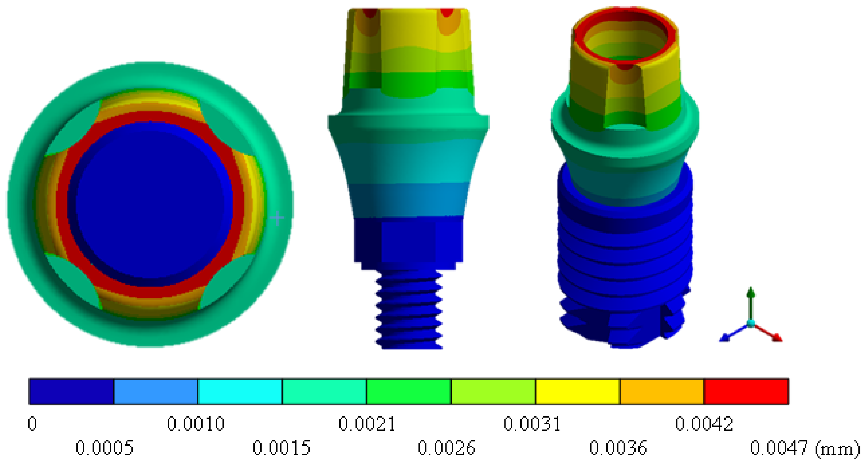
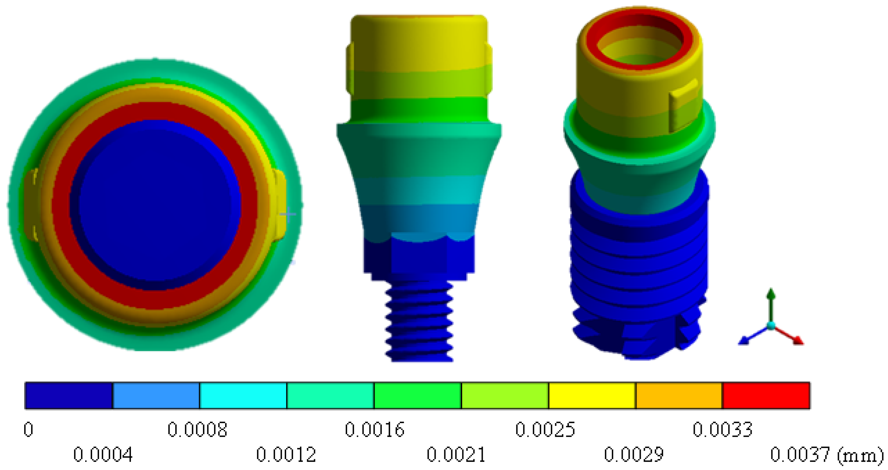
| Total displacement (mm) | 0.0206 Max | 0.0081 Max | 0.0064 Max | 0.0060 Max | 0.0047 Max | 0.0037 Max | 0.0032 Max |
| 0 Min | 0 Min | 0 Min | 0 Min | 0 Min | 0 Min | 0 Min | |
| Displacement nominal X (mm) | 0.0064 Max | 0.0012 Max | 0.0008 Max | 0.0006 Max | 0.0007 Max | 0.0004 Max | 0.0003 Max |
| −0.0025 Min | −0.0014 Min | −0.0009 Min | −0.0009 Min | −0.0008 Min | −0.0005 Min | −0.0006 Min | |
| Displacement nominal Y (mm) | 0.0002 Max | 0.0002 Max | 3.72 × 10 Max | 3.90 × 10 Max | 9.13 × 10 Max | 3.86 × 10 Max | 3.90 × 10 Max |
| −0.0181 Min | −0.0081 Min | −0.0064 Min | −0.0060 Min | −0.0047 Min | −0.0037 Min | −0.0032 Min | |
| Displacement nominal Z (mm) | 0.0016 Max | 0.0013 Max | 0.0009 Max | 0.0008 Max | 0.0008 Max | 0.0004 Max | 0.0004 Max |
| −0.0106 Min | −0.0013 Min | −0.0008 Min | −0.0007 Min | −0.0007 Min | −0.0004 Min | −0.0003 Min | |
| Stress nominal X (MPa) | 188.2 Max | 61.56 Max | 55.02 Max | 57.67 Max | 57.40 Max | 51.1 Max | 58.24 Max |
| −294.1 Min | −139.8 Min | −107.6 Min | −105.5 Min | −137.2 Min | −102.9 Min | −103.1 Min | |
| Stress nominal Y (MPa) | 101.1 Max | 54.79 Max | 33.12 Max | 27.75 Max | 55.33 Max | 30.47 Max | 26.49 Max |
| −617.1 Min | −282.9 Min | −184.2 Min | −174.7 Min | −280.8 Min | −183.7 Min | −202.8 Min | |
| Stress nominal Z (MPa) | 242.9 Max | 61.62 Max | 54.99 Max | 56.49 Max | 57.43 Max | 50.40 Max | 46.56 Max |
| −468.7 Min | −136.4 Min | −103.2 Min | −107.7 Min | −133.4 Min | −98.39 Min | −104.7 Min | |
| Maximum principal stress (MPa) | 310.1 Max | 63.81 Max | 57.94 Max | 58.02 Max | 58.86 Max | 55.64 Max | 58.75 Max |
| −291.2 Min | −75.78 Min | −58.21 Min | −52.13 Min | −75.85 Min | −57.96 Min | −54.95 Min | |
| Minimum principal stress (MPa) | 36.28 Max | 11.67 Max | 9.80 Max | 11.94 Max | 9.83 Max | 8.56 Max | 11.84 Max |
| −710.7 Min | −282.9 Min | −198.8 Min | −174.8 Min | −280.8 Min | −194.9 Min | −204.8 Min | |
| Shear stress XY (MPa) | 126.8 Max | 51.80 Max | 61.25 Max | 42.55 Max | 52.75 Max | 62.27 Max | 39.88 Max |
| −228.5 Min | −53.07 Min | −55.85 Min | −40.43 Min | −53.74 Min | −56.46 Min | −41.32 Min | |
| Shear stress YZ (MPa) | 225.5 Max | 54.07 Max | 46.70 Max | 41.63 Max | 55.11 Max | 47.67 Max | 40.67 Max |
| −116.6 Min | −50.04 Min | −40.76 Min | −40.73 Min | −50.79 Min | −42.01 Min | −41.27 Min | |
| Shear stress XZ (MPa) | 114.4 Max | 131.4 Max | 51.80 Max | 49.81 Max | 127.5 Max | 50.44 Max | 49.28 Max |
| −106.3 Min | −130.8 Min | −51.48 Min | −49.90 Min | −127.3 Min | −50.14 Min | −49.33 Min | |
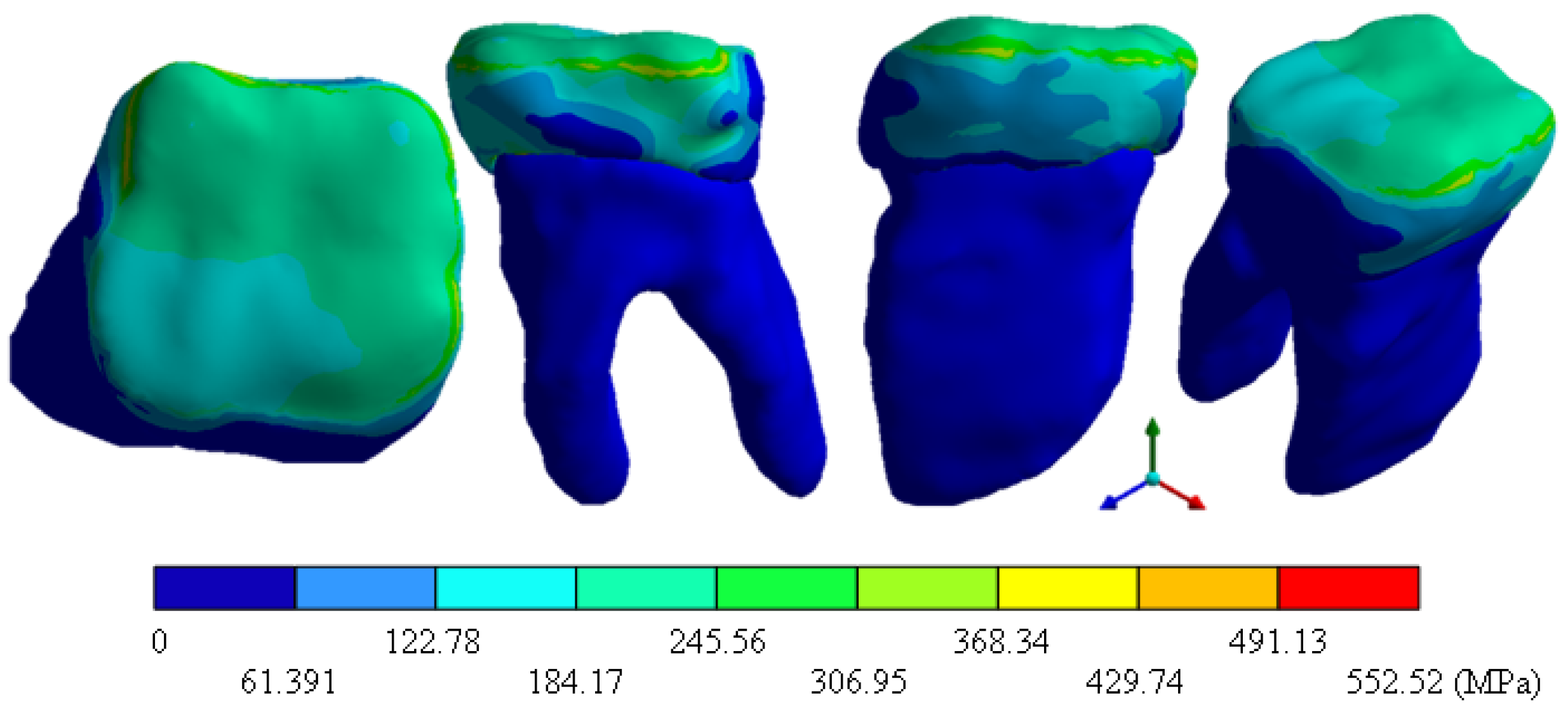
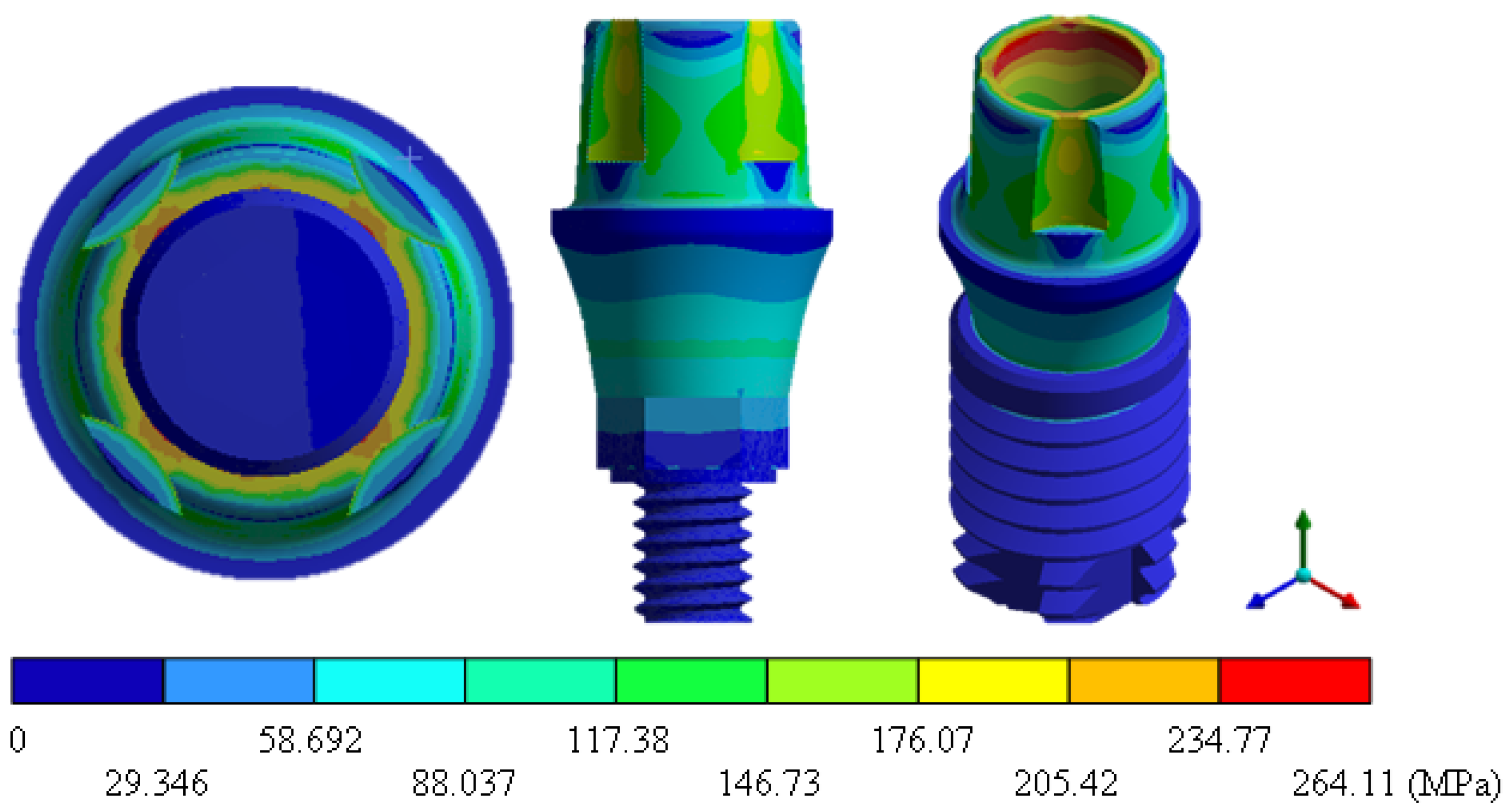
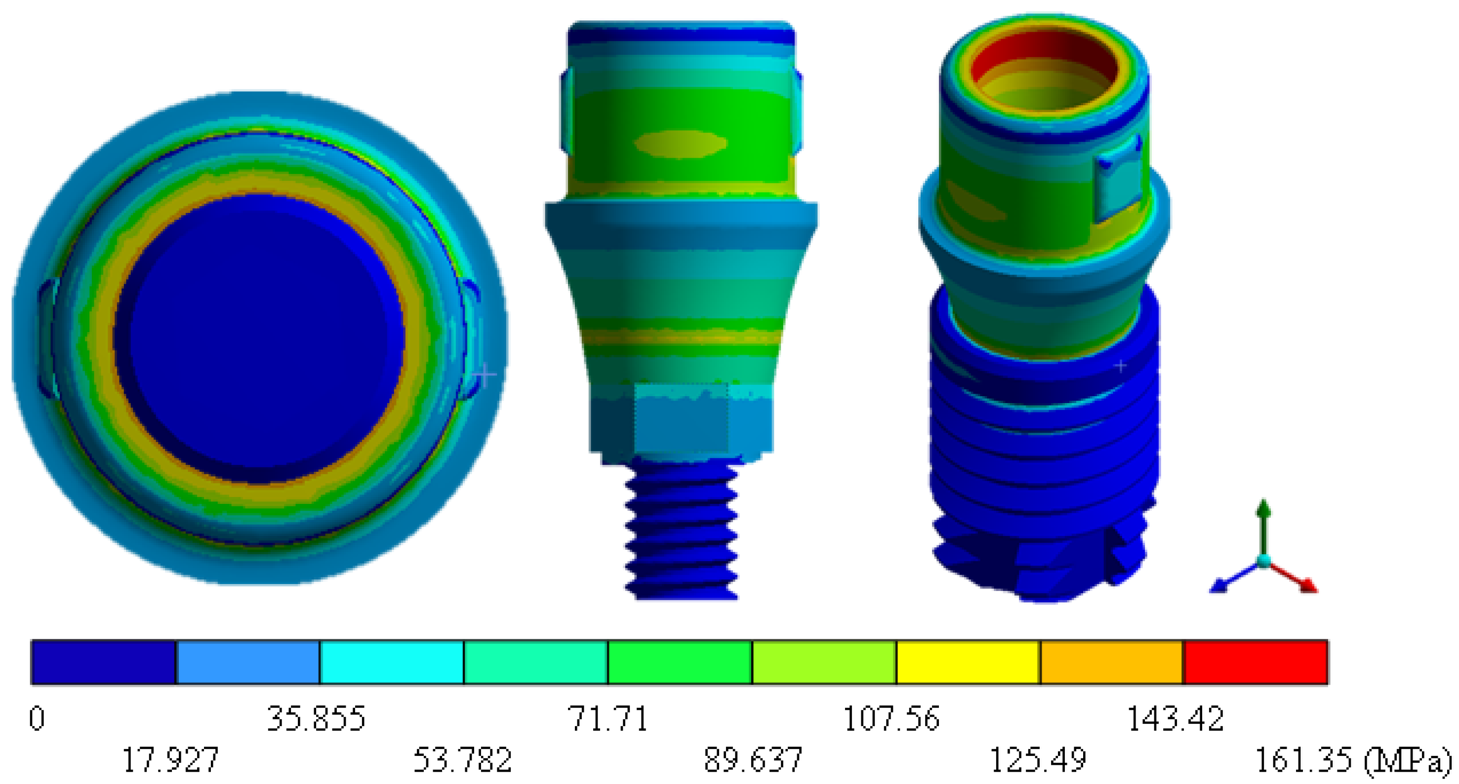
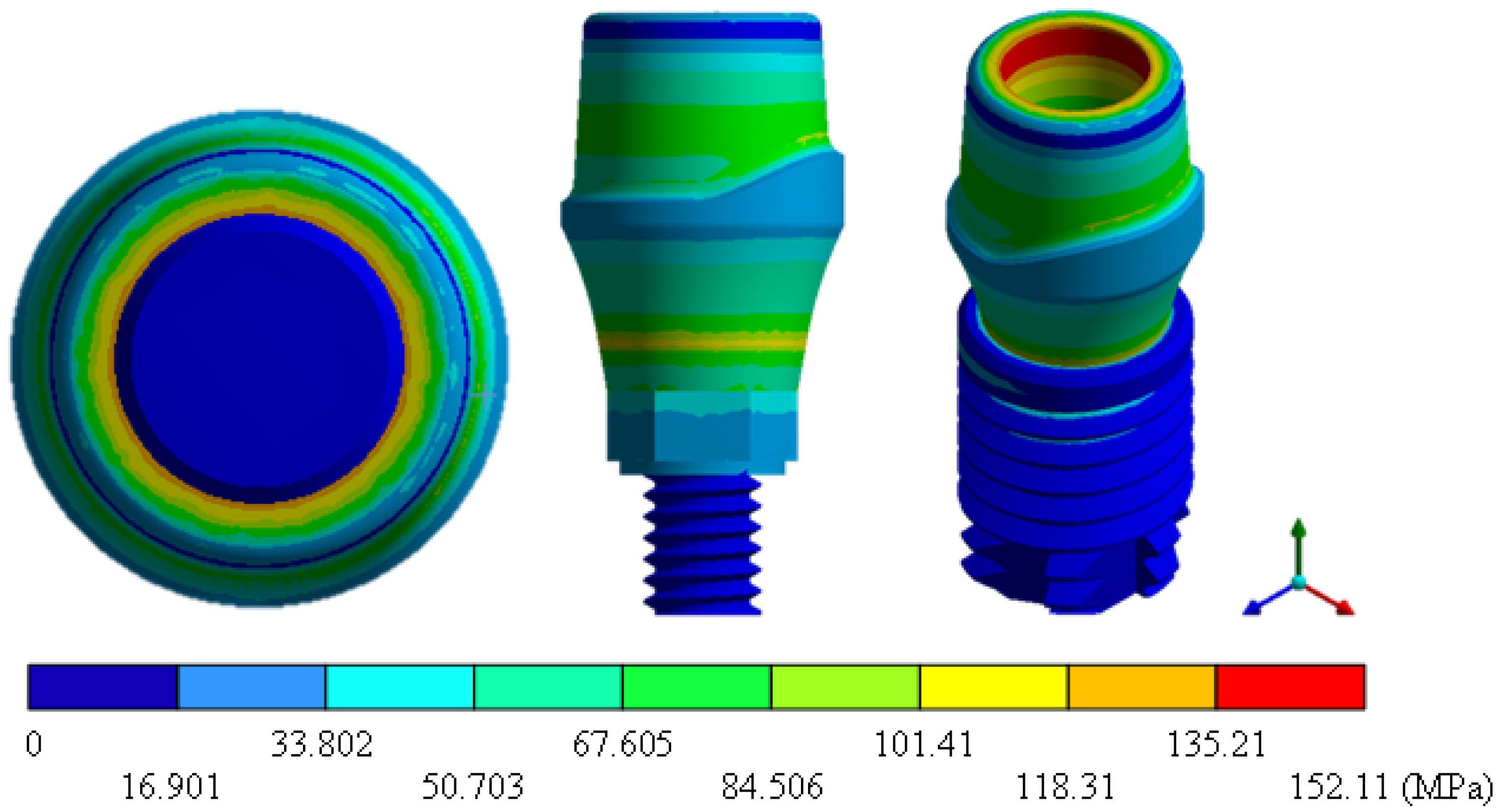
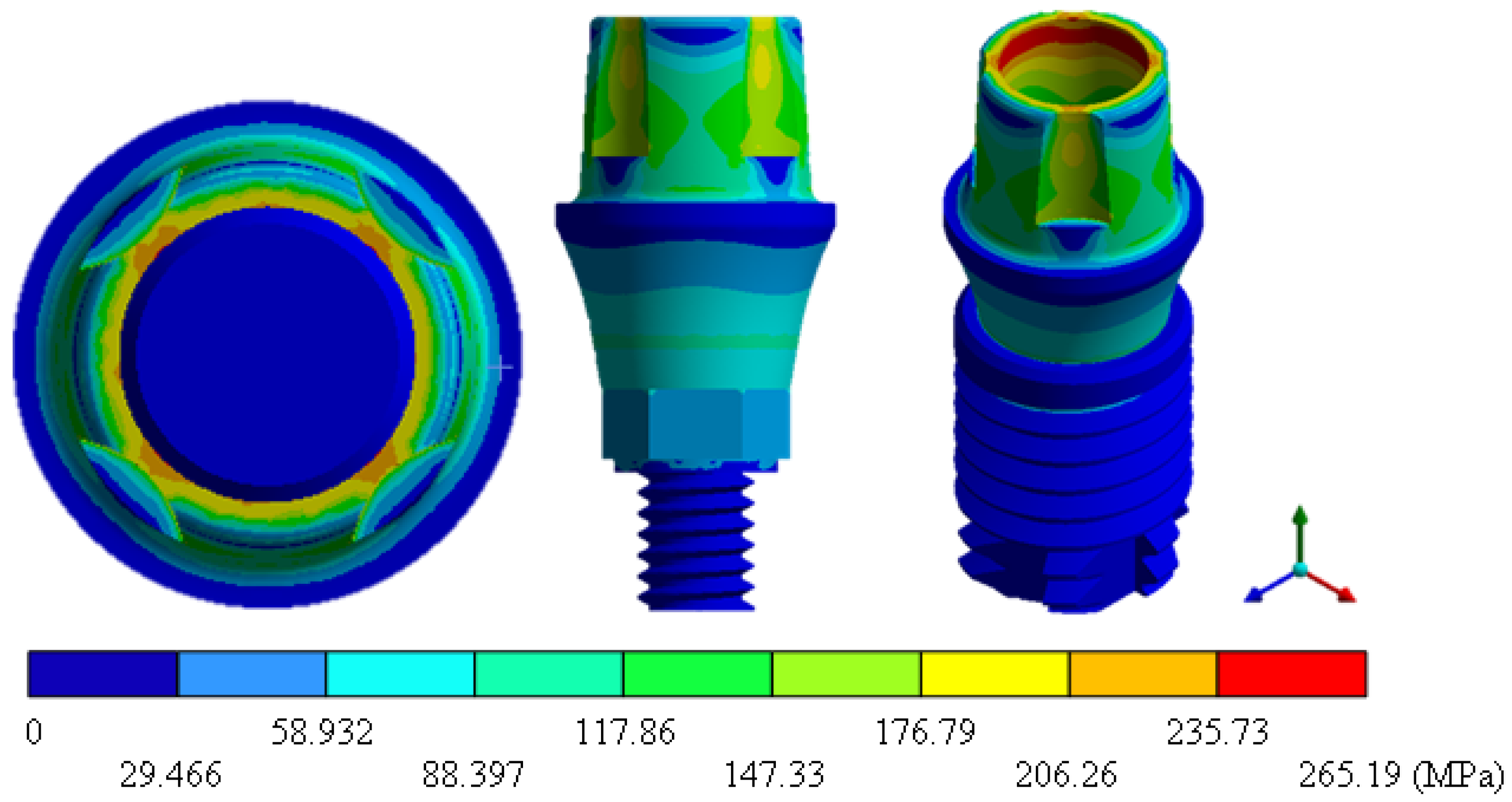
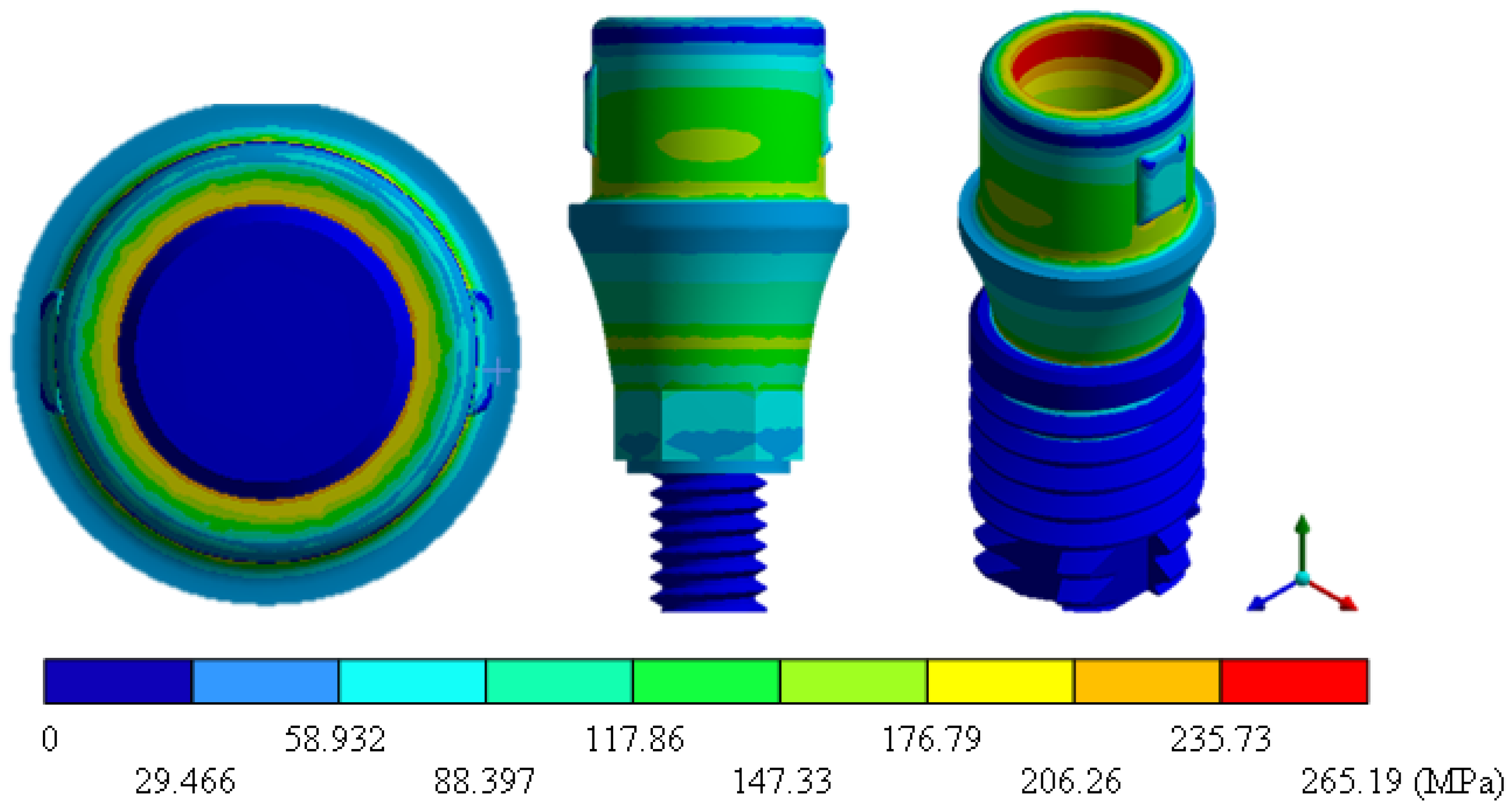
| Von Mises Stress (MPa) | 552.5 Max | 264.1 Max | 161.3 Max | 152.1 Max | 265.1 Max | 162.0 Max | 157.6 Max |
| 0 Min | 0 Min | 0 Min | 0 Min | 0 Min | 0 Min | 0 Min | |
References
- Haugen, H.J.; Chen, H. Is there a Better Biomaterial for Dental Implants than Titanium?—A Review and Meta-Study Analysis. J. Funct. Biomater. 2022, 13, 46. [Google Scholar] [CrossRef] [PubMed]
- Coli, P.; Jemt, T. On marginal bone level changes around dental implants. Clin. Implant. Dent. Relat. Res. 2021, 23, 159–169. [Google Scholar] [CrossRef] [PubMed]
- Kim, J.J.; Lee, J.H.; Kim, J.C.; Lee, J.B.; Yeo, I.S.L. Biological responses to the transitional area of dental implants: Material-and structure-dependent responses of peri-implant tissue to abutments. Materials 2019, 13, 72. [Google Scholar] [CrossRef] [Green Version]
- Bohara, S.; Suthakorn, J. Surface coating of orthopedic implant to enhance the osseointegration and reduction of bacterial colonization: A review. Biomater. Res. 2022, 26, 26. [Google Scholar] [CrossRef]
- Hao, C.P.; Cao, N.J.; Zhu, Y.H.; Wang, W. The osseointegration and stability of dental implants with different surface treatments in animal models: A network meta-analysis. Sci. Rep. 2021, 11, 13849. [Google Scholar] [CrossRef]
- Kim, J.C.; Lee, M.; Yeo, I.S.L. Three interfaces of the dental implant system and their clinical effects on hard and soft tissues. Mater. Horizons 2022, 9, 1387–1411. [Google Scholar] [CrossRef] [PubMed]
- Prados-Privado, M. Predicción de vida a la fatiga de implantes dentales y sus conexiones protésicas. Un estudio de elementos finitos probabilistas. Rev. Esp. Odontoestomatol. Implant. 2018, 22, 9–14. [Google Scholar]
- Hernández, J.L. Efecto del Tipo de Comportamiento de Hueso en la Biomecánica de Implantes Dentales. Bachelor’s Thesis, Universidad de Zaragoza, Zaragoza, Spain, 2019; pp. 7–10. [Google Scholar]
- Kligman, S.; Ren, Z.; Chung, C.H.; Perillo, M.A.; Chang, Y.C.; Koo, H.; Li, C. The impact of dental implant surface modifications on osseointegration and biofilm formation. J. Clin. Med. 2021, 10, 1641. [Google Scholar] [CrossRef]
- Eftekhar-Ashtiani, R.; Alam, M.; Tavakolizadeh, S.; Abbasi, K. The role of biomaterials and biocompatible materials in implant-supported dental prosthesis. Evid.-Based Complement. Altern. Med. 2021, 2021, 3349433. [Google Scholar] [CrossRef]
- Nicholson, J.W. Titanium alloys for dental implants: A review. Prosthesis 2020, 2, 100–116. [Google Scholar] [CrossRef]
- Chopra, D.; Jayasree, A.; Guo, T.; Gulati, K.; Ivanovski, S. Advancing dental implants: Bioactive and therapeutic modifications of zirconia. Bioact. Mater. 2022, 13, 161–178. [Google Scholar] [CrossRef] [PubMed]
- Nicola, M.; Francesco, M.; Santo, C. Ceramic materials as an alternative to titanium for dental implant fabrication. In Dental Implantology and Biomaterial; IntechOpen: London, UK, 2016; pp. 91–105. [Google Scholar]
- Comisso, I.; Arias-Herrera, S.; Gupta, S. Zirconium dioxide implants as an alternative to titanium: A systematic review. J. Clin. Exp. Dent. 2021, 13, e511–e519. [Google Scholar] [CrossRef] [PubMed]
- Mihai, L.L.; Parlatescu, I.; Gheorghe, C.; Andreescu, C.; Bechir, A.; Pacurar, M.; Cumpata, C.N. In vitro study of the effectiveness to fractures of the aesthetic fixed restorations achieved from zirconium and alumina. Rev. Chim. 2014, 65, 725–729. [Google Scholar]
- Nistor, L.; Grădinaru, M.; Rîcă, R.; Mărășescu, P.; Stan, M.; Manolea, H.; Ionescu, A.; Moraru, I. Zirconia use in dentistry—Manufacturing and properties. Curr. Health Sci. J. 2019, 45, 28–35. [Google Scholar] [PubMed]
- Bona, A.D.; Pecho, O.E.; Alessandretti, R. Zirconia as a Dental Biomaterial. Materials 2015, 8, 4978–4991. [Google Scholar] [CrossRef] [Green Version]
- Lohfeld, S.; Barron, V.; McHugh, P.E. Biomodels of bone: A review. Ann. Biomed. Eng. 2005, 33, 1295–1311. [Google Scholar] [CrossRef]
- Marquet-Rivera, R.A.; Urriolagoitia-Sosa, G.; Hernández-Vázquez, R.A.; Romero-Ángeles, B.; Mastache-Miranda, O.A.; Urriolagoitia-Calderón, G. High Biofidelity 3D Biomodel Reconstruction from Soft and Hard Tissues (Knee), FEM, and 3D Printing: A Three-Dimensional Methodological Proposal. BioMed Res. Int. 2021, 2021, 6688164. [Google Scholar] [CrossRef]
- Venkatesh, E.; Ellure, S.V. Cone beam computed tomography: Basics and applications in dentistry. J. Istanb. Univ. Fac. Dent. 2017, 51 (Suppl. S1), S102–S121. [Google Scholar] [CrossRef]
- Mastache-Miranda, O.A.; Urriolagoitia-Sosa, G.; Marquet-Rivera, R.A. Three-dimensional reconstruction for use in medicine and biomechanics. MOJ Appl. Bionics Biomech. 2018, 2, 310–311. [Google Scholar]
- Hernández-Vázquez, R.A.; Urriolagoitia-Sosa, G.; Marquet-Rivera, R.A.; Romero-Ángeles, B.; Mastache-Miranda, O.A.; Urriolagoitia-Calderón, G. Numerical analysis of a dental zirconium restoration and the stresses that occur in dental tissues. Appl. Bionics Biomech. 2019, 2019, 1049306. [Google Scholar] [CrossRef]
- Hernández-Vázquez, R.A.; Marquet-Rivera, R.A.; Romero-Ángeles, B.; Egure-Hidalgo, M.; Aburto-Barrera, J.M.; Urriolagoitia-Sosa, G. Clinical application of the finite element method in dental practice. In Proceedings of the 16th Congreso Nacional de Ingeniería Electromecánica y de Sistemas, Mexico City, Mexico, 13–17 November 2017. [Google Scholar]
- Marquet-Rivera, R.A. Numerical Analysis of Anterior Cruciate Ligament Injury in Three Different Degrees of Damage. Master’s Thesis, Instituto Politécnico Nacional, SEPI ESIME Zacatenco, Mexico City, Mexico, 2017. [Google Scholar]
- Mastache-Miranda, O.A. Modeling by Means of Tomography of Volumes of Bone Structures for Numerical Simulation Under the Action of Loads and/or External Agents. Master’s Thesis, Instituto Politécnico Nacional, SEPI ESIME Zacatenco, Mexico City, Mexico, 2016. [Google Scholar]
- Hernández-Vázquez, R.A.; Romero-Ángeles, B.; Urriolagoitia-Sosa, G.; Vázquez-Feijoo, J.A.; Vázquez-López, A.J. Urriolagoitia-Calderón, G. Numerical Analysis of Masticatory Forces on a Lower First Molar considering the Contact between Dental Tissues. Appl. Bionics Biomech. 2018, 2018, 4196343. [Google Scholar] [CrossRef] [PubMed] [Green Version]
- Urriolagoitia-Sosa, G. Analysis of Prior Strain History Effect on Mechanical Properties and Residual Stresses in Beams. Ph.D. Thesis, Oxford Brookes University, Oxford, UK, 2005. [Google Scholar]
- Marquet-Rivera, R.A.; Urriolagoitia-Sosa, G.; Romero-Ángeles, B.; Vázquez-Feijoo, J.A.; Urriolagoitia-Calderón, G. Computational biomodelling and numerical analysis as means of diagnostic and odontological prognosis. MOJ Appl. Bionics Biomech. 2018, 2, 262–263. [Google Scholar]
- Nobel Biocare. 2022. Available online: https://www.nobelbiocare.com/es-mx (accessed on 9 September 2022).
- Henao, J.I.A.; Ossa, J.A.V.; Correa, F.L. Evaluación de la micro-deformación de la zona maxilar anterior con regeneración; Análisis de elementos finitos. Odontol. Sanmarquina 2019, 22, 110–116. [Google Scholar] [CrossRef]
- Hanawa, T. Zirconia versus titanium in dentistry: A review. Dent. Mater. J. 2020, 39, 24–36. [Google Scholar] [CrossRef] [PubMed]


| Material | Young’s Modulus (MPa) | Poisson Ratio | Density (Kg/m3) |
|---|---|---|---|
| Enamel | 70,000 | 0.30 | 250 |
| Dentin | 18,300 | 0.30 | 310 |
| Pulp | 2000 | 0.45 | 100 |
| Material | Young’s Modulus (MPa) | Poisson Ratio | Density (Kg/m3) | Elastic Limit (MPa) | Hardness (Hv) | Fracture Toughness (MPa m1/2) |
|---|---|---|---|---|---|---|
| Titanium | 114 | 0.36 | 4430 | 1100 | 320 | 50 |
| Zirconium | 210 | 0.31 | 6100 | 900 | 1200 | 6–8 |
| Total General Displacement (mm) | Von Mises Stress (MPa) | |||
|---|---|---|---|---|
| Min | Max | Min | Max | |
| Tooth (Molar) | 0 | 0.0206 | 0 | 552.52 |
| Implant Snappy (TiAlV) | 0 | 0.0081 | 0 | 264.11 |
| Implant Universal (TiAlV) | 0 | 0.0064 | 0 | 161.35 |
| Implant On1 Esthetic (TiAlV) | 0 | 0.0060 | 0 | 152.11 |
| Implant Snappy (ZrO) | 0 | 0.0047 | 0 | 265.19 |
| Implant Universal (ZrO) | 0 | 0.0037 | 0 | 162.01 |
| Implant On1 Esthetic (ZrO) | 0 | 0.0032 | 0 | 157.65 |
Publisher’s Note: MDPI stays neutral with regard to jurisdictional claims in published maps and institutional affiliations. |
© 2022 by the authors. Licensee MDPI, Basel, Switzerland. This article is an open access article distributed under the terms and conditions of the Creative Commons Attribution (CC BY) license (https://creativecommons.org/licenses/by/4.0/).
Share and Cite
Martinez-Mondragon, M.; Urriolagoitia-Sosa, G.; Romero-Ángeles, B.; Maya-Anaya, D.; Martínez-Reyes, J.; Gallegos-Funes, F.J.; Urriolagoitia-Calderón, G.M. Numerical Analysis of Zirconium and Titanium Implants under the Effect of Critical Masticatory Load. Materials 2022, 15, 7843. https://doi.org/10.3390/ma15217843
Martinez-Mondragon M, Urriolagoitia-Sosa G, Romero-Ángeles B, Maya-Anaya D, Martínez-Reyes J, Gallegos-Funes FJ, Urriolagoitia-Calderón GM. Numerical Analysis of Zirconium and Titanium Implants under the Effect of Critical Masticatory Load. Materials. 2022; 15(21):7843. https://doi.org/10.3390/ma15217843
Chicago/Turabian StyleMartinez-Mondragon, Miguel, Guillermo Urriolagoitia-Sosa, Beatriz Romero-Ángeles, Daniel Maya-Anaya, Jacobo Martínez-Reyes, Francisco Javier Gallegos-Funes, and Guillermo Manuel Urriolagoitia-Calderón. 2022. "Numerical Analysis of Zirconium and Titanium Implants under the Effect of Critical Masticatory Load" Materials 15, no. 21: 7843. https://doi.org/10.3390/ma15217843