Effects of Copper Metallic Nanoparticles on Structural and Optical Properties of Antimony Phosphate Glasses Co-Doped with Samarium Ions

 and
and
Abstract
:1. Introduction
2. Materials and Methods
3. Results and Discussion
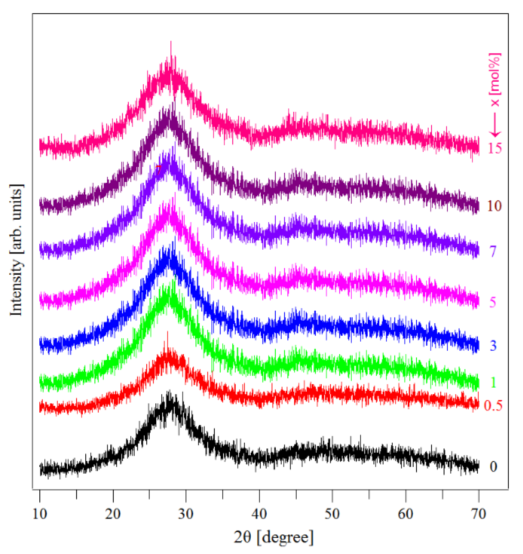
3.1. XRD Data
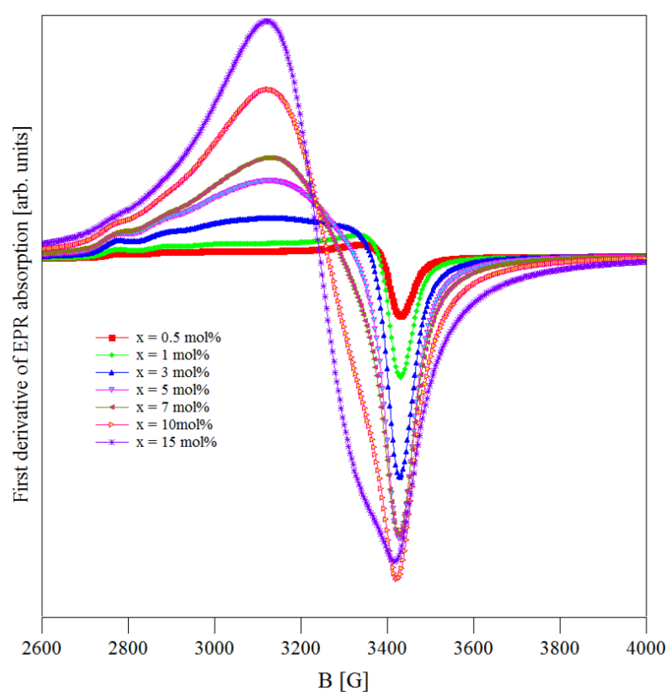
3.2. EPR Data
3.3. UV–Vis Data
3.4. Luminescence Data
- 562 nm assigned to the 4G5/2 → to the 6H5/2 ground state transition; it is called also the zero-zero band and represents a forbidden transition;
- 599 nm assigned to the 4G5/2 → to the 6H7/2 (excited level) transition, a magnetic dipole transition;
- 643 nm assigned to the 4G5/2 → to the 6H9/2 (excited level) transition, an electric dipole transition.
4. Conclusions
Author Contributions
Funding
Acknowledgments
Conflicts of Interest
References
- Som, T.; Karmakar, B. Nano silver: Antimony glass hybrid nanocomposite and their enhanced flourescence application. Solid State Sci. 2011, 13, 887–895. [Google Scholar] [CrossRef]
- Minelly, J.; Ellison, A. Applications of antimony-silicate glasses for fiber optic amplifiers. Opt. Fiber Technol. 2002, 8, 123–138. [Google Scholar] [CrossRef]
- Nalin, M.; Poulain, M.; Poulain, M.; Ribeiro, S.J.L.; Messaddeq, Y. Antimony oxide based glasses. J. Non-Cryst. Solids 2001, 284, 110–116. [Google Scholar] [CrossRef]
- Franco, D.F.; Carvajal, E.E.; Donoso, J.P.; Silva, M.A.P.; Sant’Ana, A.C.; Fares, H.; Magon, C.J.; Nalin, M. Structural and EPR studies of Cu2+ ions in NaPO3-Sb2O3-CuO. J. Non-Cryst. Solids 2019, 503–504, 169–175. [Google Scholar] [CrossRef]
- Ouannes, K.; Lebbou, K.; Walsh, B.M.; Poulain, M.; Alombert-Goget, G.; Guyot, Y. New Er3+ doped antimony oxide based glasses: Thermal analysis, structural and spectral properties. J. Alloy. Compd. 2015, 649, 564–572. [Google Scholar] [CrossRef]
- Ouannes, K.; Lebbou, K.; Walsh, B.M.; Poulain, M.; Alombert-Goget, G.; Guyot, Y. Antimony oxide based glasses, novel laser materials. Opt. Mater. 2017, 65, 8–14. [Google Scholar] [CrossRef]
- Ouannes, K.; Soltani, M.T.; Poulain, M.; Boulon, G.; Alombert-Goget, G.; Guyot, Y.; Pillonnet, A.; Lebbou, K. Spectroscopic properties of Er3+-doped antimony oxide glass. J. Alloy. Compd. 2014, 603, 132–135. [Google Scholar] [CrossRef] [Green Version]
- Franco, D.F.; Sant’Ana, A.C.; De Oliveira, L.F.C.; Silva, M.A.P. The Sb2O3 redox route to obtain copper nanoparticles in glasses with plasmonic properties. J. Mater. Chem. C 2015, 3, 3803–3808. [Google Scholar] [CrossRef]
- Machado, T.M.; Silva, M.A.P. The reduction of tellurium in binary glasses in the system TeO2-Sb2O3. Mater. Chem. Phys. 2017, 201, 86–91. [Google Scholar] [CrossRef]
- Kesavulu, C.R.; Jayasankar, C.K. Spectroscopic properties of Sm3+ ions in lead fluorophosphate glasses. J. Lumin. 2012, 132, 2802–2809. [Google Scholar] [CrossRef]
- Kindrat, I.I.; Padlyak, B.V.; Drzewiecki, A. Luminescence properties of the Sm-doped borate glasses. J. Lumin. 2015, 166, 264–275. [Google Scholar] [CrossRef]
- Herrera, A.; Fernandes, R.G.; de Camargo, A.S.S.; Hernandes, A.C.; Buchner, S.; Jacinto, C.; Balzaretti, N.M. Visible-NIR emission and structural properties of Sm3+ doped heavy-metal oxide glass with composition B2O3-PbO-Bi2O3-GeO2. J. Lumin. 2016, 171, 106–111. [Google Scholar] [CrossRef]
- Som, T.; Karmakar, B. Enhancement of Er3+ upconverted luminescence in Er3+: Au-antimony glass dichroic nanocomposites containing hexagonal Au nanoparticles. JOSA B 2009, 26, B21–B27. [Google Scholar] [CrossRef]
- Ghoshal, S.K.; Sahar, M.R.; Dousti, M.R.; Arifin, R.; Rohani, M.; Hamzah, K. A model for enhanced up-conversion luminescence in Erbium-doped tellurite glass containing Silver nanoparticles. Adv. Mater. Res. Trans. Tech. Publ. 2012, 501, 61–65. [Google Scholar] [CrossRef]
- Mahraz, Z.A.S.; Sahar, M.R.S.; Ghoshal, K.; Dousti, M.R.; Amjad, R.J. Silver nanoparticles enhanced luminescence of Er3+ ions in boro-tellurite glasses. Mater. Lett. 2013, 112, 136–138. [Google Scholar] [CrossRef]
- Chen, S.; Akai, T.; Kadono, K.; Yazawa, T. Reversible control of silver nanoparticle generation and dissolution in soda-lime silicate glass through x-ray irradiation and heat treatment. Appl. Phys. Lett. 2001, 79, 3687–3689. [Google Scholar] [CrossRef]
- Chen, S.; Akai, T.; Kadono, K.; Yazawa, T. A silver-containing. Chem. Commun. 2001, 20, 2090–2091. [Google Scholar] [CrossRef]
- Zeng, H.; Qiu, J.; Ye, Z.; Zhu, C.; Gan, F. Irradiation assisted fabrication of gold nanoparticles-doped glasses. J. Cryst. Growth 2004, 267, 156–160. [Google Scholar] [CrossRef]
- Culea, E.; Pascuta, P.; Pustan, M.; Tamas-Gavrea, D.R.; Pop, L.; Vida-Simiti, I. Effects of Eu: Ag codoping on structural magnetic and mechanical properties of lead tellurite glass ceramics. J. Non-Cryst. Solids 2015, 408, 18–25. [Google Scholar] [CrossRef]
- Bolundut, L.; Culea, E.; Borodi, G.; Stefan, R.; Munteanu, C.; Pascuta, P. Influence of Sm3+: Ag codoping on structural and spectroscopic properties of lead tellurite glass ceramics. Ceram. Int. 2015, 41, 2931–2939. [Google Scholar] [CrossRef]
- Bolundut, L.; Pop, L.; Bosca, M.; Tothazan, N.; Borodi, G.; Culea, E.; Pascuta, P.; Stefan, R. Structural, spectroscopic and magnetic properties of Nd3+ doped lead tellurite glass ceramics containing silver. J. Alloy. Comp. 2017, 692, 934–940. [Google Scholar] [CrossRef]
- Bosca, M.; Pop, L.; Bolundut, L.; Tothazan, N.; Borodi, G.; Vida-Simiti, I.; Stefan, R.; Popa, A.; Culea, E.; Pascuta, P. Effects of Gd3+: Ag co-doping on structural and magnetic properties of lead tellurite glass ceramics. Ceram. Int. 2016, 42, 1169–1176. [Google Scholar] [CrossRef]
- Ciorcas, F.; Mendiratta, S.K.; Ardelean, I.; Valente, M.A. Structural and magnetic studies of CuO-TeO2 and CuO-TeO2-B2O3 glasses. Eur. Phys. J. B 2001, 20, 235–240. [Google Scholar]
- Sandhya Rani, P.; Singh, R. Electrical and magnetic properties of copper tellurite glasses. J. Mater. Sci. 2010, 45, 2868–2873. [Google Scholar] [CrossRef]
- Brant, A.T.; Halliburton, L.E.; Basun, S.A.; Grabar, A.A.; Odoulov, S.G.; Shumelyuk, A.; Giles, N.C.; Evans, D.R. Photoinduced EPR study of Sb2+ ions in photorefractive Sn2P2S6 crystals. Phys. Rev. B 2012, 86, 134109. [Google Scholar] [CrossRef]
- Schreurs, J.W.H.; Davis, D.H. EPR spectrum of Sb4+ in a silicate glass. J. Chem. Phys. 1979, 71, 557. [Google Scholar] [CrossRef]
- Imagawa, H. ESR studies of cupric ion in various oxide glasses. Phys. Status Solidi 1968, 30, 469–478. [Google Scholar] [CrossRef]
- Kawazoe, H.; Hosono, H.; Kanazawa, T. ESR and optical absorption of Cu2+ in Na2O-SiO2 glasses. J. Non-Cryst. Solids 1979, 33, 103–115. [Google Scholar]
- Griscom, D.L. Electron spin resonance in glasses. J. Non-Cryst. Solids 1980, 40, 211–272. [Google Scholar] [CrossRef]
- Deka, U.; Lezcano-Gonzalez, I.; Weckhuysen, B.M.; Beale, A.M. Local environment and nature of Cu active sites in Zeolite-Based Catalysts for the selective catalytic reduction of NOx. ACS Catal. 2013, 3, 413–427. [Google Scholar] [CrossRef]
- Ardelean, I.; Peteanu, M.; Simon, V.; Ciorcas, F.; Ioncu, V. Structural and magnetic investigations of the xCuO(100-x)[70TeO2·25B2O3·5SrF2] glasses. Appl. Phys. A Mater. Sci. Process. 2001, 73, 481–484. [Google Scholar] [CrossRef]
- Andronenko, S.I.; Andronenko, R.R.; Vasiliev, A.V.; Zagrebelnyi, O.A. Local symmetry of Cu2+ ions in sodium silicate glasses from data of EPR spectroscopy. Glas. Phys. Chem. 2004, 30, 230–235. [Google Scholar] [CrossRef]
- Dehelean, A.; Popa, A.; Rada, S.; Culea, E. EPR and magnetic characterization of Fe2O3-TeO2 and CuO-TeO2 glasses obtained by melt-quenching and sol-gel proccesses. J. Magn. Magn. Mater. 2015, 381, 131–137. [Google Scholar] [CrossRef]
- Zamyatin, O.A.; Plotnichenko, V.G.; Churbanov, M.F.; Zamyatina, E.V.; Karzanov, V.V. Optical properties of zinc tellurite glasses doped with Cu2+ions. J. Non-Cryst. Solids 2018, 480, 81–89. [Google Scholar] [CrossRef]
- Zhang, L.; Peng, M.; Dong, G.; Qiu, J. Spectroscopic properties of Sm3+-doped phosphate glasses. J. Mater. Res. 2012, 27, 2111–2115. [Google Scholar] [CrossRef]
- Carnall, W.T.; Fields, P.R.; Rajnak, K. Electronic energy levels in the trivalent lanthanide aquo ions. I. Pr3+, Nd3+, Pm3+, Sm3+, Dy3+, Ho3+, Er3+ and Tm3+. J. Chem. Phys. 1968, 49, 4424–4442. [Google Scholar] [CrossRef]
- Som, T.; Karmakar, B. Infrared-to-red upconversion luminescence in samarium-doped antimony glasses. J. Lumin. 2008, 128, 1989–1996. [Google Scholar] [CrossRef]
- Bae, B.S.; Weinberg, M.C. Optical absorption of copper phosphate glasses in the visible spectrum. J. Non-Cryst. Solids 1994, 168, 223–231. [Google Scholar] [CrossRef]
- Kaufmann, J.; Russel, C. Thermodynamics of the Cu+/Cu2+- redox equilibrium in aluminosilicate melts. J. Non-Cryst. Solids 2010, 356, 1615–1619. [Google Scholar] [CrossRef]
- Sinha, S.P. Complexes of the Rare Earths; Pergamon Press: Oxford, UK, 1966. [Google Scholar]
- Sa-Ardsin, W.; Yasaka, P.; Kaewkhao, J.; Boonin, K. Luminescence and optical properties of Li2O:Gd2O3:B2O3:Sm2O3 glasses system. Adv. Mater. Res. 2014, 979, 479–482. [Google Scholar] [CrossRef]
- Thomas, S.; George, R.; Rasool, S.N.; Rathaiah, M.; Venkatramu, V.; Joseph, C.; Unnikrishman, N. Optical properties of Sm3+ ions in zinc potassium fluorophosphate glasses. Opt. Mater. 2013, 36, 242–250. [Google Scholar] [CrossRef]
- Zhang, L.Y.; Hu, L.L.; Jiang, Z.H. Yb3+ doped fluorophosphate glasses: A good candidate for high energy, ultra short pulse, tunable fiber lasers. Prog. Phys. 2003, 23, 473–483. [Google Scholar]
- Douglas Faza, F.; Fares, H.; de Souza, A.E.; Santagneli, S.E.; Nalin, M. Glass formation and the structural study of the Sb2O3-SbPO4-WO3 system. Eclética Quim. 2017, 42, 51–59. [Google Scholar]


| x (mol %) | g|| | g⊥ | g |
|---|---|---|---|
| 0.5 | 2.398 | 2.083 | |
| 1 | 2.404 | 2.087 | |
| 3 | 2.407 | 2.097 | |
| 5 | 2.410 | 2.104 | |
| 7 | 2.145 | ||
| 10 | 2.161 | ||
| 15 | 2.174 |
Publisher’s Note: MDPI stays neutral with regard to jurisdictional claims in published maps and institutional affiliations. |
© 2020 by the authors. Licensee MDPI, Basel, Switzerland. This article is an open access article distributed under the terms and conditions of the Creative Commons Attribution (CC BY) license (http://creativecommons.org/licenses/by/4.0/).
Share and Cite
Pascuta, P.; Stefan, R.; Olar, L.E.; Bolundut, L.C.; Culea, E. Effects of Copper Metallic Nanoparticles on Structural and Optical Properties of Antimony Phosphate Glasses Co-Doped with Samarium Ions. Materials 2020, 13, 5040. https://doi.org/10.3390/ma13215040
Pascuta P, Stefan R, Olar LE, Bolundut LC, Culea E. Effects of Copper Metallic Nanoparticles on Structural and Optical Properties of Antimony Phosphate Glasses Co-Doped with Samarium Ions. Materials. 2020; 13(21):5040. https://doi.org/10.3390/ma13215040
Chicago/Turabian StylePascuta, Petru, Razvan Stefan, Loredana Elena Olar, Liviu Calin Bolundut, and Eugen Culea. 2020. "Effects of Copper Metallic Nanoparticles on Structural and Optical Properties of Antimony Phosphate Glasses Co-Doped with Samarium Ions" Materials 13, no. 21: 5040. https://doi.org/10.3390/ma13215040