The Potential Selective Cytotoxicity of Poly (L- Lactic Acid)-Based Scaffolds Functionalized with Nanohydroxyapatite and Europium (III) Ions toward Osteosarcoma Cells


 and
and
Abstract
:
1. Introduction
2. Materials and Methods
2.1. Biomaterial Preparation
2.2. Cell Cultures
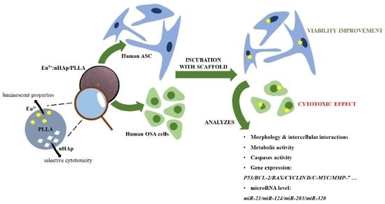
2.3. The Experiment
2.4. Analysis of Biomaterial Impact on Cells’ Morphology and Eu3+ Ions-Doped nHAp Colocalization
2.5. Analysis of Biomaterial Impact on Cells’ Adhesion and Intercellular Interactions
2.6. Analysis of Biomaterial Impact on Cells’ Metabolic Activity
2.6.1. MTS Test
2.6.2. Mitochondria Depolarization Status
2.7. Analysis of Biomaterial Impact on Caspases Activation
2.8. Analysis of Biomaterial Impact on Genes Expression Involved in Apoptosis and Cell Cycle
2.9. Analysis of Biomaterial Impact at the miRNA Level
2.10. Statistical Analysis
3. Results
3.1. Biomaterial Impact on Cells’ Morphology
3.2. Biomaterial Impact on Cells’ Adhesion and Intercellular Interaction
3.3. Analysis of Cells Viability Based on Caspase Activation
3.4. Biomaterial Impact on Cells’ Metabolic Activity
3.5. Biomaterial Impact on Transcriptional Activity
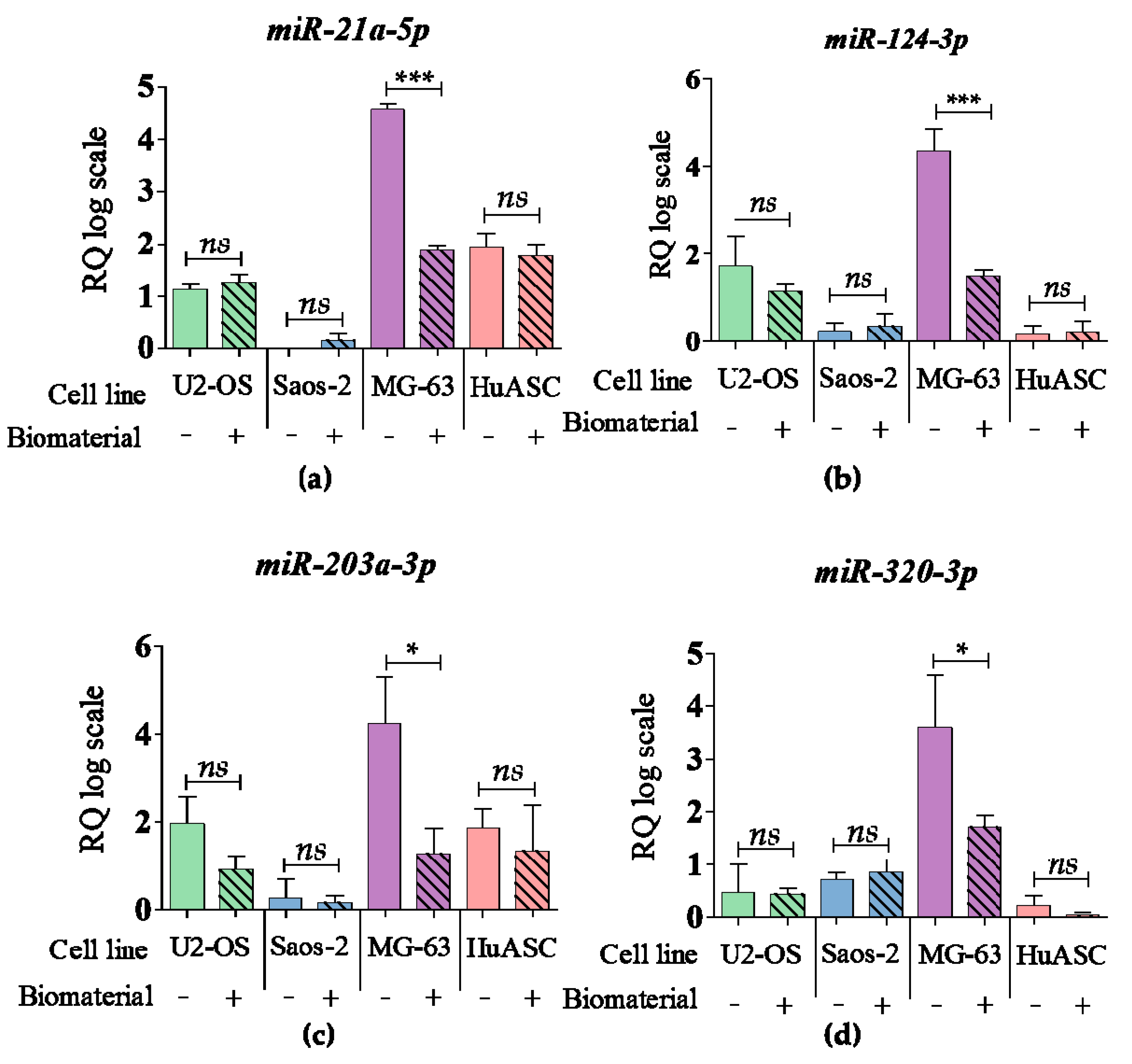
3.6. Biomaterial Impact at the miRNA Level
4. Discussion
Author Contributions
Funding
Conflicts of Interest
References
- Savage, S.A.; Mirabello, L. Using Epidemiology and Genomics to Understand Osteosarcoma Etiology. Sarcoma 2011, 2011, 1–13. [Google Scholar] [CrossRef] [PubMed]
- Simpson, S.; Dunning, M.D.; de Brot, S.; Grau-Roma, L.; Mongan, N.P.; Rutland, C.S. Comparative review of human and canine osteosarcoma: Morphology, epidemiology, prognosis, treatment and genetics. Acta Vet. Scand. 2017, 59, 71. [Google Scholar] [CrossRef] [PubMed]
- Eyre, R.; Feltbower, R.G.; James, P.W.; Blakey, K.; Mubwandarikwa, E.; Forman, D.; McKinney, P.A.; Pearce, M.S.; McNally, R.J. The epidemiology of bone cancer in 0–39 year olds in northern England, 1981–2002. BMC Cancer 2010, 10, 1–7. [Google Scholar] [CrossRef] [PubMed]
- Mirabello, L.; Troisi, R.J.; Savage, S.A. Osteosarcoma incidence and survival rates from 1973 to 2004: Data from the Surveillance, Epidemiology, and End Results Program. Cancer 2009, 115, 1531–1543. [Google Scholar] [CrossRef]
- Duchman, K.R.; Gao, Y.; Miller, B.J. Prognostic factors for survival in patients with high-grade osteosarcoma using the Surveillance, Epidemiology, and End Results (SEER) Program database. Cancer Epidemiol. 2015, 39, 593–599. [Google Scholar] [CrossRef]
- Misaghi, A.; Goldin, A.; Awad, M.; Kulidjian, A.A. Osteosarcoma: A comprehensive review. SICOT J. 2018, 4, 12. [Google Scholar] [CrossRef]
- Wang, Y.; Qin, N.; Zhao, C.; Yuan, J.; Lu, S.; Li, W.; Xiang, H.; Hao, H. The correlation between the methylation of PTEN gene and the apoptosis of osteosarcoma cells mediated by SeHA nanoparticles. Colloids Surf. B Biointerfaces 2019, 184, 110499. [Google Scholar] [CrossRef]
- Wu, V.M.; Huynh, E.; Tang, S.; Uskoković, V. Brain and bone cancer targeting by a ferrofluid composed of superparamagnetic iron-oxide/silica/carbon nanoparticles (earthicles). Acta Biomater. 2019, 88, 422–447. [Google Scholar] [CrossRef]
- Yang, Y.; Han, L.; He, Z.; Li, X.; Yang, S.; Yang, J.; Zhang, Y.; Li, D.; Yang, Y.; Yang, Z. Advances in limb salvage treatment of osteosarcoma. J. Bone Oncol. 2017, 10, 36–40. [Google Scholar] [CrossRef]
- Simon, M.; Aschliman, M.; Thomas, N.; Mankin, H. Limb-salvage treatment versus amputation for osteosarcoma of the distal end of the femur. J. Bone Jt. Surg. 1986, 68, 1331–1337. [Google Scholar] [CrossRef]
- Gherlinzoni, F.; Picci, P.; Bacci, G.; Campanacci, D. Limb sparing versus amputation in osteosarcomaCorrelation between local control, surgical margins and tumor necrosis: Istituto Rizzoli experience. Ann. Oncol. 1992, 3, S23–S27. [Google Scholar] [CrossRef] [PubMed]
- Loh, A.H.P.; Wu, H.; Bahrami, A.; Navid, F.; McCarville, M.B.; Wang, C.; Wu, J.; Bishop, M.W.; Daw, N.C.; Neel, M.D.; et al. Influence of Bony Resection Margins and Surgicopathological Factors on Outcomes in Limb-Sparing Surgery for Extremity Osteosarcoma. Pediatr. Blood Cancer 2015, 62, 246–251. [Google Scholar] [CrossRef] [PubMed]
- Sirichativapee, W.; Wisanuyotin, T.; Pattanittum, P.; Paholpak, P.; Laupattarakasem, P.; Srisodaphol, W.; Tsuchiya, H.; Laopaiboon, M.; Kosuwon, W.; Wiangnon, S. Chemotherapy for treating high-grade osteosarcoma in children and young adults. Cochrane Database Syst. Rev. 2016, 2016, 1465–1858. [Google Scholar] [CrossRef]
- Han, G.; Bi, W.-Z.; Xu, M.; Jia, J.-P.; Wang, Y. Amputation Versus Limb-Salvage Surgery in Patients with Osteosarcoma: A Meta-analysis. World J. Surg. 2016, 40, 2016–2027. [Google Scholar] [CrossRef]
- Yu, D.; Zhang, S.; Feng, A.; Xu, D.; Zhu, Q.; Mao, Y.; Zhao, Y.; Lv, Y.; Han, C.; Liu, R.; et al. Methotrexate, doxorubicin, and cisplatinum regimen is still the preferred option for osteosarcoma chemotherapy. Medicine 2019, 98, e15582. [Google Scholar] [CrossRef]
- Zhang, Y.; Yang, J.; Zhao, N.; Wang, C.; Kamar, S.; Zhou, Y.; He, Z.; Yang, J.; Sun, B.; Shi, X.; et al. Progress in the chemotherapeutic treatment of osteosarcoma. Oncol. Lett. 2018, 16, 6228–6237. [Google Scholar] [CrossRef]
- Wang, J.J.; Li, G.J. Relationship between RFC gene expression and intracellular drug concentration in methotrexate-resistant osteosarcoma cells. Genet. Mol. Res. 2014, 13, 5313–5321. [Google Scholar] [CrossRef]
- Kim, J.Y.; Ohn, J.; Yoon, J.-S.; Kang, B.M.; Park, M.; Kim, S.; Lee, W.; Hwang, S.; Kim, J.-I.; Kim, K.H.; et al. Priming mobilization of hair follicle stem cells triggers permanent loss of regeneration after alkylating chemotherapy. Nat. Commun. 2019, 10, 3694. [Google Scholar] [CrossRef]
- Nair, P.R. Delivering Combination Chemotherapies and Targeting Oncogenic Pathways via Polymeric Drug Delivery Systems. Polymers 2019, 11, 630. [Google Scholar] [CrossRef]
- Zhang, Y.; He, Z.; Duan, Y.; Wang, C.; Kamar, S.; Shi, X.; Yang, J.; Yang, J.; Zhao, N.; Han, L.; et al. Does intensified chemotherapy increase survival outcomes of osteosarcoma patients? A meta-analysis. J. Bone Oncol. 2018, 12, 54–60. [Google Scholar] [CrossRef]
- Li, X.; Zhang, Y.; Wan, S.; Li, H.; Li, D.; Xia, J.; Yuan, Z.; Ren, M.; Yu, S.; Li, S.; et al. A comparative study between limb-salvage and amputation for treating osteosarcoma. J. Bone Oncol. 2016, 5, 15–21. [Google Scholar] [CrossRef] [PubMed]
- Mavrogenis, A.F.; Abati, C.N.; Romagnoli, C.; Ruggieri, P. Similar Survival but Better Function for Patients after Limb Salvage versus Amputation for Distal Tibia Osteosarcoma. Clin. Orthop. Relat. Res. 2012, 470, 1735–1748. [Google Scholar] [CrossRef] [PubMed]
- Asghari, F.; Samiei, M.; Adibkia, K.; Akbarzadeh, A.; Davaran, S. Biodegradable and biocompatible polymers for tissue engineering application: A review. Artif. Cells Nanomed. Biotechnol. 2017, 45, 185–192. [Google Scholar] [CrossRef] [PubMed]
- Scaffaro, R.; Lopresti, F.; Maio, A.; Sutera, F.; Botta, L. Development of polymeric functionally graded scaffolds: A brief review. JABFM 2017, 15, 107–121. [Google Scholar] [CrossRef]
- Wang, C.; Feng, N.; Chang, F.; Wang, J.; Yuan, B.; Cheng, Y.; Liu, H.; Yu, J.; Zou, J.; Ding, J.; et al. Injectable Cholesterol-Enhanced Stereocomplex Polylactide Thermogel Loading Chondrocytes for Optimized Cartilage Regeneration. Adv. Healthc. Mater. 2019, 8, 1900312. [Google Scholar] [CrossRef]
- Yu, X.; Huang, W.; Zhao, D.; Yang, K.; Tan, L.; Zhang, X.; Li, J.; Zhang, M.; Zhang, S.; Liu, T.; et al. Study of engineered low-modulus Mg/PLLA composites as potential orthopaedic implants: An in vitro and in vivo study. Colloids Surf. B Biointerfaces 2019, 174, 280–290. [Google Scholar] [CrossRef]
- Smieszek, A.; Marycz, K.; Szustakiewicz, K.; Kryszak, B.; Targonska, S.; Zawisza, K.; Watras, A.; Wiglusz, R.J. New approach to modification of poly (l-lactic acid) with nano-hydroxyapatite improving functionality of human adipose-derived stromal cells (hASCs) through increased viability and enhanced mitochondrial activity. Mater. Sci. Eng. C 2019, 98, 213–226. [Google Scholar] [CrossRef]
- Marycz, K.; Sobierajska, P.; Smieszek, A.; Maredziak, M.; Wiglusz, K.; Wiglusz, R.J. Li+ activated nanohydroxyapatite doped with Eu 3+ ions enhances proliferative activity and viability of human stem progenitor cells of adipose tissue and olfactory ensheathing cells. Further perspective of nHAP: Li+, Eu 3+ application in theranostics. Mater. Sci. Eng. C 2017, 78, 151–162. [Google Scholar] [CrossRef]
- Belka, J.; Nickel, J.; Kurth, D.G. Growth on Metallo-Supramolecular Coordination Polyelectrolyte (MEPE) Stimulates Osteogenic Differentiation of Human Osteosarcoma Cells (MG63) and Human Bone Marrow Derived Mesenchymal Stem Cells. Polymers 2019, 11, 1090. [Google Scholar] [CrossRef]
- Catauro, M.; Papale, F.; Sapio, L.; Naviglio, S. Biological influence of Ca/P ratio on calcium phosphate coatings by sol-gel processing. Mater. Sci. Eng. C 2016, 65, 188–193. [Google Scholar] [CrossRef]
- Dalgic, A.D.; Atila, D.; Karatas, A.; Tezcaner, A.; Keskin, D. Diatom shell incorporated PHBV/PCL-pullulan co-electrospun scaffold for bone tissue engineering. Mater. Sci. Eng. C 2019, 100, 735–746. [Google Scholar] [CrossRef] [PubMed]
- Lin, L.; Hao, R.; Xiong, W.; Zhong, J. Quantitative analyses of the effect of silk fibroin/nano-hydroxyapatite composites on osteogenic differentiation of MG-63 human osteosarcoma cells. J. Biosci. Bioeng. 2015, 119, 591–595. [Google Scholar] [CrossRef] [PubMed]
- Saravanan, S.; Nethala, S.; Pattnaik, S.; Tripathi, A.; Moorthi, A.; Selvamurugan, N. Preparation, characterization and antimicrobial activity of a bio-composite scaffold containing chitosan/nano-hydroxyapatite/nano-silver for bone tissue engineering. Int. J. Biol. Macromol. 2011, 49, 188–193. [Google Scholar] [CrossRef] [PubMed]
- Vohra, S.; Hennessy, K.M.; Sawyer, A.A.; Zhuo, Y.; Bellis, S.L. Comparison of mesenchymal stem cell and osteosarcoma cell adhesion to hydroxyapatite. J. Mater. Sci. Mater. Med. 2008, 19, 3567–3574. [Google Scholar] [CrossRef] [PubMed] [Green Version]
- Han, Y.; Li, S.; Cao, X.; Yuan, L.; Wang, Y.; Yin, Y.; Qiu, T.; Dai, H.; Wang, X. Different Inhibitory Effect and Mechanism of Hydroxyapatite Nanoparticles on Normal Cells and Cancer Cells In Vitro and In Vivo. Sci. Rep. 2015, 4, 7134. [Google Scholar] [CrossRef] [PubMed] [Green Version]
- Wu, V.M.; Mickens, J.; Uskoković, V. Bisphosphonate-Functionalized Hydroxyapatite Nanoparticles for the Delivery of the Bromodomain Inhibitor JQ1 in the Treatment of Osteosarcoma. ACS Appl. Mater. Interfaces 2017, 9, 25887–25904. [Google Scholar] [CrossRef] [PubMed]
- Shi, Z.; Huang, X.; Liu, B.; Tao, H.; Cai, Y.; Tang, R. Biological Response of Osteosarcoma Cells to Size-Controlled Nanostructured Hydroxyapatite. J. Biomater. Appl. 2010, 25, 19–37. [Google Scholar]
- Bauer, I.W.; Li, S.-P.; Han, Y.-C.; Yuan, L.; Yin, M.-Z. Internalization of hydroxyapatite nanoparticles in liver cancer cells. J. Mater. Sci. Mater. Med. 2008, 19, 1091–1095. [Google Scholar] [CrossRef]
- Li, G.; Huang, J.; Li, Y.; Zhang, R.; Deng, B.; Zhang, J.; Aoki, H. In vitro study on influence of a discrete nano-hydroxyapatite on leukemia P388 cell behavior. Bio. Med. Mater. Eng. 2007, 17, 321–327. [Google Scholar]
- Xiong, H.; Du, S.; Ni, J.; Zhou, J.; Yao, J. Mitochondria and nuclei dual-targeted heterogeneous hydroxyapatite nanoparticles for enhancing therapeutic efficacy of doxorubicin. Biomaterials 2016, 94, 70–83. [Google Scholar] [CrossRef]
- Li, J.; Yang, Z.; Li, Y.; Xia, J.; Li, D.; Li, H.; Ren, M.; Liao, Y.; Yu, S.; Chen, Y.; et al. Cell apoptosis, autophagy and necroptosis in osteosarcoma treatment. Oncotarget 2016, 7, 44763–44778. [Google Scholar] [CrossRef] [PubMed] [Green Version]
- Chomczynski, P. Single-Step Method of RNA Isolation by Acid Guanidinium Thiocyanate-Phenol-Chloroform Extraction. Anal. Biochem. 1987, 162, 156–159. [Google Scholar] [CrossRef]
- Marycz, K.; Smieszek, A.; Trynda, J.; Sobierajska, P.; Targonska, S.; Grosman, L.; Wiglusz, R.J. Nanocrystalline Hydroxyapatite Loaded with Resveratrol in Colloidal Suspension Improves Viability, Metabolic Activity and Mitochondrial Potential in Human Adipose-Derived Mesenchymal Stromal Stem Cells (hASCs). Polymers 2019, 11, 92. [Google Scholar] [CrossRef] [PubMed] [Green Version]
- Wang, X.; Lin, M.; Kang, Y. Engineering Porous β-Tricalcium Phosphate (β-TCP) Scaffolds with Multiple Channels to Promote Cell Migration, Proliferation, and Angiogenesis. ACS Appl. Mater. Interfaces 2019, 11, 9223–9232. [Google Scholar] [CrossRef]
- Lin, M.; Firoozi, N.; Tsai, C.-T.; Wallace, M.B.; Kang, Y. 3D-printed flexible polymer stents for potential applications in inoperable esophageal malignancies. Acta Biomater. 2019, 83, 119–129. [Google Scholar] [CrossRef]
- Choi, S.; Oh, Y.-I.; Park, K.-H.; Lee, J.-S.; Shim, J.-H.; Kang, B.-J. New clinical application of three-dimensional-printed polycaprolactone/β-tricalcium phosphate scaffold as an alternative to allograft bone for limb-sparing surgery in a dog with distal radial osteosarcoma. J. Vet. Med. Sci. 2019, 81, 434–439. [Google Scholar] [CrossRef] [Green Version]
- Fang, Z.; Sun, Y.; Xiao, H.; Li, P.; Liu, M.; Ding, F.; Kan, W.; Miao, R. Targeted osteosarcoma chemotherapy using RGD peptide-installed doxorubicin-loaded biodegradable polymeric micelle. Biomed. Pharmacother. 2017, 85, 160–168. [Google Scholar] [CrossRef]
- Palamà, I.E.; Arcadio, V.; D’Amone, S.; Biasiucci, M.; Gigli, G.; Cortese, B. Therapeutic PCL scaffold for reparation of resected osteosarcoma defect. Sci. Rep. 2017, 7, 12672. [Google Scholar] [CrossRef] [Green Version]
- He, H.; Ni, J.; Huang, J. Molecular mechanisms of chemoresistance in osteosarcoma (Review). Oncol. Lett. 2014, 7, 1352–1362. [Google Scholar] [CrossRef] [Green Version]
- Szewczyk, M.; Lechowski, R.; Zabielska, K. What do we know about canine osteosarcoma treatment?—Review. Vet. Res. Commun. 2015, 39, 61–67. [Google Scholar] [CrossRef] [Green Version]
- Rong, Z.-J.; Yang, L.-J.; Cai, B.-T.; Zhu, L.-X.; Cao, Y.-L.; Wu, G.-F.; Zhang, Z.-J. Porous nano-hydroxyapatite/collagen scaffold containing drug-loaded ADM–PLGA microspheres for bone cancer treatment. J. Mater. Sci. Mater. Med. 2016, 27, 89. [Google Scholar] [CrossRef] [PubMed]
- Raucci, M.G.; Fasolino, I.; Caporali, M.; Serrano-Ruiz, M.; Soriente, A.; Peruzzini, M.; Ambrosio, L. Exfoliated Black Phosphorus Promotes in Vitro Bone Regeneration and Suppresses Osteosarcoma Progression through Cancer-Related Inflammation Inhibition. ACS Appl. Mater. Interfaces 2019, 11, 9333–9342. [Google Scholar] [CrossRef] [PubMed]
- Murugan, S.; Rajan, M.; Alyahya, S.A.; Alharbi, N.S.; Kadaikunnan, S.; Kumar, S.S. Development of self-repair nano-rod scaffold materials for implantation of osteosarcoma affected bone tissue. New J. Chem. 2017, 42, 725–734. [Google Scholar] [CrossRef]
- Cojocaru, F.D.; Balan, V.; Popa, I.M.; Munteanu, A.; Anghelache, A.; Verestiuc, L. Magnetic Composite Scaffolds for Potential Applications in Radiochemotherapy of Malignant Bone Tumors. Medicina 2019, 55, 153. [Google Scholar] [CrossRef] [Green Version]
- Gregor, A.; Filová, E.; Novák, M.; Kronek, J.; Chlup, H.; Buzgo, M.; Blahnová, V.; Lukášová, V.; Bartoš, M.; Nečas, A.; et al. Designing of PLA scaffolds for bone tissue replacement fabricated by ordinary commercial 3D printer. J. Biol. Eng. 2017, 11, 31. [Google Scholar] [CrossRef]
- Cai, Y.; Liu, Y.; Yan, W.; Hu, Q.; Tao, J.; Zhang, M.; Shi, Z.; Tang, R. Role of hydroxyapatite nanoparticle size in bone cell proliferation. J. Mater. Chem. 2007, 17, 3780. [Google Scholar] [CrossRef]
- Shi, M.; Xia, L.; Chen, Z.; Lv, F.; Zhu, H.; Wei, F.; Han, S.; Chang, J.; Xiao, Y.; Wu, C. Europium-doped mesoporous silica nanosphere as an immune-modulating osteogenesis/angiogenesis agent. Biomaterials 2017, 144, 176–187. [Google Scholar] [CrossRef]
- Bastos, A.R.; da Silva, L.P.; Maia, F.R.; Pina, S.; Rodrigues, T.; Sousa, F.; Oliveira, J.M.; Cornish, J.; Correlo, V.M.; Reis, R.L. Lactoferrin-Hydroxyapatite Containing Spongy-Like Hydrogels for Bone Tissue Engineering. Materials 2019, 12, 2074. [Google Scholar] [CrossRef] [Green Version]
- Seyedmajidi, S.; Seyedmajidi, M.; Zabihi, E.; Hajian-Tilaki, K. A comparative study on cytotoxicity and genotoxicity of the hydroxyapatite-bioactive glass and fluorapatite-bioactive glass nanocomposite foams as tissue scaffold for bone repair. J. Biomed. Mater. Res. 2018, 106, 2605–2612. [Google Scholar] [CrossRef]
- Ito, T.; Sasaki, M.; Taguchi, T. Enhanced ALP activity of MG63 cells cultured on hydroxyapatite-poly(ethylene glycol) hydrogel composites prepared using EDTA-OH. Biomed. Mater. 2015, 10, 015025. [Google Scholar] [CrossRef]
- Begam, H.; Kundu, B.; Chanda, A.; Nandi, S.K. MG63 osteoblast cell response on Zn doped hydroxyapatite (HAp) with various surface features. Ceram. Int. 2017, 43, 3752–3760. [Google Scholar] [CrossRef]
- Pietilä, M.; Palomäki, S.; Lehtonen, S.; Ritamo, I.; Valmu, L.; Nystedt, J.; Laitinen, S.; Leskelä, H.-V.; Sormunen, R.; Pesälä, J.; et al. Mitochondrial Function and Energy Metabolism in Umbilical Cord Blood- and Bone Marrow-Derived Mesenchymal Stem Cells. Stem Cells Dev. 2012, 21, 575–588. [Google Scholar] [CrossRef] [PubMed]
- Kostova, I. Lanthanides as Anticancer Agents. Available online: http://www.eurekaselect.com/90178/article (accessed on 4 October 2019).
- Teo, R.D.; Termini, J.; Gray, H.B. Lanthanides: Applications in Cancer Diagnosis and Therapy. J. Med. Chem. 2016, 59, 6012–6024. [Google Scholar] [CrossRef] [PubMed] [Green Version]
- Chen, G. The relationship between the expression of TAM, survivin and the degree of necrosis of the tumor after cisplatin treatment in osteosarcoma. Eur. Rev. Med. Pharmacol. Sci. 2017, 21, 490–497. [Google Scholar] [PubMed]
- Pautke, C.; Schieker, M.; Tischer, T.; Kolk, A.; Neth, P.; Mutschler, W.; Milz, S. Characterization of osteosarcoma cell lines MG-63, Saos-2 and U-2 OS in comparison to human osteoblasts. Anticancer. Res. 2004, 24, 3743–3748. [Google Scholar]


| Gene | Primer Sequence 5’-3’ | Loci | Amplicon Lenght [bp] | Accesion No. |
|---|---|---|---|---|
| P21 | F:GGCAGACCAGCATGACAGATTTC | 705-727 | 72 | NM_001291549.1 |
| R:CGGATTAGGGCTTCCTCTTGG | 776-756 | |||
| P53 | F:AGATAGCGATGGTCTGGC | 868-885 | 381 | NM_001126118.1 |
| R:TTGGGCAGTGCTCGCTTAGT | 1229-1248 | |||
| BCL-2 | F:ATCGCCCTGTGGATGACTGAG | 1010-1030 | 129 | NM_000633.2 |
| R:CAGCCAGGAGAAATCAAACAGAGG | 1138-1115 | |||
| BAX | F:ACCAAGAAGCTGAGCGAGTGTC | 235-256 | 414 | NM_001291428.1 |
| R:ACAAAGATGGTCACGGTCTGCC | 648-627 | |||
| CYCLIN D | F:GATGCCAACCTCCTCAACGA | 264-283 | 211 | NM_053056.2 |
| R:GGAAGCGGTCCAGGTAGTTC | 474-455 | |||
| C-MYC | F:CTTCTCTCCGTCCTCGGATTCT | 1847-1868 | 204 | NM_001354870.1 |
| R:GAAGGTGATCCAGACTCTGACCTT | 2050-2027 | |||
| SURV | F: ACCGCATCTCTACATTCAAG | 114-143 | 113 | NM_001168.3 |
| R: CAAGTCTGGCTCGTTCTC | 226-209 | |||
| MMP-7 | F:TGTATGGGGAACTGCTGACA | 488-507 | 151 | NM_002423.5 |
| R:GCGTTCATCCTCATCGAAGT | 638-619 | |||
| MMP-14 | F: TCGGCCCAAAGCAGCAGCTTC | 312-332 | 180 | NM_004995.4 |
| R: CTTCATGGTGTCTGCATCAGC | 491-471 | |||
| APAF | F:CTTCTTCCAGTGTAAGGACAGT | 861-882 | 243 | NM_013229.2 |
| R:CTGAAACCCAATGCACTCCC | 1103-1084 | |||
| CASP3 | F:AATACCAGTGGAGGCCGACT | 650-669 | 128 | NM_001354779.1 |
| R:TGTCGGCATACTGTTTCAGC | 777-758 | |||
| CASP6 | F:TCATGAGAGGTTCTTTTGGCAC | 231-252 | 197 | NM_001226.3 |
| R:CACACACAAAGCAATCGGCA | 427-408 | |||
| CASP8 | F:TGCTGAGCACGTGGAGTTAG | 282-301 | 178 | NM_001080125.1 |
| R:CAGGCTCAGGAACTTGAGGG | 459-440 | |||
| CASP9 | F:CTGCGTGGTGGTCATTCTCT | 763-782 | 130 | NM_032996.3 |
| R:GCAGCTGGTCCCATTGAAGA | 892-873 | |||
| GAPDH | F: GTCAGTGGTGGACCTGACCT | 894-913 | 256 | NM_001289746.1 |
| R: CACCACCCTGTTGCTGTAGC | 1149-1130 |
| Gene | Primer Sequence 5’-3’ | Loci | Accesion No. |
|---|---|---|---|
| miR-21a-5p | UAGCUUAUCAGACUGAUGUUGA | 18-39 | MIMAT0000530 |
| miR-124-3p | UAAGGCACGCGGUGAAUGCC | 44-63 | MIMAT0000134 |
| miR-203a-3p | GUGAAAUGUUUAGGACCACUAG | 65-86 | MI0000283 |
| miR-320-3p | AAAAGCUGGGUUGAGAGGGCGA | 48-69 | MI0000704 |
© 2019 by the authors. Licensee MDPI, Basel, Switzerland. This article is an open access article distributed under the terms and conditions of the Creative Commons Attribution (CC BY) license (http://creativecommons.org/licenses/by/4.0/).
Share and Cite
Sikora, M.; Marcinkowska, K.; Marycz, K.; Wiglusz, R.J.; Śmieszek, A. The Potential Selective Cytotoxicity of Poly (L- Lactic Acid)-Based Scaffolds Functionalized with Nanohydroxyapatite and Europium (III) Ions toward Osteosarcoma Cells. Materials 2019, 12, 3779. https://doi.org/10.3390/ma12223779
Sikora M, Marcinkowska K, Marycz K, Wiglusz RJ, Śmieszek A. The Potential Selective Cytotoxicity of Poly (L- Lactic Acid)-Based Scaffolds Functionalized with Nanohydroxyapatite and Europium (III) Ions toward Osteosarcoma Cells. Materials. 2019; 12(22):3779. https://doi.org/10.3390/ma12223779
Chicago/Turabian StyleSikora, Mateusz, Klaudia Marcinkowska, Krzysztof Marycz, Rafał Jakub Wiglusz, and Agnieszka Śmieszek. 2019. "The Potential Selective Cytotoxicity of Poly (L- Lactic Acid)-Based Scaffolds Functionalized with Nanohydroxyapatite and Europium (III) Ions toward Osteosarcoma Cells" Materials 12, no. 22: 3779. https://doi.org/10.3390/ma12223779