Article

Characterization of the Ensemble of Lignin-Remodeling DyP-Type Peroxidases from Streptomyces coelicolor A3(2)

Department of Chemistry and Biotechnology, Tallinn University of Technology, Akadeemia tee 15, 12618 Tallinn, Estonia
*
Author to whom correspondence should be addressed.
Energies 2023, 16(3), 1557; https://doi.org/10.3390/en16031557
Submission received: 30 December 2022 / Revised: 21 January 2023 / Accepted: 25 January 2023 / Published: 3 February 2023
(This article belongs to the Special Issue Biofuels Production from Lignocellulosic Biomass)

Abstract

:
Lignin is Nature’s major source of aromatic chemistry and is by many seen as the green entry-point alternative to the fossil-based chemical industry. Due to its chemically recalcitrant structure, the utilization of lignin is challenging, wherein enzymes might be the key to overcome this challenge. Here, we focus on the characterization of dye-decolorizing peroxidases from Streptomyces coelicolor A3(2) (ScDyPs) in the context of enzymatic modification of organosolv lignins from aspen and Miscanthus × giganteus. In this study, we show that the ScDyPB can remodel organosolv lignins from grassy biomass, leading to higher molecular weight species, while ScDyPAs can deconstruct hardwood lignin, leading to an overall reduction in its molecular weight. Additionally, we show that ScDyPB is effective in polymerizing low-molecular-weight phenolics, leading to their removal from the solution.

1. Introduction

Lignocellulosic biomass (LCB) is Earth’s most abundant renewable raw material [1]. This sustainable feedstock makes an attractive alternative to fossil-based resources to obtain bio-based materials, second-generation biofuels, bioenergy, value-added products, and chemicals [2,3,4]. LCB comprises three biopolymers: cellulose, hemicellulose, and lignin, with a typical ratio of 4:3:3, which varies among different species [5,6].
Lignin represents the second most abundant natural polymer after cellulose and constitutes the most abundant phenolic biopolymer on Earth [7]. Therefore, lignin is by many seen as a green entry-point alternative to fossil-based sources for the chemical industry [8,9]. The content of lignin varies significantly among hardwoods, softwoods, and herbaceous plants, wherein the highest content of lignin is in softwoods and the lowest in grasses [10].
Lignin is constructed from three monolignols—coniferyl alcohol, sinapyl alcohol, and p-coumaryl alcohol—which differ by the degree of methoxylation. When incorporated into lignin, those monolignols make up guaiacyl (G), syringyl (S), and hydroxyphenyl (H) units, respectively (Figure 1) [7,11]. The composition and type of lignin depend on the plant type. While softwood lignins primarily comprise guaiacyl units (G-type lignin), hardwood lignins also contain syringyl units (G/S-type lignin). Lignins in grassy biomass are built up from all three sub-units and, in addition to the previous two, contain substantial amounts of hydroxyphenyl units (G/S/H-type lignin) [10,12].
Since monolignol coupling involves radicals, there are many possibilities for linkages between lignin monomers. These sub-units are primarily connected by ether and carbon-carbon linkages, wherein β-aryl ether (β-O-4) linkages are the most common ones in softwood and hardwood lignins, approximately 50% and 60%, respectively (Figure 1). C-C linkages are less common in hardwood than in softwood lignins, as the additional methoxy groups, chiefly in the S units, hamper the formation of these linkages [12,13].
In addition to the source of lignin, the composition and molecular weight depend on the extraction method [12]. Most of the technical lignins are obtained by recovery as an industrial by-product [14]. The pulp and paper industries are the primary producers of technical lignin generating 50–70 million tons annually [15]. In contrast to the more traditional Kraft process, organosolv pulping utilizes organic solvents to dissolve lignin from the biomass [12]. It is an potent method for biorefineries for the utilization of biomass from hard- and softwoods as well as from grasses [12,16,17]. Organosolv lignin is highly pure, homogeneous, sulfur-free, with low content of carbohydrates and ash, and has a low molecular weight and dispersity. It is highly similar to native lignin, containing fewer modifications, and has an increased number of aryl ether linkages. So far, this technology has only reached pilot and demonstration scale, and not the industrial scale, mainly due to the high cost of solvent recovery [6,12,18].
Approximately only 2% of industrial lignin leftovers are used for commercial products [4,19]. Instead of being a mere by-product, lignin could be a useful co-product to produce valuable chemicals, such as vanillin [20]. Structurally extensively modified technical lignins with rearranged structures hinder its depolymerization into smaller fragments, a crucial and critical step for lignin utilization [21,22].
In nature, lignin decomposition is accomplished by numerous organisms, such as white-, brown-, and soft-rot fungi [23], and several Gram-positive and Gram-negative bacteria, mostly Actinobacteria and Proteobacteria [24]. In addition to other Streptomycetes, Streptomyces coelicolor has been identified as a lignin-degrading bacterial species, which harbors genes responsible for the catabolism of naturally occurring phenolics [25].
Lignin degraders, in nature, employ sophisticated metabolic and enzyme systems to attack and depolymerize lignin. Lignin-modifying enzymes, the key enzymes in lignin depolymerization, are phenol oxidases (laccases, EC 1.10.3.2) and heme-containing peroxidases—lignin-modifying peroxidases (LMPs): lignin peroxidase (LiP, EC 1.11.1.14), manganese(-dependent) peroxidase (MnP, EC 1.11.1.13), and versatile peroxidase (VP, EC 1.11.1.16). LMPs are part of the fungal class II heme-containing peroxidases in the catalase-peroxidase superfamily. These three ligninolytic peroxidases share noticeably high sequence identity and similarities in their structure [26,27].
Dye-decolorizing peroxidases (DyPs) also belong to LMPs but are phylogenetically unrelated to other LMPs (LiP, MnP, and VP). These peroxidases have heme as the attached prosthetic group and require hydrogen peroxide as the co-substrate that is reduced to water during its catalytic cycle [28]. DyPs (EC 1.11.1.19), systematically reactive-blue-5: hydrogen-peroxide oxidoreductases, were first identified in the fungus Bjerkandera adusta (previously Geotrichum candidum 1 Dec) in 1999 [29], later in bacteria [26]. Because of their distinctive structural and catalytic characteristics, DyPs form a new group of heme peroxidases [30]. They were named based on their ability to degrade and decolorize anthraquinone and azo dyes, such as Reactive Blue 5 [29,31,32]. The substrate specificity of DyPs is broad; in addition to dyes, they also oxidize other typical peroxidase substrates such as 2,2′-azino-bis(3-ethylbenzothiazoline-6-sulfonic acid (ABTS) and 2,6-dimethoxyphenol (2,6-DMP) [26,33]. DyPs can oxidize phenolic [34] and non-phenolic lignin fragments without requiring a mediator [35,36].
According to the phylogenetic analysis of genomic sequences, DyPs have been classified into four sub-families in PeroxiBase. Sub-families A, B, and C contain predominately bacterial peroxidases, while sub-family D peroxidases are from fungi [37]. A newer classification was proposed in 2015, which divides DyPs into classes P (primitive, former class B), I (intermediate, former class A), and V (advanced, former classes C and D) [38]. DyP-type peroxidases are more or less considered bacterial counterparts to fungal LMPs [39]. The ability to decompose lignin has been identified from several DyPs. It includes peroxidases from fungi Auricularia auricula-judae [35], Irpex lacteus [40], and bacterial species such as Rhodococcus jostii [41], Thermobifida fusca [42], and Pseudomonas fluorescens [43]. S. coelicolor A3(2) genome was sequenced in 2002 [44] and has three genes encoding DyPs, two A-type DyPs and a B-type DyP. We hypothesize that this bacterium requires dye-decolorizing peroxidases to metabolize lignin.
Here, we cloned, expressed, and purified all three DyPs encoded by S. coelicolor A3(2). The H2O2-dependent activity of the enzymes were characterized using a soluble model substrate ABTS as well as water-insoluble organosolv lignins from Miscanthus × giganteus and aspen. The activity of DyPs on ABTS did not correlate with their ability in modifying lignin, a phenomenon that may reflect different pH optima observed with these two substrates. Furthermore, different DyPs had distinctly different effects in lignin modification. Treatment of lignins with type-A DyPs, ScDyP1A and ScDyP2A, resulted in depolymerization of lignin, whereas treatment with ScDyPB increased the molecular weight of lignin.

2. Materials and Methods

In order to conserve space, portions containing detailed information about the experiments can be found in the Supplementary Materials.

2.1. Gene Cloning

Genes encoding dye-decolorizing peroxidases (DyPs) (accession numbers: NP_626524.1, NP_628147.1, and NP_631251.1 for locus tags SCO2276 (ScDyP1A), SCO3963 (ScDyP2A), and SCO7193 (ScDyPB), respectively) were amplified from Streptomyces coelicolor A3(2) genomic DNA using the oligonucleotide primers with restriction endonuclease sites for NdeI and XhoI, respectively (Table S1). Plasmid DNAs were purified by alkaline lysis. The amplified genes (1263 bp, 1338 bp, and 951 bp for SCO2276, SCO3963, and SCO7193, respectively) were cloned into pET15b expression vector coding for an N-terminal His6-tag followed by thrombin protease site. Additionally, codon-optimized genes for SCO2276 and SCO3963 were obtained and pre-cloned into the pET28a expression vector (Twist Bioscience, San Francisco, CA, USA). Each clone (codon-optimized) carried the Kan resistance gene, N-terminal His6-tag, and thrombin cleavage site.

2.2. Overexpression

Recombinant ScDyPs were overproduced in E. coli BL21 (DE3) as His6-fusion proteins (without the N-terminal 40-amino acid putative twin-arginine translocation (Tat) signal peptides in the case of ScDyPAs). The overnight starter cultures were used for inoculation. The culture media (LB prepared in tap water) supplemented with antibiotics was incubated at 37 °C until the OD600 reached 0.6–0.8. The overexpression of recombinant protein was induced by 1 mM isopropyl-β-D-1-thiogalactopyranoside. The culture was grown overnight, at 30 °C and 180 rpm. The cells were harvested at 4000 g, 15 min, and 4 °C.

2.3. Purification

The harvested cells were resuspended in buffer A (20 mM Tris-HCl, pH 7.5, 0.5 M NaCl, 5 mM imidazole) and lysed using sonication (Bandelin Sonopuls, Bandelin GmbH & Co, Berlin, Germany) or high-pressure homogenization (EmulsifFex-C5, Avestin, Ottawa, ON, Canada). The lysed cells were centrifuged at 35,000 g for 1 h, at 4 °C. The supernatant was loaded onto the HisTrap FF column (GE Healthcare, Chicago, IL, USA). The column was washed with 10 column volumes (CV) of 10% buffer B (20 mM Tris-HCl, pH 7.5, 0.5 M NaCl, 0.5 M imidazole) to remove non-specifically bound proteins. The His6-tagged protein was eluted with the linear gradient of imidazole of 10 to 100% buffer B (20 CV). The fractions were analyzed with SDS-PAGE, and the protein of interest was pooled. His6-tag was cleaved by overnight incubation with thrombin (2 U per 1 mg protein) at 4 °C. The heme reconstitution was conducted with hemin (2-fold molar excess) at room temperature for 2 h using 20 mg/mL hemin (98%, porcine, Acros Organics, Fair Lawn, NJ, USA) dissolved in DMSO. The protein was exchanged to 20 mM Tris-HCl buffer (pH 7.5) containing 0.1 M NaCl (HiPrep 26/10 Desalting column) (GE Healthcare); the resulting protein fractions were pooled and concentrated using centrifugal filters with a molecular weight cutoff of 10 kDa (Sartorius, Göttingen, Germany). The protein was flash-frozen in liquid nitrogen and stored at −80 °C.

2.4. Determination of Enzyme Concentration

The total protein concentration was determined spectrophotometrically at 280 nm (UV-2700 UV-Visible Spectrophotometer, Shimadzu, Japan). The extinction coefficient ε280 nm values, calculated using the Expasy ProtParam tool (https://web.expasy.org/protparam/, accessed on 15 August 2022), were 51,450; 38,960; and 18,450 M−1cm−1, for ScDyP1A, ScDyP2A, and ScDyPB, respectively. To estimate the total protein concentration before heme reconstitution, the buffer was exchanged using ultrafiltration filters to prevent concentration overestimation due to imidazole absorption.
The amount of active enzyme (with heme in the active site) was calculated using characteristic absorbance at 406 nm (ε406 = 100,000 M−1 cm−1) for heme proteins [45]. The total and the active enzyme content was calculated before and after the reconstitution with hemin. The heme content of DyPs was evaluated by Reinheitszahl value (Rz, A406/A280). In all experiments, the enzymes were dosed based on the active enzyme concentration.

2.5. Enzyme Assays

All enzyme assays with ABTS (ChemCruzTM, Santa Cruz, CA, USA) were performed in triplicate using Shimadzu UV-2700 UV-Visible Spectrophotometer at room temperature (except for the investigation of temperature dependence). All measurements with 100 nM ScDyP1A, 50 nM ScDyP2A, or 15 nM ScDyPB were used. Oxidation of ABTS was carried out with 1 mM H2O2, and the formation of ABTS cation radical was measured at 420 nm (ε420 = 36,000 M−1 cm−1). A fresh stock solution of H2O2 was prepared daily before use. The stock of 2,6-DMP (Acros Organics, Fair Lawn, NJ, USA) was prepared in dimethylformamide. The oxidation of 2,6-DMP was measured at 470 nm (ε470 = 53,200 M−1 cm−1) [46]. Optimal pH and temperature dependence parameters were determined (Supplementary Materials). The data analysis was carried out using OriginPro 2021 software (OriginLab, Northampton, MA, USA).

2.6. Organosolv Extraction of Lignin

Ethanol organosolv lignin from bleached chemi-thermomechanical aspen pulp was extracted as described in Jõul et al., 2022 [16]. Ethanol organosolv lignin from Miscanthus × giganteus (Mxg-lignin) was provided by Stefan Bauer [17]. Klason analysis was conducted as described in Jõul et al., 2022 [16] to determine the total lignin content for aspen (86.8 ± 0.7%) and Mxg (85.0 ± 1.1%) lignins, respectively.

2.7. Enzymatic Treatment of Organosolv Lignins

Ethanol organosolv lignin suspension at 1 mg/mL in aqueous HEPES (Sigma-Aldrich, St. Louis, MO, USA), buffer (50 mM, pH 8.0) with added 2 mM H2O2 was treated with 10 μM of ScDyP1A, ScDyP2A, or ScDyPB, incubated for 24 h, at 28 °C. For turbidity, the reaction mixtures were agitated at 200 rpm. The reaction mixtures and controls were centrifuged, and the insoluble portions were lyophilized for storage. The supernatants were extracted with ethyl acetate for further analysis. The water-insoluble portions of lignin were analyzed via gel permeation chromatography (GPC) and water-soluble portions with gas chromatography-mass spectrometry (GC-MS).

3. Results

3.1. The Ensemble of DyP-Peroxidases of Streptomyces coelicolor

The genome of S. coelicolor A3(2) (Sc) carries three genes encoding for DyPs: two type-A DyPs, ScDyP1A (SCO2276) and ScDyP2A (SCO3963), and a type-B DyP, ScDyPB (SCO7193) with molecular weights of 45 kDa, 47 kDa, and 34 kDa, respectively. The three genes are located in three distinct loci on the single linear chromosome of S. coelicolor. Regarding the operonic context, it is not entirely clear what specific biological role ScDyPB might be involved with. However, in the case of ScDyP1A, the annotations of the neighboring genes in its operon suggest its role in iron homeostasis (i.e., iron permease, high-affinity iron-transporter, etc.). In contrast, in the case of ScDyP2A, the operon contains a putative prephenate dehydrogenase and an aminoacyl-tRNA synthetase, thus suggesting a role in the biosynthesis of aromatic amino acids, which is well aligned with lignin biochemistry (Table 1). The genes of ScDyP1A and ScDyP2A contain an N-terminal Tat secretion tag, suggesting a role outside of the cytosole, whereas ScDyPB lacks the secretion signal, suggesting a biochemical role inside the cell.

3.2. Production and Purification of Recombinant ScDyPs

ScDyPs were expressed as His-tagged fusion proteins in E. coli, purified, and the concentration of apo- and holo-enzymes determined using spectrophotometry. The ScDyPs showed the typical heme-peroxidase spectrum with a Soret band maxima at 408 nm, 405 nm and 401 nm for ScDyP1A, ScDyP2A, and ScDyPB, respectively (Figure S1). The heme content of peroxidases was evaluated by Rz value (Table 2). Before reconstitution with hemin, the highest heme content was observed with ScDyPB followed by ScDyP1A and ScDyP2A (Table 2). The reconstitution improved the proportion of holo-enzyme (Table 2)—1.7-fold for ScDyP1A and 2.2-fold for ScDyP2A—while for ScDyPB, the Rz ratio remained relatively the same. In order to tackle the low expression yields of ScDyPAs (about 1 mg of protein from 1 L culture) from plasmids containing inserts of PCR-amplified genomic DNA (i.e., 1 mg of protein for ScDyP1A/ScDyP2A from 1 L culture), synthetic genes that were codon-optimized for expression from an E. coli host, were obtained. The proteins were then expressed and purified, where ScDyP1A had an Rz value of 2.01 and ScDyP2A 2.02 before reconstitution with hemin. The reconstitution of DyPAs from the codon-optimized system with hemin did not show a particular effect, as the Rz values remained the same. The codon-optimization of genes considerably improved the protein expression yield for both ScDyP1A and ScDyP2A.

3.3. Biochemical Characterization

3.3.1. Optimal pH for ScDyPs

The pH dependency of the oxidation of ABTS is shown in Figure 2. ScDyP1A and ScDyP2A exhibited the highest activity at pH 3.5, while the optimal pH for ScDyPB was determined to be 3.0 (Figure 2). At pH 5.0, all ScDyPs showed almost no activity with ABTS. Contrary to ABTS, the oxidation of phenolic substrate (2,6-DMP) by ScDyP2A and ScDyPB had a pH optimum around 8.5 (data not shown). ScDyP1A did not catalyze the oxidation of 2,6-DMP.

3.3.2. Temperature Dependency

The temperature dependency on enzyme activity was measured at pH 3.5 using ABTS as the substrate. Peroxidase activity for all DyPs were at a decline or started decreasing already at 30 °C and decreased with further temperature increase (Figure 3).

3.3.3. Thermostability

To further estimate the temperature stability of ScDyPs, the enzyme solutions were incubated, at 30 °C, for 48 h, and at fixed time intervals, sample aliquots were withdrawn, and tested for activity. Within 2 h, ScDyP2A had lost most activity compared to other ScDyPs, approximately one-third (Figure 4). After 24 h, ScDyP1A and ScDyPB had lost ~20% of activity, and ScDyP2A had around half of its activity. ScDyP1A is the most thermoresistant ScDyP and had retained ~70% of its activity within 48 h, wherein ScDyP2A had lost ~70% of its activity.

3.3.4. Steady-State Kinetics

Steady-state kinetic measurements were conducted at pH 3.5, varying the concentration of ABTS and using 1 mM H2O2 as a co-substrate. All ScDyPs displayed non-Michaelis-Menten kinetics (Figure S2), and the results were analyzed using the equation accounting for substrate inhibition (Equation S1 in the Supplementary Materials). Kinetic parameters are summarized in Table 3. Highest kcat was measured with ScDyPB, followed by ScDyP2A and ScDyP1A. The ScDyPB had also the lowest apparent KM values for ABTS. Owing to its high kcat and low KM value, the ScDyPB had by far the highest catalytic efficiency (kcat/KM) in oxidation of ABTS (Table 3). All three DyPs are inhibited by increased substrate concentrations, wherein ScDyPB is more prone to inhibition by ABTS with a Ki of 0.4 mM (Table 3; Figure S2). 2,6-DMP was also tested as a substrate but proved to be a poor substrate for ScDyPs, especially for ScDyP1A, which did not oxidize this substrate (data not shown). However, the apparent specificities (kcat/KM) were approximately 140-fold lower for ScDyP2A (~0.1 mM−1·s−1) and approximately 200-fold lower for ScDyPB (~3.1 mM−1·s−1). Similarly, substrate inhibition was seen with 2,6-DMP (Figure S3).

3.4. Lignin Remodeling Activity of ScDyPs

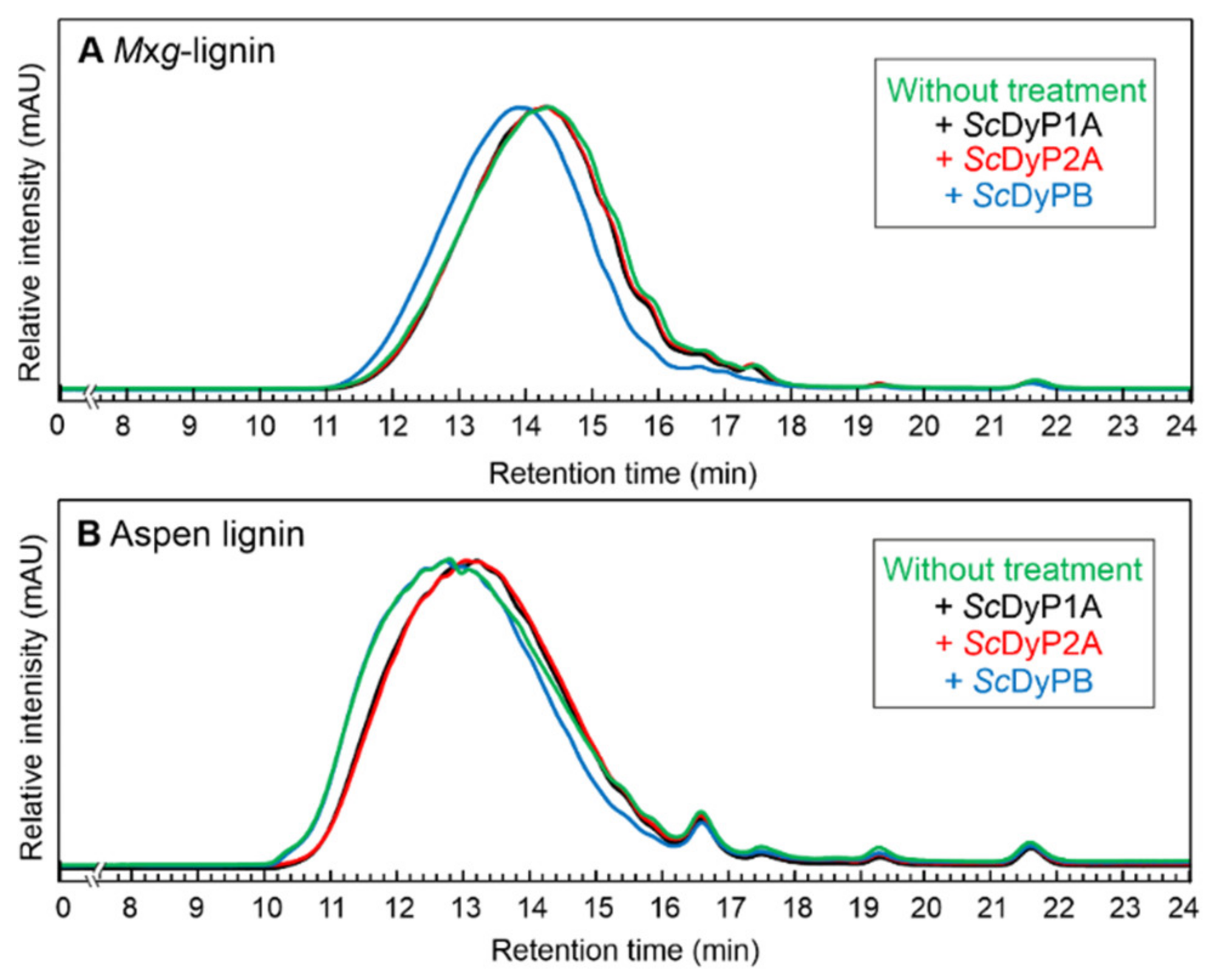
3.4.1. Identification of Molecular Weight Distribution of Lignin Samples by GPC Analysis

GPC analysis was utilized to observe the changes in the relative size distribution of ScDyPs-treated organosolv Mxg- and aspen lignins. Lignins were incubated in the presence of ScDyPs for 24 h, then centrifuged, and the pellets were lyophilized. For the GPC analysis, the lyophilized pellets were dissolved in THF, and the molecular weights of lignins were observed. The number-average molecular weight (Mn) of Mxg-lignin was 1509 Da, and the weight-average molecular weight (Mw) 2435 Da (Table 4).
At first, pH 3.5 was used for the lignin experiments, but the lignin treatment at acidic media did not show an effect. Acidic to alkaline pH ranges were tested for optimal lignin-remodeling activity, where a pH range of 7.5 to 8.5 was deemed the most active (data not shown). The treatment of Mxg-lignin with ScDyPB led to the polymerization of lignin polymer up to 19%. Similar behavior was seen with aspen lignin. The enzymatic treatment with ScDyPB caused the increase in lignin molecular weight, whereas ScDyPAs showed the ability to decrease the molecular weight of lignin. The Mn of Mxg organosolv lignin treated with ScDyP2A and with ScDyP1A remained the same as with untreated material. However, the Mw of aspen lignin after 24 h treatment with ScDyP2A decreased by approximately 20%. Similarly, a reduction in the average molecular weight of aspen lignin after ScDyP1A treatment could also be detected. The ĐM of Mxg-lignin fragments decreased from 1.61 to 1.57. The ĐM of aspen lignin decreased from 2.00 to 1.87 (ScDyPB), 1.80 (ScDyP2A), and 1.82 (ScDyP1A). According to these results, all three ScDyPs can remodel organosolv lignin, wherein the Mxg-lignin is a better substrate for ScDyPB and aspen lignin for ScDyPAs (Figure 5).

3.4.2. Identification of Low-Molecular-Weight Compounds in Lignin Samples by GC-MS Analysis

GC-MS analysis was used to determine the low-molecular-weight (LMW) compounds in lignin samples. The organosolv lignins were incubated with and without ScDyPs for 24 h and centrifuged; the supernatants were extracted with ethyl acetate, lyophilized, dissolved in methanol, and analyzed with GC-MS. According to the GC-MS results, the LMW proportion of Mxg-lignin contains mostly H-units, fewer G-units, and a small portion of S-units, wherein aspen lignin is in the most part composed of S-units, fewer G-units, and a minute proportion of H-units (based on the integrated peak area). The treatment of both lignins with ScDyPs decreased the amount of LMW compounds found in supernatants, where ScDyPB had the highest effect (Table 5).

4. Discussion

The purpose of this study was to characterize the full ensemble of DyP peroxidases from S. coelicolor A3(2), which consists of two A-type and one B-type DyP peroxidase in the context of enzymatic modification of organosolv lignins. The physiological roles of DyPs are unclear to date. It has been shown previously that some type-A DyPs are components of tripartite ferrous iron transporters and serve a role in iron uptake [47,48]. There are reports of DyPs functioning as deferrochelatases by releasing heme iron without tetrapyrrole degradation, such as EfeB (class A) from E. coli [49]. According to the operonic context, ScDyP1A might be a ferrous iron transport peroxidase. B-type DyPs are putative cytoplasmic enzymes indicating a function in intracellular metabolism [50]. However, the operonic context of ScDyPB does not hint at its specific role for S. coelicolor.
For the biochemical characterization, the three DyPs from S. coelicolor A3(2) were heterologously expressed and purified. The efficiency of heterologous expression of proteins is often hampered by differences in the codon usage between the organism of origin and the chosen host expression organism for a particular gene. In addition to differences in the abundance of specific tRNAs, the genome of S. coelicolor has 72% G + C content, when compared to that of about 51% of E. coli. [44,51]. Although the expression of ScDyPB yielded sufficient amounts of enzyme for further experiments (20 mg per liter culture), the expression of ScDyPAs resulted in roughly 20-fold less protein. Successful protein expression in E. coli as the host depends on several factors, such as the choice of the expression plasmid and the strain, the optimization of expression conditions, etc. [52,53]. However, codon usage seems to have the highest impact on obtaining high protein yields from recombinant expression [54]. It is also known that S. coelicolor has remarkably different codon usage profile than E. coli [55]. Codon optimization is often used to enhance the translation efficiency of a gene of interest [56]. It has been seen that codon-optimization results in an even 140-fold improvement of protein expression yield [57]. Here, the expression with codon-optimized genes enabled the production of higher quantities of protein when compared to the non-optimized construct, resulting in a 4-fold and 43-fold increase for ScDyP1A and ScDyP2A, respectively. Additionally, DyPs require heme as a prosthetic group for its catalytic activity [29]. Recombinant bacterial expression frequently produces heme-deficient proteins [58,59,60,61], which can be combated by supplementing the growth media with extra hemin during the expression [61]. Unfortunately, the heme transport systems of most E. coli strains cannot efficiently uptake hemin and depend on the diffusion through the cell membrane [60,61]. The Reinheitszahl (Rz) value (from German “purity number”), the ratio of absorbances (ASoret/A280), is used to obtain an estimate on the extent of heme incorporation into peroxidases. Prior to supplying the DyPs with additional hemin, Rz values of ScDyP1A and ScDyP2A were below one. The reconstitution improved the Rz values by approximately 2-fold for both ScDyPAs. The Rz values for ScDyPAs obtained from codon-optimized genes and ScDyPB were over two without any extra steps. The heme reconstitution after purification did not increase the Rz values of ScDyPAs obtained from codon-optimized genes and for ScDyPB. It is fathomable that the uptake kinetics of heme by DyPAs in vivo are affected by the differences in the speed of translation between the high G + C non-optimized vs. codon-optimized DyPA constructs. Although we have no direct evidence for it, it is possible that there may be minor differences in protein folding due to ribosome stalling which may also affect the heme uptake by the purified proteins in vitro.
Having obtained sufficient amounts of heme-reconstituted ScDyPs, we set out to determine their optimal working conditions (i.e., pH and temperature). DyP-type peroxidases have demonstrated a lower pH preference than classic heme peroxidases [29,62]. Typically, rather similar pH optima for ABTS and 2,6-DMP oxidation have been identified for DyPs. Previous studies of fungal and bacterial DyPs have reported that the optimum pH for ABTS and 2,6-DMP oxidation falls in the pH range of 3–5 [63,64,65]. Our results of ScDyPs were consistent with the past findings for ABTS. The optimum pH of ScDyPs for ABTS oxidation was determined in the range of 3–3.5, but in contrast, for phenolic 2,6-DMP oxidation, the optimum pH was 8.5. Chaplin et al., 2017 reported that the optimum pH for 2,6-DMP oxidation is 9.0 for DyPA from Streptomyces lividans [66]. This kind of phenomenon has also been seen in the case of laccases, where the enzymes catalyze the oxidation of ABTS better at pH 4, but the oxidation of 2,6-DMP at alkaline pH (7–9) [67,68].
It has been shown that enzymes isolated from extremophiles can tolerate more severe conditions, such as extremes in pH, temperature, salt concentration, etc., often required for industrial processes. For instance, the optimal temperature of A-type TcDyP from a thermophilic actinomycete Thermomonospora curvata was also 30 °C; however, 1 h incubation of TcDyP at 60 °C resulted in ~70% retained activity [69]. Since S. coelicolor is a mesophilic bacterium, it was expected that ScDyPs would show only modest resistance to elevated temperatures. ScDyPB showed the highest activity at 30 °C, and the activity of all ScDyPs decreased with the rising temperature. The activities of ScDyPAs decreased more rapidly at higher temperatures compared to ScDyPB. The relative activity of ScDyPB at 40 °C was 90%, but in contrast, ScDyPAs had retained only less than 20% of activity. Further, the stability of ScDyPs at 30 °C was determined (Figure 4). The thermostability of ScDyP2A was decreasing most rapidly and, within 48 h, had lost 70% of its activity. ScDyP1A was relatively stable for 48 h with residual activity of 70%. Min et al., 2015, had a novel observation where the optimum temperature of BsDyP was dependent on the substrates the residual activity was measured with [36]. Therefore, temperature dependency and thermostability results were only compared to those where ABTS was used for the measurements.
All three DyPs possessed different apparent specificities (kcat/KM) for ABTS, 5 mM−1·s−1, 14 mM−1·s−1, and 600 mM−1·s−1 for ScDyP1A, ScDyP2A, and ScDyPB, respectively. Like ScDyPs, the peroxidase activities are not comparable to C- and D-type DyPs and show much lower activities. Previously characterized DypA and DypB from Rhodococcus jostii RHA1, a Gram-positive soil bacteria, showed lower apparent specificities with ABTS than ScDyPs, approximately 2 mM−1·s−1 [41]. Substrate inhibition is a frequent phenomenon and occurs in roughly 25% of known enzymes [70]. Substrate inhibition was observed in case of all ScDyPs, with both substrates, wherein with ABTS, ScDyPA1 was inhibited the most and ScDyPB the least.
According to in silico analysis by Benslama et al., 2022, it was proposed that a DyP from S. coelicolor (ScDyP1A) has the potential to efficiently biodegrade lignin [71]. Lignin remodeling experiments with ScDyPs were conducted with organosolv lignins from aspen and Miscanthus × giganthus. The acidic conditions (pH 3.5) initially used in lignin remodeling experiments were unsuitable, most probably due to the aggregation of DyPs at lower pH. Additionally, alkaline pH was preferred due to the substantially increased solubility of lignin in alkaline media (deprotonation of phenolic OH-groups) [9]. A range of alkaline pHs was tested, and pH 8 was chosen as the most promising pH for lignin experiments.
GPC analysis determined that the enzymatic treatment of lignin decreased the dispersity—ĐM [72] for both lignins—indicating that lignin becomes more homogeneous due to treatment with ScDyPs. Neither of the A-type ScDyPs had an effect on the molecular weight distribution of Mxg-lignin, while ScDyPB did not affect the molecular weight distribution of aspen lignin. ScDyP2A had the highest effect on lignin, and Mw of aspen lignin was reduced by ~20%. Treating Mxg-lignin with ScDyPB led to lignin polymerization and led to the increase in the molecular weight of Mxg-lignin. This is a common problem, which is brought about by several condensation and polymerization routes, such as radical coupling, vinyl condensation, generation of reactive fragments involving polymerization, and reactive functional group-induced repolymerization reactions [73]. It has been observed that the success of lignin depolymerization is highly dependent on the nature of the lignin sample. In addition to the radical species generated, the ratio of phenolic/aliphatic OH-groups in lignin determines the fate of lignin [74]. A possible strategy to combat polymerization is to combine different enzymes. For instance, Rahmanpour et al., 2017, have identified an extracellular bacterial flavoenzyme that can prevent the repolymerization of lignin [75].
GC-MS analysis was used to analyze the supernatant of enzymatically treated or untreated lignins. Although this analysis does not determine the entire monolignol content of these lignin preparations, it was used to verify the main LMW compounds solely in the supernatant. According to GC-MS, the predominant LMW peak in Mxg-lignin belongs to an H-unit (p-Hydroxycinnamic acid ethyl ester), while in aspen lignin, the spectra is dominated mostly by S-units. Several monolignols in Mxg-lignin were α-ethoxylated, a distinctive feature of ethanol organosolv lignins [17]. The results from GC-MS analysis provide one possible explanation for the results obtained from GPC analysis—the tendency of aspen lignin to be depolymerized and Mxg-lignin to be (re)polymerized. As S-units have a higher degree of methoxylation, the steric hindrance decreases the probability of crosslinking, and therefore, the depolymerization of lignin is promoted compared to polymerization. It has been reported previously that DyPs can be applied to coupling phenolic monomers [76]. Here, we similarly observed that after the enzymatic treatment, the number of monolignols in the supernatant decreased drastically. This observation might be explained by (i) the loss of lignin solubility caused by the enzymatic treatment and (ii) the polymerization of LMW compounds of lignins remaining in the pellet fraction after centrifugation.

5. Conclusions

Streptomyces coelicolor A3(2) is a rich source of enzymes with high potential for a variety of biotechnological applications. In addition to an extensively studied small laccase, the genome of S. coelicolor also encodes for several lytic polysaccharide monooxygenases and glycosyl hydrolases, but also an ensemble of dye-decolorizing peroxidases—ScDyPs. Here, ScDyPs were characterized in the context of their possible biotechnological application as lignin modifying enzymes. Using ABTS as the model substrate, pH and temperature dependence as well as Michaelis–Menten kinetic parameters were determined. ScDyPB showed the highest rate of catalysis with ABTS compared to other ScDyPs. Lignin remodeling activity was demonstrated with ScDyPB, which polymerized grassy biomass derived organosolv lignin, and with ScDyPAs, which depolymerized hardwood derived organosolv lignin. In addition to that, we demonstrated that ScDyPB is an effective tool for the removal of water-soluble LMW phenolics.
Our results illustrate the relevance of ligninolytic enzyme characterization alongside characterization of lignin to understand and improve the enzymatic treatment techniques of lignin.

Supplementary Materials

The following supporting information can be downloaded at: https://www.mdpi.com/article/10.3390/en16031557/s1, Supplementary Materials and Methods: Gene cloning (Transformation), Enzyme Assays (Optimal pH, Temperature Dependence & Thermostability, Enzyme Kinetics), Enzymatic Treatment of Organosolv Lignin (Gel Permeation Chromatography, Gas Chromatography-Mass Spectrometry). Table S1: Oligonucleotide primers used for gene amplification. Figure S1: Spectra of ScDyPs. Figure S2: Steady-state kinetics of ScDyPs against ABTS as a substrate. Figure S3: Steady-state kinetics of ScDyP2A and ScDyPB against 2,6-DMP.

Author Contributions

Conceptualization, T.L. and H.P.; methodology, formal analysis and investigation, T.L., H.P., P.J. and M.I.B.; writing—original draft preparation, H.P., T.L. and P.J.; funding acquisition, T.L. All authors have read and agreed to the published version of the manuscript.

Funding

This work was supported by the European Regional Development Fund and the programme Mobilitas Pluss (grant number MOBTT60).

Data Availability Statement

All data underlying the results are available as part of the article and supplementary materials. No additional source data are required.

Acknowledgments

The authors would like to thank Priit Väljamäe for helpful discussions during the preparation of this manuscript. The contribution of COST Action LignoCOST (CA17128), supported by COST (European Cooperation in Science and Technology, www.cost.eu), in promoting interaction, exchange of knowledge and collaborations in the field of lignin valorization is gratefully acknowledged.

Conflicts of Interest

The authors declare no conflict of interest.

References

  1. Dahmen, N.; Lewandowski, I.; Zibek, S.; Weidtmann, A. Integrated Lignocellulosic Value Chains in a Growing Bioeconomy: Status Quo and Perspectives. GCB Bioenergy 2019, 11, 107–117. [Google Scholar] [CrossRef]
  2. Watkins, D.; Nuruddin, M.; Hosur, M.; Tcherbi-Narteh, A.; Jeelani, S. Extraction and Characterization of Lignin from Different Biomass Resources. J. Mater. Res. Technol. 2015, 4, 26–32. [Google Scholar] [CrossRef]
  3. Chandel, A.K.; Garlapati, V.K.; Singh, A.K.; Antunes, F.A.F.; da Silva, S.S. The Path Forward for Lignocellulose Biorefineries: Bottlenecks, Solutions, and Perspective on Commercialization. Bioresour. Technol. 2018, 264, 370–381. [Google Scholar] [CrossRef]
  4. Cao, L.; Yu, I.K.M.; Liu, Y.; Ruan, X.; Tsang, D.C.W.; Hunt, A.J. Lignin Valorization for the Production of Renewable Chemicals : State-of-the- Art Review and Future Prospects. Bioresour. Technol. 2018, 269, 465–475. [Google Scholar] [CrossRef] [PubMed]
  5. Zoghlami, A.; Paës, G. Lignocellulosic Biomass: Understanding Recalcitrance and Predicting Hydrolysis. Front. Chem. 2019, 7, 874. [Google Scholar] [CrossRef] [PubMed]
  6. Kumar, A.; Anushree; Kumar, J.; Bhaskar, T. Utilization of Lignin: A Sustainable and Eco-Friendly Approach. J. Energy Inst. 2020, 93, 235–271. [Google Scholar] [CrossRef]
  7. Ragauskas, A.J.; Beckham, G.T.; Biddy, M.J.; Chandra, R.; Chen, F.; Davis, M.F.; Davison, B.H.; Dixon, R.A.; Gilna, P.; Keller, M.; et al. Lignin Valorization : Improving Lignin Processing in the Biorefinery. Science 2014, 344, 1246843. [Google Scholar] [CrossRef]
  8. Li, C.; Zhao, X.; Wang, A.; Huber, G.W.; Zhang, T. Catalytic Transformation of Lignin for the Production of Chemicals and Fuels. Chem. Rev. 2015, 115, 11559–11624. [Google Scholar] [CrossRef]
  9. Schutyser, W.; Renders, T.; Van Den Bosch, S.; Koelewijn, S.F.; Beckham, G.T.; Sels, B.F. Chemicals from Lignin: An Interplay of Lignocellulose Fractionation, Depolymerisation, and Upgrading. Chem. Soc. Rev. 2018, 47, 852–908. [Google Scholar]
  10. Becker, J.; Wittmann, C. A Field of Dreams: Lignin Valorization into Chemicals, Materials, Fuels, and Health-Care Products. Biotechnol. Adv. 2019, 37, 107360. [Google Scholar]
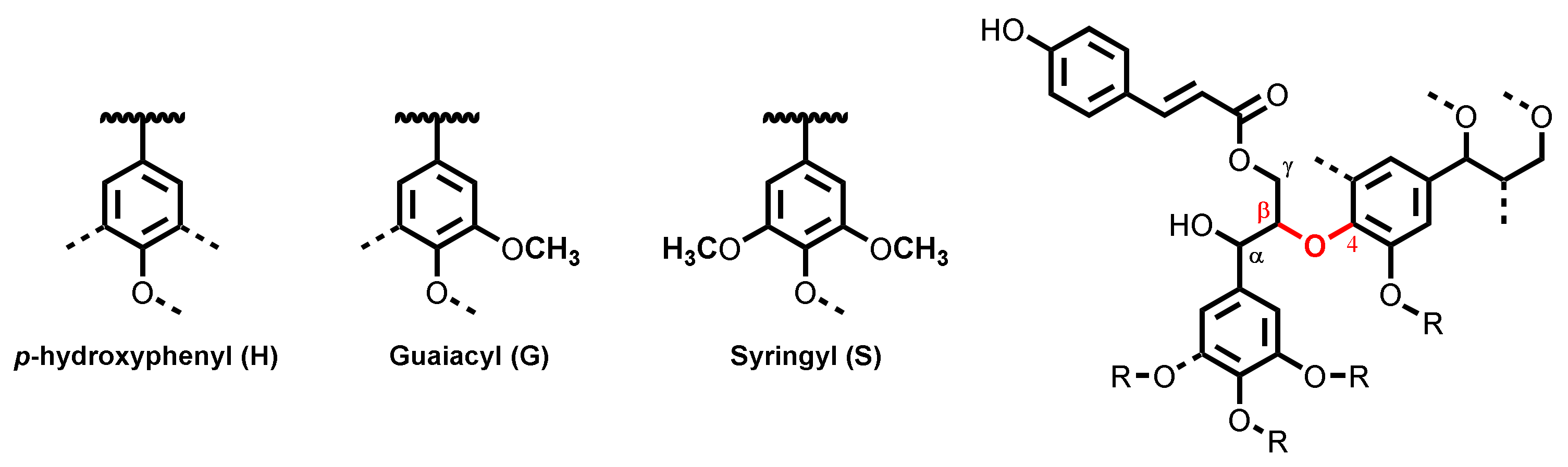
  11. Vanholme, R.; Demedts, B.; Morreel, K.; Ralph, J.; Boerjan, W. Lignin Biosynthesis and Structure. Plant Physiol. 2010, 153, 895–905. [Google Scholar] [CrossRef] [PubMed]
  12. Chio, C.; Sain, M.; Qin, W. Lignin Utilization : A Review of Lignin Depolymerization from Various Aspects. Renew. Sustain. Energy Rev. 2019, 107, 232–249. [Google Scholar] [CrossRef]
  13. Koranyi, T.I.; Fridrich, B.; Pineda, A.; Barta, K. Development of “Lignin-First” Approaches for the Valorization of Lignocellulosic Biomass. Molecules 2020, 25, 2815. [Google Scholar] [CrossRef] [PubMed]
  14. Dessbesell, L.; Paleologou, M.; Leitch, M.; Pulkki, R.; Xu, C. Global Lignin Supply Overview and Kraft Lignin Potential as an Alternative for Petroleum-Based Polymers. Renew. Sustain. Energy Rev. 2020, 123, 109768. [Google Scholar] [CrossRef]
  15. Bajwa, D.S.; Pourhashem, G.; Ullah, A.H.; Bajwa, S.G. A Concise Review of Current Lignin Production, Applications, Products and Their Environment Impact. Ind. Crops Prod. 2019, 139, 111526. [Google Scholar] [CrossRef]
  16. Jõul, P.; Ho, T.T.; Kallavus, U.; Konist, A.; Leiman, K.; Salm, O.S.; Kulp, M.; Koel, M.; Lukk, T. Characterization of Organosolv Lignins and Their Application in the Preparation of Aerogels. Materials 2022, 15, 2861. [Google Scholar] [CrossRef]
  17. Bauer, S.; Sorek, H.; Mitchell, V.D.; Ibáñez, A.B.; Wemmer, D.E. Characterization of Miscanthus Giganteus Lignin Isolated by Ethanol Organosolv Process under Reflux Condition. J. Agric. Food Chem. 2012, 60, 8203–8212. [Google Scholar] [CrossRef]
  18. Tribot, A.; Amer, G.; Abdou Alio, M.; de Baynast, H.; Delattre, C.; Pons, A.; Mathias, J.D.; Callois, J.M.; Vial, C.; Michaud, P.; et al. Wood-Lignin: Supply, Extraction Processes and Use as Bio-Based Material. Eur. Polym. J. 2019, 112, 228–240. [Google Scholar] [CrossRef]
  19. Chan, J.C.; Paice, M.; Zhang, X. Enzymatic Oxidation of Lignin: Challenges and Barriers Toward Practical Applications. ChemCatChem 2020, 12, 401–425. [Google Scholar] [CrossRef]
  20. Fache, M.; Boutevin, B.; Caillol, S. Vanillin Production from Lignin and Its Use as a Renewable Chemical. ACS Sustain. Chem. Eng. 2016, 4, 35–46. [Google Scholar] [CrossRef]
  21. Van Den Bosch, S.; Renders, T.; Kennis, S.; Koelewijn, S.F.; Van Den Bossche, G.; Vangeel, T.; Deneyer, A.; Depuydt, D.; Courtin, C.M.; Thevelein, J.M.; et al. Integrating Lignin Valorization and Bio-Ethanol Production: On the Role of Ni-Al2O3 Catalyst Pellets during Lignin-First Fractionation. Green Chem. 2017, 19, 3313–3326. [Google Scholar] [CrossRef]
  22. Shuai, L.; Talebi Amiri, M.; Questell-Santiago, Y.M.; Héroguel, F.; Li, Y.; Kim, H.; Meilan, R.; Chapple, C.; Ralph, J.; Luterbacher, J.S. Formaldehyde Stabilization Facilitates Lignin Monomer Production during Biomass Depolymerization. Science 2016, 354, 329–334. [Google Scholar] [CrossRef] [Green Version]
  23. Bugg, T.D.H.; Ahmad, M.; Hardiman, E.M.; Rahmanpour, R. Pathways for Degradation of Lignin in Bacteria and Fungi. Nat. Prod. Rep. 2011, 28, 1883–1896. [Google Scholar] [CrossRef]
  24. Tian, J.; Pourcher, A.; Bouchez, T.; Gelhaye, E.; Peu, P. Occurrence of Lignin Degradation Genotypes and Phenotypes among Prokaryotes. Appl. Microbiol. Biotechnol. 2014, 98, 9527–9544. [Google Scholar] [CrossRef] [PubMed]
  25. Majumdar, S.; Lukk, T.; Solbiati, J.O.; Bauer, S.; Nair, S.K.; Cronan, J.E.; Gerlt, J.A. Roles of Small Laccases from Streptomyces in Lignin Degradation. Biochemistry 2014, 53, 4047–4058. [Google Scholar] [CrossRef]
  26. Janusz, G.; Pawlik, A.; Sulej, J.; Świderska-Burek, U.; Jarosz-Wilkolazka, A.; Paszczyński, A. Lignin Degradation: Microorganisms, Enzymes Involved, Genomes Analysis and Evolution. FEMS Microbiol. Rev. 2017, 41, 941–962. [Google Scholar] [CrossRef] [PubMed]
  27. Li, C.; Chen, C.; Wu, X.; Tsang, C.; Mou, J.; Yan, J.; Liu, Y.; Sze, C.; Lin, K. Recent Advancement in Lignin Biorefinery: With Special Focus on Enzymatic Degradation and Valorization. Bioresour. Technol. 2019, 291, 121898. [Google Scholar] [CrossRef] [PubMed]
  28. Zámocky, M.; Hofbauer, S.; Schaffner, I.; Gasselhuber, B.; Nicolussi, A.; Soudi, M.; Pirker, K.F.; Furtmüller, P.G.; Obinger, C. Independent Evolution of Four Heme Peroxidase Superfamilies. Arch. Biochem. Biophys. 2015, 574, 108–119. [Google Scholar] [CrossRef] [PubMed]
  29. Kim, S.J.U.N.; Shoda, M. Purification and Characterization of a Novel Peroxidase from Geotrichum Candidum Dec 1 Involved in Decolorization of Dyes. Appl. Environ. Microbiol. 1999, 65, 1029–1035. [Google Scholar] [CrossRef]
  30. Sugano, Y.; Muramatsu, R.; Ichiyanagi, A.; Sato, T.; Shoda, M. DyP, a Unique Dye-Decolorizing Peroxidase, Represents a Novel Heme Peroxidase Family: ASP171 Replaces the Distal Histidine of Classical Peroxidases. J. Biol. Chem. 2007, 282, 36652–36658. [Google Scholar] [CrossRef]
  31. Kim, S.J.; Ishikawa, K.; Hirai, M.; Shoda, M. Characteristics of a Newly Isolated Fungus, Geotrichum Candidum Dec 1, Which Decolorizes Various Dyes. J. Ferment. Bioeng. 1995, 79, 601–607. [Google Scholar] [CrossRef]
  32. Singh, R.; Eltis, L.D. The Multihued Palette of Dye-Decolorizing Peroxidases. Arch. Biochem. Biophys. 2015, 574, 56–65. [Google Scholar] [CrossRef] [PubMed]
  33. Sugawara, K.; Nishihashi, Y.; Narioka, T.; Yoshida, T.; Morita, M.; Sugano, Y. Characterization of a Novel DyP-Type Peroxidase from Streptomyces Avermitilis. J. Biosci. Bioeng. 2017, 123, 425–430. [Google Scholar] [CrossRef]
  34. Ahmad, M.; Roberts, J.N.; Hardiman, E.M.; Singh, R.; Eltis, L.D.; Bugg, T.D.H. Identification of DypB from Rhodococcus Jostii RHA1 as a Lignin Peroxidase. Biochemistry 2011, 50, 5096–5107. [Google Scholar] [CrossRef] [PubMed]
  35. Liers, C.; Bobeth, C.; Pecyna, M.; Ullrich, R.; Hofrichter, M. DyP-like Peroxidases of the Jelly Fungus Auricularia Auricula-Judae Oxidize Nonphenolic Lignin Model Compounds and High-Redox Potential Dyes. Appl. Microbiol. Biotechnol. 2010, 85, 1869–1879. [Google Scholar] [CrossRef]
  36. Min, K.; Gong, G.; Woo, H.M.; Kim, Y.; Um, Y. A Dye-Decolorizing Peroxidase from Bacillus Subtilis Exhibiting Substrate-Dependent Optimum Temperature for Dyes and β-Ether Lignin Dimer. Sci. Rep. 2015, 5, 8245. [Google Scholar] [CrossRef] [PubMed]
  37. Fawal, N.; Li, Q.; Savelli, B.; Brette, M.; Passaia, G.; Fabre, M.; Mathé, C.; Dunand, C. PeroxiBase: A Database for Large-Scale Evolutionary Analysis of Peroxidases. Nucleic Acids Res. 2013, 41, 441–444. [Google Scholar] [CrossRef] [PubMed]
  38. Yoshida, T.; Sugano, Y. A Structural and Functional Perspective of DyP-Type Peroxidase Family. Arch. Biochem. Biophys. 2015, 574, 49–55. [Google Scholar] [CrossRef]
  39. Colpa, D.I.; Fraaije, M.W.; Bloois, E. Van. DyP-Type Peroxidases: A Promising and Versatile Class of Enzymes. J. Ind. Microbiol. Biotechnol. 2014, 41, 1–7. [Google Scholar] [CrossRef]
  40. Salvachúa, D.; Prieto, A.; Martínez, Á.T.; Martínez, M.J. Enzyme from Irpex Lacteus and Its Application in Enzymatic Hydrolysis of Wheat Straw. Appl. Environ. Microbiol. 2013, 79, 4316–4324. [Google Scholar] [CrossRef]
  41. Roberts, J.N.; Singh, R.; Grigg, J.C.; Murphy, M.E.P.; Bugg, T.D.H.; Eltis, L.D. Characterization of Dye-Decolorizing Peroxidases from Rhodococcus Jostii RHA1. Biochemistry 2011, 50, 5108–5119. [Google Scholar] [CrossRef] [PubMed]
  42. Van Bloois, E.; Pazmiño, D.E.T.; Winter, R.T.; Fraaije, M.W. A Robust and Extracellular Heme-Containing Peroxidase from Thermobifida Fusca as Prototype of a Bacterial Peroxidase Superfamily. Appl. Microbiol. Biotechnol. 2010, 86, 1419–1430. [Google Scholar] [CrossRef] [PubMed] [Green Version]
  43. Rahmanpour, R.; Bugg, T.D.H. Characterisation of Dyp-Type Peroxidases from Pseudomonas Fluorescens Pf-5: Oxidation of Mn(II) and Polymeric Lignin by Dyp1B. Arch. Biochem. Biophys. 2015, 574, 93–98. [Google Scholar] [CrossRef] [PubMed]
  44. Bentley, S.D.; Chater, K.F.; Cerdeño-Tárraga, A.M.; Challis, G.L.; Thomson, N.R.; James, K.D.; Harris, D.E.; Quail, M.A.; Kieser, H.; Harper, D.; et al. Complete Genome Sequence of the Model Actinomycete Streptomyces Coelicolor A3(2). Nature 2002, 417, 141–147. [Google Scholar] [CrossRef] [PubMed]
  45. Teder, T.; Lõhelaid, H.; Samel, N. Structural and Functional Insights into the Reaction Specificity of Catalase-Related Hydroperoxide Lyase: A Shift from Lyase Activity to Allene Oxide Synthase by Site-Directed Mutagenesis. PLoS ONE 2017, 12, e0185291. [Google Scholar] [CrossRef]
  46. Breslmayr, E.; Hanžek, M.; Hanrahan, A.; Leitner, C.; Kittl, R.; Šantek, B.; Oostenbrink, C.; Ludwig, R. A Fast and Sensitive Activity Assay for Lytic Polysaccharide Monooxygenase. Biotechnol. Biofuels 2018, 11, 79. [Google Scholar] [CrossRef]
  47. Ollinger, J.; Song, K.B.; Antelmann, H.; Hecker, M.; Helmann, J.D. Role of the Fur Regulon in Iron Transport in Bacillus Subtilis. J. Bacteriol. 2006, 188, 3664–3673. [Google Scholar] [CrossRef] [PubMed]
  48. Biswas, L.; Biswas, R.; Nerz, C.; Ohlsen, K.; Schlag, M.; Schäfer, T.; Lamkemeyer, T.; Ziebandt, A.K.; Hantke, K.; Rosenstein, R.; et al. Role of the Twin-Arginine Translocation Pathway in Staphylococcus. J. Bacteriol. 2009, 191, 5921–5929. [Google Scholar] [CrossRef]
  49. Létoffé, S.; Heuck, G.; Delepelaire, P.; Lange, N.; Wandersman, C. Bacteria Capture Iron from Heme by Keeping Tetrapyrrol Skeleton Intact. Proc. Natl. Acad. Sci. USA 2009, 106, 11719–11724. [Google Scholar] [CrossRef]
  50. Zitare, U.A.; Habib, M.H.; Rozeboom, H.; Mascotti, M.L.; Todorovic, S.; Fraaije, M.W. Mutational and Structural Analysis of an Ancestral Fungal Dye-Decolorizing Peroxidase. FEBS J. 2021, 288, 3602–3618. [Google Scholar] [CrossRef]
  51. Jeong, H.; Barbe, V.; Lee, C.H.; Vallenet, D.; Yu, D.S.; Choi, S.H.; Couloux, A.; Lee, S.W.; Yoon, S.H.; Cattolico, L.; et al. Genome Sequences of Escherichia Coli B Strains REL606 and BL21(DE3). J. Mol. Biol. 2009, 394, 644–652. [Google Scholar] [CrossRef] [PubMed]
  52. Rosano, G.L.; Ceccarelli, E.A. Recombinant Protein Expression in Escherichia Coli: Advances and Challenges. Front. Microbiol. 2014, 5, 172. [Google Scholar] [CrossRef] [PubMed] [Green Version]
  53. Rosano, G.L.; Morales, E.S.; Ceccarelli, E.A. New Tools for Recombinant Protein Production in Escherichia Coli: A 5-Year Update. Protein Sci. 2019, 28, 1412–1422. [Google Scholar] [CrossRef] [PubMed]
  54. Lithwick, G.; Margalit, H. Hierarchy of Sequence-Dependent Features Associated with Prokaryotic Translation. Genome Res. 2003, 13, 2665–2673. [Google Scholar] [CrossRef] [PubMed]
  55. Gustafsson, C.; Govindarajan, S.; Minshull, J. Codon Bias and Heterologous Protein Expression. Trends Biotechnol. 2004, 22, 346–353. [Google Scholar] [CrossRef]
  56. Al-Hawash, A.B.; Zhang, X.; Ma, F. Strategies of Codon Optimization for High-Level Heterologous Protein Expression in Microbial Expression Systems. Gene Rep. 2017, 9, 46–53. [Google Scholar] [CrossRef]
  57. Johansson, A.S.; Bolton-Grob, R.; Mannervik, B. Use of Silent Mutations in CDNA Encoding Human Glutathione Transferase M2-2 for Optimized Expression in Escherichia Coli. Protein Expr. Purif. 1999, 17, 105–112. [Google Scholar] [CrossRef]
  58. Jung, Y.; Kwak, J.; Lee, Y. High-Level Production of Heme-Containing Holoproteins in Escherichia Coli. Appl. Microbiol. Biotechnol. 2001, 55, 187–191. [Google Scholar] [CrossRef]
  59. Chouchane, S.; Lippai, I.; Magliozzo, R.S. Catalase-Peroxidase (Mycobacterium Tuberculosis KatG) Catalysis and Isoniazid Activation. Biochemistry 2000, 39, 9975–9983. [Google Scholar] [CrossRef]
  60. Sudhamsu, J.; Kabir, M.; Airola, M.V.; Patel, B.A.; Yeh, S.R.; Rousseau, D.L.; Crane, B.R. Co-Expression of Ferrochelatase Allows for Complete Heme Incorporation into Recombinant Proteins Produced in E. Coli. Protein Expr. Purif. 2010, 73, 78–82. [Google Scholar] [CrossRef]
  61. Varnado, C.L.; Goodwin, D.C. System for the Expression of Recombinant Hemoproteins in Escherichia Coli. Protein Expr. Purif. 2004, 35, 76–83. [Google Scholar] [CrossRef] [PubMed]
  62. Sugano, Y.; Nakano, R.; Sasaki, K.; Shoda, M. Efficient Heterologous Expression in Aspergillus Oryzae of a Unique Dye- Decolorizing Peroxidase, DyP, of Geotrichum Candidum Dec 1. Appl. Environ. Microbiol. 2000, 66, 1754–1758. [Google Scholar] [CrossRef] [PubMed] [Green Version]
  63. Sugawara, K.; Yoshida, T.; Hirashima, R.; Toriumi, R.; Akiyama, H.; Kakuta, Y.; Ishige, Y.; Sugano, Y. Characterization of Class V DyP-Type Peroxidase SaDyP1 from Streptomyces Avermitilis and Evaluation of SaDyPs Expression in Mycelium. Int. J. Mol. Sci. 2021, 22, 8683. [Google Scholar] [CrossRef] [PubMed]
  64. Linde, D.; Ayuso-Fernández, I.; Laloux, M.; Aguiar-Cervera, J.E.; de Lacey, A.L.; Ruiz-Dueñas, F.J.; Martínez, A.T. Comparing Ligninolytic Capabilities of Bacterial and Fungal Dye-Decolorizing Peroxidases and Class-Ii Peroxidase-Catalases. Int. J. Mol. Sci. 2021, 22, 2629. [Google Scholar] [CrossRef]
  65. Rodrigues, C.F.; Borges, P.T.; Scocozza, M.F.; Silva, D.; Taborda, A.; Brissos, V.; Frazão, C.; Martins, L.O. Loops around the Heme Pocket Have a Critical Role in the Function and Stability of Bsdyp from Bacillus Subtilis. Int. J. Mol. Sci. 2021, 22, 10862. [Google Scholar] [CrossRef]
  66. Chaplin, A.K.; Wilson, M.T.; Worrall, J.A.R. Kinetic Characterisation of a Dye Decolourising Peroxidase from Streptomyces Lividans: New Insight into the Mechanism of Anthraquinone Dye Decolourisation. Dalt. Trans. 2017, 46, 9420–9429. [Google Scholar] [CrossRef]
  67. Liu, Y.; Huang, L.; Guo, W.; Jia, L.; Fu, Y.; Gui, S.; Lu, F. Cloning, Expression, and Characterization of a Thermostable and PH-Stable Laccase from Klebsiella Pneumoniae and Its Application to Dye Decolorization. Process Biochem. 2017, 53, 125–134. [Google Scholar] [CrossRef]
  68. Arregui, L.; Ayala, M.; Gómez-Gil, X.; Gutiérrez-Soto, G.; Hernández-Luna, C.E.; Herrera De Los Santos, M.; Levin, L.; Rojo-Domínguez, A.; Romero-Martínez, D.; Saparrat, M.C.N.; et al. Laccases: Structure, Function, and Potential Application in Water Bioremediation. Microb. Cell Fact. 2019, 18, 200. [Google Scholar] [CrossRef]
  69. Chen, C.; Shrestha, R.; Jia, K.; Gao, P.F.; Geisbrecht, B.V.; Bossmann, S.H.; Shi, J.; Li, P. Characterization of Dye-Decolorizing Peroxidase (DyP) from Thermomonospora Curvata Reveals Unique Catalytic Properties of A-Type DyPs. J. Biol. Chem. 2015, 290, 23447–23463. [Google Scholar] [CrossRef]
  70. Kokkonen, P.; Beier, A.; Mazurenko, S.; Damborsky, J.; Bednar, D.; Prokop, Z. Substrate Inhibition by the Blockage of Product Release and Its Control by Tunnel Engineering. RSC Chem. Biol. 2021, 2, 645–655. [Google Scholar] [CrossRef]
  71. Benslama, O.; Mansouri, N.; Arhab, R. In Silico Investigation of the Lignin Polymer Biodegradation by Two Actinomycetal Peroxidase Enzymes. Mater. Today Proc. 2022, 53, 1–5. [Google Scholar] [CrossRef]
  72. Gilbert, R.G.; Hess, M.; Jenkins, A.D.; Jones, R.G.; Kratochvíl, P.; Stepto, R.F.T. Dispersity in Polymer Science (IUPAC Recommendations 2009). Pure Appl. Chem. 2009, 81, 351–353. [Google Scholar]
  73. Kim, K.H.; Kim, C.S. Recent Efforts to Prevent Undesirable Reactions from Fractionation to Depolymerization of Lignin: Toward Maximizing the Value from Lignin. Front. Energy Res. 2018, 6, 92. [Google Scholar] [CrossRef] [Green Version]
  74. Vignali, E.; Gigli, M.; Cailotto, S.; Pollegioni, L.; Rosini, E.; Crestini, C. The Laccase-Lig Multienzymatic Multistep System in Lignin Valorization. ChemSusChem 2022, 15, e202201147. [Google Scholar] [CrossRef]
  75. Rahmanpour, R.; King, L.D.W.; Bugg, T.D.H. Identification of an Extracellular Bacterial Flavoenzyme That Can Prevent Re-Polymerisation of Lignin Fragments. Biochem. Biophys. Res. Commun. 2017, 482, 57–61. [Google Scholar] [CrossRef]
  76. Musengi, A.; Durrell, K.; Prins, A.; Khan, N.; Agunbiade, M.; Kudanga, T.; Kirby-McCullough, B.; Pletschke, B.I.; Burton, S.G.; Le Roes-Hill, M. Production and Characterisation of a Novel Actinobacterial DyP-Type Peroxidase and Its Application in Coupling of Phenolic Monomers. Enzyme Microb. Technol. 2020, 141, 109654. [Google Scholar] [CrossRef]
Figure 1. Lignin monomers with potential branchpoints marked with a dashed line; a structure of a possible lignin fragment containing the β-O-4 aryl ether linkage.
Energies 16 01557 g001
Figure 2. Optimum pH of dye-decolorizing peroxidases of S. coelicolor toward oxidation of ABTS. Data are average values of three independent measurements.
Energies 16 01557 g002
Figure 3. The temperature dependency of ScDyPs. Peroxidase activity was measured at pH 3.5 (50 mM sodium citrate) using ABTS as substrate. 1 mM H2O2 was used as a co-substrate. Experiments were conducted in triplicate.
Energies 16 01557 g003
Figure 4. Thermostability of ScDyPs. The enzyme stocks were incubated in 50 mM sodium citrate (pH 3.5), at 30 °C. At selected times, the activities were measured using ABTS and 1 mM H2O2. Experiments were conducted in triplicate.
Energies 16 01557 g004
Figure 5. The characterization of lignin-remodeling activities of ScDyPs by GPC. Panel (A) Mxg-lignin, panel (B) aspen lignin. The treatment of Mxg-lignin with ScDyPB led to the polymerization of lignin polymer. The treatment of aspen lignin with ScDyPAs resulted in lignin depolymerization.
Energies 16 01557 g005
Table 1. The annotations of DyP-type peroxidases of S. coelicolor A3(2) and their corresponding operon neighbors, as assigned in the public databases.
ScDyPLocus TagAnnotation 1
ScDyPBSCO7193putative iron-dependent peroxidase
SCO7192sigma factor
SCO7194putative polysaccharide biosynthesis protein
ScDyP1ASCO2276ferrous iron transport peroxidase
SCO2272, SCO2274heme ABC transporter
SCO2273hemin transport system permease
SCO2275ferrous iron transport periplasmic protein
SCO2277ferrous iron transport permease
SCO2278hypothetical protein
ScDyP2A 2SCO3963DyP-type peroxidase family protein
SCO3971conserved hypothetical protein
SCO3970Xaa-Pro aminopeptidase
SCO3969possible ATP/GTP-binding protein
SCO3968putative integral membrane protein
SCO3967putative membrane protein
SCO3966, SCO3965putative copper metallochaperone
SCO3964copper transport protein
SCO3962prephenate dehydratase
SCO3961seryl-tRNA synthetase
SCO3960hydrolase (HAD superfamily)
1 Annotation is based on JGI IMG/M and the SEED databases. 2 The operon for ScDyP2A is encoded on the complement strand.
Table 2. Reinheitszahl values of ScDyPs before and after the heme reconstitution.
Rz ValueScDyP1AScDyP2AScDyPBScDyP1A COScDyP2A CO
Before reconstitution with hemin0.78 ± 0.020.51 ± 0.022.36 ± 0.042.01 ± 0.112.02 ± 0.01
After reconstitution with hemin1.33 ± 0.081.14 ± 0.022.32 ± 0.071.92 ± 0.032.17 ± 0.03
CO Codon optimized genes for the expression out of E. coli BL21 (DE3).
Table 3. Kinetic parameters for the oxidation of soluble model substrate ABTS by ScDyPs. Apparent steady-state kinetic parameters were measured at pH 3.5 using 1 mM H2O2 as a co-substrate. Data are average values of three independent measurements.
ScDyP1AScDyP2AScDyPB
KM (mM)1.4 ± 0.152.3 ± 0.90.08 ± 0.02
kcat (s−1)7.6 ± 1.131.7 ± 14.648.6 ± 5.7
kcat/KM (mM−1·s−1)5.4 ± 0.213.8 ± 1.3607 ± 95.2
Ki (mM)4.3 ± 1.41.6 ± 1.00.4 ± 0.1
Table 4. Mn, Mw, and the molar-mass dispersity (ĐM) of Mxg- (panel A) and aspen (panel B) ethanol organosolv lignins treated with ScDyPs determined by GPC analysis. Solutions containing lignins at 1 mg/mL in HEPES buffer (pH 8.0) and 2 mM H2O2 without enzymatic treatment, or treated with 10 μM enzyme were incubated for 24 h. Samples were centrifuged, pellets lyophilized and dissolved, and the lignins were analyzed using GPC.
Mxg-LigninAspen Lignin
MnMwĐMMnMwĐM
Without enzyme1509 ± 102435 ± 351.612772 ± 515533 ± 532.0
+ScDyP1A1502 ± 82357 ± 261.572589 ± 124705 ± 721.82
+ScDyP2A1489 ± 112332 ± 451.572429 ± 1064371 ± 2581.8
+ScDyPB1856 ± 212921 ± 281.572994 ± 335607 ± 791.87
Table 5. The most prominent LMW compounds found in Mxg-lignin (A) and aspen lignin (B), and the influence of ScDyPs treatment to the peak area reduction. The peak areas are represented in percentages where the result from the untreated sample is set to 100%, and the treated samples are defined as the percentage peak area left after the treatment.
(A)
Unit
Compound NameWithout
Treatment
ScDyP1AScDyP2AScDyPB
Hp-Hydroxycinnamic acid, ethyl ester10078782
GEthyl (E)-ferulate10067752
G(E)-4-(3-Hydroxyprop-1-en-1-yl)-2-methoxyphenol10077699
GGlyceryl ferulate1008795nd *
Strans-Sinapyl alcohol10063695
(B)
Unit
Compound NameWithout
Treatment
ScDyP1AScDyP2AScDyPB
Strans-Sinapyl alcohol100535537
G(E)-4-(3-Hydroxyprop-1-en-1-yl)-2-methoxyphenol100445216
Strans-Sinapaldehyde100646853
GButyrovanillone1009273nd *
G2-Propanone, 1-hydroxy-3-(4-hydroxy-3-methoxyphenyl)-100nd *nd *nd *
* not detected.
Disclaimer/Publisher’s Note: The statements, opinions and data contained in all publications are solely those of the individual author(s) and contributor(s) and not of MDPI and/or the editor(s). MDPI and/or the editor(s) disclaim responsibility for any injury to people or property resulting from any ideas, methods, instructions or products referred to in the content.

