Case Report

Ultra-High-Risk Gestational Choriocarcinoma of the Ovary Associated with Ectopic Pregnancy

1
Faculty of Medicine, University of Maribor, 2000 Maribor, Slovenia
2
Department of Pathology, University Medical Centre, 2000 Maribor, Slovenia
3
Clinic for Gynaecology and Perinatology, University Medical Centre, 2000 Maribor, Slovenia
*
Author to whom correspondence should be addressed.
Curr. Oncol. 2023, 30(2), 2217-2226; https://doi.org/10.3390/curroncol30020171
Submission received: 30 December 2022 / Revised: 6 February 2023 / Accepted: 7 February 2023 / Published: 11 February 2023
(This article belongs to the Section Gynecologic Oncology)

Abstract

:
Gestational choriocarcinoma of the ovary is an exceptionally rare and highly aggressive tumor. Preoperative diagnosis of extrauterine choriocarcinoma is difficult due to nonspecific clinical presentation and its resemblance to ectopic pregnancy. Without molecular genetic analysis, it is not possible to reliably differentiate gestational from non-gestational choriocarcinoma. Here, we present a case of a 44-year-old woman who presented to our emergency department with complaints of pelvic pain, vaginal bleeding, and amenorrhea. Because of a recent history of conservatively managed ectopic pregnancy, the patient underwent emergency laparoscopy. Right-sided salpingo-oophorectomy was performed due to intraoperatively suspected ovarian ectopic pregnancy. Histopathology results revealed the diagnosis of ovarian choriocarcinoma of possible gestational origin. It was classified as FIGO stage IV and WHO ultra-high-risk, and she underwent multi-agent chemotherapy without major complications. She has remained in complete remission after a 12-month follow-up. Considering the rarity of this diagnosis, we conducted a literature review including all published cases of suspected gestational choriocarcinomas of the ovary. We conclude that due to the rarity of this entity, preoperative differentiating between ovarian ectopic pregnancy and ovarian choriocarcinoma is extremely challenging, and without molecular genetic analysis, it is not possible to identify the genetic origin of the tumor.

1. Introduction

Gestational choriocarcinoma is a malignant neoplasm that belongs to a group of interrelated processes called gestational trophoblastic disease (GTD) [1]. GTD is characterized by abnormal proliferation of trophoblastic tissue. It includes the premalignant partial and complete hydatidiform mole and malignant invasive mole, choriocarcinoma, placental site trophoblastic tumor, and epithelioid trophoblastic tumor. Malignant variants of GTD are collectively known as gestational trophoblastic neoplasia [2,3].
Choriocarcinoma is the most aggressive type of GTN, and patients tend to develop early systemic metastases [4]. It can be either gestational or non-gestational in origin. However, distinguishing between gestational and non-gestational choriocarcinoma is difficult, and the tumor is generally treated without considering its origin [5].
Gestational choriocarcinoma usually originates in the uterine cavity, but it can rarely also affect fallopian tubes, ovaries, cervix, vagina, and abdominopelvic cavity [6,7]. It most commonly follows a molar pregnancy, but it can also develop after normal pregnancy, miscarriage, ectopic pregnancy, or termination of pregnancy [8].
Non-gestational choriocarcinoma is a rare type of tumor with trophoblastic differentiation originating from germ cells [9]. In contrast to gestational choriocarcinoma, it is unrelated to pregnancy with no paternal genes present. Non-gestational choriocarcinomas often present in children and young women. There are two types of non-gestational choriocarcinoma: the pure type, which consists of only choriocarcinoma with no other germ cell elements, and the mixed type. The latter contains other germ cell tumors such as immature teratomas or dysgerminomas [10].
Differences in gestational and non-gestational types are summarized in Table 1.
Neither gestational nor non-gestational choriocarcinoma are often considered in the initial differential diagnosis of an adnexal mass [10]. Extrauterine choriocarcinomas are exceedingly rare and can present with very aggressive clinical course; hence, timely diagnosis is crucial [6]. Due to clinical symptoms resembling ectopic pregnancy, correct diagnosis before commencing treatment is challenging [11]. These include amenorrhea, vaginal bleeding, pelvic pain, and increased serum β-hCG [12]. This means that the diagnosis of gestational choriocarcinoma is often incidental on histopathological examination after laparoscopy in suspected ectopic pregnancies [8,12].
Here, we present a case of an ultra-high-risk ovarian gestational choriocarcinoma following an ectopic pregnancy and discuss the diagnosis and treatment together with a brief review of the literature.

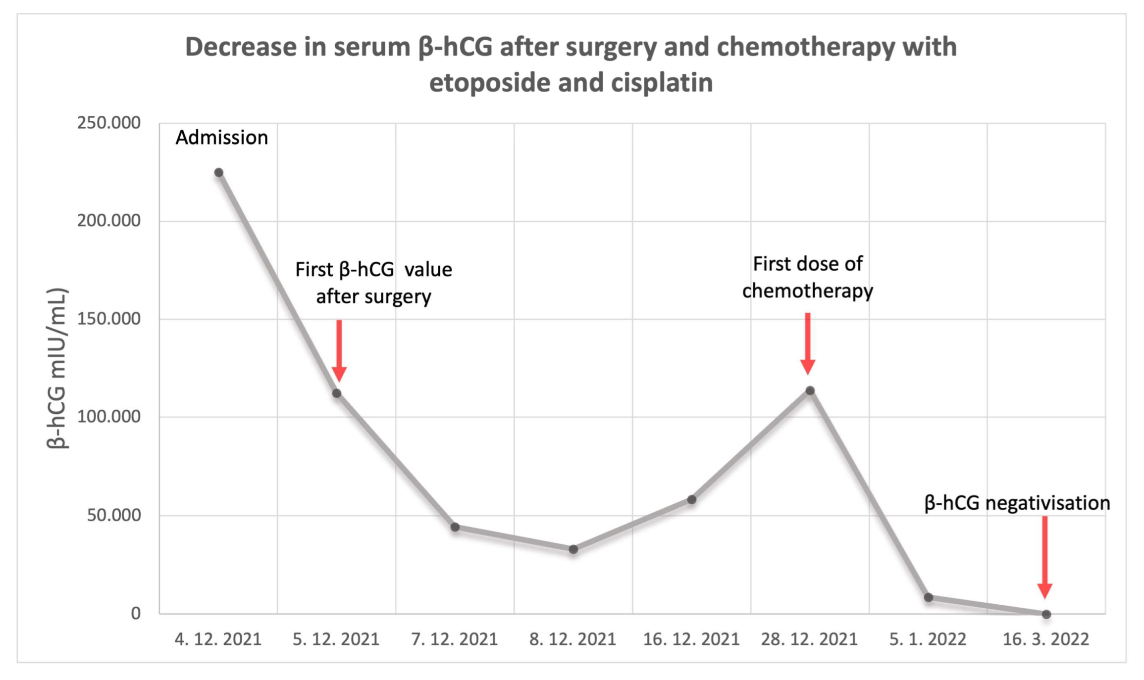
2. Case Presentation

A 44-year-old woman presented to our emergency clinic due to amenorrhea of 2 weeks, several days of vaginal bleeding, and one month history of pelvic pain. She was previously managed at our clinic 4 months before this episode due to tubal ectopic pregnancy and was then treated with an intramuscular injection of methotrexate (75 mg i.m.). In the follow-up visit one month after application, serum β-hCG level was negative. She was previously healthy without any evidence of chronic disease or any other underlying medical conditions. She had a history of one spontaneous miscarriage and one full-term pregnancy and gave birth with caesarean section five years ago. She was not taking any medications and had no known allergies. She did not use any form of hormonal contraception. Physical examination at presentation revealed abdominal pain on palpation, cervical motion tenderness and adnexal tenderness. A bloody discharge from the cervical canal was noted. Serum β-hCG was above 225,000 IU/L. The patient underwent transvaginal ultrasonography, which showed a normal uterine cavity with thin endometrium and no signs for intrauterine gestational sac or embryo. An ectopic mass of 114 × 86 × 82 mm was visualized in the right adnexal region. An area of healthy ovarian tissue was seen adjacent to the mass. Due to clinical suspicion of persistent tubal ectopic pregnancy, the patient underwent urgent laparoscopy. There was a significant hemoperitoneum and approximately 1000 mL of blood was aspirated. A large tumorous structure with pseudo-capsule was found, filling the whole pelvis. The structure was friable with a tendency to bleed on palpation. Due to excessive bleeding, the patient received a transfusion of 2 units of concentrated erythrocytes. Because the structure was indistinguishable from the ovary and because of heavy bleeding, a right-sided salpingo-oophorectomy was performed. The trophoblastic tissue was attached to the sigmoid colon, posterior uterine wall, and pelvic peritoneum. The tissue was successfully completely removed. Final histopathological diagnosis confirmed ovarian infiltration with choriocarcinoma. Histopathological case characteristics are presented in Figure 1. The pathologist could not confirm the possibility of gestational origin as no other elements of pregnancy were seen. The right fallopian tube was without signs of pregnancy or carcinoma.
After surgical management, β-hCG levels abruptly declined (Figure 2). During the 12th post-operative day, β-hCG started to rise again.
Computed tomography (CT) of the abdomen, thorax and head was performed to rule out the possibility of systemic metastasis of the tumor. Chest CT revealed small parenchymal metastases in all lung segments. Because of a suspected liver lesion, magnetic resonance imaging (MRI) scan of the liver was performed, and it showed multiple suspected metastases in several liver segments, measuring up to 10 mm. The L1 vertebrum was infiltrated with 9.5 mm lesion, suspected for metastasis. Based on these findings, the multidisciplinary medical team concluded that the patient had FIGO IV stage disease. This disease was classified as ultra-high-risk according to the WHO scoring system [3]. The patient was treated with induction chemotherapy with etoposide 150 mg i.v. and cisplatin 30 mg i.v., without acute toxic side effects. After 6 cycles of subsequent etoposide, methotrexate, actinomycin D, cyclophosphamide, and vincristine (EMA-CO) regimen chemotherapy, the follow-up period was unremarkable, and there was no evidence of disease relapse after a 12-month follow-up.

3. Discussion

Ovarian choriocarcinoma is an extremely rare but highly aggressive tumor that can be gestational or non-gestational in origin. Gestational ovarian choriocarcinoma can arise from an ectopic ovarian pregnancy or present as a metastasis from a uterine or tubal choriocarcinoma. Non-gestational ovarian choriocarcinoma originates from germ cells [9]. It is extremely rare, because most malignant germ cell tumors are mixed-type and consist of various malignant components, including immature teratoma, dysgerminoma, yolk sac tumor, and choriocarcinoma [15]. Our case presentation shows an ovarian choriocarcinoma that most likely developed from conservatively managed misdiagnosed ovarian ectopic pregnancy. Ovarian ectopic pregnancies represent less than 1% of all ectopic pregnancies, and conservative diagnosis based on ultrasound imaging is extremely challenging [16]. Often, the diagnosis is confirmed after final surgical management. As in our case, misdiagnosis of tubal ectopic pregnancy based on imaging is likely.
Pure non-gestational choriocarcinoma’s estimated incidence is 1:369 million and is significantly rarer compared to primary extrauterine gestational choriocarcinoma with a prevalence of 1:5335 ovarian pregnancies and 1:2.2 million normal intrauterine pregnancies. Specific prevalence of choriocarcinoma primarily originating from ovarian gestation is unknown [14].
Ovarian choriocarcinoma usually presents with irregular vaginal bleeding, abdominal pain, adnexal mass, and increased serum β-hCG. It can also present with metastatic manifestations to different organs, most commonly to the lungs and brain. Because clinical symptoms are non-specific, ovarian choriocarcinoma can be easily mistaken for other more common diseases, as in our case with ectopic pregnancy [14,17]. Gestational and non-gestational choriocarcinomas are difficult to differentiate based on history, clinical presentation, or histology [14]. In cases when the disease develops in girls prior to puberty, women who have never had sexual intercourse, or are definitely unable to conceive, we can confirm the diagnosis of non-gestational choriocarcinoma [15]. Non-gestational type choriocarcinoma involves the patients with an average age of 13 and is largely confined to females under 20 [13]. In women of reproductive age who are sexually active, gestational origin is significantly more likely. Both types of choriocarcinomas produce very high amounts of β-hCG. The value ranges from 3 to 100 times more than a normal pregnancy [7]. However, β-hCG levels are usually lower in non-gestational variants in comparison to gestational type [13]. Serum β-hCG elevation leads to pseudopuberty in premenarchal patients, while patients in reproductive age usually present with menstrual abnormalities (mainly amenorrhea) [18]. The presence of a well-developed corpus luteum of pregnancy adjacent to the tumor may also be indicative of a gestational origin. The corpus luteum or theca lutein cysts can also develop due to excess of hCG [13,19]. For definite differentiation of these two types of ovarian choriocarcinomas, DNA short tandem repeat (STR) analysis can be performed [14]. A STR is a microsatellite consisting of a unit of 2–10 nucleotides repeated several to hundreds of times, and STR analysis allows evaluation of the highly polymorphic STR loci where each individual shows differing numbers of repeated sequences of nucleotides. The genome in non-gestational choriocarcinoma comprises only a maternal (patient) allele, whereas gestational choriocarcinoma contains a paternal allele [15].
Since choriocarcinomas tend to have a good response to chemotherapy, this is the primary choice of treatment. However, the diagnosis of choriocarcinoma is commonly made after surgical removal of adnexal mass and histological examination. Surgery is also useful in controlling life-threatening hemorrhage from metastatic lesions [19]. Gestational choriocarcinoma can be treated with methotrexate, actinomycin D, or etoposide as a single agent, or with combined agents such as EMA-CO in high-risk cases. An ultra-high-risk GTN requires salvage chemotherapy in the form of low-dose induction chemotherapy consisting of etoposide-cisplatin (EP) as ultra-high-risk cases were associated with increased risk of early death either due to the tumor pathology itself or respiratory compromise and hemorrhage secondary to a heavy burden of disease or rapid tumor destruction with full-dose chemotherapy. Presently, there is very limited information about the ultra-high-risk subgroup due to its rarity [20]. In cases of resistant or relapsed disease, regimens including cisplatin such as TP (paclitaxel, cisplatin) and BEP (bleomyxin, etoposide, cisplatin) are used. When non-gestational choriocarcinoma develops as a component of mixed-type germ cell tumor, it responds well to treatment with surgery and BEP regimen. Pure-type non-gestational choriocarcinoma behaves more aggressively and is treated with cisplatin regimens [15,21]. Gestational ovarian choriocarcinoma has a better prognosis than their non-gestational counterpart. Gestational type is extremely sensitive to chemotherapy, with the overall cure rate approaching 98 % in specialized centers. However, some studies suggest that the surgical stage of pure ovarian choriocarcinoma may be a more important determinant of clinical outcome compared to being of gestational or non-gestational type [13,14].
We have performed a literature review and identified only 12 cases of ovarian gestational choriocarcinomas (Table 2). In five cases, the diagnosis of gestational origin was genetically confirmed (Figure 3), while in others, this was concluded based on the presence of a corpus luteum [21]. As in our case, five women were initially treated for suspected ectopic pregnancy. The remaining seven cases were already preoperatively suspected ovarian tumors. This shows that ectopic pregnancy is the main differential diagnosis, and the presentation is highly similar to extrauterine choriocarcinoma. Correct preoperative diagnosis could avoid often unnecessary surgical procedure [14]. Although staging and risk assessment based on FIGO and World Health Organization (WHO) are today the basis for management and prognosis of GTN, these were often not reported [2]. The 2000 FIGO staging system, which is the standard classification for GTN, was reported in only five cases. A modified WHO prognostic index score based on prognostic factors modified by FIGO was applied only in two cases. There was only one relapse reported out of 13 cases (7.7%), confirming excellent prognosis after standard chemotherapy treatment [21].

4. Conclusions

Ovarian choriocarcinoma is a challenging clinical diagnosis due to its nonspecific clinical presentation. It is commonly diagnosed after surgical management of suspected ectopic pregnancy. Gestational choriocarcinoma is significantly more common compared to non-gestational choriocarcinoma, and the origin can be suspected based on clinical characteristics, but the definitive diagnosis can only be confirmed after molecular genetic analysis. Although there are only few cases of ovarian choriocarcinomas published, the reported prognosis after chemotherapy is excellent.

Author Contributions

Conceptualization, E.P.M.; writing—original draft preparation, E.P.M. and N.L.; writing—review and editing, J.K., T.B. and M.S.; visualization, T.B., M.S., E.P.M.; supervision, J.K. and M.S.; project administration, J.K. All authors have read and agreed to the published version of the manuscript.

Funding

This research received funding by the Internal Research Project funding UMC Maribor, reg no. IRP-2019/01-05 and Slovenian Research Agency (grant number J3-4523).

Institutional Review Board Statement

This case report was conducted in accordance with the Declaration of Helsinki, and national legislation. Consent was taken in accordance with national guidelines.

Informed Consent Statement

Written informed consent has been obtained from the patient to publish this paper.

Data Availability Statement

The data presented in this study are available on request from the corresponding author.

Conflicts of Interest

The authors declare no conflict of interest.

References

  1. Tarney, C.M.; Tian, C.; Craig, E.R.; Crothers, B.A.; Chan, J.K.; Gist, G.D.; Bateman, N.W.; Conrads, T.P.; Hamilton, C.A.; Maxwell, G.L.; et al. Relative Effects of Age, Race, and Stage on Mortality in Gestational Choriocarcinoma. Int. J. Gynecol. Cancer 2018, 28, 338–345. [Google Scholar] [CrossRef] [PubMed]
  2. Ngan, H.Y.S.; Seckl, M.J.; Berkowitz, R.S.; Xiang, Y.; Golfier, F.; Sekharan, P.K.; Lurain, J.R.; Massuger, L. Diagnosis and management of gestational trophoblastic disease: 2021 update. Int. J. Gynecol. Obstet. 2021, 155 (Supp. 1), 86–93. [Google Scholar] [CrossRef]
  3. Lukinovic, N.; Malovrh, E.P.; Takac, I.; Sobocan, M.; Knez, J. Advances in diagnostics and management of gestational trophoblastic disease. Radiol. Oncol. 2022, 56, 430–439. [Google Scholar] [CrossRef] [PubMed]
  4. Soper, J.T. Gestational Trophoblastic Disease: Current Evaluation and Management. Obstet. Gynecol. 2021, 137, 355–370. [Google Scholar] [CrossRef]
  5. Sakurai, S.; Asano, R.; Furugori, M.; Shigeta, H. A rare case of gestational ovarian choriocarcinoma coexistent with intrauterine pregnancy. Taiwan. J. Obstet. Gynecol. 2022, 61, 708–712. [Google Scholar] [CrossRef]
  6. Karaman, E.; Çetin, O.; Kolusari, A.; Bayram, I. Primary Tubal Choriocarcinoma Presented as Ruptured Ectopic Pregnancy. J. Clin. Diagn. Res. 2015, 9, 17–18. [Google Scholar] [CrossRef]
  7. Anjum, A.S.; Maqsood, H.; Younus, S.; Anjum, S.; Fatima, M. A Rare Case of Non-Gestational Metastatic Ovarian Choriocarcinoma: Case Report and Literature Review with a Special Emphasis on Imaging. Cureus 2021, 13, 13121. [Google Scholar] [CrossRef]
  8. Savage, P.; Winter, M.; Parker, V.; Harding, V.; Sita-Lumsden, A.; Fisher, R.A.; Harvey, R.; Unsworth, N.; Sarwar, N.; Short, D.; et al. Demographics, natural history and treatment outcomes of non-molar gestational choriocarcinoma: A UK population study. BJOG Int. J. Obstet. Gynaecol. 2020, 127, 1102–1107. [Google Scholar] [CrossRef]
  9. Lv, L.; Yang, K.; Wu, H.; Lou, J.; Peng, Z. Pure choriocarcinoma of the ovary: A case report. J. Gynecol. Oncol. 2011, 22, 135–139. [Google Scholar] [CrossRef] [PubMed]
  10. Cronin, S.; Ahmed, N.; Craig, A.D.; King, S.; Huang, M.; Chu, C.S.; Mantia-Smaldone, G.M. Non-Gestational Ovarian Choriocarcinoma: A Rare Ovarian Cancer Subtype. Diagnostics 2022, 12, 560. [Google Scholar] [CrossRef]
  11. Han, V.; Kaye, S. A Rare Case of Gestational Choriocarcinoma Presenting as Cornual Ectopic Pregnancy. J. Obstet. Gynaecol. Can. 2018, 40, 351–353. [Google Scholar] [CrossRef]
  12. Xu, S.; Song, X.; Jin, C.; Li, Y. Tubal choriocarcinoma presented as ruptured ectopic pregnancy: A case report and review of the literature. World J. Surg. Oncol. 2020, 18, 245. [Google Scholar] [CrossRef]
  13. Nishino, K.; Yamamoto, E.; Ikeda, Y.; Niimi, K.; Yamamoto, T.; Kajiyama, H. A poor prognostic metastatic nongestational choriocarcinoma of the ovary: A case report and the literature review. J. Ovarian Res. 2021, 14, 56. [Google Scholar] [CrossRef]
  14. Mood, N.I.; Samadi, N.; Rahimi-Moghaddam, P.; Sarmadi, S.; Eftekhar, Z.; Yarandi, F. Pure ovarian choriocarcinoma: Report of two cases. J. Res. Med. Sci. Off. J. Isfahan Univ. Med. Sci. 2009, 14, 327–330. [Google Scholar]
  15. Knez, J.; Day, A.; Jurkovic, D. Ultrasound imaging in the management of bleeding and pain in early pregnancy. Best Pract. Res. Clin. Obstet. Gynaecol. 2014, 28, 621–636. [Google Scholar] [CrossRef]
  16. Adow, M.T.; Gebresilasie, S.F.; Abebe, N.A. Primary Ovarian Choriocarcinoma: Rare Entity. Case Rep. Obstet. Gynecol. 2021, 2021, 4545375. [Google Scholar] [CrossRef] [PubMed]
  17. Heo, E.J.; Choi, C.H.; Park, J.M.; Lee, J.W.; Bae, D.S.; Kim, B.G. Primary ovarian choriocarcinoma mimicking ectopic pregnancy. Obstet. Gynecol. Sci. 2014, 57, 330–333. [Google Scholar] [CrossRef] [PubMed]
  18. Corakçi, A.; Ozeren, S.; Ozkan, S.; Gürbüz, Y.; Ustün, H.; Yücesoy, I. Pure nongestational choriocarcinoma of ovary. Arch. Gynecol. Obstet. 2005, 271, 176–177. [Google Scholar] [CrossRef]
  19. Naniwadekar, M.R.; Desai, S.R.; Kshirsagar, N.S.; Angarkar, N.N.; Dombale, V.D.; Jagtap, S.V. Pure choriocarcinoma of ovary diagnosed by fine needle aspiration cytology. Indian J. Pathol. Microbiol. 2009, 52, 417–420. [Google Scholar] [CrossRef]
  20. Patel, S.; Arora, R.; Tiwari, R.; Poddar, P.; Desai, A.; Mankad, M.; Panchal, H.P. Management of “Ultra-High Risk” Gestational Trophoblastic Neoplasia at a Tertiary Center in India. Indian J. Med. Paediatr. Oncol. 2020, 41, 345. [Google Scholar] [CrossRef]
  21. Jia, N.; Chen, Y.; Tao, X.; Ou, E.; Lu, X.; Feng, W. A gestational choriocarcinoma of the ovary diagnosed by DNA polymorphic analysis: A case report and systematic review of the literature. J. Ovarian Res. 2017, 10, 46. [Google Scholar] [CrossRef] [Green Version]
  22. Kazemi, S.N.; Raoufi, M.; Moghaddam, N.A.; Tabatabaeefar, M.; Ganjooei, T.A. Ovarian ectopic pregnancy misdiagnosed as gestational choriocarcinoma: A case report. Ann. Med. Surg. 2022, 73, 103236. [Google Scholar] [CrossRef]
  23. Haruma, T.; Ogawa, C.; Nishida, T.; Kusumoto, T.; Nakamura, K.; Seki, N.; Katayama, T.; Hiramatsu, Y. Pure Choriocarcinoma of the Ovary in Silver-Russell Syndrome. Acta Med. Okayama. 2015, 69, 183–188. [Google Scholar] [PubMed]
  24. Gerson, R.F.; Lee, E.Y.; Gorman, E. Primary extrauterine ovarian choriocarcinoma mistaken for ectopic pregnancy: sonographic imaging findings. AJR Am. J. Roentgenol. 2007, 189, W280–W283. [Google Scholar] [CrossRef]
  25. Vautier-Rit, S.; Ducarme, G.; Devisme, L.; Vinatier, D.; Leroy, J.-L. Primary choriocarcinoma of the ovary: A case report. Obstet. Fertil. 2004, 32, 620–623. [Google Scholar] [CrossRef]
  26. Aucouturier, J.S.; Bader, G.; El Fata, G.; Guyot, B.; Louboutin, A.; Camus, E. Ovarian choriocarcinoma: About one case. Gynecol. Obstet. Fertil. 2003, 31, 539–542. [Google Scholar] [CrossRef] [PubMed]
  27. Namba, A.; Nakagawa, S.; Nakamura, N.; Takazawa, Y.; Kugu, K.; Tsutsumi, O.; Taketani, Y. Ovarian choriocarcinoma arising from partial mole as evidenced by deoxyribonucleic acid microsatellite analysis. Obstet. Gynecol. 2003, 102, 991–994. [Google Scholar] [CrossRef] [PubMed]
  28. Lorigan, P.C.; Grierson, A.J.; Goepel, J.R.; Coleman, R.E.; Goyns, M.H. Gestational choriocarcinoma of the ovary diagnosed by analysis of tumour DNA. Cancer Lett. 1996, 104, 27–30. [Google Scholar] [CrossRef] [PubMed]
Figure 1. Histopathological features of the gestational choriocarcinoma. (a) Microscopic appearance of the ovary shows infiltration with pure gestational choriocarcinoma with widespread necrosis (H&E). (b) The tumor was positive for b-human chorionic gonadothropin., which is relatively specific for choriocarcinoma. (c) Positive inhibin stain indicates the presence of syncytiotrophoblast. (d) Intermediate magnification micrograph of choriocarcinoma (H&E). Choriocarcinomas consist of two cell populations: cytotrophoblast with pale/clear cytoplasm and syncytiotrophoblast with hyperchromatic cytoplasm and typically multinucleated cells.
Curroncol 30 00171 g001
Figure 2. Decrease in serum β-hCG levels from admission to negativization. Biochemical follow-up was performed during the initial treatment follow as well as after systemic therapy treatment.
Curroncol 30 00171 g002
Figure 3. Identified cases of ovarian choriocarcinoma and their genetic origin.
Curroncol 30 00171 g003
Table 1. Differences in gestational and non-gestational choriocarcinoma [9,13,14].
CharacteristicsGestational TypeNon-Gestational Type
AgeReproductive periodAverage age of 13 years, most patients are under 20
History of normal, molar, or ectopic pregnancy or miscarriageYesNo
Histology/Elements of other germ cell tumours are significant for mixed-type
Corpus luteumYesNo
GenomeTotally or partially different from the patientIdentical to the patient
Serum β-hCGHigherLower
TreatmentLow-risk: single agent (methotrexate, actinomycin D or etoposide)
High-risk: combination chemotherapy (e.g., EMA-CO)
Mixed-type: surgery and BEP regimen
Pure type: cisplatin regimens (e.g., BEP)
PrognosisBetterWorse (especially pure type)
EMA-CO = etoposide, methotrexate, actinomycin D, cyclophosphamide, vincristine; BEP = bleomyxin, etoposide, cisplatin.
Table 2. Published cases of gestational ovarian choriocarcinoma.
AgeClinical PresentationβhCG (mlU/mL)SurgeryMetastasis at the Time of DiagnosisFIGO Grade/WHO Risk ScoreChemotherapyOutcomeGestational Origin Confirmed
Our case44Abdominal pain, vaginal bleeding>225,000Laparoscopic right-sided adnexectomyLiver, lung, boneFIGO IV
WHO 16
EP, EMA-COcomplete remissionno
Sakurai et al., 2022 [5]38Lower left abdominal pain and abdominal distension2.7 × 1061st surgery: left salpingo-oophorectomy and right ovarian biopsy. Artificial abortion of viable intrauterine pregnancy.
2nd surgery: total hysterectomy including the residual tumor, right salpingo-oophorectomy, and omentectomy.
noFIGO II
WHO 13
EMA-COcomplete remissionyes
Kazemi et al., 2022 [22]35Severe pelvic pain, fatigue, nausea, vomiting, cough33,827Laparotomy, not specifiedlung, brain, kidney, spleenFIGO IVEMA-EP, EMA-CO,
Relapse: 3 cycles of paclitaxel, cisplatin, etoposide, 4 cycles of liposomal doxorubicin and carboplatin,
5 cycles of fluorouracil and dactinomycin
Relapse, death 8 months from the initial diagnosisno
Adow et al., 2021 [16]25Lower abdominal swelling and pain1,000,000Total abdominal
hysterectomy and bilateral salpingo-oophorectomy
not mentioned/BEPComplete remissionno
Jia et al., 2017 [21]27Amenorrhea, lower abdominal pain and vaginal bleeding>200,000Laparoscopic exploration, dissection of the cystic mass of the right ovaryno/EP-EMAComplete remission, patient gave birth 25 months after
chemotherapy
yes
Haruma et al., 2015 [23]19Lower abdominal pain, amenorrhea373,170Left salpingo-oophorectomylung, peritoneum, pelvisFIGO III, WHO > 7 (high risk)EMA-COComplete remissionno
Naniwadekar et al., 2009 [19]19Abdominal pain, vaginal bleeding, palpable abdominal mass380,000Total hysterectomy with removal of bilateral ovarian masses with omentectomyno/EMA-COLost to follow-up after second course of chemotherapyno
Mood et al., 2009 [14]31Signs of acute abdomen and spotting>1000Right salpingo-oophorectomyno/EMA-CEcomplete remissionno
Gerson et al., 2005 [24]33Right lower quadrant abdominal pain564,000First surgery: laparoscopic right salpingo-oophorectomy and resection of a right adnexal mass
Second surgery: total abdominal hysterectomy and left salpingectomy
spleen/EMA-COcomplete remissionno
Vautier-Rit et al., 2004 [25]32Pelvic pain, vaginal bleeding315,000Left-sided ovariectomynoFIGO IcEPcomplete remissionyes
Aucouturier et al., 2003 [26]43Abdominal pain37,260Total hysterectomy with left-sided adnexectomy and omentectomy, multiple peritoneal biopsieslungT3c NOEPcomplete remissionno
Namba et al., 2003 [27]37Amenorrhea990,000Right salpingo-oophorectomy and a curettage of the uterusno/Methotrexate, actinomycin D, cyclophosphamide as neoadjuvant therapy; methotrexate, actinomycin
D, cyclophosphamide as consolidation therapy
The patient remains after follow-up with no signs of recurrenceyes
Lorigan et al., 1996 [28]41Amenorrhea, vaginal bleeding151,500Total abdominal hysterectomy, bilateral salpingo-oophorectomy, and omentectomyno/BEP, salvage therapy Ifosfamide and etoposidecomplete remissionyes
Disclaimer/Publisher’s Note: The statements, opinions and data contained in all publications are solely those of the individual author(s) and contributor(s) and not of MDPI and/or the editor(s). MDPI and/or the editor(s) disclaim responsibility for any injury to people or property resulting from any ideas, methods, instructions or products referred to in the content.

