Article

Microarray Profiling of Differentially Expressed Genes in Coronary Artery Bypass Grafts of High-Risk Patients with Postoperative Cognitive Dysfunctions

by
Noor Anisah Abu Yazit
1,
Norsham Juliana
1,*,
Suhaini Kadiman
2,
Kamilah Muhammad Hafidz
2,
Nur Islami Mohd Fahmi Teng
3,
Nazefah Abdul Hamid
1,
Nadia Effendy
1,
Sahar Azmani
1,
Izuddin Fahmy Abu
4,
Nur Adilah Shuhada Abd Aziz
1 and
Srijit Das
5
1
Faculty Medicine and Health Sciences, Universiti Sains Islam Malaysia, Nilai 71800, Malaysia
2
Anaesthesia and Intensive Care Unit, National Heart Institute, Kuala Lumpur 50400, Malaysia
3
Faculty of Health Sciences, Universiti Teknologi MARA, Puncak Alam 42300, Malaysia
4
Institute of Medical Science Technology, Universiti Kuala Lumpur, Kajang 43000, Malaysia
5
Department of Human & Clinical Anatomy, College of Medicine & Health Sciences, Sultan Qaboos University, Al-Khoud, Muscat 123, Oman
*
Author to whom correspondence should be addressed.
Int. J. Environ. Res. Public Health 2023, 20(2), 1457; https://doi.org/10.3390/ijerph20021457
Submission received: 7 December 2022 / Revised: 7 January 2023 / Accepted: 10 January 2023 / Published: 13 January 2023

Abstract

:
Postoperative cognitive dysfunction (POCD) is cognitive decline after surgery. The authors hypothesized that gene-level changes could be involved in the pathogenesis of POCD. The present study evaluated the incidence of POCD and its associated differentially expressed genes. This was a prospective cohort study conducted on high-risk coronary artery bypass graft patients aged 40 to 75 years. POCD classification was based on a one standard deviation decline in the postoperative scores compared to the preoperative scores. The differentially expressed genes were identified using microarray analysis and validated using quantitative RT-PCR. Forty-six patients were recruited and completed the study. The incidence of POCD was identified using a set of neurocognitive assessments and found to be at 17% in these high-risk CABG patients. Six samples were selected for the gene expression analyses (3 non-POCD and 3 POCD samples). The findings showed five differentially expressed genes in the POCD group compared to the non-POCD group. The upregulated gene was ERFE, whereas the downregulated genes were KIR2DS2, KIR2DS3, KIR3DL2, and LIM2. According to the results, the gene expression profiles of POCD can be used to find potential proteins for POCD diagnostic and predictive biomarkers. Understanding the molecular mechanism of POCD development will further lead to early detection and intervention to reduce the severity of POCD, and hence, reduce the mortality and morbidity rate due to the condition.

1. Introduction

Postoperative cognitive dysfunction (POCD) is a mental impairment that arises after a surgical procedure. It is defined by a range of changes in neurocognitive patients’ condition and behavior for weeks or even months from the time anesthesia is introduced in the intensive care unit until a few months after the surgery [1]. Comparing types of surgery, the prevalence of POCD in cardiac surgery is much higher than in non-cardiac surgery. A study reported that cardiac surgery has a 40% prevalence of POCD compared to 20% for non-cardiac surgery [2]. A meta-analysis discovered that adjuncts in anesthesia, such as dexmedetomidine, may alleviate or reduce the prevalence of POCD in cardiac surgery [3].
Patients are diagnosed with POCD when there are post-operative deficits in their mental states, such as attention, concentration, executive function, memory, visuospatial ability, and psychomotor speed. POCD brings disadvantages to patients’ postoperative care, such as prolonged rehabilitation and hospitalization, reduced quality of life, increased functional recovery time, and increased healthcare costs [4]. The diagnosis requires sensitive and tedious pre-operative and post-operative neuropsychiatric tests. Up to date, multiple standard cognitive tests were utilized to indicate the problem, however, there are no gold standard tests being exclusively set only for POCD. The complication may result in an increased risk of losing their jobs or their independence and leads to a significant reduction in their quality of life [5]. However, the exact mechanism of POCD is unclear due to conflicting results and a lack of evidence [6]. Despite the fact that POCD was discovered a long time ago, there is an ongoing debate regarding the risk factors, diagnostic criteria, pathophysiology, outcomes, and various effects on the effective postoperative management of patients.
The study of POCD continues as researchers actively study gene changes in POCD. Changes in gene levels caused by surgery and anesthesia, according to the authors, play a critical role in the pathogenesis of POCD [7,8]. Often, the diagnosis of POCD is difficult and tedious, with no gold standard methods, classification, or molecular diagnosis [9]. This opens up the opportunity for researchers to explore its gene expression profiling, identification of target genes, construction of interaction network, and pathway analysis. Studies focusing on gene changes with regard to POCD were still lacking, thus allowing authors to explore the area.
The urge to find how POCD occurs grows, as the mechanisms of POCD development are still vague as they are related to various factors. The focus among researchers in the past few years has been neuroinflammation and POCD. Li et al. (2022) describe the pathways by which peripheral inflammation leads to neuroinflammation. Inflammation and immune activation due to surgical stress will release the damage-associated molecular patterns (DAMPs), which include high mobility group protein B1 (HMGB1). HMGB1 secretions were enhanced by proinflammatory factors that induce and maintain the peripheral inflammatory response [10]. The peripheral inflammation may spread to the central nervous system via a specific transporter, periventricular area, blood–brain barrier disruption, homoreceptor, and vagal nerve [6], thereby causing neuroinflammation and cognitive dysfunction. The finding was in accordance with a study where HMGB1 levels in serum were reported to be higher in POCD patients compared to the control group, showing the potential correlation between HMGB1 and POCD [11].
With current technological advancements, many medical breakthroughs have been discovered, especially in understanding disease pathophysiology at a molecular level. Gene studies have been involved in disease diagnosis, prognosis, and risk factor assessment [12]. Apolipoprotein Epsilon-4 (APOE4), one of the genes closely associated with Alzheimer’s disease, was thoroughly studied to determine its relationship with POCD. However, the results were conflicting and had a poor prognosis [13]. Hence, current studies focus on differentially expressed genes to understand the mechanism involved in POCD. A study has summarized different types of microribonucleic acid (miRNA) expressions involved in POCD mechanisms, mainly through neuroinflammation [7]. Another study also found that long non-coding RNA (lncRNA) and messenger RNA (mRNA) were differentially expressed, suggesting their involvement in POCD pathogenesis [9]. In addition, Wang et al. (2019) identified an abnormal expression of circular RNA (circRNA_089763) that was found in POCD patients using the microarray technique [14].
The present study aimed to identify the incidence of POCD using specific neurocognitive assessments and to understand its associated key differentially expressed genes using the microarray technique, in high-risk coronary artery bypass graft patients that received dexmedetomidine in the perioperative period. The findings from this study are novel as understanding the molecular mechanism of POCD development will further lead to early detection and intervention to reduce the severity of POCD, hence, reducing the mortality and morbidity rate due to the condition.

2. Materials and Methods

2.1. Study Design and Patients Selection

This prospective cohort study on high-risk CABG patients was conducted in order to elucidate the key differences in gene expressions for the incidence of POCD. The sampling frame includes CABG patients aged 40 to 75 years. We included patients that were categorized as high risk according to the doctor in charge, able to understand Malay or English language, and not diagnosed as having dementia. The exclusion criteria included; Mini-Mental State Examination (MMSE) score < 24 points; patients with planned off-pump surgery; dialysis-dependent patients; patients with severe hepatic impairment; and pregnant women. Written informed consent was obtained from patients prior to the recruitment. This study was conducted with prior ethical approval by the National Heart Institute Ethical Committee (IJNREC/441/2019).

2.2. Neurocognitive Assessment

Patients were screened for eligibility using the Malay version of the Mini-Mental State Examination (MMSE) [15]. Only patients with MMSE scores of more than 24 were recruited to eliminate patients with poor cognitive performance at baseline. The recruited patients completed the neurocognitive assessment one day before surgery and seven days after. Patients were admitted 2 days prior to the surgery to allow routine preoperative assessments (blood test, echocardiogram, physiotherapy) to be carried out. The patients were enlightened about all the procedures during their clinic sessions and subsequently, all patients selected in the study were screened to assure they were psychologically stable enough to conduct all the preoperative assessments including the cognitive testing. Furthermore, consent from all patients was taken. Hence, they were free to withdraw from the projects at any point without any discrimination on the standard treatment or patient management protocol. On average, patients were allowed to discharge one week after surgery, or even earlier (depending on the doctor’s assessment); hence, day 7 postoperative is the best time to conduct a cognitive assessment. The neurocognitive assessments utilized in this study were: (1) Trail Making Test (TMT) Part A and Part B for visuoperceptual abilities, secondary task switching, and working memory; (2) Digit Span Forward and Backward to test attention and working memory, (3) Digit Symbol Substitution Test (DSST) to test for speed, attention, and manual dexterity [16]; and (4) Clock Drawing Test (CDT) to assess visuoconstructive abilities. To discriminate on patients’ POCD statuses, the 1 SD method was adopted. The method defines a decline in cognitive function based on more than one standard deviation of the postoperative scores compared to the preoperative scores. The SD was calculated from population norms or published samples. Our population norms were taken from Ibrahim et al. (2009), who validated Malay MMSE in our population, where 1 SD equals 3.6 scores [17].

2.3. Data Collection

Demographic data such as age, gender, body mass index (BMI), education level, and intraoperative data such as bypass and cross-clamp times were collected. Blood samples were collected one day before surgery and three days after surgery. Ten ml of blood were collected in anticoagulant tubes and centrifuged at 4 °C at 400× g for 5 min. The samples were then subjected to RNA extraction on the same day.

2.4. RNA Extraction

The RNA extraction protocol was conducted to extract total RNA from samples. GeneJETTM (Thermo Fisher Scientific Inc., Waltham, MA, USA) Whole Blood RNA Purification Mini Kit was used. Briefly, 50–500 µL of blood was centrifuged at 4 °C at 400× g for 5 min, and the supernatant was discarded. The cells were lysed by adding 600 µL of lysis buffer and 450 µL of ethanol using a vortex. The lysate was purified by transferring it to a collection tube inserted with the column, centrifuged at 12,000× g for 1 min. Then, the flow-through solution was discarded, and the purification column was transferred to a new collection tube. To eliminate excess protein and impurities, 700 µL of Wash Buffer 1 was added and centrifuged for 1 min at 12,000× g. The flow-through solution was discarded, and the process was repeated with 500 µL Wash Buffer 2. Another 500 µL Wash Buffer 2 was added and centrifuged for 2 min at 12,000× g. The purification column was then placed into a new 1.5 mL collection tube to collect the RNA. By adding 50 µL of nuclease-free water to the center of the purification column membrane, centrifuge for 1 min at 12,000× g. The purified RNA collected in the tube was immediately stored at −80 °C until further analysis.

2.5. Sample Selection and Microarray Analysis

Six samples were randomly selected: three non-POCD (labeled A, B, C) and 3 POCD (labeled D, E, F). Microarray analysis was conducted using the Agilent SureScan Microarray Scanner (Agilent Sureprint and SureScan Technologies, Santa Clara, CA, USA). The sample was sent to Neoscience Sdn Bhd Lab (Selangor, Malaysia) for all protocols to be carried out in service provider facilities. The samples were checked for purity and concentration using an Agilent 2100 Bioanalyzer (Agilent Technologies, Santa Clara, CA, USA). The microarray protocol includes labeling the samples, hybridizing, washing, and staining before analysis using fluorescence techniques. The raw data were extracted and analyzed using GeneSpring GX Bioinformatic Software version 13.0. Differentially expressed genes (DEGs) were validated using real-time PCR (qRT-PCR). Our genes of interest were selected for qRT-PCR. The selection of genes of interest was made through a literature search.

2.6. Statistical Analysis

Statistical analysis was conducted using SPSS 26.0 software (SPSS, Inc., Chicago, IL, USA). Normality tests were conducted, and normally distributed data were presented as mean ± SD. Data were analyzed using an independent t-test for demographic and intraoperative analysis between the POCD and non-POCD groups, and a dependent t-test for comparison of before and after surgery. Categorical data were presented in frequency and percentage and analyzed using the Pearson Chi-square test. A p-value of <0.05 was considered statistically significant.

3. Results

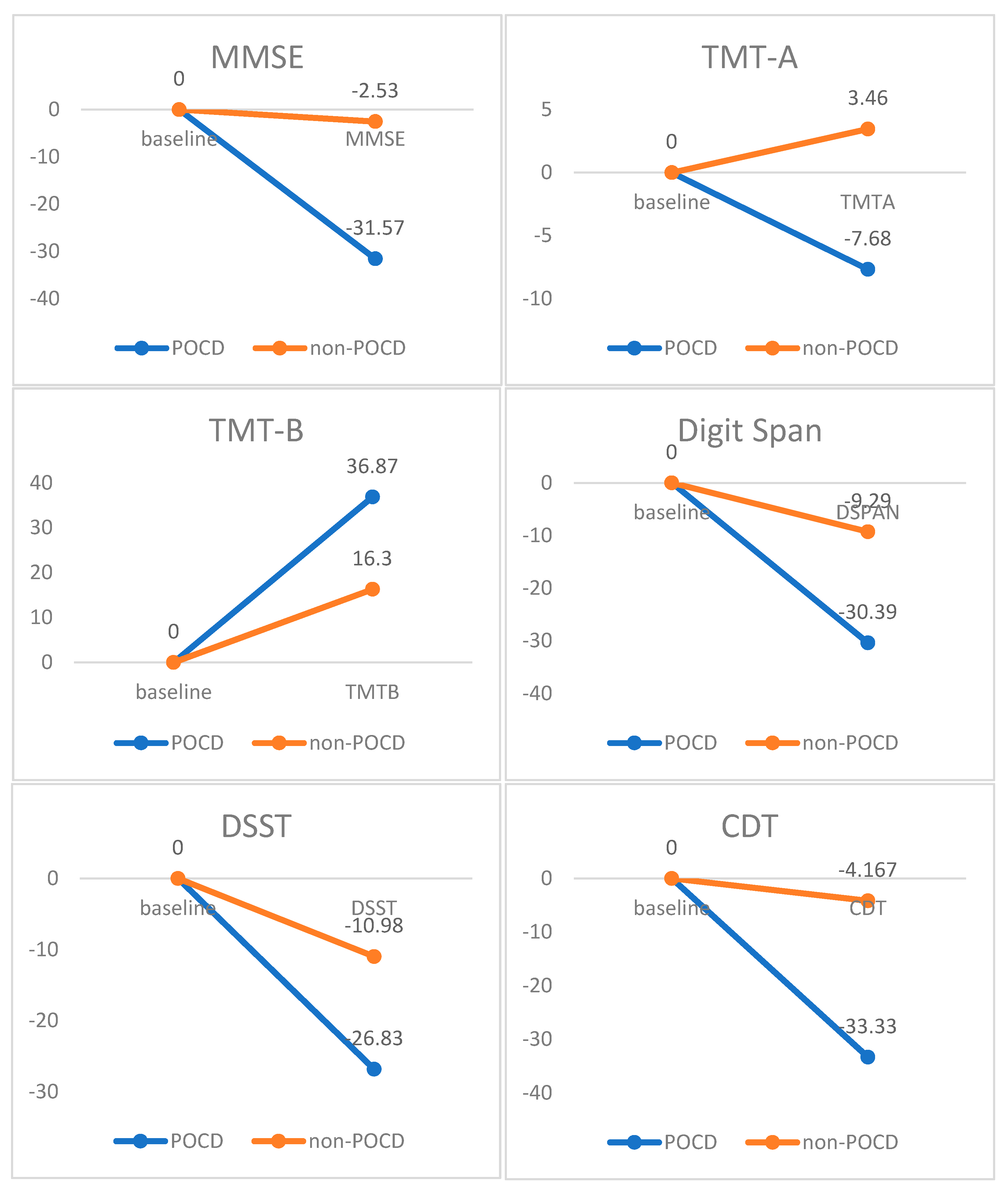
3.1. Neurocognitive Assessment

Forty-six patients were recruited and successfully completed the study. Out of 46, 17% (n = 8) patients were found to have decreased cognitive assessment scores, hence classified as POCD at discharge. Patients in the POCD group were significantly older and had lower education levels than non-POCD patients (p < 0.05). Postoperative cognitive assessment reveals significant differences between the POCD and non-POCD groups, with the POCD group having lower MMSE, digit span, DSST, and CDT scores (p > 0.05). All data are shown in Table 1.
The percentage differences between POCD and non-POCD groups for baseline and postoperative scores were calculated for each cognitive test. MMSE, digit span, digit symbol substitution test, and clock drawing test showed that POCD patients had a more significant percentage decrease from baseline compared to non-POCD patients. Regarding the Trail Making Test, an increased percentage difference means the time taken was longer. A longer time taken to complete the Trail Making Test reflects slower visuoperceptual abilities as seen in POCD patients. The graph of the percentage differences for all tests is shown in Figure 1.

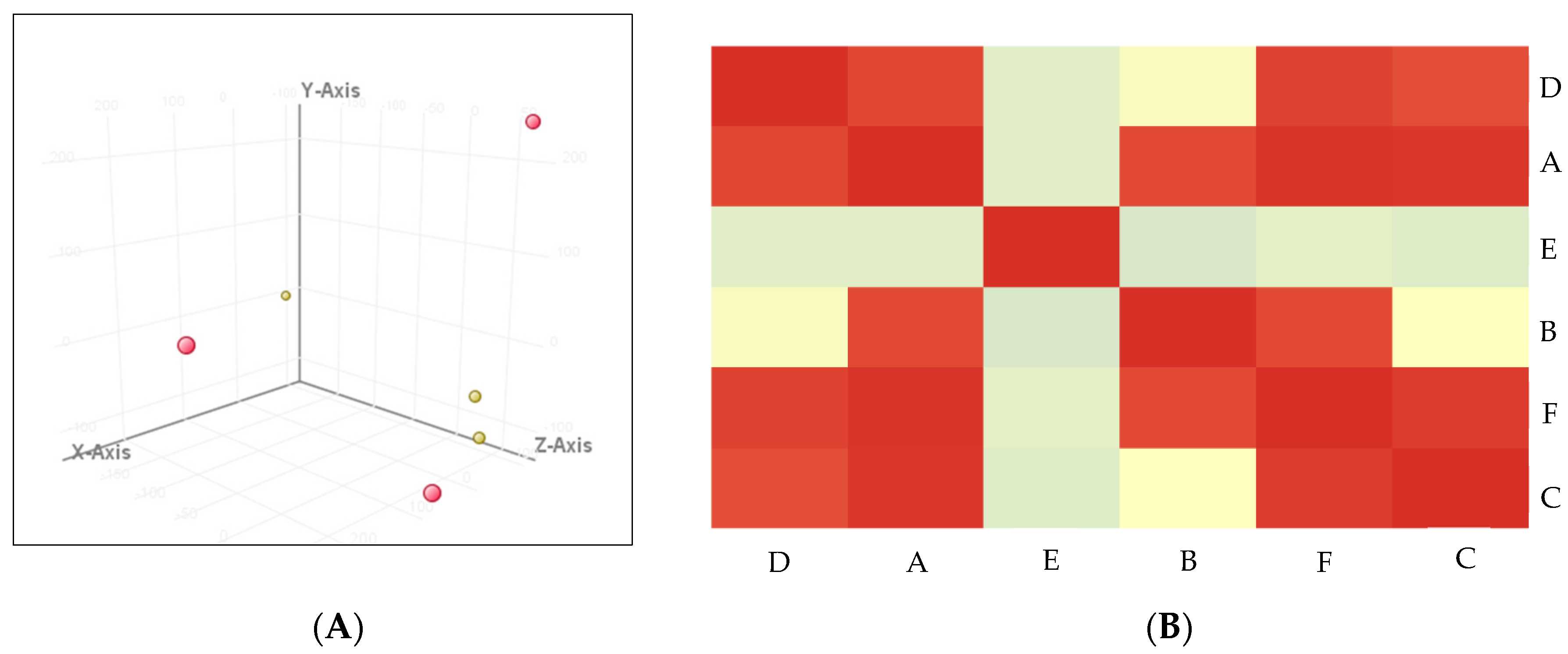
3.2. Samples Quality Detection

The detection of outliers in our samples was conducted using PCA plots. Our models show a normal distribution and good sample correlations, as illustrated in Figure 2. The purity, concentration, and RNA integrity numbers were tabulated in Table 2.

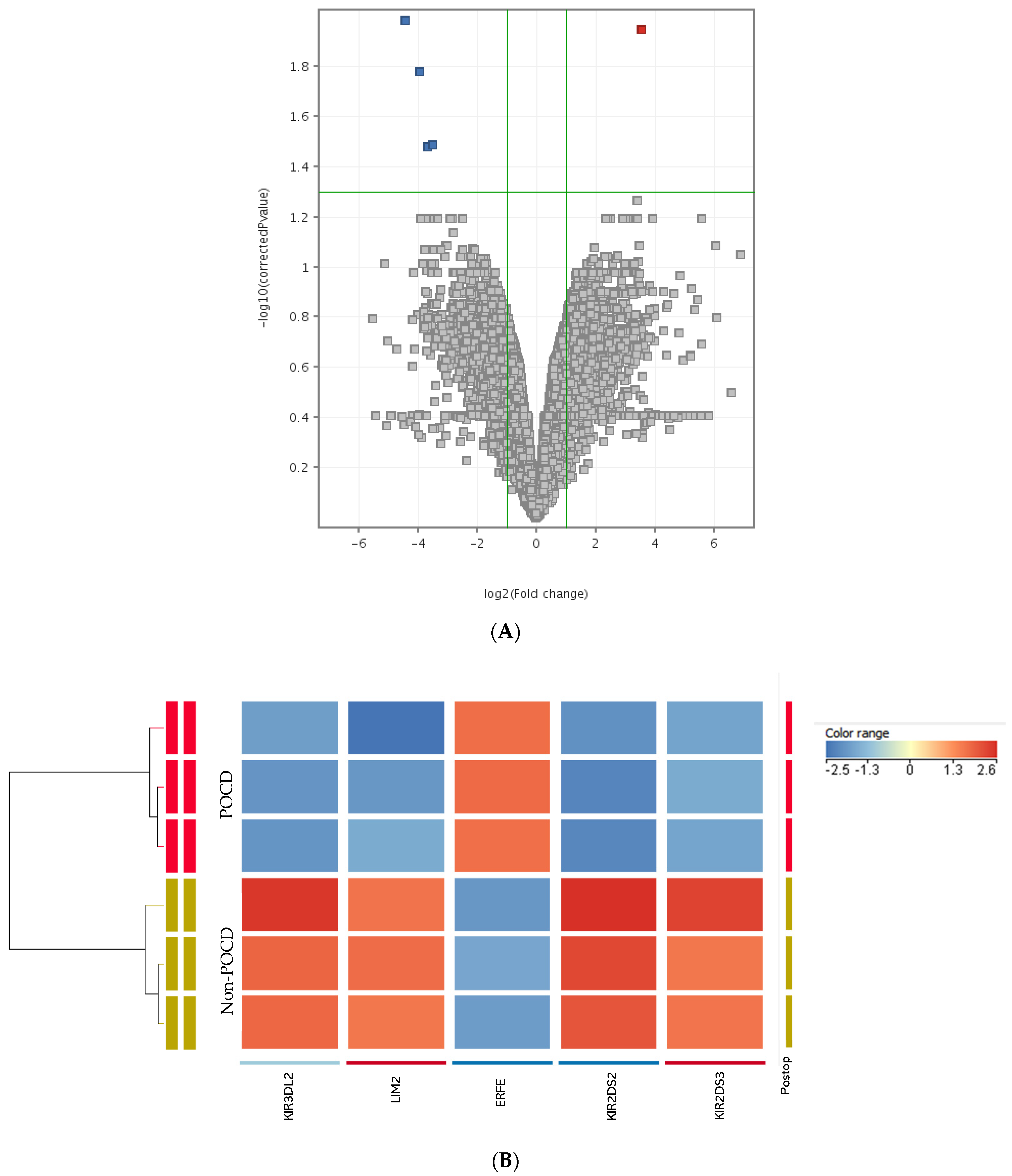
3.3. Differentially Expressed Genes

Microarray analysis showed that five genes were differentially expressed, four were downregulated, and one was upregulated (Table 3). The volcano plot and clustering of the genes are shown in Figure 3. The upregulated gene was ERFE, whereas the downregulated genes include KIR2DS2, LIM2, KIR3DL2, and KIR2DS3. Differential expression was set at fold change > 2 and statistically significant at p < 0.05.

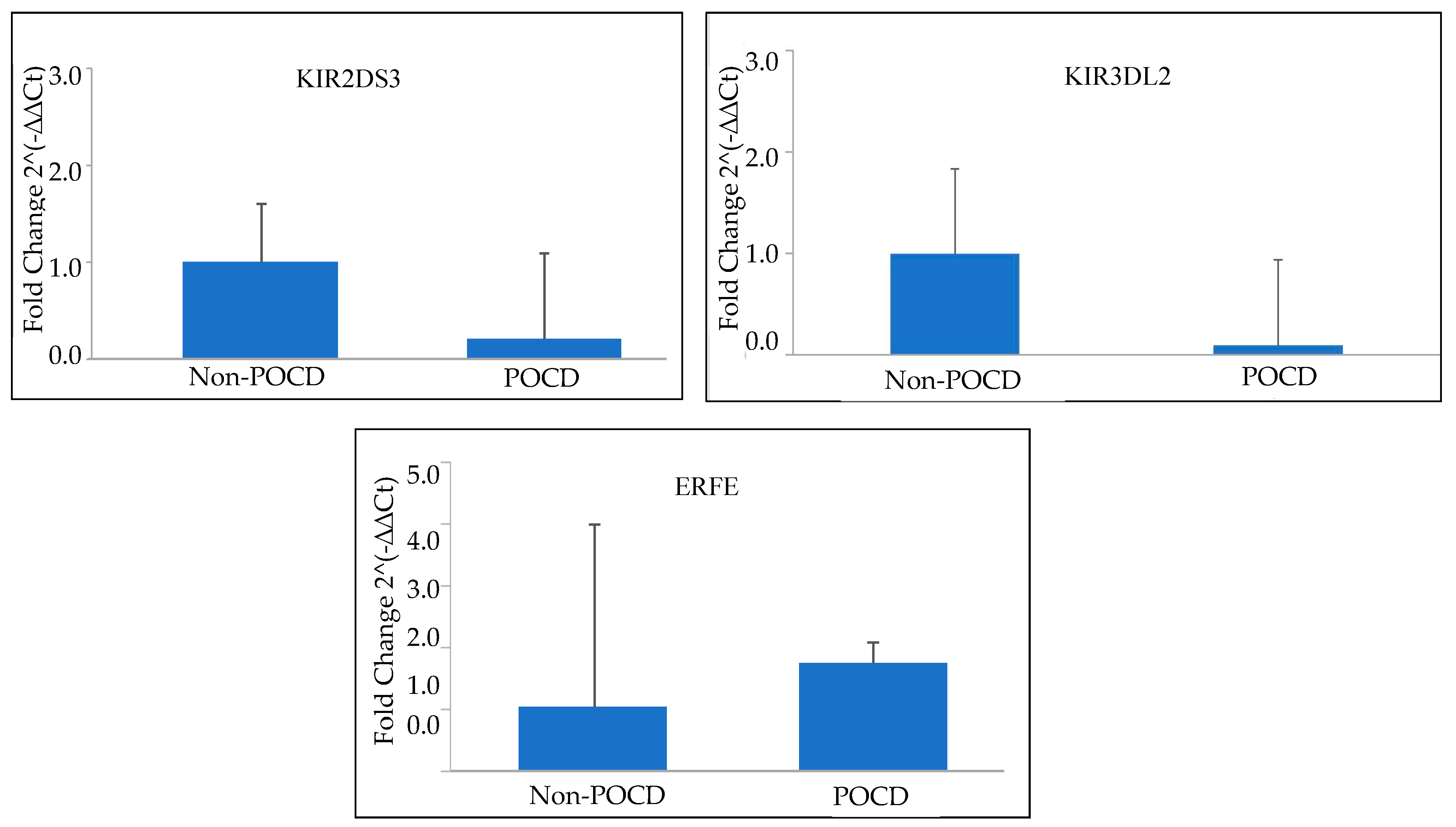
3.4. Quantitative RT-PCR Validation

mRNA expression levels of three genes (ERFE, KIR2DS3, and KIR3DL2) were analyzed by qRT-PCR. However, two genes were not analyzed; LIM2 was identified as not the gene of interest, whereas KIR2DS2 had insufficient samples to proceed after optimization, respectively, and hence, was discarded from validation.
Microarray analysis revealed that gene expression levels correlated with qRT-PCR analysis, with both showing a similar trend in POCD patients’ genes. Both KIR2DS3 (p = 0.021) and KIR3DL2 (p = 0.008) genes were found to be significantly different between POCD and non-POCD patients. Gene ERFE (p = 0.672), on the other hand, offered a higher expression in POCD patients without ever reaching statistical significance (Figure 4).

4. Discussion

Cognitive decline postoperatively is a concern, especially in cardiac surgery. A meta-analysis of 215 studies involving 91,829 patients discovered that 43% of patients experienced cognitive impairment immediately following coronary artery bypass surgery, which decreased to 19% at 4–6 months postoperatively [18]. The meta-analysis involves patients receiving multiple ranges of standard care perioperatively, depending on the clinical site. Compared to our study, the prevalence of POCD at discharge was 17%, and this study homogenously included high-risk patients who received dexmedetomidine sedation.
Yazit et al. (2020) discussed that advancing age would cause degenerative changes, hence becoming one of the risk factors for POCD [13]. Current meta-analyses prove that preoperative depression and old age are significant risk factors for POCD after CABG [19]. Since healthcare advancements improve longevity, many older individuals were able to undergo cardiac surgery. Besides, higher education levels were found to reduce the probability of cognitive decline. Individuals with more exposure to mental challenges and complex tasks were said to have a higher cognitive reserve, lessening the likelihood of POCD [20]. These findings correspond well with our results, whereby patients in the POCD group were significantly older and had lower education levels than non-POCD patients (p < 0.05).
Cognitive screening in the perioperative setting presents significant challenges related to time and efficiency. The tools selected for this study are well-validated and reliable neuropsychological measures with excellent sensitivity and specificity for detecting subtle impairment. However, they are still time-consuming and require expertise and experience to administer. Because we recognized the importance of accuracy in cognitive screening and the importance of time management during sampling, the tests chosen for this study supplement the need for cognitive assessment while requiring minimal administration time [21].
A total of five genes were differentially expressed in POCD groups: ERFE, KIR2DS2, LIM2, KIR3DL2, and KIR2DS3. LIM2 is the lens intrinsic membrane 2, a gene that encodes the protein specifically for the eye lens. It is a receptor for calmodulin and functions in cataractogenesis and lens development. A study found a potential relationship between old age and the LIM2 gene, as it is involved in the development of age-related cataracts [22]. This explained the recent findings in POCD patients of an older age group.
Erythroferrone (ERFE) is a protein coded by the gene ERFE. As the primary erythroid regulator of hepcidin, erythroferrone plays a role in iron homeostasis. Hepcidin regulates total body iron content and plasma levels in normal conditions. This ensures iron flows are available whenever there is a need to develop red blood cells while preventing the potential toxicity of excessive extracellular iron. The release of erythropoietin induced the ERFE expression, with erythroblast being the highest ERFE-expressing cell type [23]. During a bypass procedure, iron homeostasis is disturbed by several means, such as blood loss through the incision, blood shunt to a bypass machine, and hemolysis. This hemopoietic stress will cause an iron loss in blood plasma, jeopardizing the most iron-extensive process, erythropoiesis. In the situation of high iron demand, erythroblast produced ERFE, which inhibited the production of hepcidin, which then allowed ferroportin to transport iron into the bloodstream [24]. This ensures iron regulation during surgery is maintained at an optimum level to avoid the risk of multi-organ failure due to hypoxia.
A study proves that preserving blood flow to the brain has lesser adverse outcomes post-open-heart surgery [25]. Maintaining cerebral blood perfusion will ensure a stable blood flow to the brain. This will control a continuous oxygen supply. The meta-analysis found that high oxygen levels contribute to a significantly lower risk of getting POCD [26]. Hence, we hypothesized that massive blood loss during surgery reduced blood flow to the brain, thereby reducing oxygen supply that may cause tissue hypoxia and inflammatory reactions, which led to POCD afterward. In the case of POCD, the demand for erythroferrone is higher to maintain iron homeostasis, as reflected by the expression postoperatively.
KIR2DS2, KIR3DL2, and KIR2DS3 genes are killer cell immunoglobulin-like receptors (KIR), with 2 Ig domains and short cytoplasmic tail 2, 3 Ig domains and long cytoplasmic tail 2, and 2 Ig domains with short cytoplasmic tail 3, respectively. They are protein-coding genes that are classified according to their extracellular immunoglobulin number (either 2D or 3D) and by long (L) or short (S) cytoplasmic domain. KIR are receptors on natural killer (NK) cells together with their ligands, human leukocyte antigen (HLA). NK cells with the KIR2DS2 immunogenotype were found to be efficient killers of glioblastomas, a brain cancer cell [27]. NK cells were known to be involved in multiple neurodegenerative diseases such as Alzheimer’s disease (AD), Parkinson’s disease (PD), and amyotrophic lateral sclerosis (ALS) [28,29,30]. A study found a correlation between the haplotype combination of KIR2DS2/KIR2DL2/HLA-C1 with lower MMSE scores, representing the condition of AD [28]. On the other hand, KIR2DS3 was found to correlate with acute myeloid leukemia (AML), where the frequency was significantly lower in the AML group compared to the control [31]. Besides, KIR3DL2 was recently found to be the biomarker for Sezary syndrome, a potent type of cutaneous T-cell lymphoma. The percentage of KIR3DL2+ cells at diagnosis with >85% correlated with reduced disease-specific survival [32]. In ALS, the knockdown of KIR3DL2 receptors on the HLA-F molecule on human astrocytes enhanced motor neuron death [33].
All KIR genes play an essential role in the regulation of immune responses. The genes were downregulated in our POCD group. Although the link between these downregulations and POCD occurrence was unclear, clinicians anticipated the condition due to the possibility that the intervention during surgery would impact patients’ immune responses. Blood contact with foreign surfaces from the bypass circuit, surgical trauma, endotoxin, and ischemia may provoke systemic inflammatory response syndrome (SIRS) [34]. SIRS during surgery may develop complications such as myocardial dysfunction, neurological and renal dysfunction, respiratory failure, bleeding disorders, and multi-organ failure [35]. Multiple interventions during cardiopulmonary bypass (CPB) may prevent the secretion of pro-inflammatory cytokines and lessen the complement-mediated activation of neutrophils [34]. As a result, immune response regulator genes such as KIRs were downregulated, mirroring the actual clinical intervention performed on selected CABG patients. We hypothesized that these genes may be related to neuroinflammation. Studies showed that NK cells play a role in Parkinson’s disease, where NK cells inhibit microglial transactivation, which later reduces neuroinflammation [36]. It is suggested that NK cells have a neuroprotective effect on PD pathogenesis [27]. We speculate that the downregulation of KIR genes may interfere with the neuroprotective effect of NK cells, hence increasing the risk of POCD.
The limitation of this study includes the time of blood collection that differs for every patient, which might change the gene expression level. Blood collections were set for day three postoperatively, without a specific time. The day for postoperative assessment also differed from patient to patient, which may influence the overall results. Furthermore, the significantly dysregulated genes in our results were not as numerous as in other studies, implying that potential genes of interest may be overlooked.

5. Conclusions

It was found that there were altered gene expressions postoperatively in high-risk POCD patients receiving dexmedetomidine. The current study provides the gene expression profiles obtained from microarray analysis, which can be used to find potential proteins for predictive biomarkers. The dysregulated genes were an actual representation of cardiopulmonary bypass, which is probably involved in the mechanism of POCD. The genes found in this study were associated with POCD development, yet further elucidation is required for a thorough understanding of its importance in clinical settings. Pathway analysis for these genes is suggested to further understand their roles, particularly in the mechanisms and networks involved.

Author Contributions

Conceptualization, S.K. and N.J.; methodology, K.M.H. and S.K.; software, N.A.H.; validation, N.E., N.J. and S.K.; formal analysis, N.A.A.Y. and N.A.H.; investigation, N.A.S.A.A.; resources, K.M.H. and N.A.S.A.A.; data curation, N.I.M.F.T.; writing—original draft preparation, N.A.A.Y.; writing—review and editing, S.D. and S.A.; visualization, N.J.; supervision, I.F.A.; project administration, N.J.; funding acquisition, S.K. All authors have read and agreed to the published version of the manuscript.

Funding

This project received a grant from National Heart Institute (IJNREC/44/2019).

Institutional Review Board Statement

The study was conducted in accordance with the Declaration of Helsinki, and approved by the Institutional Review Board (or Ethics Committee) of National Heart Institute Ethical Committee (IJNREC/441/2019).

Informed Consent Statement

Informed consent was obtained from all subjects involved in the study. Written informed consent was obtained from the patients to publish this paper.

Data Availability Statement

All data are available within the article and publicly accessible.

Acknowledgments

We would like to acknowledge all staff nurses that helped with blood collection in intensive care units and wards.

Conflicts of Interest

The authors declare no conflict of interest.

Abbreviations

Listed here are the abbreviations utilized throughout this manuscript:
POCDPostoperative Cognitive Dysfunction
RT-PCRReal-Time Polymerase Chain Reaction
CABGCoronary Artery Bypass Graft
RNARibonucleic Acid
MMSEMini–Mental State Examination
TMTTrail Making Test
DSSTDigit Symbol Substitution Test
CDTClock Drawing Test
ERFEErythroferrone
KIRKiller Immunoglobulin-like Receptor
NKNatural Killer
PDParkinson’s Disease

References

  1. Evered, L.; Silbert, B.; Knopman, D.S.; Scott, D.A.; DeKosky, S.T.; Rasmussen, L.S.; Oh, E.S.; Crosby, G.; Berger, M.; Eckenhoff, R.G. Nomenclature Consensus Working Group. Recommendations for the nomenclature of cognitive change associated with anaesthesia and surgery. Anesthesiology 2018, 129, 872–879. [Google Scholar] [CrossRef] [PubMed]
  2. Nemeth, E.; Vig, K.; Racz, K.; Koritsanszky, K.B.; Ronkay, K.I.; Hamvas, F.P.; Gal, J. Influence of the postoperative inflammatory response on cognitive decline in elderly patients undergoing on-pump cardiac surgery: A controlled, prospective observational study. BMC Anesth. 2017, 17, 113. [Google Scholar] [CrossRef] [PubMed]
  3. Liu, X.; Xie, G.; Zhang, K.; Song, S.; Song, F.; Jin, Y.; Fang, X. Dexmedetomidine vs. propofol sedation reduces delirium in patients after cardiac surgery: A meta-analysis with trial sequential analysis of randomized controlled trials. J. Crit. Care 2017, 38, 190–196. [Google Scholar] [CrossRef]
  4. Duan, X.; Zhu, T.; Chen, C.; Zhang, G.; Zhang, J.; Wang, L.; Wang, X. Serum glial cell line–derived neurotrophic factor levels and postoperative cognitive dysfunction after surgery for rheumatic heart disease. J. Thorac. Cardiovasc. Surg. 2018, 155, 958–965. [Google Scholar] [CrossRef] [Green Version]
  5. Pappa, M.; Theodosiadis, N.; Tsounis, A.; Sarafis, P. Pathogenesis and treatment of post-operative cognitive dysfunction. Electron. Physician 2017, 9, 3768–3775. [Google Scholar] [CrossRef] [Green Version]
  6. Li, Z.; Zhu, Y.; Kang, Y.; Qin, S.; Chai, J. Neuroinflammation as the underlying mechanism of postoperative cognitive dysfunction and therapeutic strategies. Front. Cell Neurosci. 2022, 16, 843069. [Google Scholar] [CrossRef] [PubMed]
  7. He, B.; Wang, J. METTL3 regulates hippocampal gene transcription via N6-methyladenosine methylation in sevoflurane-induced postoperative cognitive dysfunction mouse. Aging 2021, 13, 23108–23118. [Google Scholar] [CrossRef]
  8. Schenning, K.J.; Holden, S.; Davis, B.A.; Mulford, A.; Nevonen, K.A.; Quinn, J.F.; Raber, J.; Carbone, L.; Alkayed, N.J. Gene-specific DNA methylation linked to postoperative cognitive dysfunction in apolipoprotein E3 and E4 Mice. J. Alzheimers Dis. 2021, 83, 1251–1268. [Google Scholar] [CrossRef]
  9. Zhang, Y.; Liu, Y.X.; Xiao, Q.X.; Liu, Q.; Deng, R.; Bian, J.; Yu, F.X. Microarray expression profiles of lncRNAs and mRNAs in postoperative cognitive dysfunction. Front. Neurosci. 2018, 12, 694. [Google Scholar] [CrossRef]
  10. Skvarc, D.R.; Berk, M.; Byrne, L.K.; Dean, O.M.; Dodd, S.; Lewis, M.; Gray, L. Post-operative cognitive dysfunction: An exploration of the inflammatory hypothesis and novel therapies. Neurosci. Biobehav. Rev. 2018, 84, 116–133. [Google Scholar] [CrossRef]
  11. Yu, H.; Dong, R.; Lu, Y.; Yang, X.; Chen, C.; Zhang, Z.; Peng, M. Shortterm postoperative cognitive dysfunction and inflammatory response in patients undergoing cytoreductive surgery and hyperthermic intraperitoneal chemotherapy: A pilot study. Mediat. Inflamm. 2017, 2017, 3605350. [Google Scholar] [CrossRef] [PubMed] [Green Version]
  12. Wu, L.; Mickey, W.P.; Koch, W.H. Clinical applications of microarray-based diagnostic tests. Biotechniques 2005, 39, S557–S582. [Google Scholar] [CrossRef]
  13. Yazit, N.A.; Juliana, N.; Das, S.; Teng, N.I.; Fahmy, N.M.; Azmani, S.; Kadiman, S. Association of micro RNA and postoperative cognitive dysfunction: A review. Mini Rev. Med. Chem. 2020, 20, 1781–1790. [Google Scholar] [CrossRef] [PubMed]
  14. Wang, M.; Su, P.; Liu, Y.; Zhang, X.; Yan, J.; An, X.; Gu, S. Abnormal expression of circRNA_089763 in the plasma exosomes of patients with post-operative cognitive dysfunction after coronary artery bypass grafting. Mol. Med. Rep. 2019, 20, 2549–2562. [Google Scholar] [CrossRef] [Green Version]
  15. Zarina, Z.A.; Zahiruddin, O.; Che, W.A. Validation of Malay mini-mental state examination. Malays. J. Psychiatry 2007, 16, 16–19. [Google Scholar]
  16. Jaeger, J. Digit symbol substitution test: The case for sensitivity over specificity in neuropsychological testing. J. Clin. Psychopharmacol. 2018, 38, 513. [Google Scholar] [CrossRef]
  17. Ibrahim, N.M.; Shohaimi, S.; Chong, H.T.; Rahman, A.H.A.; Razali, R.; Esther, E.; Basri, H.B. Validation study of the Mini-Mental State Examination in a Malay-speaking elderly population in Malaysia. Dement. Geriatr. Cogn. Disord. 2009, 27, 247–253. [Google Scholar] [CrossRef]
  18. Greaves, D.; Psaltis, P.J.; Ross, T.J.; Davis, D.; Smith, A.E.; Boord, M.S.; Keage, H.A. Cognitive outcomes following coronary artery bypass grafting: A systematic review and meta-analysis of 91,829 patients. Int. J. Cardiol. 2019, 289, 43–49. [Google Scholar] [CrossRef] [Green Version]
  19. Greaves, D.; Psaltis, P.J.; Davis, D.H.; Ross, T.J.; Ghezzi, E.S.; Lampit, A.; Keage, H.A. Risk factors for delirium and cognitive decline following coronary artery bypass grafting surgery: A systematic review and meta-analysis. J. Am. Heart Assoc. 2020, 9, e017275. [Google Scholar] [CrossRef]
  20. Newman, M.F.; Kirchner, J.L.; Phillips-Bute, B.; Gaver, V.; Grocott, H.; Jones, R.H.; Blumenthal, J.A. Longitudinal assessment of neurocognitive function after coronary-artery bypass surgery. N. Engl. J. Med. 2001, 344, 395–402. [Google Scholar] [CrossRef]
  21. Harrington, M.B.; Kraft, M.; Grande, L.J.; Rudolph, J.L. Independent association between preoperative cognitive status and discharge location after cardiac surgery. Am. J. Crit. Care 2011, 20, 129–137. [Google Scholar] [CrossRef] [PubMed] [Green Version]
  22. Zhou, Z.; Wang, B.; Hu, S.; Zhang, C.; Ma, X.; Qi, Y. Genetic variations in GJA3, GJA8, LIM2, and age-related cataract in the Chinese population: A mutation screening study. Mol. Vis. 2011, 17, 621. [Google Scholar] [PubMed]
  23. Srole, D.N.; Ganz, T. Erythroferrone structure, function, and physiology: Iron homeostasis and beyond. J. Cell Physiol. 2021, 236, 4888–4901. [Google Scholar] [CrossRef] [PubMed]
  24. Coffey, R.; Ganz, T. Erythroferrone: An erythroid regulator of hepcidin and iron metabolism. Hemasphere 2018, 2, e35. [Google Scholar] [CrossRef]
  25. Juliana, N.; Abu Yazit, N.A.; Kadiman, S.; Muhammad Hafidz, K.; Azmani, S.; Mohd Fahmi Teng, N.I.; Das, S. Intraoperative cerebral oximetry in open heart surgeries reduced postoperative complications: A retrospective study. PLoS ONE 2021, 16, e0251157. [Google Scholar] [CrossRef]
  26. Wang, L.; Lang, Z.; Gao, H.; Liu, Y.; Dong, H.; Sun, X. The relationship between the incidence of postoperative cognitive dysfunction and intraoperative regional cerebral oxygen saturation after cardiovascular surgery: A systematic review and meta-analysis of randomized controlled trials. Rev. Cardiovasc. Med. 2022, 23, 388. [Google Scholar] [CrossRef]
  27. Navarro, A.G.; Kmiecik, J.; Leiss, L.; Zelkowski, M.; Engelsen, A.; Bruserud, Ø.; Chekenya, M. NK cells with KIR2DS2 immunogenotype have a functional activation advantage to efficiently kill glioblastoma and prolong animal survival. J. Immunol. 2014, 193, 6192–6206. [Google Scholar] [CrossRef] [Green Version]
  28. Rizzo, R.; Bortolotti, D.; Gentili, V.; Rotola, A.; Bolzani, S.; Caselli, E.; Di Luca, D. Kir2ds2/kir2dl2/hla-c1 haplotype is associated with Alzheimer’s disease: Implication for the role of herpesvirus infections. J. Alzheimers Dis. 2019, 67, 1379–1389. [Google Scholar] [CrossRef]
  29. Earls, R.H.; Lee, J.K. The role of natural killer cells in Parkinson’s disease. Exp. Mol. Med. 2020, 52, 1517–1525. [Google Scholar] [CrossRef]
  30. Garofalo, S.; Cocozza, G.; Porzia, A.; Inghilleri, M.; Raspa, M.; Scavizzi, F.; Limatola, C. Natural killer cells modulate motor neuron-immune cell cross talk in models of Amyotrophic Lateral Sclerosis. Nat. Commun. 2020, 11, 1–6. [Google Scholar] [CrossRef] [Green Version]
  31. Shahsavar, F.; Tajik, N.; Entezami, K.Z.; Radjabzadeh, M.F.; Asadifar, B.; Alimoghaddam, K.; Ghavamzadeh, A. KIR2DS3 is associated with protection against acute myeloid leukemia. Iran J. Immunol. 2010, 7, 8–17. [Google Scholar] [PubMed]
  32. Hurabielle, C.; Thonnart, N.; Ram-Wolff, C.; Sicard, H.; Bensussan, A.; Bagot, M.; Marie-Cardine, A. Usefulness of KIR3DL2 to Diagnose, Follow-Up, and Manage the Treatment of Patients with Sézary Syndrome. Clin. Cancer Res. 2017, 23, 3619–3627. [Google Scholar] [CrossRef] [PubMed] [Green Version]
  33. Song, S.; Miranda, C.J.; Braun, L.; Meyer, K.; Frakes, A.E.; Ferraiuolo, L.; Kaspar, B.K. Major histocompatibility complex class I molecules protect motor neurons from astrocyte-induced toxicity in amyotrophic lateral sclerosis. Nat. Med. 2016, 22, 397–403. [Google Scholar] [CrossRef] [Green Version]
  34. Kats, S.; Schönberger, J.P.; Brands, R.; Seinen, W.; van Oeveren, W. Endotoxin release in cardiac surgery with cardiopulmonary bypass: Pathophysiology and possible therapeutic strategies. An update. Eur. J. Cardiothorac. Surg. 2011, 39, 451–458. [Google Scholar] [CrossRef] [PubMed] [Green Version]
  35. Paparella, D.; Yau, T.M.; Young, E. Cardiopulmonary bypass induced inflammation: Pathophysiology and treatment. An update. Eur. J. Cardiothorac. Surg. 2002, 21, 232–244. [Google Scholar] [CrossRef] [Green Version]
  36. Waggoner, S.N.; Kumar, V. Evolving role of 2B4/CD244 in T and NK cell responses during virus infection. Front. Immunol. 2012, 3, 377. [Google Scholar] [CrossRef]
Figure 1. Percentage differences between POCD and non−POCD patients in all tests. MMSE = Mini−Mental State Examination; TMT = Trail Making Test; DSST = digit symbol substitution test; CDT = clock drawing test.
Ijerph 20 01457 g001
Figure 2. Sample quality. (A) PCA plot. Yellow dots are non−POCD samples; red dots are POCD samples. (B) Correlation analysis. A, B, and C are non−POCD samples, while D, E, and F are POCD samples. Darker red means higher correlation between samples.
Ijerph 20 01457 g002
Figure 3. Distribution of differentially expressed genes. (A) Volcano plot showing upregulated (red) genes on the top right and downregulated (blue) genes on the top left of the plot. Genes in grey color are not significantly expressed. (B) Heatmap showing the upregulated (red) and downregulated (blue) genes in POCD and non−POCD condition.
Ijerph 20 01457 g003
Figure 4. Bar graphs representing expression levels of KIR2DS3, KIR3DL2, and ERFE genes by quantitative real-time RT-PCR in POCD and non-POCD groups define by their fold change.
Ijerph 20 01457 g004
Table 1. Demographic, intraoperative characteristics, and cognitive assessment scores of patients in the POCD and non-POCD groups.
ItemNon-POCD (n = 38), Mean ± SD/n (%)POCD (n = 8), Mean ± SD/n (%)p-Value
Age (years)56.21 ± 13.19166.00 ± 5.9760.047 *
Gender
 Male23 (79.3)6 (20.7)0.366
 Female15 (88.2)2 (11.8)
Education (years)11.82 ± 2.7108.00 ± 4.0360.002 *
Body Mass Index (BMI)26.44 ± 4.19928.34 ± 13.072.44
Bypass time (minutes)127.24 ± 72.431123.86 ± 26.550.905
Cross Clamp time (minutes)99.35 ± 63.14184.86 ± 28.9560.559
Preoperative scores
 MMSE28.34 ± 2.09625.50 ± 2.5630.002 *
 TMT-A (seconds)49.16 ± 26.05469.38 ± 29.3060.57
 TMT-B (seconds)107.53 ± 56.125162.50 ± 62.4520.017 *
 Digit Span13.63 ± 3.18310.75 ± 3.3700.026 *
 DSST 53.39 ± 16.83929.75 ± 14.4690.001 *
 CDT1.92 ± 0.2731.75 ± 0.7070.250
Postoperative scores
 MMSE27.63 ± 2.45418.00 ± 7.483<0.001 **
 TMT-A (seconds)47.8 ± 22.51271.80 ± 47.4840.064
 TMT-B (seconds)123.31 ± 71.160184.20 ± 45.0690.072
 Digit Span12.08 ± 3.6316.88 ± 4.8530.001 *
 DSST 47.56 ± 19.16623.14 ± 14.5650.003 *
 CDT1.86 ± 0.4871.14 ± 1.0690.007 *
POCD, postoperative cognitive dysfunction; MMSE, Mini–Mental State Examination; TMT, Trail Making Test; DSST, digit symbol substitution test; CDT, clock drawing test. * p-value significant at p < 0.05 using independent t-test. ** p-value significant at p < 0.001 using independent t-test.
Table 2. Purity, concentration, and integrity of RNA samples.
SamplesPurityConcentrationRIN Number
A1.83243.28.80
B1.85561.66.80
C1.85746.88.00
D1.89225.28.80
E1.842135.68.00
F1.81663.28.40
Table 3. Differentially expressed genes in the POCD group.
NoFold Changep-ValueRegulationGene SymbolDescription
111.6066330.01127UpERFEerythroferrone
2−11.3874190.03253DownKIR2DS2killer cell immunoglobulin-like receptor, two Ig domains, and short cytoplasmic tail 2
3−12.6478940.03299DownLIM2lens intrinsic membrane protein 2
4−15.3811290.01660DownKIR3DL2killer cell immunoglobulin-like receptor, three Ig domains, and long cytoplasmic tail 2
5−21.7303280.01028DownKIR2DS3killer cell immunoglobulin-like receptor, two Ig domains, and short cytoplasmic tail 3
Disclaimer/Publisher’s Note: The statements, opinions and data contained in all publications are solely those of the individual author(s) and contributor(s) and not of MDPI and/or the editor(s). MDPI and/or the editor(s) disclaim responsibility for any injury to people or property resulting from any ideas, methods, instructions or products referred to in the content.

