
Assessment of CVD Risk Factors in Secondary Prevention after Ischemic Stroke Using the ICF

 , , ,
, , ,
Abstract
:1. Introduction
2. Objectives
- (1)
- Assessment of the frequency of risk factors for recurrent CVD events between the group of patients after ischemic stroke (SG) and the control group without previous CVD (CG) events.
- (2)
- Use of ICF classification categories to represent the frequency of risk factors for recurrent CVD events between SG and CG.
3. Materials and Method
3.1. Study Design
3.2. ICF Profile
3.3. Statistical Analysis
3.4. Ethical Approval
4. Results
4.1. Study Population
4.2. Risk Factors for Recurrent CVD Incidents
4.2.1. SG vs. CG
4.2.2. SG vs. CG in Relation to Female Gender
4.2.3. SG vs. CG in Relation to Male Gender
4.3. CVD Risk Factor Profile in Secondary Prevention According to the ICF Classification
4.3.1. SG vs. CG
4.3.2. SG vs. CG in Relation to Female Gender
4.3.3. SG vs. CG in Relation to Male Gender
4.4. Concomitance Occurrence of CVD Risk Factors
5. Discussion
6. Limitations
7. Conclusions
Author Contributions
Funding
Institutional Review Board Statement
Informed Consent Statement
Data Availability Statement
Conflicts of Interest
References
- Roth, G.A.; Forouzanfar, M.H.; Moran, A.E.; Barber, R.; Nguyen, G.; Feigini, V.L.; Naghavi, M.; Mensah, G.A.; Murray, C.J. Demographic and epidemiologic drivers of global cardiovascular mortality. N. Engl. J. Med. 2015, 372, 1333–1341. [Google Scholar] [CrossRef] [PubMed] [Green Version]
- Skajaa, N.; Adelborg, K.; Horváth-Puhó, E.; Rothman, K.J.; Henderson, V.W.; Thygesen, L.C.; Sørensen, H.T. Risks of Stroke Recurrence and Mortality After First and Recurrent Strokes in Denmark: A Nationwide Registry Study. Neurology 2022, 98, e329–e342. [Google Scholar] [CrossRef] [PubMed]
- Mendis, S.; Abegunde, D.; Yusuf, S.; Ebrahim, S.; Shaper, G.; Ghannem, H.; Shengelia, B. WHO study on Prevention of Recurrences of Myocardial Infarction and StrokE (WHO-PREMISE). Bull. World Health Organ. 2005, 83, 820–829. [Google Scholar] [PubMed]
- Stewart, J.; Manmathan, G.; Wilkinson, P. Primary prevention of cardiovascular disease: A review of contemporary guidance and literature. JRSM Cardiovasc Dis. 2017, 6. [Google Scholar] [CrossRef] [Green Version]
- Yuan, H.W.; Wang, C.X.; Zhang, N.; Bai, Y.; Shi, Y.Z.; Zhou, Y.; Wang, Y.L.; Zhang, T.; Zhou, J.; Yu, X.; et al. Poststroke depression and risk of recurrent stroke at 1 year in a Chinese cohort study. PLoS ONE 2012, 7, e46906. [Google Scholar] [CrossRef]
- Cappuccio, F.P.; Cooper, D.; D’Elia, L.; Strazzullo, P.; Miller, M.A. Sleep duration predicts cardiovascular outcomes: A systematic review and meta-analysis of prospective studies. Eur. Heart J. 2011, 32, 1484–1492. [Google Scholar] [CrossRef] [Green Version]
- Woodward, M.; Webster, R.; Murakami, Y.; Barzi, F.; Lam, T.H.; Fang, X.; Suh, I.; Batty, G.D.; Huxley, R.; Rodgers, A.; et al. The association between resting heart rate, cardiovascular disease and mortality: Evidence from 112,680 men and women in 12 cohorts. Eur. J. Prev. Cardiol. 2014, 21, 719–726. [Google Scholar] [CrossRef]
- Lip, G.Y.; Hunter, T.D.; Quiroz, M.E.; Ziegler, P.D.; Turakhia, M.P. Atrial fibrillation diagnosis timing, ambulatory ECG monitoring utilization and risk of recurrent stroke. Circ. Cardiovasc. Qual. Outcomes 2017, 10, e002864. [Google Scholar] [CrossRef]
- Orrapin, S.; Rerkasem, K. Carotid endarterectomy for symptomatic carotid stenosis. Cochrane Database Syst. Rev. 2017, 6, CD001081. [Google Scholar] [CrossRef]
- Liu, L.; Wang, Z.; Gong, L.; Zhang, Y.; Thijs, L.; Staessen, J.A.; Wang, J. Blood pressure reduction for the secondary prevention of stroke: A Chinese trial and a systematic review of the literature. Hypertens. Res. 2009, 32, 1032–1040. [Google Scholar] [CrossRef] [Green Version]
- Weiner, D.E.; Tighiouart, H.; Stark, P.C.; Amin, M.G.; MacLeod, B.; Griffith, J.L.; Salem, D.N.; Levey, A.S.; Sarnak, M.J. Kidney disease as a risk factor for recurrent cardiovascular disease and mortality. Am. J. Kidney Dis. 2004, 44, 198–206. [Google Scholar] [CrossRef] [PubMed]
- Pisters, R.; Lane, D.A.; Nieuwlaat, R.; de Vos, C.B.; Crijns, H.J.; Lip, G.Y. A novel userfriendly score (HAS-BLED) to assess 1-year risk of major bleeding in patients with atrial fibrillation: The Euro Heart Survey. Chest 2010, 138, 1093–1100. [Google Scholar] [CrossRef] [Green Version]
- Gerner, S.T.; Kuramatsu, J.B.; Sembill, J.A.; Sprügel, M.I.; Hagen, M.; Knappe, R.U.; Endres, M.; Haeusler, K.G.; Sobesky, J.; Schurig, J.; et al. Characteristics in non-vitamin K antagonist oral anticoagulant-related intracerebral hemorrhage. Stroke 2019, 50, 1392–1402. [Google Scholar] [CrossRef] [PubMed]
- Wu, S.; Shi, Y.; Wang, C.; Jia, Q.; Zhang, N.; Zhao, X.; Liu, G.; Wang, Y.; Liu, L.; Wang, Y.; et al. Glycated hemoglobin independently predicts stroke recurrence within one year after acute first-ever non-cardioembolic strokes onset in A Chinese cohort study. PLoS ONE 2013, 8, e80690. [Google Scholar] [CrossRef] [PubMed] [Green Version]
- Amarenco, P.; Bogousslavsky, J.; Callahan, A., 3rd; Goldstein, L.B.; Hennerici, M.; Rudolph, A.E.; Sillesen, H.; Simunovic, L.; Szarek, M.; Welch, K.M.; et al. Stroke Prevention by Aggressive Reduction in Cholesterol Levels (SPARCL) Investigators. High-dose atorvastatin after stroke or transient ischemic attack. N. Engl. J. Med. 2006, 355, 549–559. [Google Scholar] [PubMed]
- Ois, A.; Gomis, M.; Rodríguez-Campello, A.; Cuadrado-Godia, E.; Jiménez-Conde, J.; Pont-Sunyer, C.; Cuccurella, G.; Roquer, J. Factors associated with a high risk of recurrence in patients with transient ischemic attack or minor stroke. Stroke 2008, 39, 1717–1721. [Google Scholar] [CrossRef] [Green Version]
- Narum, S.; Solhaug, V.; Myhr, K.; Brørs, O.; Kringen, M.K. Characterisation of non-warfarin-associated bleeding events reported to the Norwegian spontaneous reporting system. Eur. J. Clin. Pharmacol. 2013, 69, 1445–1452. [Google Scholar] [CrossRef]
- Epstein, K.A.; Viscoli, C.M.; Spence, J.D.; Young, L.H.; Inzucchi, S.E.; Gorman, M.; Gerstenhaber, B.; Guarino, P.D.; Dixit, A.; Furie, K.L.; et al. Smoking cessation and outcome after ischemic stroke or TIA. Neurology 2017, 89, 1723–1729. [Google Scholar] [CrossRef]
- Słowik, A.; Wnuk, M.; Brzegowy, P.; Chrzanowska-Wásko, J.; Golenia, A.; Łasocha, B.; Włoch-Kopéc, D.; Ferens, A.; Serednicki, W.; Jarocki, P.; et al. Mechanical thrombectomy in acute stroke—Five years of experience in Poland. Neurol. Neurochir. Pol. 2017, 51, 339–346. [Google Scholar] [CrossRef]
- Strazzullo, P.; D’Elia, L.; Cairella, G.; Garbagnati, F.; Cappuccio, F.P.; Scalfi, L. Excess body weight and incidence of stroke: Meta-analysis of prospective studies with 2 million participants. Stroke 2010, 41, e418–e426. [Google Scholar] [CrossRef] [Green Version]
- Arboix, A.; Jiménez, C.; Massons, J.; Parra, O.; Besses, C. Hematological disorders: A commonly unrecognized cause of acute stroke. Expert Rev. Hematol. 2016, 9, 891–901. [Google Scholar] [CrossRef]
- Adams, R.J.; Chimowitz, M.I.; Alpert, J.S.; Awad, I.A.; Cerqueria, M.D.; Fayad, P.; Taubert, K.A.; American Heart Association/American Stroke Association; Stroke Council and the Council on Clinical Cardiology of the American Heart Association; American Stroke Association. Coronary risk evaluation in patients with transient ischemic attack and ischemic stroke: A scientific statement for healthcare professionals from the Stroke Council and the Council on Clinical Cardiology of the American Heart Association/American Stroke Association. Stroke 2003, 34, 2310–2322. [Google Scholar] [PubMed] [Green Version]
- Maritz, R.; Aronsky, D.; Prodinger, B. The International Classification of Functioning, Disability and Health (ICF) in Electronic Health Records. Appl. Clin. Inform. 2017, 8, 964–980. [Google Scholar] [CrossRef]
- Chen, S.; Tao, J.; Tao, Q.; Fang, Y.; Zhou, X.; Chen, H.; Chen, Z.; Huang, J.; Chen, L.; Chan, C.C. Rater experience influences reliability and validity of the Brief International Classification of Functioning, Disability, and Health Core Set for Stroke. J. Rehabil. Med. 2016, 48, 265–272. [Google Scholar] [CrossRef] [PubMed] [Green Version]
- Lucki, M.; Chlebuś, E.; Wareńczak, A.; Lisiński, P. The ICF Classification System to Assess Risk Factors for CVD in Secondary Prevention after Ischemic Stroke and Intracerebral Hemorrhage. Medicina 2021, 57, 190. [Google Scholar] [CrossRef]
- Ceccarini, M.; Manzoni, G.M.; Castelnuovo, G. Assessing depression in cardiac patients: What measures should be considered? Depress Res. Treat. 2014, 2014, 148–256. [Google Scholar] [CrossRef] [PubMed] [Green Version]
- Cuspidi, C.; Tadic, M.; Grassi, G.; Mancia, G. Treatment of hypertension: The ESH/ESC guidelines recommendations. Pharmacol. Res. 2018, 128, 315–321. [Google Scholar] [CrossRef] [PubMed]
- Inker, L.A.; Astor, B.C.; Fox, C.H.; Isakova, T.; Lash, J.P.; Peralta, C.A.; Kurella Tamura, M.; Feldman, H.I. KDOQI US commentary on the 2012 KDIGO clinical practice guideline for the evaluation and management of CKD. Am. J. Kidney Dis. 2014, 63, 713–735. [Google Scholar] [CrossRef] [Green Version]
- Inzucchi, S.E.; Bergenstal, R.M.; Buse, J.B.; Diamant, M.; Ferrannini, E.; Nauck, M.; Peters, A.L.; Tsapas, A.; Wender, R.; Matthews, D.R. Management of hyperglycemia in type 2 diabetes, 2015: A patient-centered approach: Update to a position statement of the American Diabetes Association and the European Association for the Study of Diabetes. Diabetes Care 2015, 38, 140–149. [Google Scholar] [CrossRef] [PubMed] [Green Version]
- Schwartz, G.G.; Steg, P.G.; Szarek, M.; Bhatt, D.L.; Bittner, V.A.; Diaz, R.; Edelberg, J.M.; Goodman, S.G.; Hanotin, C.; Harrington, R.A.; et al. ODySSEy OUTCOMES Committees and Investigators. Alirocumab and car-diovascular outcomes after acute coronary syndrome. N. Engl. J. Med. 2018, 379, 2017–2097. [Google Scholar] [CrossRef]
- Wood, A.M.; Kaptoge, S.; Butterworth, A.S.; Willeit, P.; Warnakula, S.; Bolton, T.; Paige, E.; Paul, D.S.; Sweeting, M.; Burgess, S.; et al. Risk thresholds for alcohol consumption: Combined analysis of individual-participant data for 599,912 current drinkers in 83 prospective. Lancet 2018, 391, 1513–1523. [Google Scholar] [CrossRef] [Green Version]
- Mozaffarian, D.; Benjamin, E.J.; Go, A.S.; Arnett, D.K.; Blaha, M.J.; Cushman, M.; Das, S.R.; de Ferranti, S.; Després, J.P.; Fullerton, H.J.; et al. American Heart Association Statistics Committee; Stroke Statistics Subcommittee. Heart Disease and Stroke Statistics-2016 Update: A Report from the American Heart Association. Circultion 2016, 133, e38–e209. [Google Scholar]
- Blanco-Rojas, L.; Arboix, A.; Canovas, D.; Grau-Olivares, M.; Oliva Morera, J.C.; Parra, O. Cognitive profile in patients with a first-ever lacunar infarct with and without silent lacunes: A comparative study. BMC Neurol. 2013, 13, 203. [Google Scholar] [CrossRef] [PubMed] [Green Version]
- Zhang, J.; Wang, Y.; Wang, G.N.; Sun, H.; Sun, T.; Shi, J.Q.; Xiao, H.; Zhang, J.S. Clinical factors in patients with ischemic versushemorrhagic stroke in East China. World J. Emerg. Med. 2011, 2, 18–23. [Google Scholar] [CrossRef] [Green Version]
- Wang, S.L.; Wang, C.L.; Wang, P.L.; Xu, H.; Du, J.P.; Zhang, D.W.; Gao, Z.Y.; Zhang, L.; Fu, C.G.; Chen, K.J.; et al. Resting heart rate associates with one-year risk of major adverse cardiovascular events in patients with acute coronary syndrome after percutaneous coronary intervention. Exp. Biol. Med 2016, 241, 478–484. [Google Scholar] [CrossRef] [Green Version]
- Lewington, S.; Clarke, R.; Qizilbash, N.; Peto, R.; Collins, R. Prospective Studies Collaboration. Age-specific relevance of usual blood pressure to vascular mortality: A meta-analysis of individual data for one million adults in 61 prospective studies. Lancet 2003, 360, 1903–1913. [Google Scholar]


| SG | CG | p | ||
|---|---|---|---|---|
| Sex n (%) | Males | 28 (50.9%) | 28 (50.9%) | 1.0 a |
| Females | 27 (49.1%) | 27 (49.1%) | ||
| Age (years) | Mean ± SD | 63.2 ± 8.8 | 65.9 ± 6.3 | 0.066 b |
| Median | 63.4 | 67 | ||
| Min–Max | 42.7–78.9 | 47.0–77.0 | ||
| BMI | Mean ± SD | 28.9 ± 4.6 | 28.6 ± 4.9 | 0.630 b |
| Median | 28.7 | 28 | ||
| Min–Max | 18.6–37.8 | 21.0–42.8 | ||
| Comorbidites | ||||
| Hypertension | n (%) | 50 (90.9%) | 32 (58.2%) | <0.001 a |
| Coronary heart disease | n (%) | 16 (29.1%) | 6 (10.9%) | 0.017 a |
| Chronic heart failure | n (%) | 18 (32.7%) | 7 (12.7%) | <0.001 a |
| Diabetes melitus type 2 | n (%) | 24 (43.6) | 17 (30.9%) | 0.168 a |
| Carotid artery disease | n (%) | 30 (54.5%) | 3 (5.5) | <0.001 a |
| SG vs. CG | Females | Males | ||||||||
|---|---|---|---|---|---|---|---|---|---|---|
| SG | CG | p | SG | CG | p | SG | CG | p | ||
| b152 Emotional functions—Depression [26] | n (%) | 12 (21.8%) | 5 (9.3%) | 0.071 | 7 (25.0%) | 4 (14.3%) | 0.314 | 5 (18.5%) | 1 (3.8%) | 0.092 |
| b134 Sleep functions—Insomia [6] | n (%) | 12 (21.8%) | 20 (36.4%) | 0.092 | 7 (25.0%) | 9 (32.1%) | 0.556 | 5 (18.5%) | 11 (40.7%) | 0.074 |
| b4100 Heart rate [7] | HR > 80/min, n (%) | 25 (46.3%) | 12 (21.8%) | 0.007 | 15 (53.6%) | 10 (35.7%) | 0.179 | 10 (38.5%) | 2 (7.4%) | 0.007 |
| b4101 Heart rhythm [8] | Atrial fibrillation, n (%) | 15 (27.3%) | 5 (9.1%) | 0.013 | 6 (21.4%) | 4 (14.3%) | 0.485 | 9 (33.3%) | 1 (3.8%) | 0.005 |
| b4150 Functions of arteries [9] | Stenosis 50–69%, n (%) | 15 (27.3%) | 1 (1.8%) | <0.001 | 8 (28.6%) | 1 (3.6%) | 0.011 | 7 (25.9%) | 0 (0.0%) | 0.005 |
| Stenosis > 70%, n (%) | 15 (27.3%) | 0 (0.0%) | <0.001 | 7 (25.0%) | 0 (0.0%) | 0.005 | 8 (29.6%) | 0 (0.0%) | 0.002 | |
| b4200 Increased blood pressure [27] | >140/90 mmHg, n (%) | 50 (90.9%) | 32 (58.2%) | <0.001 | 25 (92.6%) | 16 (59.3%) | 0.004 | 25 (92.6%) | 16 (59.3%) | 0.004 |
| b4302 Metabolite-carrying functions of the blood [11,12] | eGFR (mL/min/1.73 m2) < 15, n (%) | 6 (10.9%) | 5 (9.1%) | 0.753 | 3 (10.7%) | 4 (14.3%) | 0.686 | 3 (11.1%) | 1 (3.8%) | 0.299 |
| Bilirubin > 2x ULN, ALT/AST/ALP > 3x ULN, n (%) | 5 (9.1%) | 1 (1.8%) | 0.092 | 4 (14.3%) | 1 (3.6%) | 0.16 | 1 (3.8%) | 0 (0.0%) | 0.313 | |
| b4303 Functions related to the coagulation of blood [13] | VKA, n (%) | 11 (20.0%) | 9 (16.4%) | 0.625 | 6 (21.4%) | 7 (25.0%) | 0.752 | 5 (18.5%) | 2 (7.4%) | 0.224 |
| NOAC, n (%) | 9 (16.4%) | 1 (1.8%) | 0.008 | 5 (17.9%) | 1 (3.6%) | 0.084 | 4 (14.8%) | 0 (0.0%) | 0.038 | |
| b5401 Carbohydrate metabolism [29] | HbA1 >7%, n (%) | 23 (41.8%) | 14 (25.5%) | 0.07 | 9 (32.1%) | 5 (17.9%) | 0.217 | 14 (51.9%) | 9 (33.3%) | 0.169 |
| b7302 Lipid metabolism [30] | LDL 55–70 mg/dL, n (%) | 6 (10.9%) | 8 (14.5%) | 0.571 | 3 (10.7%) | 5 (17.9%) | 0.442 | 3 (11.1%) | 3 (11.1%) | 1 |
| LDL 71–115 mg/dL, n (%) | 20 (36.4%) | 4 (7.3%) | <0.001 | 12 (42.9%) | 1 (3.6%) | <0.001 | 8 (29.6%) | 3 (11.1%) | 0.091 | |
| LDL > 116 mg/dL, n (%) | 25 (45.5%) | 0 (0.0%) | <0.001 | 12 (42.9%) | 0 (0.0%) | <0.001 | 13 (48.1%) | 0 (0.0%) | <0.001 | |
| e1100 Food [31] | Alcohol consumption > 10 g (>1 unit), n (%) | 1 (1.8%) | 1 (1.8%) | 1 | 0 (0.0%) | 0 (0.0%) | 1 | 1 (3.8%) | 1 (3.8%) | 1 |
| e1101 Drugs [17] | NSAIDs, n (%) | 46 (83.6%) | 35 (63.6%) | 0.017 | 21 (75.0%) | 19 (67.9%) | 0.554 | 25 (92.6%) | 16 (59.3%) | 0.004 |
| e1109 Products or substances for personal consumption, other specified [18] | Smoking, n (%) | 19 (34.5%) | 5 (9.1%) | 0.001 | 8 (28.6%) | 2 (7.1%) | 0.036 | 11 (40.7%) | 3 (11.1%) | 0.013 |
| ICF Category | Complete | Severe | Modrate | Mild | No | |||||
|---|---|---|---|---|---|---|---|---|---|---|
| Body Functions | Scoring | Percentage Distribution of Occurrence of Qualifiers | ||||||||
| b152 Emotional functions [26] | Depresion BDI | 26–63 | 20–25 | 10–12 | 0–9 | |||||
| SG | 21.80% | 78.20% | ||||||||
| CG | 9.30% | 90.70% | ||||||||
| SG Females | 25.00% | 75.00% | ||||||||
| CG Females | 14.30% | 85.70% | ||||||||
| SG Males | 18.50% | 81.50% | ||||||||
| CG Males | 3.80% | 96.20% | ||||||||
| b134 Sleep functions [6] | Sleep time (h) | <6 and >9 | 6 to 9 | |||||||
| SG | 36.40% | 63.60% | ||||||||
| CG | 21.80% | 78.20% | ||||||||
| SG Females | 32.10% | 67.90% | ||||||||
| CG Females | 25.00% | 75.00% | ||||||||
| SG Males | 40.70% | 59.30% | ||||||||
| CG Males | 18.50% | 81.50% | ||||||||
| b4100 Heart rate [7] | HR | >80/min | <80/min | |||||||
| SG | 46.30% | 53.70% | ||||||||
| CG | 21.80% | 78.20% | ||||||||
| SG Females | 53.60% | 46.40% | ||||||||
| CG Females | 35.70% | 64.30% | ||||||||
| SG Males | 38.50% | 61.50% | ||||||||
| CG Males | 7.40% | 92.60% | ||||||||
| b4101 Heart rhythm [8] | Heart Rhythm | Atrial Fibrillation | Normal sinus rhythm | |||||||
| SG | 27.30% | 72.70% | ||||||||
| CG | 9.10% | 90.90% | ||||||||
| SG Females | 21.40% | 78.60% | ||||||||
| CG Females | 14.30% | 85.70% | ||||||||
| SG Males | 33.30% | 66.70% | ||||||||
| CG Males | 3.80% | 96.20% | ||||||||
| b4150 Functions of arteries [9] | Stenosis (%) | >70 | 50–69 | <50 | ||||||
| SG | 27.30% | 27.30% | 45.40% | |||||||
| CG | 1.80% | 98.20% | ||||||||
| SG Females | 25.00% | 28.60% | 46.40% | |||||||
| CG Females | 3.60% | 96.40% | ||||||||
| SG Males | 29.60% | 25.90% | 44.50% | |||||||
| CG Males | 100.00% | |||||||||
| b4200 Increased blood pressure [27] | BP (mmHg) | >180/110 | >160/90 | >140/90 | >130/80 | <130/80 | ||||
| SG | 4.50% | 15.00% | 25.50% | 25.00% | 30.50% | |||||
| CG | 18.50% | 34.20% | 47.30% | |||||||
| SG Females | 4.50% | 35.50% | 45.50% | 4.50% | 2.50% | |||||
| CG Females | 20.50% | 39.30% | 40.70% | |||||||
| SG Males | 3.80% | 30.50% | 25.50% | 25.00% | 7.80% | |||||
| CG Males | 30.00% | 29.30% | 40.70% | |||||||
| b4302 Metabolite-carrying functions of the blood [12,28] | eGFR (mL/min/1.73 m2) | <15 | 15–29 | 30–59 | 60–89 | >90 | ||||
| SG | 5.00% | 5.90% | 89.10% | |||||||
| CG | 4.10% | 5.00% | 91.00% | |||||||
| SG Females | 5.00% | 5.70% | 89.30% | |||||||
| CG Females | 4.30% | 10.00% | 86.00% | |||||||
| SG Males | 5.00% | 6.10% | 88.90% | |||||||
| CG Males | 3.80% | 96.20% | ||||||||
| Bilirubin (ULN) | >2× | <2× | ||||||||
| ALT/AST/Alkaline phosphatase (ULN) | >3× | <3× | ||||||||
| SG | 9.10% | 90.90% | ||||||||
| CG | 1.80% | 98.20% | ||||||||
| SG Females | 14.30% | 85.70% | ||||||||
| CG Females | 3.60% | 96.40% | ||||||||
| SG Males | 3.80% | 96.00% | ||||||||
| CG Males | 100.00% | |||||||||
| b4303 Functions related to the coagulation of blood [13] | VKA | YES | NO | |||||||
| SG | 20.00% | 80.00% | ||||||||
| CG | 16.40% | 83.60% | ||||||||
| SG Females | 21.40% | 78.60% | ||||||||
| CG Females | 25.00% | 75.00% | ||||||||
| SG Males | 18.50% | 81.50% | ||||||||
| CG Males | 7.40% | 92.60% | ||||||||
| NOAC | YES | NO | ||||||||
| SG | 16.40% | 83.60% | ||||||||
| CG | 1.80% | 98.20% | ||||||||
| SG Females | 17.90% | 82.10% | ||||||||
| CG Females | 3.60% | 96.40% | ||||||||
| SG Males | 14.80% | 85.20% | ||||||||
| CG Males | 100.00% | |||||||||
| b5401 Carbohydrate metabolism [29] | HbA1 (%) | >7 | <7 | |||||||
| SG | 41.80% | 58.20% | ||||||||
| CG | 25.50% | 74.50% | ||||||||
| SG Females | 32.10% | 67.90% | ||||||||
| CG Females | 17.90% | 82.10% | ||||||||
| SG Males | 51.90% | 48.10% | ||||||||
| CG Males | 33.10% | 66.70% | ||||||||
| b7302 Lipid metabolism [30] | LDL-C (mg/dL) | >116 | 115–71 | 70–55 | <55 | |||||
| SG | 45.50% | 36.40% | 10.90% | 7.20% | ||||||
| CG | 7.30% | 14.50% | 78.20% | |||||||
| SG Females | 42.90% | 42.90% | 10.70% | 3.50% | ||||||
| CG Females | 3.60% | 17.90% | 78.50% | |||||||
| SG Males | 48.10% | 29.60% | 11.10% | 11.20% | ||||||
| CG Males | 11.10% | 11.10% | 77.80% | |||||||
| Environmental factors | ||||||||||
| e1100 Food [31] | Alcohol consumption (g) | >10 | <10 | |||||||
| SG | 1.80% | 98.20% | ||||||||
| CG | 1.80% | 98.20% | ||||||||
| SG Females | 100.00% | |||||||||
| CG Females | 100.00% | |||||||||
| SG Males | 3.80% | 96.20% | ||||||||
| CG Males | 3.80% | 96.20% | ||||||||
| e1101 Drugs [17] | NSAIDs | YES | NO | |||||||
| SG | 83.60% | 16.40% | ||||||||
| CG | 63.60% | 36.40% | ||||||||
| SG Females | 75.00% | 25.00% | ||||||||
| CG Females | 67.90% | 32.10% | ||||||||
| SG Males | 92.60% | 7.40% | ||||||||
| CG Males | 59.30% | 40.70% | ||||||||
| e1109 Products or substances for personal consumption, other specified [18] | Smoking | YES | NO | |||||||
| SG | 34.50% | 65.50% | ||||||||
| CG | 9.10% | 90.90% | ||||||||
| SG Females | 28.60% | 71.40% | ||||||||
| CG Females | 7.10% | 92.90% | ||||||||
| SG Males | 40.70% | 59.30% | ||||||||
| CG Males | 11.10% | 88.90% | ||||||||
| CG vs. SG | Females | Males | |||||||
|---|---|---|---|---|---|---|---|---|---|
| SG | CG | p | SG | CG | p | SG | CG | p | |
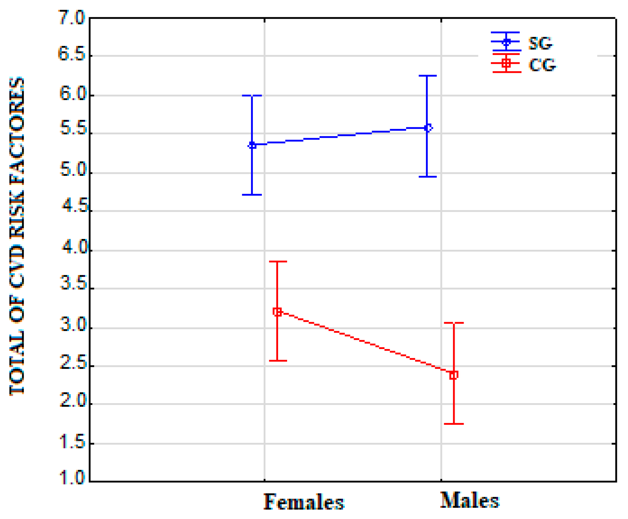
| Mean ± SD | 5.5 ± 1.9 | 2.8 ± 1.5 | <0.001 | 5.4 ± 1.7 | 3.2 ± 1.5 | <0.001 | 5.6 ± 2.2 | 2.4 ± 1.4 | <0.001 |
| Median | 5 | 3 | 5 | 3 | 6 | 2 | |||
| Min–Max | 2–10 | 0–6 | 3–9 | 1–6 | 2–10 | 0–5 | |||
Publisher’s Note: MDPI stays neutral with regard to jurisdictional claims in published maps and institutional affiliations. |
© 2022 by the authors. Licensee MDPI, Basel, Switzerland. This article is an open access article distributed under the terms and conditions of the Creative Commons Attribution (CC BY) license (https://creativecommons.org/licenses/by/4.0/).
Share and Cite
Lucki, M.; Chlebuś, E.; Wareńczak, A.; Daroszewski, P.; Lisiński, P. Assessment of CVD Risk Factors in Secondary Prevention after Ischemic Stroke Using the ICF. Int. J. Environ. Res. Public Health 2022, 19, 3368. https://doi.org/10.3390/ijerph19063368
Lucki M, Chlebuś E, Wareńczak A, Daroszewski P, Lisiński P. Assessment of CVD Risk Factors in Secondary Prevention after Ischemic Stroke Using the ICF. International Journal of Environmental Research and Public Health. 2022; 19(6):3368. https://doi.org/10.3390/ijerph19063368
Chicago/Turabian StyleLucki, Mateusz, Ewa Chlebuś, Agnieszka Wareńczak, Przemysław Daroszewski, and Przemysław Lisiński. 2022. "Assessment of CVD Risk Factors in Secondary Prevention after Ischemic Stroke Using the ICF" International Journal of Environmental Research and Public Health 19, no. 6: 3368. https://doi.org/10.3390/ijerph19063368