Inflamed Phylloides Tumour in a Girl: A Challenging Diagnosis in Paediatric Breast Lesions

 ,
,  and
and 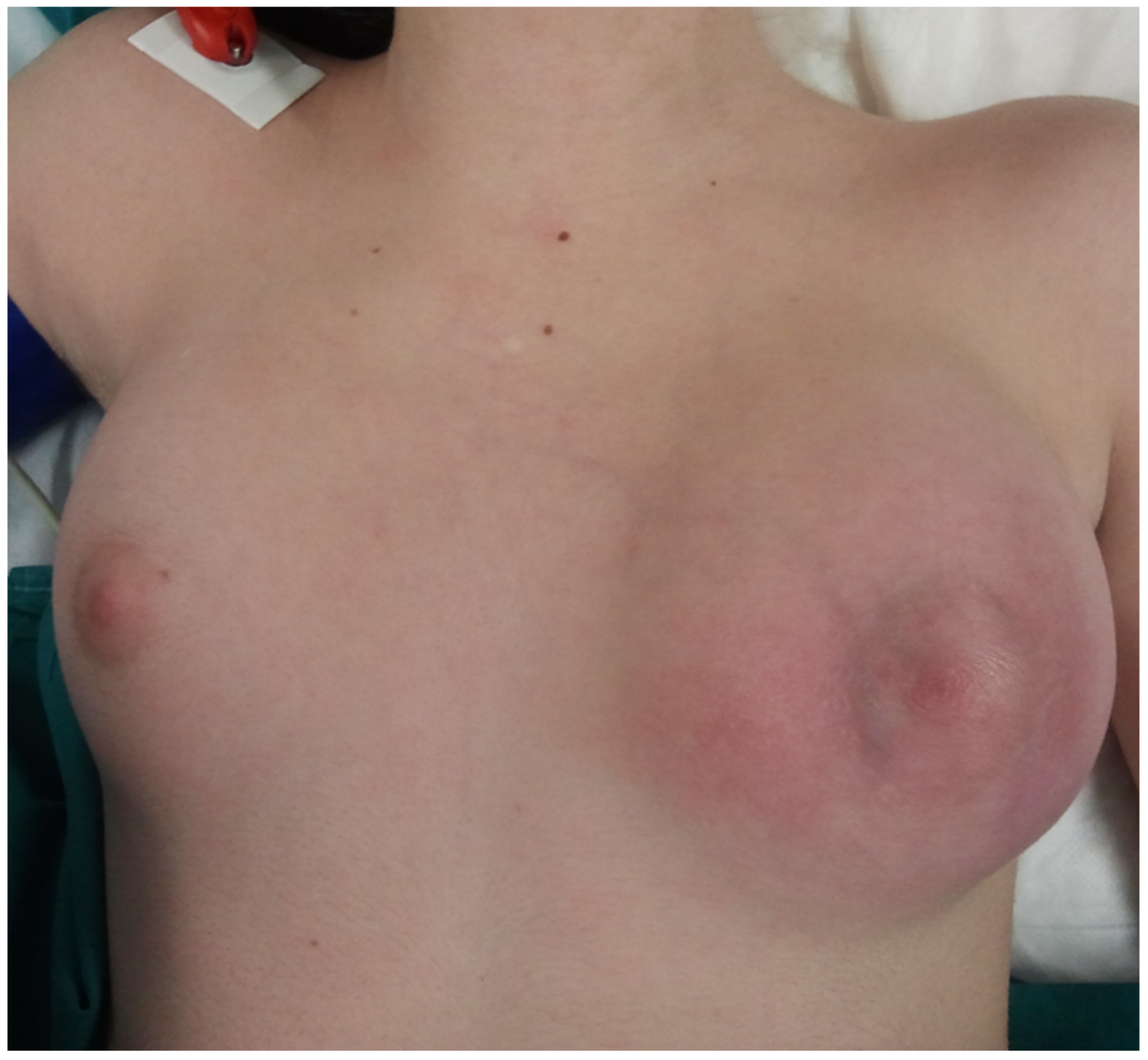
Abstract
:1. Introduction
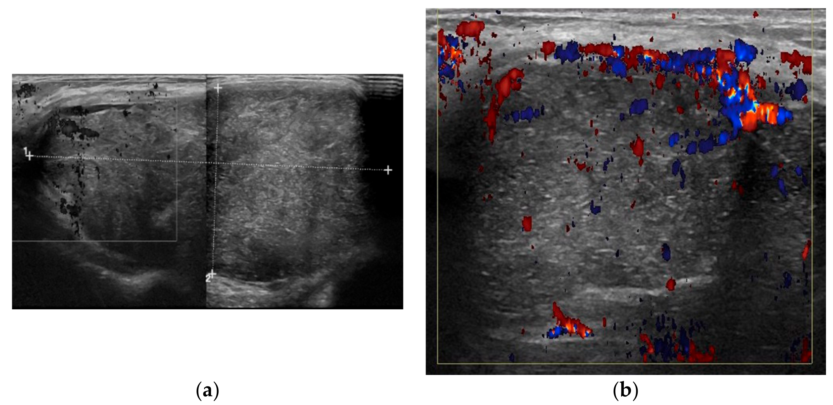
2. Case Presentation
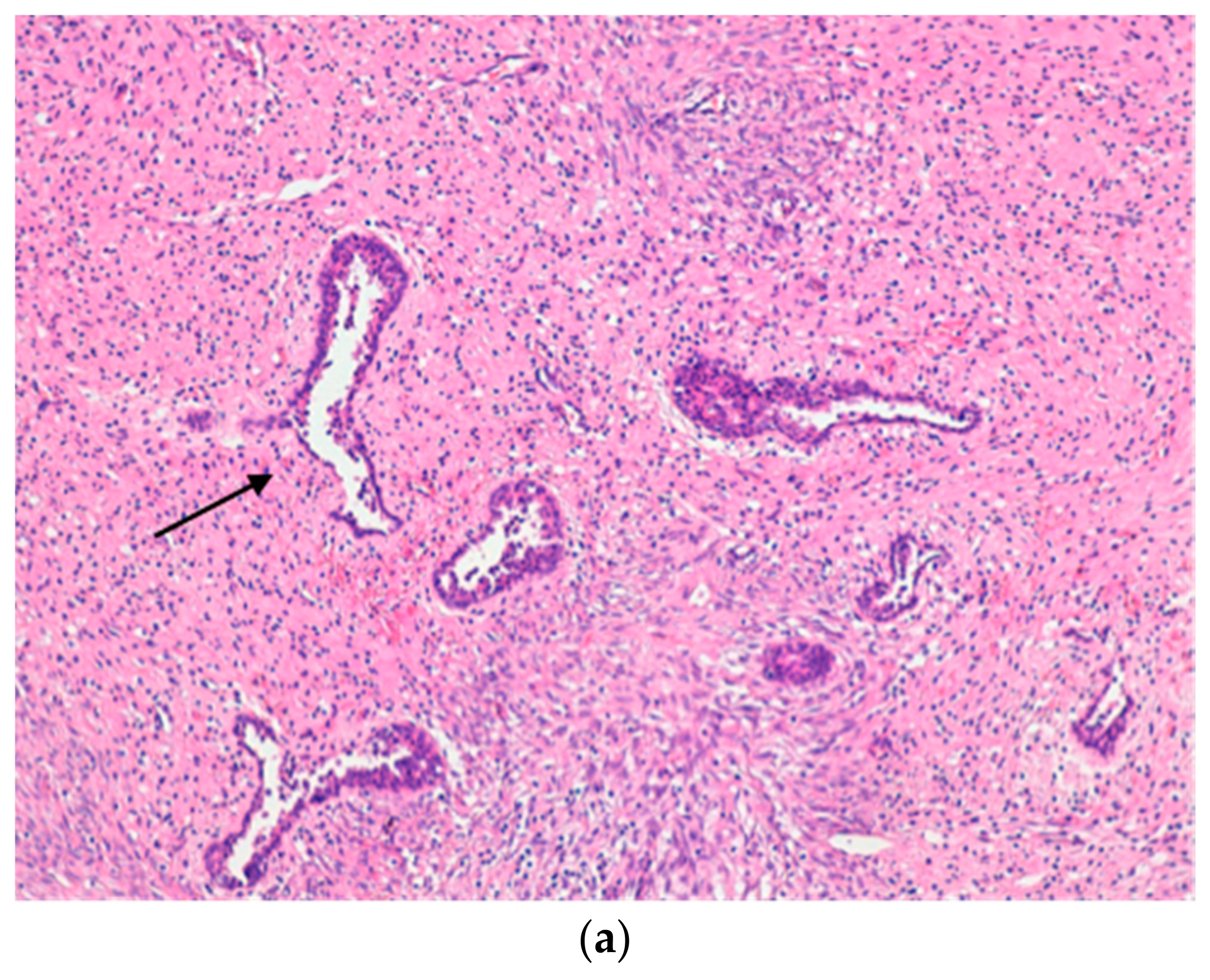
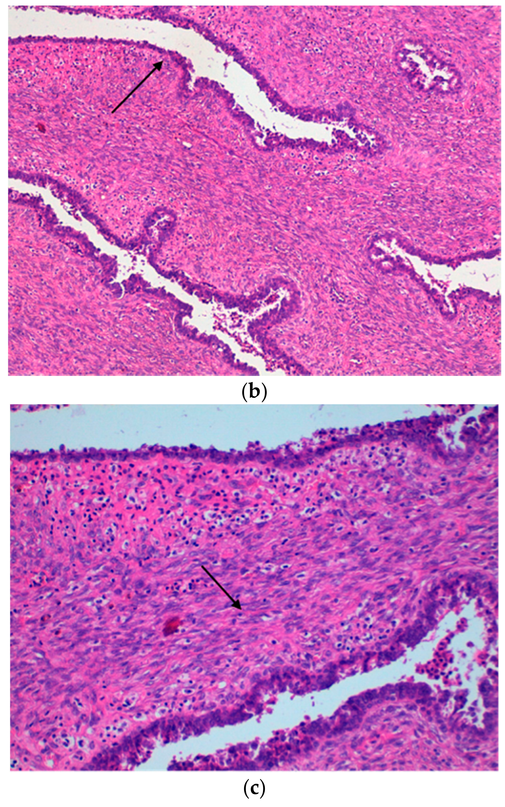
3. Discussion
4. Conclusions
Author Contributions
Funding
Acknowledgments
Conflicts of Interest
References
- Co, M.; Chen, C.; Tsang, J.Y.; Tse, G.; Kwong, A. Mammary phyllodes tumour: A 15-year multicentre clinical review. J. Clin. Pathol. 2017. [Google Scholar] [CrossRef] [PubMed]
- Parker, S.J.; Harries, S.A. Phyllodes tumours. Postgrad. Med. J. 2001, 77, 428–435. [Google Scholar] [CrossRef] [PubMed]
- Kaneda, H.J.; Mack, J.; Kasales, C.J.; Schetter, S. Pediatric and adolescent breast masses: A review of pathophysiology, imaging, diagnosis, and treatment. Am. J. Roentgenol. 2013, 200, W204–W212. [Google Scholar] [CrossRef] [PubMed]
- Martino, A.; Zamparelli, M.; Santinelli, A.; Cobellis, G.; Rossi, L.; Amici, G. Unusual clinical presentation of a rare case of phyllodes tumor of the breast in an adolescent girl. J. Pediatr. Surg. 2001, 36, 941–943. [Google Scholar] [CrossRef] [PubMed]
- Pistolese, C.A.; Tanga, I.; Cossu, E.; Perretta, T.; Yamgoue, M.; Bonanno, E.; Simonetti, G. A phyllodes tumor in a child. J. Pediatr. Adolesc. Gynecol. 2009, 22, e21–e24. [Google Scholar] [CrossRef] [PubMed]
- Galazios, G.; Dafopoulos, K.; Gardikis, S.; Sigalas, J.; Tamiolakis, D.; Liberis, V.; Sivridis, E. Cystosarcoma phyllodes in a 13-year-old Muslim girl treated with conservative surgery: A case report. Eur. J. Gynaecol. Oncol. 2003, 24, 89–90. [Google Scholar] [PubMed]
- Makhija, D.; Shah, H.; Bothra, J.; Jayaswal, S. An adolescent with phyllodes tumour: Case report and review. Int. J. Pediatr. Adolesc. Med. 2016, 3, 280–283. [Google Scholar] [CrossRef]
- Akakpo, P.K.; Der, E.M.; Cooper, P.; Anim, J.T. Childhood phyllodes tumour of the breast. J. Clin. Case Rep. 2015, 5, 495–499. [Google Scholar] [CrossRef]
- Tse, G.M.; Niu, Y.; Shi, H.J. Phyllodes tumor of the breast: An update. Breast Cancer 2010, 17, 29–34. [Google Scholar] [CrossRef] [PubMed]
- Tan, B.Y.; Acs, G.; Apple, S.K.; Badve, S.; Bleiweiss, I.J.; Brogi, E.; Calvo, J.P.; Dabbs, D.J.; Ellis, I.O.; Eusebi, V.; et al. Phyllodes tumours of the breast: A consensus review. Histopathology 2016, 68, 5–21. [Google Scholar] [CrossRef] [PubMed]
- Gutierrez, J.C.; Housri, N.; Koniaris, L.G.; Fischer, A.C.; Sola, J.E. Malignant breast cancer in children: A review of 75 patients. J. Surg. Res. 2008, 147, 182–188. [Google Scholar] [CrossRef] [PubMed]
- Jaing, T.H.; Yang, C.P.; Hung, I.J.; Chen, S.H.; Tang, T.C.; Shih, H.N.; Hsueh, C. Phyllodes tumor in survivors of childhood osteosarcoma: A single institution’s experience. J. Pediatr. Hematol. Oncol. 2014, 36, e36–e38. [Google Scholar] [CrossRef] [PubMed]
- Cucinotta, M.; Quartuccio, N.; Coppolino, P.; La Delfa, V.; Bianchi, M.; Fagioli, F.; Cistaro, A. A strange case of phyllodes tumor detected using (18)F-FDG PET/CT in an adolescent patient affected by Hodgkin lymphoma: A possible pitfall. Clin. Lymphoma Myeloma Leuk. 2014, 14, e201–e205. [Google Scholar] [CrossRef] [PubMed]
- Jayasinghe, Y.; Simmons, P.S. Occurrence of two rare malignant neoplasms (breast and ovarian) in an adolescent female. J. Pediatr. Adolesc. Gynecol. 2009, 22, e99–e103. [Google Scholar] [CrossRef] [PubMed]
- Bielack, S.S.; Hecker-Nolting, S.; Kevric, M.; Juergens, H. More on osteosarcoma and phylloides tumor. J. Pediatr. Hematol. Oncol. 2015, 37, 158–159. [Google Scholar] [CrossRef] [PubMed]
- Howard, B.A.; Gusterson, B.A. Human breast development. J. Mammary Gland Biol. Neoplasia 2000, 5, 119–137. [Google Scholar] [CrossRef] [PubMed]
- Sosin, M.; Pulcrano, M.; Feldman, E.D.; Patel, K.M.; Nahabedian, M.Y.; Weissler, J.M.; Rodriguez, E.D. Giant juvenile fibroadenoma: A systematic review with diagnostic and treatment recommendations. Gland Surg. 2015, 4, 312–321. [Google Scholar] [PubMed]
- Kalambo, M.; Adrada, B.E.; Adeyefa, M.M.; Krishnamurthy, S.; Hess, K.; Carkaci, S.; Whitman, G.J. Phyllodes tumour of the breast: Ultrasound-pathology correlation. Am. J. Roentgenol. 2018. [Google Scholar] [CrossRef] [PubMed]
- Tse, G.M.; Cheung, H.S.; Pang, L.M.; Chu, W.C.; Law, B.K.; Kung, F.Y.; Yeung, D.K. Characterization of lesions of the breast with proton MR spectroscopy: Comparison of carcinomas, benign lesions, and phyllodes tumors. Am. J. Roentgenol. 2003, 181, 1267–1272. [Google Scholar] [CrossRef] [PubMed]
- Wadasadawala, T.; Swain, M.; Bajpai, J. Phyllodes tumor of the breast: Inside the sheep’s skin. Oncol. Targets 2016, 3, 1–9. [Google Scholar] [CrossRef]


© 2018 by the authors. Licensee MDPI, Basel, Switzerland. This article is an open access article distributed under the terms and conditions of the Creative Commons Attribution (CC BY) license (http://creativecommons.org/licenses/by/4.0/).
Share and Cite
Testa, I.; Salvatori, C.; Prestipino, M.; Laurenti, M.E.; Gerli, P.; Di Cara, G.; Principi, N.; Esposito, S.; Bertozzi, M. Inflamed Phylloides Tumour in a Girl: A Challenging Diagnosis in Paediatric Breast Lesions. Int. J. Environ. Res. Public Health 2018, 15, 959. https://doi.org/10.3390/ijerph15050959
Testa I, Salvatori C, Prestipino M, Laurenti ME, Gerli P, Di Cara G, Principi N, Esposito S, Bertozzi M. Inflamed Phylloides Tumour in a Girl: A Challenging Diagnosis in Paediatric Breast Lesions. International Journal of Environmental Research and Public Health. 2018; 15(5):959. https://doi.org/10.3390/ijerph15050959
Chicago/Turabian StyleTesta, Ilaria, Cristina Salvatori, Marco Prestipino, Maria Elena Laurenti, Paolo Gerli, Giuseppe Di Cara, Nicola Principi, Susanna Esposito, and Mirko Bertozzi. 2018. "Inflamed Phylloides Tumour in a Girl: A Challenging Diagnosis in Paediatric Breast Lesions" International Journal of Environmental Research and Public Health 15, no. 5: 959. https://doi.org/10.3390/ijerph15050959