

First Report of Pinnatoxin-G (PnTX-G) in a Marine–Coastal Area of the Adriatic Sea Associated with the Presence of the Dinoflagellate Vulcanodinium rugosum

 , , , , , and
Abstract
:1. Introduction
2. Results
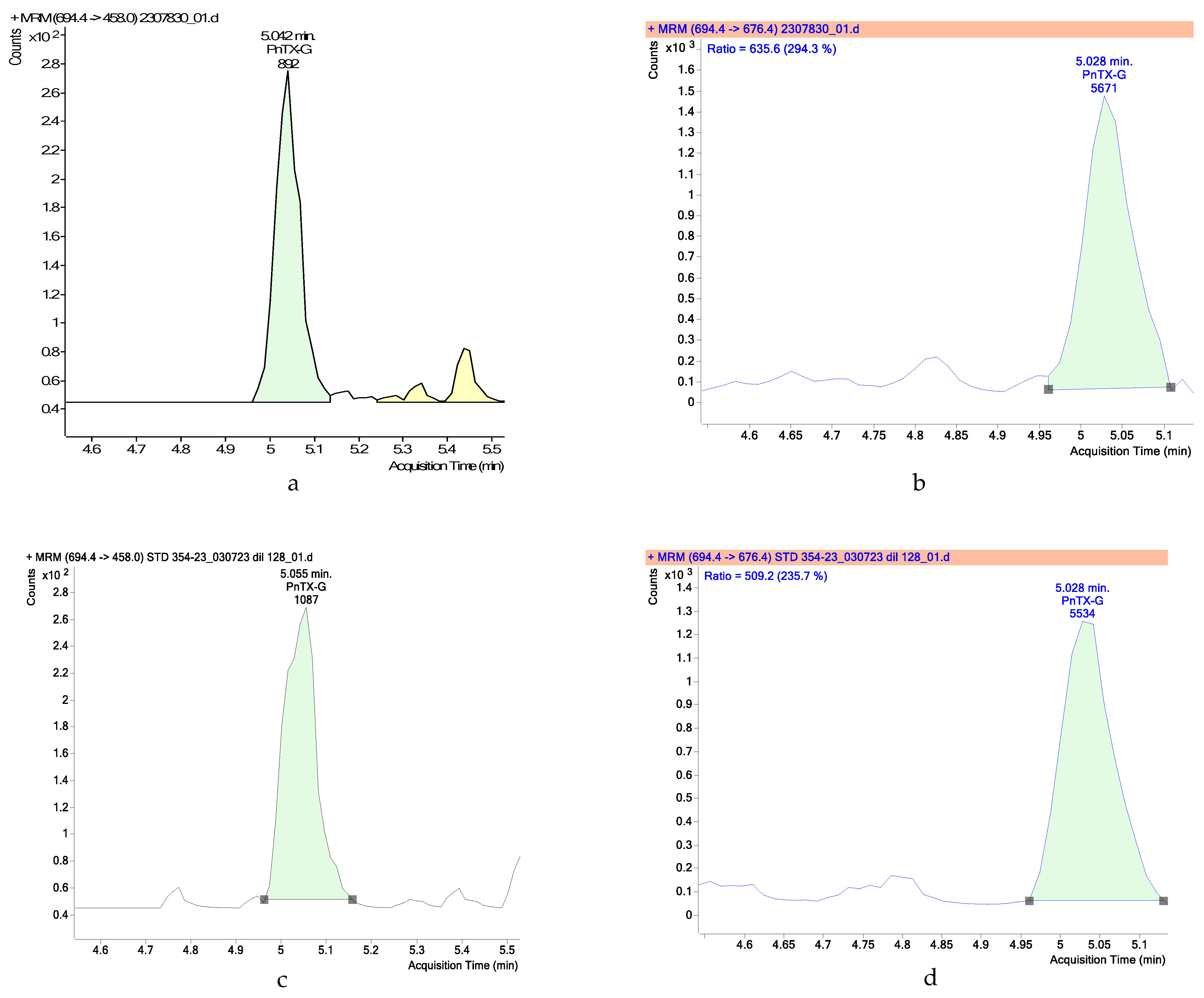
2.1. Toxin Analyses
2.2. Microscopy
3. Discussion
4. Materials and Methods
4.1. Study Area and Sampling
4.2. Solvent and Reagents
4.3. Toxin Analyses
4.3.1. Extraction
4.3.2. Liquid Chromatography with Tandem Mass Spectrometry (LC-MS/MS)
4.4. Identification of Vulcanodinium rugosum
Author Contributions
Funding
Institutional Review Board Statement
Data Availability Statement
Acknowledgments
Conflicts of Interest
References
- Zheng, S.Z.; Huang, F.L.; Chem, S.C.; Tan, X.F.; Zuo, J.B.; Peng, J.; Xie, R.W. The isolation and bioactivities of pinnatoxin. Chin. J. Mar. Drugs 1990, 9, 33–55. [Google Scholar]
- Chou, T.; Haino, T.; Kuramoto, M.; Uemura, D. Isolation and structure of pinnatoxin D, a new shellfish poison from the okinawan bivalve Pinna muricata. Tetrahedron Lett. 1996, 37, 4027–4030. [Google Scholar] [CrossRef]
- Uemura, D.; Chou, T.; Haino, T.; Nagatsu, A.; Fukuzawa, S.; Zheng, S.Z.; Chen, H. Pinnatoxin A: A toxic amphoteric macrocycle from the Okinawan bivalve Pinna muricata. J. Am. Chem. Soc. 1995, 117, 1155–1156. [Google Scholar] [CrossRef]
- Takada, N.; Umemura, N.; Suenaga, K.; Chou, T.; Nagatsu, A.; Haino, T.; Yamada, K.; Uemura, D. Pinnatoxins B and C, the most toxic components in the pinnatoxin series from the Okinawan bivalve Pinna muricata. Tetrahedron Lett. 2001, 42, 3491–3494. [Google Scholar] [CrossRef]
- McNabb, P.S.; McCoubrey, D.J.; Rhodes, L.; Smith, K.; Selwood, A.I.; van Ginkel, R.; MacKenzie, A.L.; Munday, R.; Holland, P.T. New perspectives on biotoxin detection in Rangaunu Harbour, New Zealand arising from the discovery of pinnatoxins. Harmful Algae 2012, 13, 34–39. [Google Scholar] [CrossRef]
- Selwood, A.I.; Miles, C.O.; Wilkins, A.L.; van Ginkel, R.; Munday, R.; Rise, F.; McNabb, P. Isolation, Structural Determination and Acute Toxicity of Pinnatoxins E, F and G. J. Agric. Food Chem. 2010, 58, 6532–6542. [Google Scholar] [CrossRef] [PubMed]
- Zeng, N.; Gu, H.; Smith, K.; Rhodes, L.; Selwood, A.; Yang, W. The first report of Vulcanodinium rugosum (Dinophyceae) from the South China Sea with a focus on the life cycle. N. Z. J. Mar. Freshw. Res. 2012, 46, 511–521. [Google Scholar] [CrossRef]
- Selwood, A.I.; Wilkins, A.L.; Munday, R.; Gu, H.F.; Smith, K.F.; Rhodes, L.L.; Rise, F. Pinnatoxin H: A new pinnatoxin analogue from a South China Sea Vulcanodinium rugosum isolate. Tetrahedron Lett. 2014, 55, 5508–5510. [Google Scholar] [CrossRef]
- Arnich, N.; Abadie, E.; Delcourt, N.; Fessard, V.; Fremy, J.M.; Hort, V.; Lagrange, E.; Maignien, T.; Molgó, J.; Peyrat, M.B.; et al. Health risk assessment related to pinnatoxins in French shellfish. Toxicon 2020, 180, 1–10. [Google Scholar] [CrossRef] [PubMed]
- Otero, P.; Silva, M. Emerging marine biotoxins in European waters: Potential risks and analytical challenges. Mar. Drugs 2022, 20, 199. [Google Scholar] [CrossRef]
- Nezan, E.; Chomerat, N. Vulcanodinium rugosum gen. Nov., sp. Nov.(dinophyceae): A new marine dinoflagellate from the French mediterranean coast. Cryptogam. Algol. 2011, 32, 3–18. [Google Scholar] [CrossRef]
- Rhodes, L.; Smith, K.; Selwood, A.; McNabb, P.; Munday, R.; Suda, S.; Molenaar, S.; Hallegraeff, G. Dinoflagellate Vulcanodinium rugosum identified as the causative organism of pinnatoxins in Australia, New Zealand and Japan. Phycologia 2011, 50, 624–628. [Google Scholar] [CrossRef]
- Geiger, M.; Deslanglois, G.; Hogeveen, K.; Fessard, V.; Leprêtre, T.; Mondeguer, F.; Guitton, Y.; Hervé, F.; Séchet, V.; Grovel, O.; et al. Cellular models and cytotoxicity of pinnatoxin-G and extracts of the dinoflagellate Vulcanodinium rugosum recently isolated from the French mediterranean lagoon of Ingril. Toxicon 2013, 75, 215–216. [Google Scholar] [CrossRef]
- Smith, K.F.; Rhodes, L.L.; Suda, S.; Selwood, A.I. A dinoflagellate producer of pinnatoxin G, isolated from sub-tropical Japanese waters. Harmful Algae 2011, 10, 702–705. [Google Scholar] [CrossRef]
- Möller, K.; Pinto-Torres, M.; Mardones, J.I.; Krock, B. Distribution of phycotoxins in Última Esperanza Province during the PROFAN expedition 2019. Prog. Oceanogr. 2022, 206, 102851. [Google Scholar] [CrossRef]
- Norambuena, L.; Mardones, J.I. Emerging phycotoxins in the Chilean coast: First localized detection of the neurotoxic cyclic imine Pinnatoxin-G in shellfish banks. Mar. Pollut. Bull. 2023, 190, 114878. [Google Scholar] [CrossRef]
- Hernández-Becerril, D.U.; Rodríguez-Palacio, M.C.; Lozano-Ramírez, C. Morphology and life stages of the potentially pinnatoxin-producing thecate dinoflagellate Vulcanodinium rugosum from the tropical Mexican Pacific. Bot. Mar. 2013, 56, 535–540. [Google Scholar] [CrossRef]
- Fu, Z.; Piumsomboon, A.; Punnarak, P.; Uttayarnmanee, P.; Leaw, C.P.; Lim, P.T.; Wang, A.; Gu, H. Diversity and distribution of harmful microalgae in the Gulf of Thailand assessed by DNA metabarcoding. Harmful Algae 2021, 106, 102063. [Google Scholar] [CrossRef]
- Al Muftah, A.; Selwood, A.I.; Foss, A.J.; Al-Jabri, H.M.S.J.; Potts, M.; Yilmaz, M. Algal toxins and producers in the marine waters of Qatar, Arabian Gulf. Toxicon 2016, 122, 54–66. [Google Scholar] [CrossRef]
- McCarron, P.; Rourke, W.A.; Hardstaff, W.; Pooley, B.; Quilliam, M.A. Identification of pinnatoxins and discovery of their fatty acid ester metabolites in mussels (Mytilus edulis) from Eastern Canada. J. Agric. Food Chem. 2012, 60, 1437–1446. [Google Scholar] [CrossRef]
- Rundberget, T.; Aasen, J.A.B.; Selwood, A.I.; Miles, C.O. Pinnatoxins and spirolides in norwegian blue mussels and seawater. Toxicon 2011, 58, 700–711. [Google Scholar] [CrossRef]
- Hess, P.; Abadie, E.; Hervé, F.; Berteaux, T.; Séchet, V.; Aráoz, R.; Molgó, J.; Zakarian, A.; Sibat, M.; Rundberget, T.; et al. Pinnatoxin G is responsible for atypical toxicity in mussels (Mytilus galloprovincialis) and clams (Venerupis decussata) from Ingril, a French Mediterranean lagoon. Toxicon 2013, 75, 16–26. [Google Scholar] [CrossRef] [PubMed]
- Abadie, E.; Chiantella, C.; Grottier, A.; Rhodes, L.; Masseret, E.; Berteaux, T.; Laabir, M. What are the main environmental factors driving the development of the neurotoxic dinoflagellate Vulcanodinium rugosum in a Mediterranean ecosystem (Ingril lagoon, France)? Harmful Algae 2018, 75, 75–86. [Google Scholar] [CrossRef]
- Bouquet, A.; Thébault, A.; Arnich, N.; Foucault, E.; Caillard, E.; Gianaroli, C.; Bellamy, C.; Rolland, J.L.; Laabir, M.; Abadie, E. Modelling spatiotemporal distributions of Vulcanodinium rugosum and pinnatoxin G in French Mediterranean lagoons: Application to human health risk characterisation. Harmful Algae 2023, 129, 102500. [Google Scholar] [CrossRef] [PubMed]
- Satta, C.T.; Anglès, S.; Lugliè, A.; Guillén, J.; Sechi, N.; Camp, J.; Garcés, E. Studies on dinoflagellate cyst assemblages in two estuarine Mediterranean bays: A useful tool for the discovery and mapping of harmful algal species. Harmful Algae 2013, 24, 65–79. [Google Scholar] [CrossRef]
- McCarthy, M.; Bane, V.; García-Altares, M.; van Pelt, F.N.; Furey, A.; O’Halloran, J. Assessment of emerging biotoxins (pinnatoxin G and spirolides) at Europe’s first marine reserve: Lough Hyne. Toxicon 2015, 108, 202–209. [Google Scholar] [CrossRef] [PubMed]
- Lamas, J.P.; Arévalo, F.; Moroño, Á.; Correa, J.; Muñíz, S.; Blanco, J. Detection and spatio-temporal distribution of pinnatoxins in shellfish from the Atlantic and Cantabrian coasts of Spain. Toxins 2019, 11, 340. [Google Scholar] [CrossRef] [PubMed]
- Varriale, F.; Tartaglione, L.; Cinti, S.; Milandri, A.; Dall’Ara, S.; Calfapietra, A.; Dell’Aversano, C. Development of a data dependent acquisition-based approach for the identification of unknown fast-acting toxins and their ester metabolites. Talanta 2021, 224, 121842. [Google Scholar] [CrossRef]
- Munday, R.; Selwood, A.I.; Rhodes, L. Acute toxicity of pinnatoxins E, F and G to mice. Toxicon 2012, 60, 995–999. [Google Scholar] [CrossRef]
- Servent, D.; Malgorn, C.; Bernes, M.; Gil, S.; Simasotchi, C.; Hérard, A.S.; Delzescaux, T.; Thai, R.; Barbe, P.; Keck, M.; et al. First evidence that emerging pinnatoxin-G, a contaminant of shellfish, reaches the brain and crosses the placental barrier. Sci. Total Environ. 2021, 790, 148125. [Google Scholar] [CrossRef]
- Efsa European Food Safety Authority. Scientific Opinion on marine biotoxins in shellfish—Cyclic imines (spirolides, gymnodimines, pinnatoxins and pteriatoxins). EFSA J. 2010, 8, 1628. [Google Scholar]
- Moreira-González, A.R.; Comas-González, A.; Valle-Pombrol, A.; Seisdedo-Losa, M.; Hernández-Leyva, O.; Fernandes, L.F.; Chomérat, N.; Bilien, G.; Hervé, F.; Rovillon, G.A.; et al. Summer bloom of Vulcanodinium rugosum in Cienfuegos Bay (Cuba) associated to dermatitis in swimmers. Sci. Total Environ. 2021, 757, 143782. [Google Scholar] [CrossRef] [PubMed]
- Aráoz, R.; Barnes, P.; Séchet, V.; Delepierre, M.; Zinn-Justin, S.; Molgó, J.; Zakarian, A.; Hess, P.; Servent, D. Cyclic imine toxins survey in coastal european shellfish samples: Bioaccumulation and mode of action of 28-O-palmitoyl ester of pinnatoxin-G. first report of portimine-A bioaccumulation. Harmful Algae 2020, 98, 101887. [Google Scholar] [CrossRef] [PubMed]
- Estévez, P.; Leao, J.M.; Gago-Martinez, A. Marine biotoxins as natural contaminants in seafood: European perspective. In Present Knowledge in Food Safety; Academic Press: Cambridge, MA, USA, 2023; pp. 115–127. [Google Scholar]
- Funari, E.; Manganelli, M.; Testai, E. Ostreopsis cf. ovata: Linee Guida per la Gestione Delle Fioriture Negli Ambienti Marino-Costieri in Relazione a Balneazione e Altre Attività Ricrative, ISTISAN Reports; Istituto Superiore di Sanità: Rome, Italy, 2014; (full report only in Italian). [Google Scholar]
- Garrett, M.J.; Puchulutegui, C.; Selwood, A.I.; Wolny, J.L. Identification of the harmful dinoflagellate Vulcanodinium rugosum recovered from a ballast tank of a globally traveled ship in Port Tampa Bay, Florida, USA. Harmful Algae 2014, 39, 202–209. [Google Scholar] [CrossRef]
- Rambla-Alegre, M.; Miles, C.O.; de la Iglesia, P.; Fernandez-Tejedor, M.; Jacobs, S.; Sioen, I.; Verbeke, W.; Samdal, I.A.; Sandvik, M.; Barbosa, V.; et al. Occurence of cyclic imines in European commercial seafood and consumers risk assessment. Environ. Res. 2018, 161, 392–398. [Google Scholar] [CrossRef]
- Hort, V.; Bastardo-Fernández, I.; Nicolas, M. Exploration of Vulcanodinium rugosum toxins and their metabolism products in mussels from the Ingril Lagoon hotspot in France. Mar. Drugs 2023, 21, 429. [Google Scholar] [CrossRef]
- ANSES Agence Nationale de Sécurité Sanitaire de L’alimentation, de L’environnement et du Travail, 2019. Risques Liés Aux Pinnatoxines dans les Coquillages. Avis de l’Anses et Rapport D’expertise Collective, p. 136. Mars 2019—Edition Scientifique. (Full Report only in French). Available online: https://www.anses.fr/fr/system/files/ERCA2016SA0013Ra.pdf (accessed on 27 February 2024).
- Mendez, S.; Alonso, R.; Moreira, A.; Reguera, B. Chapter 2, IOC Manuals and Guides. In Guide for Designing and Implementing a Plan to Monitor Toxin-Producing Microalgae, 2nd ed.; Reguera, B., Alonso, R., Moreira, A., Méndez, S., Dechraoui-Bottein, M.-Y., Eds.; Intergovernmental Oceanographic Commission (IOC) of UNESCO: Paris, France; International Atomic Energy Agency (IAEA): Vienna, Austria, 2016; p. 66. [Google Scholar]
- Anon 2019. Monitoring of Toxin-producing Phytoplankton in Bivalve Mollusc Harvesting Areas. Guide to Good Practice: Technical Application. Issue, 1. Available online: https://www.aesan.gob.es/en/CRLMB/web/home.html (accessed on 27 February 2024).
- EU-Harmonised Standard Operating Procedure for Determination of Lipophilica Marine Biotoxins in Molluscs by LC-MS/MS. Version 5. Available online: https://www.aesan.gob.es/en/CRLMB/docs/docs/metodos_analiticos_de_desarrollo/EU-Harmonised-SOP-LIPO-LCMSMS_Version5.pdf (accessed on 27 February 2024).
- Fritz, L.; Triemer, E. A rapid simple technique utilizing calcofluor white M2R for the visualization of dinoflagellate thecal plates. J. Phycol. 1985, 21, 662–664. [Google Scholar] [CrossRef]
- Hoppenrath, M.; Murray, S.A.; Chomérat, N.; Horiguchi, T. Marine benthic dinoflagellates—Unveiling their worldwide biodiversity. Kleine Senckenberg-Reihe Band 2015, 54, 276. [Google Scholar]
- Lassus, P.; Chomérat, N.; Hess, P.; Nézan, E. Toxic and Harmful Microalgae of the World Ocean/Micro-Algues Toxiques et Nuisibles de L’océan Mondial; IOC Manuals and Gudes, 68 (Bilingual English/French); International Society for the Study of Harmful Algae: Dalian, China; Intergovernmental Oceanografiphic Commission of UNESCO: Copenhagen, Denmark, 2016. [Google Scholar]


| Toxin | Two-Dimensional Structure |
|---|---|
| Pinnatoxin-A |  |
| Pinnatoxin-B, -C |  |
| Pinnatoxin-D |  |
| Pinnatoxin-E |  |
| Pinnatoxin-F |  |
| Pinnatoxin-G |  |
| Pinnatoxin-H |  |
| Sampling Date | PnTX-G (µg/kg SM) | V. rugosum (Presence/Absence) |
|---|---|---|
| 30 August 2022 | 6.7 | Presence |
| 13 September 2022 | <LOD | Absence |
| 11 October 2022 | <LOD | Absence |
| 21 June 2023 | <LOD | Absence |
| 27 June 2023 | <LOD | Absence |
| 25 July 2023 | <LOD | Absence |
| 22 August 2023 | 4.5 | Absence |
| 29 August 2023 | Trace | Absence |
| 04 October 2023 | Not collected | Absence |
| 12 December 2023 | Not collected | Absence |
| Time (Min) | A% | B% | Flow (mL/min) | Max. Pressure (bar) |
|---|---|---|---|---|
| 0 | 95 | 5 | 0.4 | 1000 |
| 1 | 95 | 5 | 0.4 | 1000 |
| 3 | 37 | 63 | 0.4 | 1000 |
| 6 | 13.8 | 86.2 | 0.4 | 1000 |
| 10 | 13.8 | 86.2 | 0.4 | 1000 |
| 10.01 | 0 | 100 | 0.4 | 1000 |
| 11.0 | 0 | 100 | 0.4 | 1000 |
| 11.01 | 95 | 5 | 0.4 | 1000 |
| 14 | 95 | 5 | 0.4 | 1000 |
Disclaimer/Publisher’s Note: The statements, opinions and data contained in all publications are solely those of the individual author(s) and contributor(s) and not of MDPI and/or the editor(s). MDPI and/or the editor(s) disclaim responsibility for any injury to people or property resulting from any ideas, methods, instructions or products referred to in the content. |
© 2024 by the authors. Licensee MDPI, Basel, Switzerland. This article is an open access article distributed under the terms and conditions of the Creative Commons Attribution (CC BY) license (https://creativecommons.org/licenses/by/4.0/).
Share and Cite
Cangini, M.; Dall’Ara, S.; Rubini, S.; Bertasi, B.; Rizzi, P.; Dell’Orfano, G.; Milandri, S.; Manfredini, S.; Baldini, E.; Vertuani, S. First Report of Pinnatoxin-G (PnTX-G) in a Marine–Coastal Area of the Adriatic Sea Associated with the Presence of the Dinoflagellate Vulcanodinium rugosum. Mar. Drugs 2024, 22, 122. https://doi.org/10.3390/md22030122
Cangini M, Dall’Ara S, Rubini S, Bertasi B, Rizzi P, Dell’Orfano G, Milandri S, Manfredini S, Baldini E, Vertuani S. First Report of Pinnatoxin-G (PnTX-G) in a Marine–Coastal Area of the Adriatic Sea Associated with the Presence of the Dinoflagellate Vulcanodinium rugosum. Marine Drugs. 2024; 22(3):122. https://doi.org/10.3390/md22030122
Chicago/Turabian StyleCangini, Monica, Sonia Dall’Ara, Silva Rubini, Barbara Bertasi, Paolo Rizzi, Giovanni Dell’Orfano, Stefania Milandri, Stefano Manfredini, Erika Baldini, and Silvia Vertuani. 2024. "First Report of Pinnatoxin-G (PnTX-G) in a Marine–Coastal Area of the Adriatic Sea Associated with the Presence of the Dinoflagellate Vulcanodinium rugosum" Marine Drugs 22, no. 3: 122. https://doi.org/10.3390/md22030122