
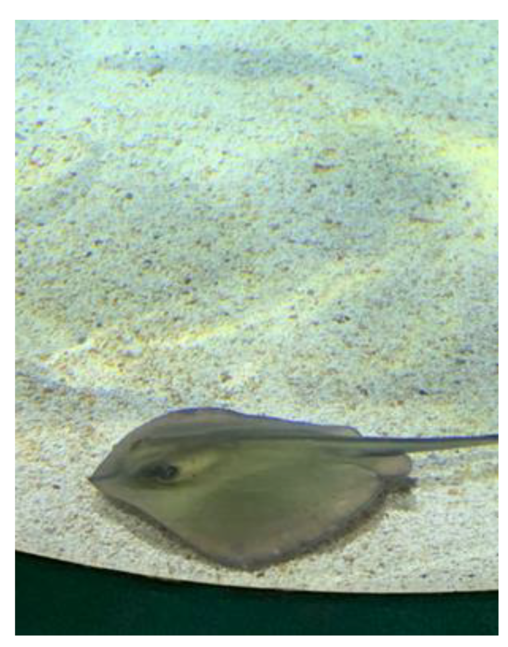
Characterization of Some Dermato-Cosmetic Preparations with Marine Lipids from Black Sea Wild Stingray
 , , , , ,
 , ,
, ,
Abstract
:1. Introduction
2. Results
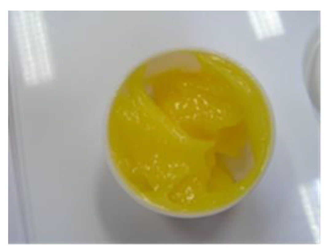
2.1. Characteristics of Stingray Liver Oil
2.2. Characteristics of Stingray Liver Oil Ointment
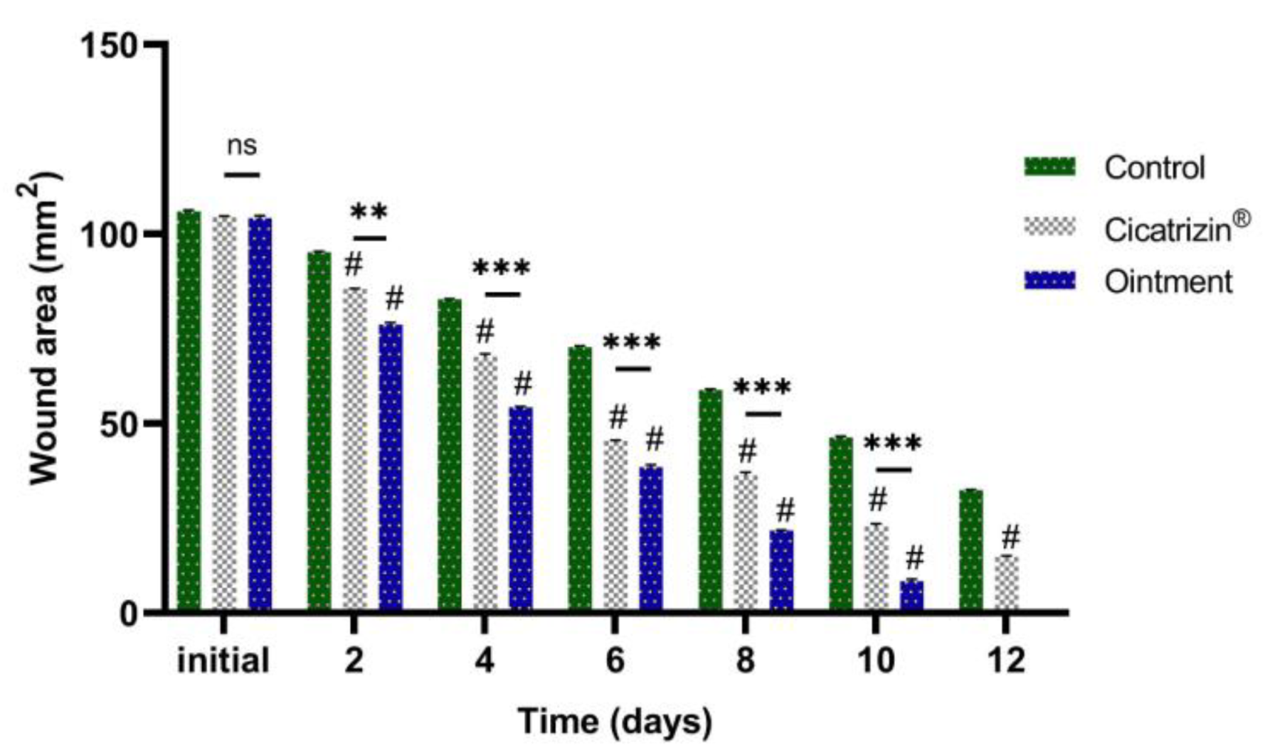
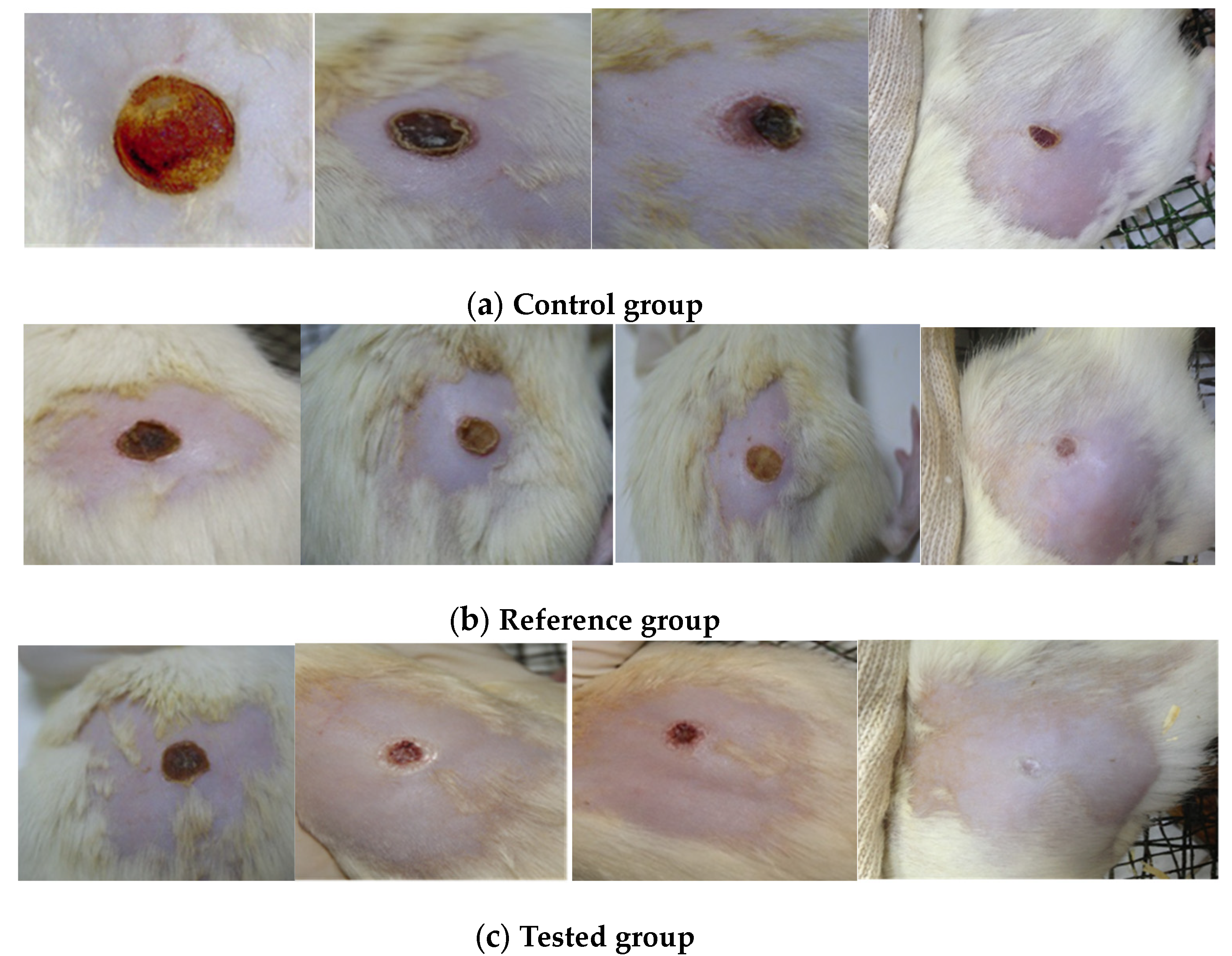
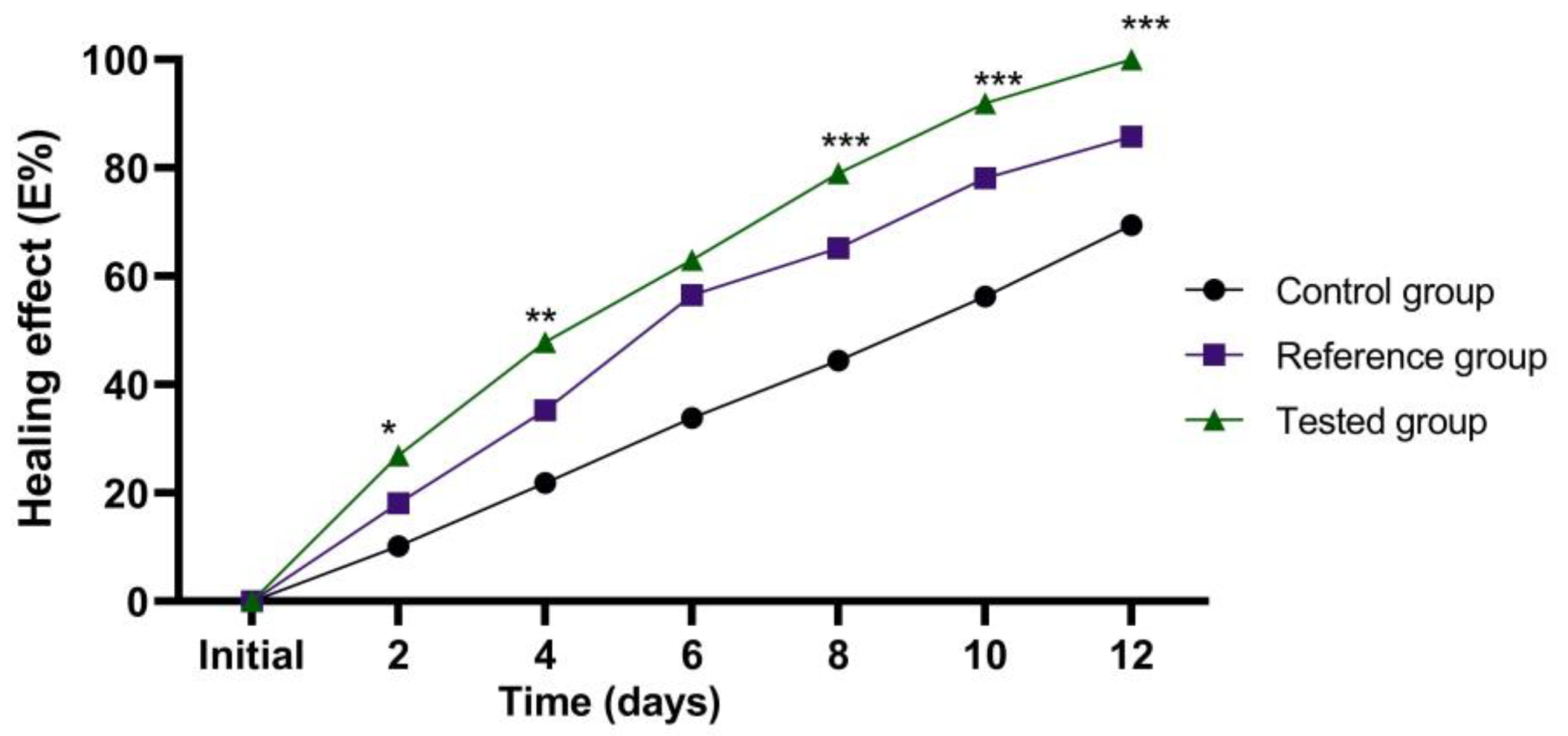
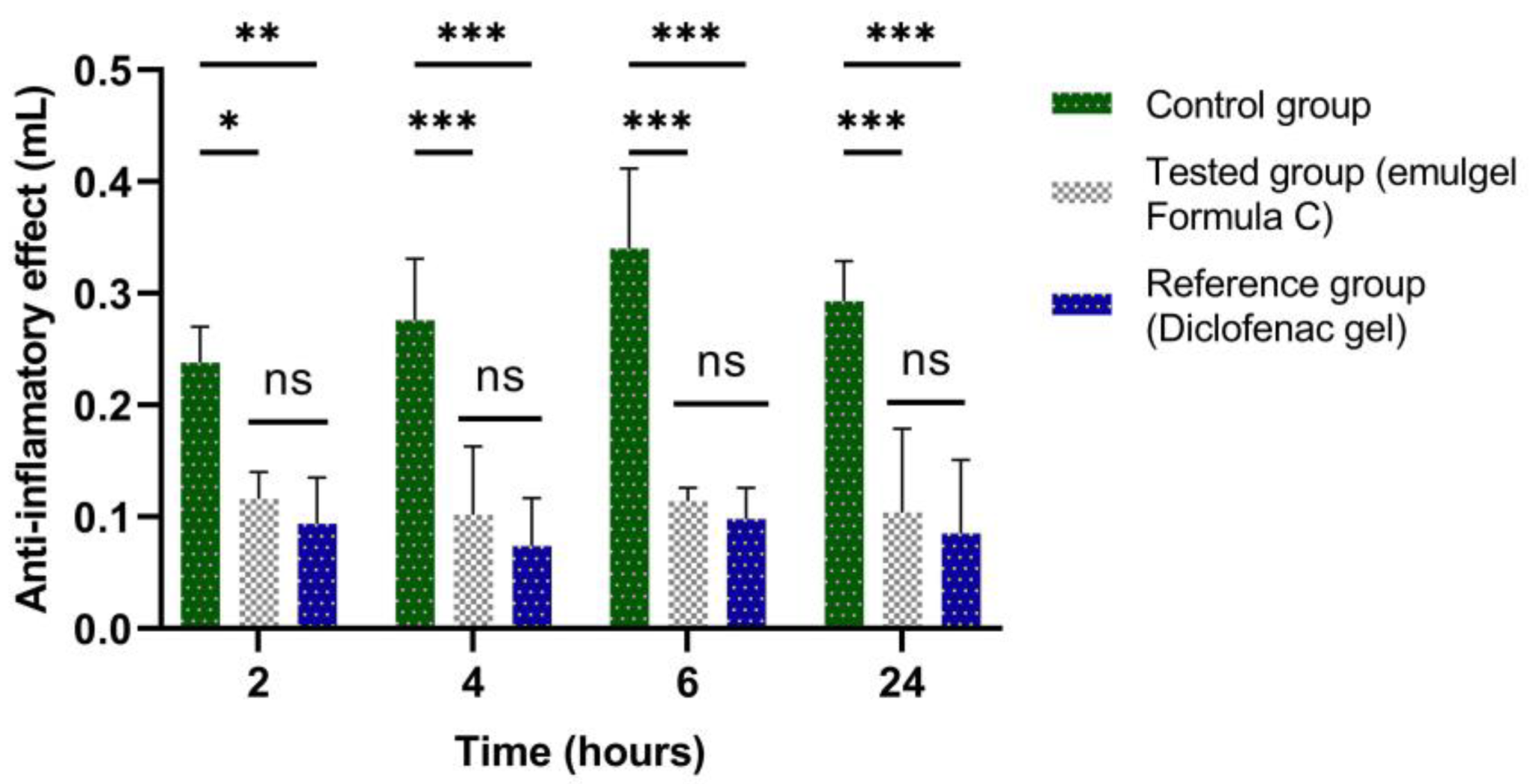
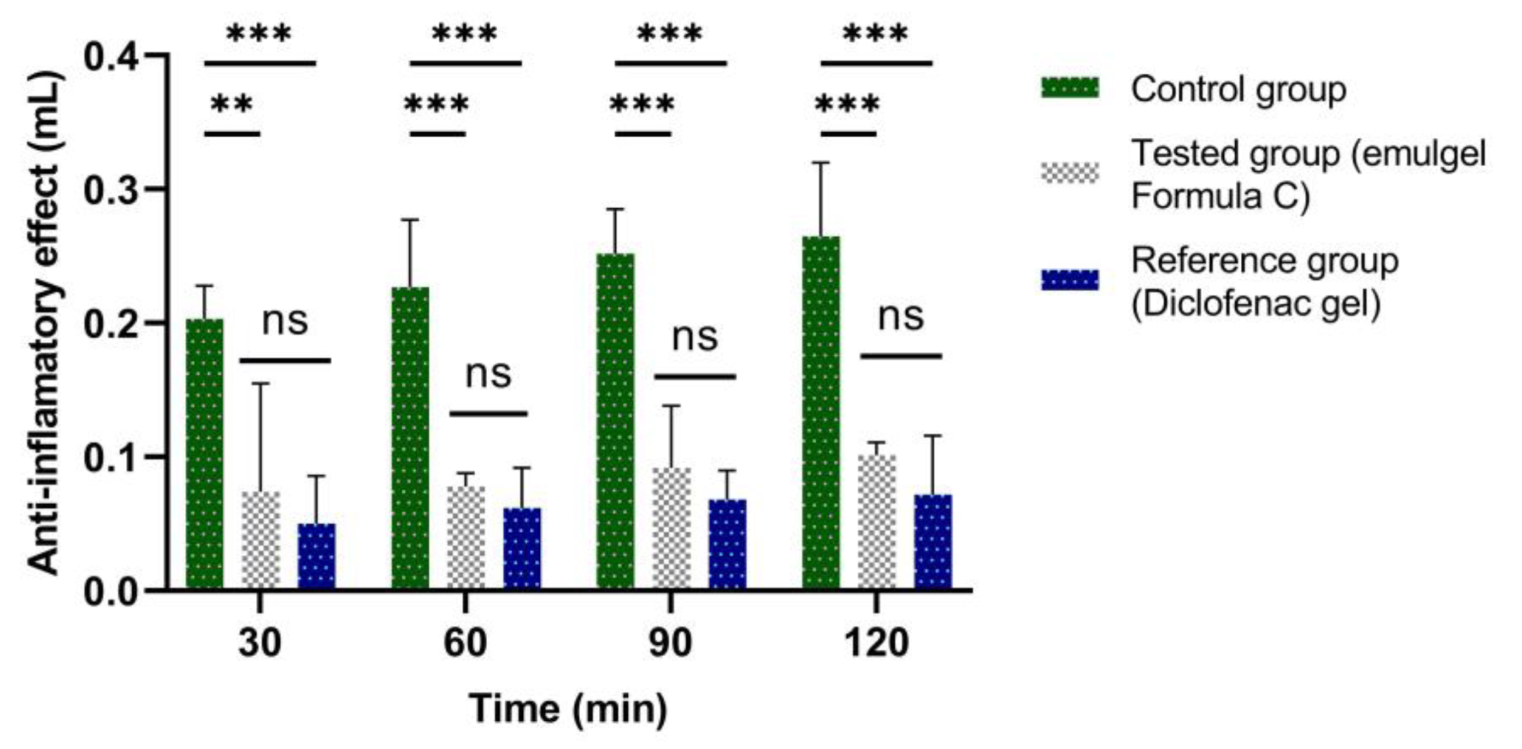
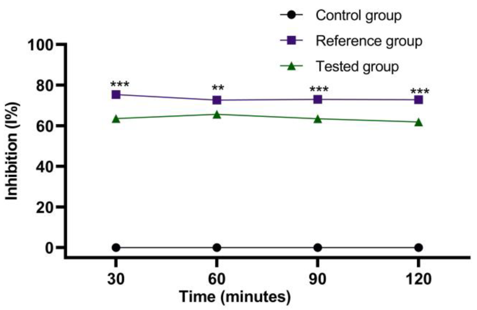
2.3. Evaluation of the Healing Action of Stingray Liver Oil Ointment
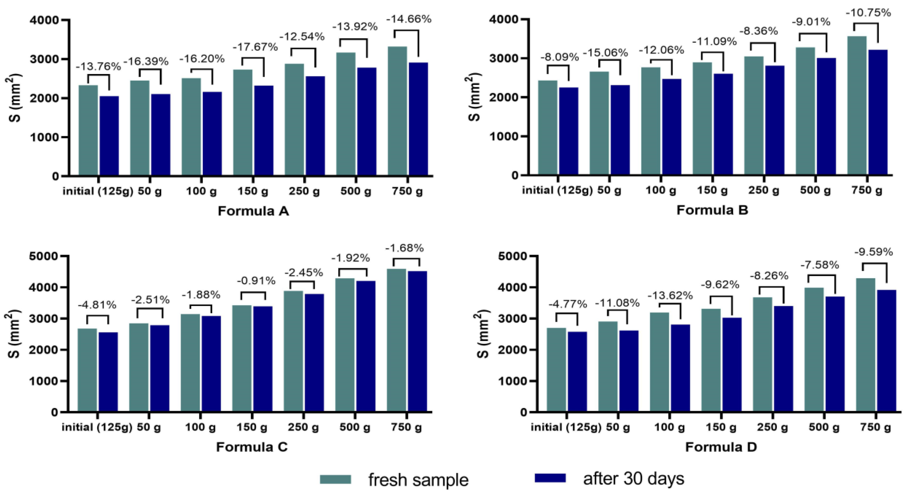
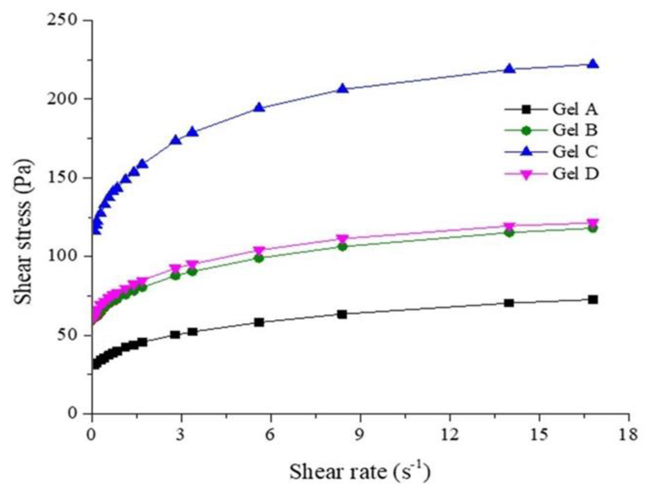
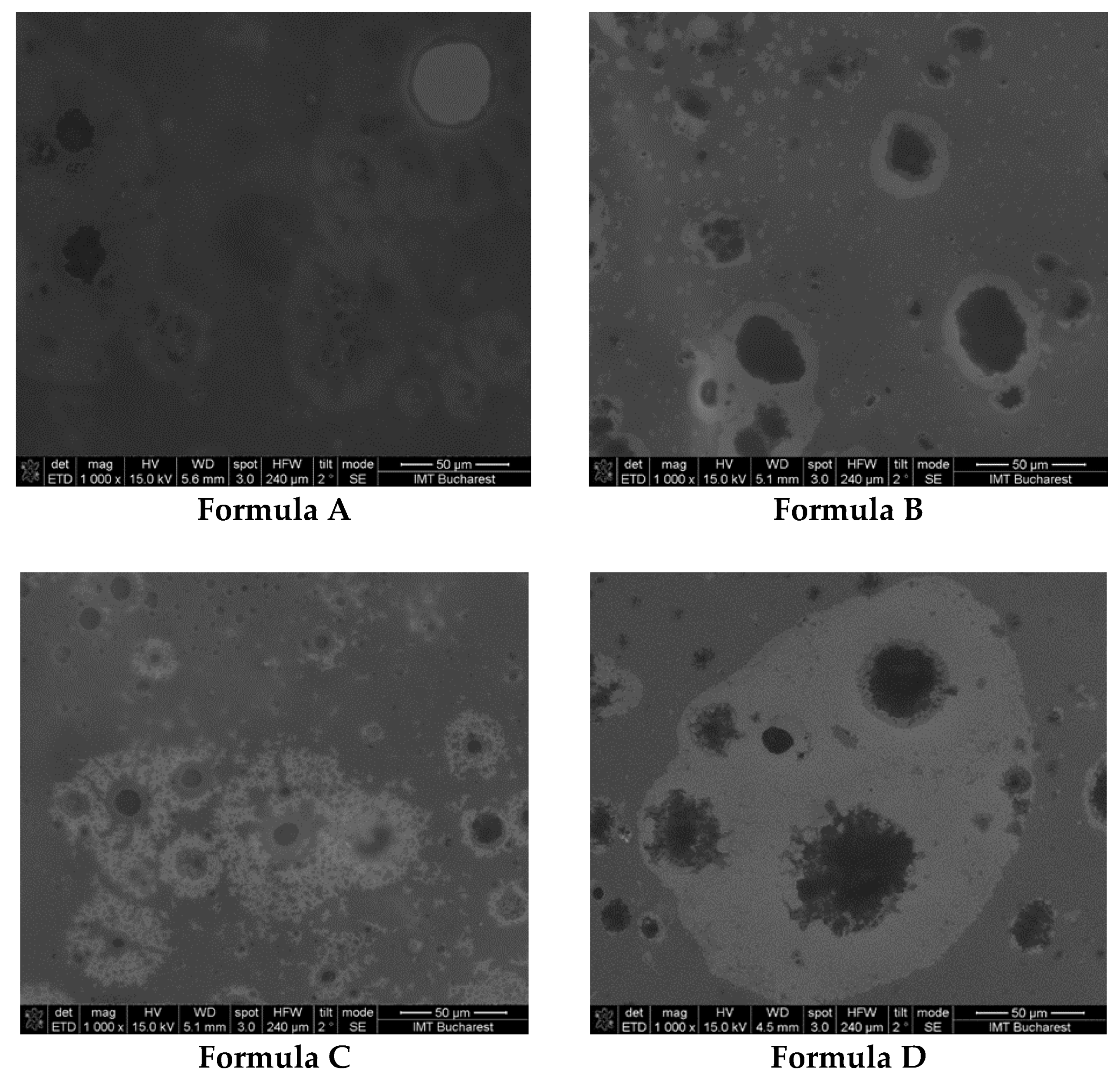
2.4. Characteristics of Catfish Liver Oil Emulgels
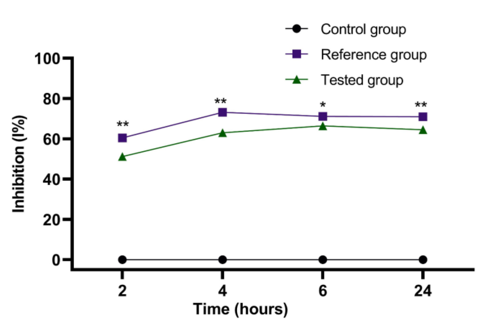
2.5. Evaluation of the Anti-Inflammatory Action of Stingray Liver Emulgel (Formula C)
3. Discussion
4. Materials and Methods
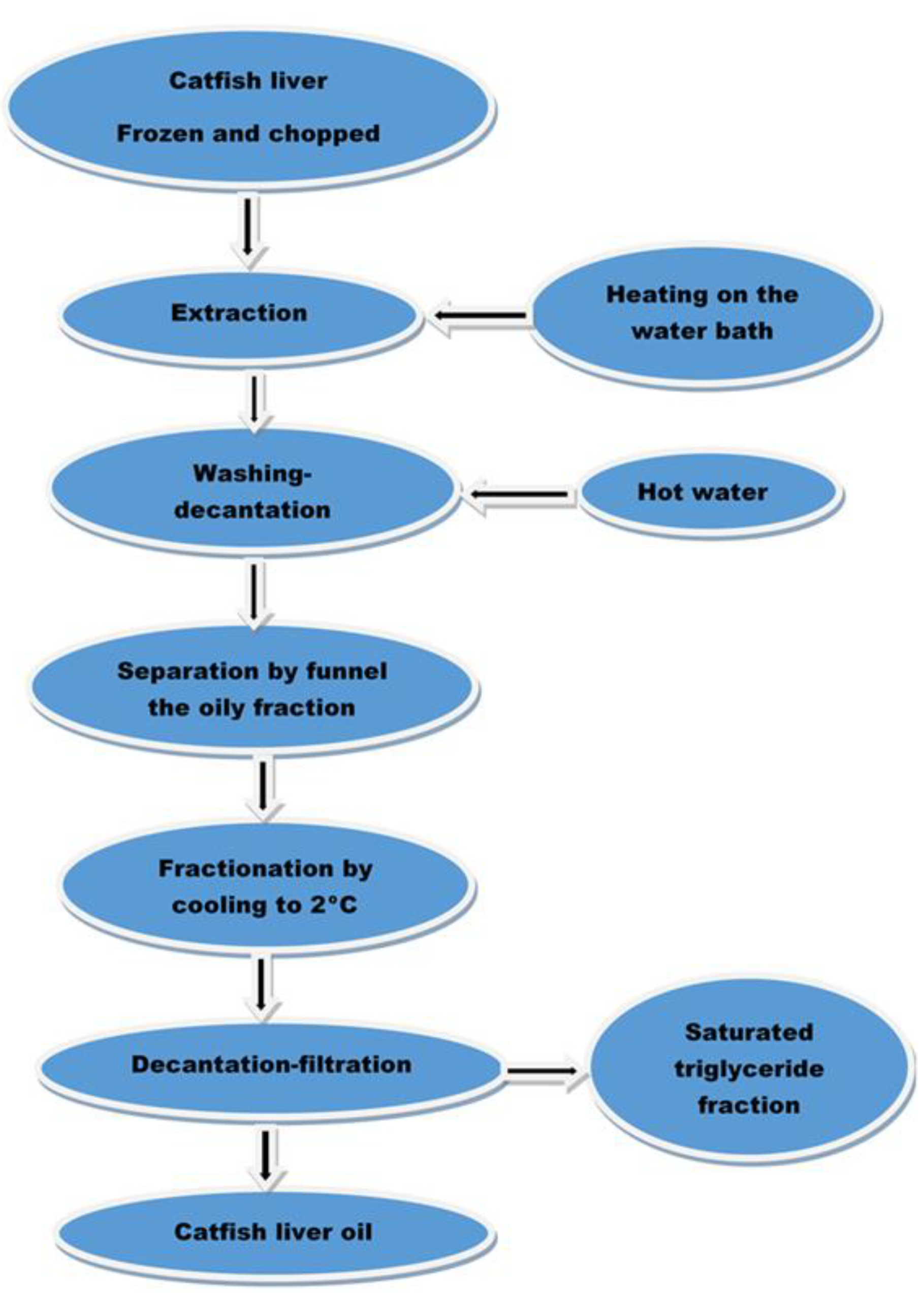
4.1. Extraction of Black Sea Stingray (Dasyatis pastinaca) Liver Oil
4.2. Analysis of Black Sea Stingray (Dasyatis pastinaca) Liver Oil
4.3. Formulation of Ointment with Stingray (Dasyatis pastinaca) Liver Oil
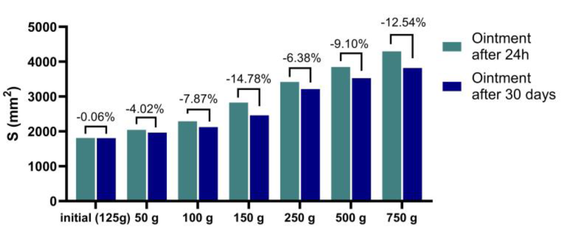
- Si is the spreading area (mm2) resulting from the applied mass “i” (g), and
- di is the mean diameter (mm) reached by the sample.
4.4. Testing the Healing Action of Stingray (Dasyatis pastinaca) Liver Oil Ointment
4.5. Formulation of Emulgels with Stingray (Dasyatis pastinaca) Liver Oil
4.6. Testing the Anti-Inflammatory Action of Stingray (Dasyatis pastinaca) Liver Oil Emulgel
5. Conclusions
Author Contributions
Funding
Institutional Review Board Statement
Data Availability Statement
Acknowledgments
Conflicts of Interest
References
- Bouchaâla, E.; BouAli, M.; Ben Ali, Y.; Miled, N.; Gargouri, Y.; Fendri, A. Biochemical Characterization and Molecular Modeling of Pancreatic Lipase from a Cartilaginous Fish, the Common Stingray (Dasyatis pastinaca). Appl. Biochem. Biotechnol. 2015, 176, 151–169. [Google Scholar] [CrossRef] [PubMed]
- Sathivel, S. Production, Process Design and Quality Characterization of Catfish Visceral Oil. Ph. D. Thesis, Louisiana State University and Agricultural & Mechanical College, Baton Rouge, LA, USA, 2001. Available online: https://digitalcommons.lsu.edu/gradschool_disstheses/434 (accessed on 15 March 2023).
- Sathivel, S.; Yin, H.; Prinyawiwatkul, W.; King, J.M. Comparison of chemical and physical properties of catfish oils prepared from different extraction processes. J. Food Sci. 2009, 74, E70–E76. [Google Scholar] [CrossRef] [PubMed]
- Sathivel, S.; Prinyawiwatkul, W.; King, J.M.; Grimm, C.C.; Lloyd, S. Oil production from catfish viscera. J. Am. Oil Chem. Soc. 2003, 80, 377–382. [Google Scholar] [CrossRef]
- Toti, E.; Oliver Chen, C.Y.; Palmery, M.; Valencia, D.V.; Peluso, I. Non-Provitamin A and Provitamin A Carotenoids as Immunomodulators: Recommended Dietary Allowance, Therapeutic Index, or Personalized Nutrition? Oxidative Med. Cell. Longev. 2018, 2018, 4637861. [Google Scholar] [CrossRef] [Green Version]
- Yin, K.; Agrawal, D.K. Vitamin D and Inflammatory Diseases. J. Inflamm. Res. 2014, 7, 69. [Google Scholar] [CrossRef] [Green Version]
- Veldurthy, V.; Wei, R.; Oz, L.; Dhawan, P.; Jeon, Y.H.; Christakos, S. Vitamin D, Calcium Homeostasis and Aging. Bone Res. 2016, 4, 16041. [Google Scholar] [CrossRef] [Green Version]
- Gruenwald, J.; Graubaum, H.J.; Harde, A. Effect of Cod Liver Oil on Symptoms of Rheumatoid Arthritis. Adv. Ther. 2002, 19, 101–107. [Google Scholar] [CrossRef]
- Galarraga, B.; Ho, M.; Youssef, H.M.; Hill, A.; McMahon, H.; Hall, C.; Ogston, S.; Nuki, G.; Belch, J.J.F. Cod Liver Oil (n-3 Fatty Acids) as an Non-Steroidal Anti-Inflammatory Drug Sparing Agent in Rheumatoid Arthritis. Rheumatology 2008, 47, 665–669. [Google Scholar] [CrossRef] [Green Version]
- Baxter-Jones, A.D.G.; Faulkner, R.A.; Forwood, M.R.; Mirwald, R.L.; Bailey, D.A. Bone Mineral Accrual from 8 to 30 Years of Age: An Estimation of Peak Bone Mass. J. Bone Miner. Res. 2011, 26, 1729–1739. [Google Scholar] [CrossRef]
- Downie, L.E.; Vingrys, A.J. Oral Omega-3 Supplementation Lowers Intraocular Pressure in Normotensive Adults. Transl. Vis. Sci. Technol. 2018, 7, 1. [Google Scholar] [CrossRef] [Green Version]
- Cartolano, F.D.C.; Dias, G.D.; Miyamoto, S.; Damasceno, N.R.T. Omega-3 Fatty Acids Improve Functionality of High-Density Lipoprotein in Individuals with High Cardiovascular Risk: A Randomized, Parallel, Controlled and Double-Blind Clinical Trial. Front. Nutr. 2022, 8, 767535. [Google Scholar] [CrossRef] [PubMed]
- Ravitchandirane, V.; Yogamoorthi, A. Studies on the Analgesic and Anti-Inflammatoryproperties of Crude Extracts of Sting Ray, Dasyatis zugei (Muller and Henle 1841). Biosci. Biotechnol. Res. Asia 2008, 5, 343–348. [Google Scholar]
- Ravitchandirane, V.; Yogamoorthi, A.; Thangaraj, M. Pharmacological Investigation and Spectral Characterization of Bioactive Compounds from Crude Extracts of Sting Ray, Dasyatis jenkinsii (Annandale, 1909). Chin. J. Nat. Med. 2014, 11, 500–505. [Google Scholar] [CrossRef] [PubMed]
- Pan American Leathers, Inc. Stingray Skin Guide. Available online: www.panamleathers.com (accessed on 7 February 2023).
- Mititelu, M.; Stanciu, G.; Drăgănescu, D.; Ioniță, A.C.; Neacșu, S.M.; Dinu, M.; Stefanvan Staden, R.-I.; Moroșan, E. Mussel Shells, a Valuable Calcium Resource for the Pharmaceutical Industry. Mar. Drugs 2022, 20, 25. [Google Scholar] [CrossRef]
- Mititelu, M.; Moroșan, E.; Nicoară, A.C.; Secăreanu, A.A.; Musuc, A.M.; Atkinson, I.; Cusu, J.P.; Nițulescu, G.M.; Ozon, E.A.; Sarbu, I.; et al. Development of immediate release tablets containing calcium lactate synthetized from Black Sea mussel shells. Mar. Drugs 2022, 20, 45. [Google Scholar] [CrossRef]
- Mititelu, M.; Ioniţă, A.C.; Moroşan, E. Research regarding integral processing of mussels from Black Sea. Farmacia 2014, 62, 625–632. [Google Scholar]
- Rodríguez-Cruz, M.; Serna, D.S. Nutrigenomics of ω-3 Fatty Acids: Regulators of the Master Transcription Factors. Nutrition 2017, 41, 90–96. [Google Scholar] [CrossRef]
- Fodor, J.G.; Helis, E.; Yazdekhasti, N.; Vohnout, B. “Fishing” for the Origins of the “Eskimos and Heart Disease” Story: Facts or Wishful Thinking? Can. J. Cardiol. 2014, 30, 864–868. [Google Scholar] [CrossRef]
- Gahche, J.J.; Bailey, R.L.; Potischman, N.; Dwyer, J.T. Dietary Supplement Use Was Very High among Older Adults in the United States in 2011–2014. J. Nutr. 2017, 147, 1968–1976. [Google Scholar] [CrossRef] [Green Version]
- Villani, A.M.; Crotty, M.; Cleland, L.G.; James, M.J.; Fraser, R.J.; Cobiac, L.; Miller, M.D. Fish Oil Administration in Older Adults: Is There Potential for Adverse Events? A Systematic Review of the Literature. BMC Geriatr. 2013, 13, 41. [Google Scholar] [CrossRef] [Green Version]
- Hardy, M.S.; Kekic, A.; Graybill, N.L.; Lancaster, Z.R. A Systematic Review of the Association between Fish Oil Supplementation and the Development of Asthma Exacerbations. SAGE Open Med. 2016, 4. [Google Scholar] [CrossRef]
- Balk, E.M.; Lichtenstein, A.H. Omega-3 Fatty Acids and Cardiovascular Disease: Summary of the 2016 Agency of Healthcare Research and Quality Evidence Review. Nutrients 2017, 9, 865. [Google Scholar] [CrossRef] [Green Version]
- Eslick, G.D.; Howe, P.R.C.; Smith, C.; Priest, R.; Bensoussan, A. Benefits of Fish Oil Supplementation in Hyperlipidemia: A Systematic Review and Meta-Analysis. Int. J. Cardiol. 2009, 136, 4–16. [Google Scholar] [CrossRef]
- Porojnicu, A.C.; Bruland, Ø.S.; Aksnes, L.; Grant, W.B.; Moan, J. Sun Beds and Cod Liver Oil as Vitamin D Sources. J. Photochem. Photobiol. B 2008, 91, 125–131. [Google Scholar] [CrossRef]
- Chee, K.M.; Gong, J.X.; Good Rees, D.M.; Meydanl, M.; Ausman, L.; Johnson, J.; Siguel, E.N.; Schaefer, E.J. Fatty Acid Content of Marine Oil Capsules. Lipids 1990, 25, 523–528. [Google Scholar] [CrossRef]
- Breiden, B.; Sandhoff, K. The Role of Sphingolipid Metabolism in Cutaneous Permeability Barrier Formation. Biochim. Biophys. Acta 2014, 1841, 441–452. [Google Scholar] [CrossRef]
- Huang, T.H.; Wang, P.W.; Yang, S.C.; Chou, W.L.; Fang, J.Y. Cosmetic and Therapeutic Applications of Fish Oil’s Fatty Acids on the Skin. Mar. Drugs 2018, 16, 256. [Google Scholar] [CrossRef] [Green Version]
- Baum, C.L.; Arpey, C.J. Normal Cutaneous Wound Healing: Clinical Correlation with Cellular and Molecular Events. Dermatol. Surg. 2005, 31, 674–686. [Google Scholar] [CrossRef]
- Calder, P.C. Omega-3 Polyunsaturated Fatty Acids and Inflammatory Processes: Nutrition or Pharmacology? Br. J. Clin. Pharmacol. 2013, 75, 645–662. [Google Scholar] [CrossRef] [Green Version]
- Kiecolt-Glaser, J.K.; Glaser, R.; Christian, L.M. Omega-3 Fatty Acids and Stress-Induced Immune Dysregulation: Implications for Wound Healing. Mil. Med. 2014, 179, 129–133. [Google Scholar] [CrossRef] [Green Version]
- Shingel, K.I.; Faure, M.P.; Azoulay, L.; Roberge, C.; Deckelbaum, R.J. Solid Emulsion Gel as a Vehicle for Delivery of Polyunsaturated Fatty Acids: Implications for Tissue Repair, Dermal Angiogenesis and Wound Healing. J. Tissue Eng. Regen. Med. 2008, 2, 383–393. [Google Scholar] [CrossRef]
- Arantes, E.L.; Dragano, N.; Ramalho, A.; Vitorino, D.; de-Souza, G.F.; Lima, M.H.M.; Velloso, L.A.; Araújo, E.P. Topical Docosahexaenoic Acid (DHA) Accelerates Skin Wound Healing in Rats and Activates GPR120. Biol. Res. Nurs. 2016, 18, 411–419. [Google Scholar] [CrossRef] [PubMed]
- Wu, D.C.; Goldman, M.P. A Topical Anti-Inflammatory Healing Regimen Utilizing Conjugated Linolenic Acid for Use Post-Ablative Laser Resurfacing of the Face: A Randomized, Controlled Trial. J. Clin. Aesthetic Dermatol. 2017, 10, 12–17. [Google Scholar]
- Komprda, T.; Sládek, Z.; Vícenová, M.; Simonová, J.; Franke, G.; Lipový, B.; Matejovičová, M.; Kacvinská, K.; Sabliov, C.; Astete, C.E.; et al. Effect of Polymeric Nanoparticles with Entrapped Fish Oil or Mupirocin on Skin Wound Healing Using a Porcine Model. Int. J. Mol. Sci. 2022, 23, 7663. [Google Scholar] [CrossRef] [PubMed]
- Rodrigues, H.G.; Vinolo, M.A.R.; Sato, F.T.; Magdalon, J.; Kuhl, C.M.C.; Yamagata, A.S.; Pessoa, A.F.M.; Malheiros, G.; dos Santos, M.F.; Lima, C.; et al. Oral Administration of Linoleic Acid Induces New Vessel Formation and Improves Skin Wound Healing in Diabetic Rats. PLoS ONE 2016, 11, e0165115. [Google Scholar] [CrossRef] [Green Version]
- Pereira, L.M.; Hatanaka, E.; Martins, E.F.; Oliveira, F.; Liberti, E.A.; Farsky, S.H.; Curi, R.; Pithon-Curi, T.C. Effect of Oleic and Linoleic Acids on the Inflammatory Phase of Wound Healing in Rats. Cell Biochem. Funct. 2008, 26, 197–204. [Google Scholar] [CrossRef]
- Cardoso, C.R.B.; Souza, M.A.; Ferro, E.A.V.; Favoreto, S.; Pena, J.D.O. Influence of Topical Administration of N-3 and n-6 Essential and n-9 Nonessential Fatty Acids on the Healing of Cutaneous Wounds. Wound Repair Regen. 2004, 12, 235–243. [Google Scholar] [CrossRef]
- Lu, Y.; Tian, H.; Hong, S. Novel 14,21-Dihydroxy-Docosahexaenoic Acids: Structures, Formation Pathways, and Enhancement of Wound Healing. J. Lipid Res. 2010, 51, 923–932. [Google Scholar] [CrossRef] [Green Version]
- Tian, H.; Yao, X.; Zeng, R.; Sun, R.; Tian, H.; Shi, C.; Li, L.; Tian, J.; Yang, K. Safety and Efficacy of a New Parenteral Lipid Emulsion (SMOF) for Surgical Patients: A Systematic Review and Meta-Analysis of Randomized Controlled Trials. Nutr. Rev. 2013, 71, 815–821. [Google Scholar] [CrossRef]
- Peng, Y.C.; Yang, F.L.; Subeq, Y.M.; Tien, C.C.; Chao, Y.F.C.; Lee, R.P. Lipid Emulsion Enriched in Omega-3 PUFA Accelerates Wound Healing: A Placebo-Controlled Animal Study. World J. Surg. 2018, 42, 1714–1720. [Google Scholar] [CrossRef]
- Mititelu, M.; Moroşan, E.; Neacsu, S.M.; Ioniţă, E.I. Research regarding the pollution degree from romanian Black Sea coast. Farmacia 2018, 66, 1059–1063. [Google Scholar] [CrossRef] [Green Version]
- Ioniţă, A.C.; Mititelu, M.; Moroşan, E. Analysis of heavy metals and organic pollutants from some Danube river fishes. Farmacia 2014, 62, 299–305. [Google Scholar]
- Mititelu, M.; Nicolescu, T.O.; Ioniţă, C.A.; Nicolescu, F. Study of Heavy Metals and Organic Polluants from Some Fisches of Danube River. J. Environ. Prot. Ecol. 2012, 13, 869–874. [Google Scholar]
- Jacobsen, C.; Warncke, S.A.; Hansen, S.H.; Sørensen, A.-D.M. Fish Liver Discards as a Source of Long-Chain Omega-3 Polyunsaturated Fatty Acids. Foods 2022, 11, 905. [Google Scholar] [CrossRef]
- Malcorps, W.; Newton, R.W.; Sprague, M.; Glencross, B.D.; Little, D.C. Nutritional Characterisation of European Aquaculture Processing By-Products to Facilitate Strategic Utilisation. Front. Sustain. Food Syst. 2021, 5, 720595. [Google Scholar] [CrossRef]
- Mititelu, M.; Udeanu, D.I.; Nedelescu, M.; Neacsu, S.M.; Nicoară, A.C.; Oprea, E.; Ghica, M. Quality Control of Different Types of Honey and Propolis Collected from Romanian Accredited Beekeepers and Consumer’s Risk Assessment. Crystals 2022, 12, 87. [Google Scholar] [CrossRef]
- Sawada, Y.; Saito-Sasaki, N.; Nakamura, M. Omega 3 Fatty Acid and Skin Diseases. Front. Immunol. 2021, 11, 623052. [Google Scholar] [CrossRef]
- Stupin, A.; Mihalj, M.; Kolobarić, N.; Šušnjara, P.; Kolar, L.; Mihaljević, Z.; Matić, A.; Stupin, M.; Jukić, I.; Kralik, Z.; et al. Anti-Inflammatory Potential of n-3 Polyunsaturated Fatty Acids Enriched Hen Eggs Consumption in Improving Microvascular Endothelial Function of Healthy Individuals—Clinical Trial. Int. J. Mol. Sci. 2020, 21, 4149. [Google Scholar] [CrossRef]
- Paduraru, D.N.; Coman, F.; Ozon, E.A.; Gherghiceanu, F.; Andronic, O.; Ion, D.; Stanescu, M.; Bolocan, A. The use of nutritional supplement in romanian patients—Attitudes and beliefs. Farmacia 2019, 67, 1060–1065. [Google Scholar] [CrossRef]
- Ioniță-Mîndrican, C.-B.; Ziani, K.; Mititelu, M.; Oprea, E.; Neacșu, S.M.; Moroșan, E.; Dumitrescu, D.-E.; Roșca, A.C.; Drăgănescu, D.; Negrei, C. Therapeutic Benefits and Dietary Restrictions of Fiber Intake: A State of the Art Review. Nutrients 2022, 14, 2641. [Google Scholar] [CrossRef]
- Stoicescu, I.; Lupu, E.C.; Radu, M.D.; Popescu, A.; Mihai, S. High-Performance Liquid Chromatography—Diode Array Detection (HPLC-DAD) Method for the Determination of Phenolic Compounds of Water Chesnut (Trapa natans L.). Anal. Lett. 2022, 55, 2147–2159. [Google Scholar] [CrossRef]
- Méndez, L.; Dasilva, G.; Taltavull, N.; Romeu, M.; Medina, I. Marine Lipids on Cardiovascular Diseases and Other Chronic Diseases Induced by Diet: An Insight Provided by Proteomics and Lipidomics. Mar. Drugs 2017, 15, 258. [Google Scholar] [CrossRef] [Green Version]
- Carrera, I.; Corzo, L.; Naidoo, V.; Martínez-Iglesias, O.; Cacabelos, R. Cardiovascular and lipid-lowering effects of a marine lipoprotein extract in a high-fat diet-induced obesity mouse model. Int. J. Med. Sci. 2023, 20, 292–306. [Google Scholar] [CrossRef]
- Mititelu, M.; Neacsu, S.M.; Oprea, E.; Dumitrescu, D.-E.; Nedelescu, M.; Drăgănescu, D.; Nicolescu, T.O.; Rosca, A.C.; Ghica, M. Black Sea Mussels Qualitative and Quantitative Chemical Analysis: Nutritional Benefits and Possible Risks through Consumption. Nutrients 2022, 14, 964. [Google Scholar] [CrossRef]
- National Drug Agency. Romanian Pharmacopoeia, 10th ed.; Editura Medicală: Bucharest, Romania, 1993; pp. 67–68, 335, 419–421, 921–922. [Google Scholar]
- Popovici, I.; Lupuleasa, D. Tehnologie Farmaceutică, 2nd ed.; Polirom: Iaşi, Romania, 2017; Volume 2, ISBN 978-973-46-6920-2. [Google Scholar]
- AVMA. Guidelines for the Euthanasia of Animals; American Veterinary Medical Association: Schaumburg, IL, USA, 2013. [Google Scholar]
- OECD. Guidelines for Testing of Chemicals. In Acute Oral Toxicities Up and Down Procedure; OECD: Paris, France, 2001; Volume 425, pp. 1–26. [Google Scholar]
- Mihele, D.; Manea, S.; Raiciu, D.; Udeanu, D. The cicatrizing action of some new gemmotherapy products. Farmacia 2006, 6, 24–28. [Google Scholar]
- Mishra, P.; Singh, U.; Pandey, C.M.; Mishra, P.; Pandey, G. Application of student’s t-test, analysis of variance, and covariance. Ann. Card. Anaesth. 2019, 22, 407–411. [Google Scholar] [CrossRef]
- Leahu, A.; Lupu, E.C. Statistical simulation and prediction in software reliability. Analele Univ. Ovidius Constanţa Ser. Mat. 2008, 16, 81–90. [Google Scholar]
- Atyim, P.; Olah, N.K.; Osser, G.; Toma, C.C.; Morgovan, C.; Atyim, E. Practical Investigation of Gels Containing Aristolochia (Aristolochia clematitis) Extract. Stud. UBB Chem. 2017, 62, 153–164. [Google Scholar] [CrossRef]
- Puri, A.; Nguyen, H.X.; Banga, A.K. Microneedle-mediated intradermal delivery of epigallocatechin-3-gallate. Int. J. Cosmet. Sci. 2016, 38, 512–523. [Google Scholar] [CrossRef]
- Castardo, J.C.; Prudente, A.S.; Ferreira, J.; Guimarães, C.L.; Monache, F.D.; Filho, V.C.; Otuki, M.F.; Cabrini, D.A. Anti-inflammatory effects of hydroalcoholic extract and two biflavonoids from Garcinia gardneriana leaves in mouse paw oedema. J. Ethnopharmacol. 2008, 118, 405–411. [Google Scholar] [CrossRef]


| Parameter | Value ± SD |
|---|---|
| Iodine value (g I2/100 g fatty acids) | 111.85 ± 0.66 |
| Acid value (mg KOH/g sample) | 4.93 ± 0.33 |
| Saponification value (mg KOH/g sample) | 179.07 ± 0.25 |
| Peroxide index (mEq O2/kg) | 0.8 ± 0.55 |
| Density at 20 °C (g/mL) | 0.921 ± 0.33 |
| Refractive index | 1.479 ± 0.55 |
| Fatty Acid | mg/g ± SD (%) |
|---|---|
| C 10:0 | 3.42 ± 0.23 |
| C 12:0 | 0.06 ± 0.13 |
| C 14:0 | 4.23 ± 0.45 |
| C 14:1 | 2.12 ± 0.22 |
| C 15:0 | 0.16 ± 0.16 |
| C 16:0 | 10.15 ± 1.84 |
| C 16:1 | 12.23 ± 1.52 |
| C 16:1ω-7 | 6.12 ± 0.81 |
| C 17:0 | 1.15± 0.12 |
| C 17:1 | 0.23 ± 0.18 |
| C 18:0 | 8.85 ± 0.65 |
| C 18:1 | 15.46 ± 1.73 |
| C 18:1ω-7 | 4.75 ± 0.33 |
| C 18:1ω-9 | 9.81 ± 0.74 |
| C 18:2ω-6 | 2.18 ± 0.52 |
| C 18:3 | 1.15 ± 0.25 |
| C 18:3ω-3 | 2.12 ± 0.14 |
| C 20:1 | 0.19 ± 0.38 |
| C 20:3 | 0.08 ± 0.04 |
| C 20:4ω-6 | 2.68 ± 0.55 |
| C 20:5ω-3 | 3.86 ± 0.28 |
| C 22:1 | 0.07 ± 0.14 |
| C 22:5ω-3 | 1.49 ± 0.16 |
| C 22:6ω-3 | 6.44 ± 0.31 |
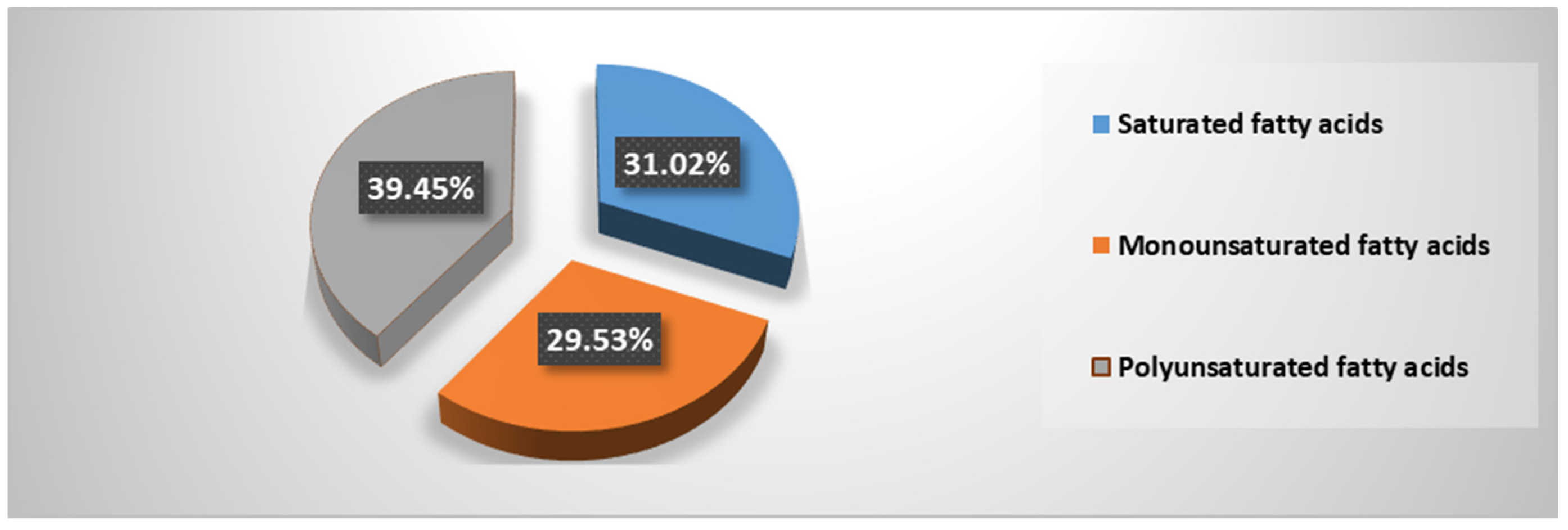
| Σ saturated fatty acids | 31.02 |
| Σ ω-3 | 13.91 |
| Σ ω-6 | 4.86 |
| Σ monounsaturated fatty acids | 29.53 |
| Σ polyunsaturated fatty acids | 39.45 |
| ω-3/ω-6 | 3.68 |
| Polyunsaturated fatty acids/Saturated fatty acids | 1.27 |
| Polyunsaturated fatty acids/Monounsaturated fatty acids | 1.33 |
| Parameter | Result |
|---|---|
| Appearance | Homogeneous appearance, yellow-orange color, characteristic smell |
| pH | 6.0 |
| Thermal stability | Good stability, the samples remained homogeneous at the subjected temperature steps without separating into several phases |
| Viscosity | 285 mPa/s |
| Characteristic | Formula A | Formula B | Formula C | Formula D |
|---|---|---|---|---|
| Initial macroscopic characteristics | appearance: homogenous; color: yellowish; smell: specific | appearance: homogenous; color: yellow; smell: specific | appearance: homogenous; color: orange-yellow; smell: specific | appearance: homogenous; color: orange-yellow; smell: specific |
| Macroscopic characteristics after 30 days | appearance: homogenous; color: yellowish; smell: specific | appearance: homogenous; color: yellow; smell: specific | appearance: homogenous; color: orange-yellow; smell: specific | appearance: homogenous; color: orange-yellow; smell: specific |
| Initial pH | 6.4 | 6.1 | 5.8 | 5.6 |
| pH after 30 days | 6.2 | 6.0 | 5.7 | 5.6 |
| Initial thermal stability | Good stability without a tendency to separate | Good stability without a tendency to separate | Good stability without a tendency to separate | Good stability without a tendency to separate |
| Thermal stability after 30 days | Good stability without a tendency to separate | Good stability without a tendency to separate | Good stability without a tendency to separate | Good stability without a tendency to separate |
| Gel/Flow Parameters | Yield Stress (Pa) (τ0 - Pa) | Consistency Index (K - Pa·sn) | Flow Index (n) | Viscosity at 0.3 rpm (η0.3 - Pa·s) Initial | Viscosity at 0.3 rpm (η0.3 - Pa·s) after 30 Days |
|---|---|---|---|---|---|
| Formula A | 27.318 | 21.081 | 0.35 | 499.200 | 482.100 |
| Formula B | 47.233 | 33.738 | 0.29 | 872.700 | 810.200 |
| Formula C | 77.154 | 66.884 | 0.27 | 553.000 | 432.800 |
| Formula D | 53.366 | 35.114 | 0.28 | 861.600 | 755.600 |
| Emulgel/Rheological Model | Casson | Herschel-Bulkley |
|---|---|---|
| Formula A | 0.832 | 0.992 |
| Formula B | 0.975 | 0.997 |
| Formula C | 0.919 | 0.995 |
| Formula D | 0.969 | 0.996 |
| Components | Formula A | Formula B | Formula C | Formula D |
|---|---|---|---|---|
| Carbopol 940 | 1 g | 1 g | 1 g | 1 g |
| Glycerin | 5 g | 5 g | 5 g | 5 g |
| Triethanolamine | 0.5 g | 0.5 g | 0.5 g | 0.5 g |
| Tween 80 | 0.5 g | 0.5 g | 0.5 g | 0.5 g |
| Stingray liver oil | 3 g | 5 g | 7 g | 10 g |
| Purified water | until 100 g | until 100 g | until 100 g | until 100 g |
Disclaimer/Publisher’s Note: The statements, opinions and data contained in all publications are solely those of the individual author(s) and contributor(s) and not of MDPI and/or the editor(s). MDPI and/or the editor(s) disclaim responsibility for any injury to people or property resulting from any ideas, methods, instructions or products referred to in the content. |
© 2023 by the authors. Licensee MDPI, Basel, Switzerland. This article is an open access article distributed under the terms and conditions of the Creative Commons Attribution (CC BY) license (https://creativecommons.org/licenses/by/4.0/).
Share and Cite
Mititelu, M.; Licu, M.; Lupu, C.E.; Neacșu, S.M.; Olteanu, G.; Stanciu, G.; Drăgănescu, D.; Oancea, C.-N.; Busnatu, Ș.S.; Hîncu, L.; et al. Characterization of Some Dermato-Cosmetic Preparations with Marine Lipids from Black Sea Wild Stingray. Mar. Drugs 2023, 21, 408. https://doi.org/10.3390/md21070408
Mititelu M, Licu M, Lupu CE, Neacșu SM, Olteanu G, Stanciu G, Drăgănescu D, Oancea C-N, Busnatu ȘS, Hîncu L, et al. Characterization of Some Dermato-Cosmetic Preparations with Marine Lipids from Black Sea Wild Stingray. Marine Drugs. 2023; 21(7):408. https://doi.org/10.3390/md21070408
Chicago/Turabian StyleMititelu, Magdalena, Monica Licu, Carmen Elena Lupu, Sorinel Marius Neacșu, Gabriel Olteanu, Gabriela Stanciu, Doina Drăgănescu, Carmen-Nicoleta Oancea, Ștefan Sebastian Busnatu, Lucian Hîncu, and et al. 2023. "Characterization of Some Dermato-Cosmetic Preparations with Marine Lipids from Black Sea Wild Stingray" Marine Drugs 21, no. 7: 408. https://doi.org/10.3390/md21070408