

Jellyfishes—Significant Marine Resources with Potential in the Wound-Healing Process: A Review
 ,
,
Abstract
:1. Introduction
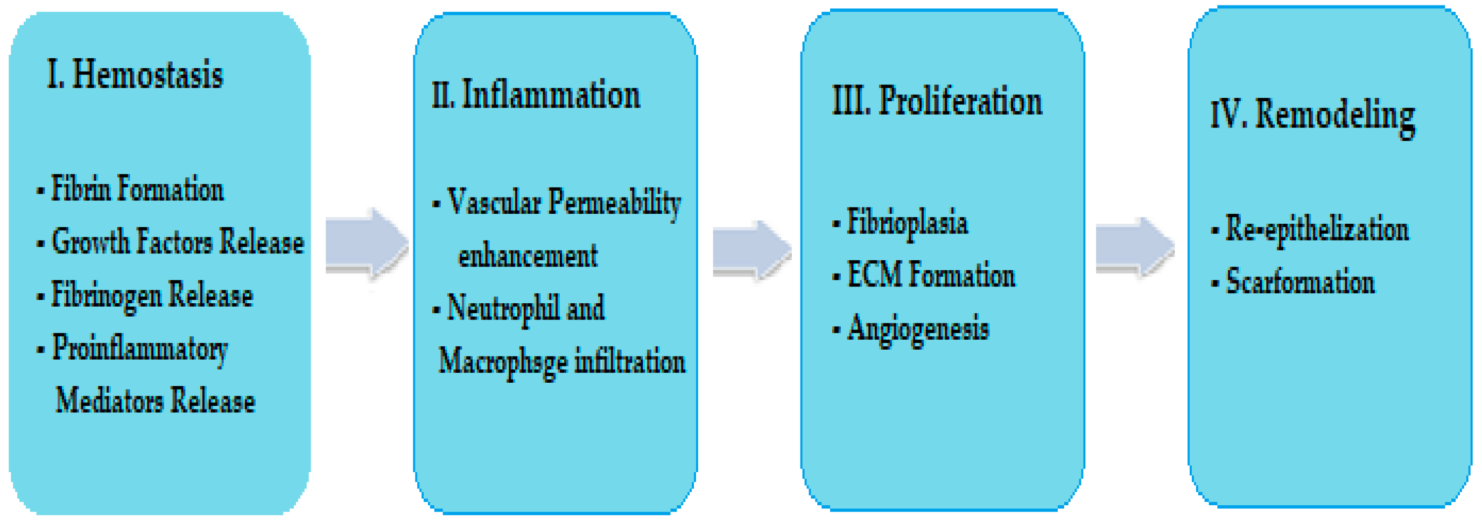
2. Wound Healing
- -
- Cheng et al. reported that collagen extracts from jellyfish demonstrate haemostatic action and could intervene to stop bleeding as it occurs in the haemostasis phase [48]. An important immunostimulatory effect of jellyfish collagens that may stimulate growth factors was reported by Krishnan et al. [11]. Singh et al. reported that in the haemostasis phase, under the actions of the growth factor and the proinflammatory mediators released in the wound, fibrinogen is converted to fibrin (a clot) which stops bleeding [2]. Jellyfish extracts could stimulate these molecular processes that occur in the wound through immunostimulatory effects;
- -
- Morishige et al. reported that collagen extracts and collagen peptides from jellyfish can exert immunostimulatory effects on growth factors such as (TNF-α), (IFN-γ) and (TGF), which are involved in phase II (inflammation) and phase III (proliferation) of wound healing [49];
- -
- Mapoung et al. and Yu H. et al. showed that glycosaminoglycan (GAG) biocompounds, in addition to the proteins, amino acids and phenolic compounds present in aqueous and hydroalcoholic extracts of certain jellyfish species, exhibit antioxidant and antibacterial activities [50,51]. These activities could be beneficial in phase III (proliferation) of the wound-healing process;
- -
- Li et al. reported that jellyfish extracts containing compounds with GAG-like structures may contribute to tissue regeneration, which also occurs in phase IV (remodelling) [52].
- -
3. Jellyfish: Important Bioresource Compound for Wound Healing Found in European Marine Habitats
4. Wound-Healing Biochemical Compounds of Interest from Jellyfish
4.1. Polysacharides from Jellyfish (JSP)
4.2. Proteins from Jellyfish
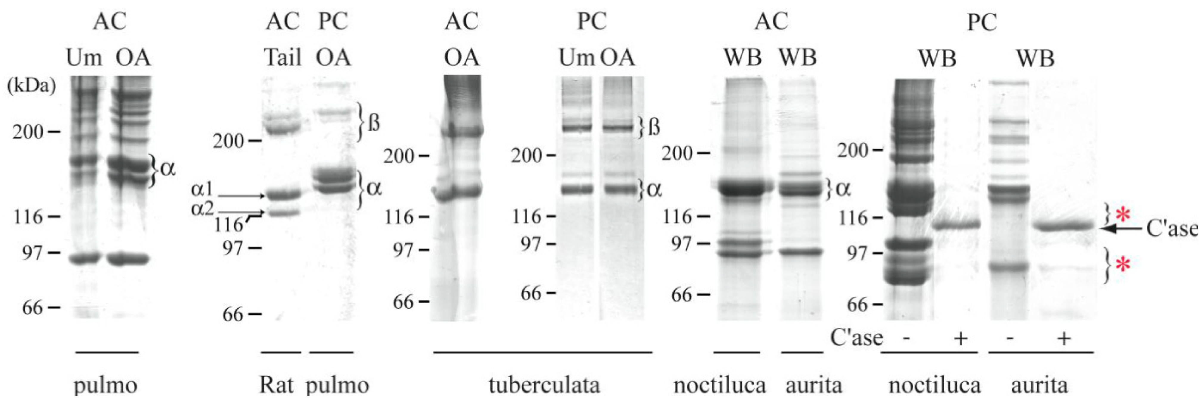
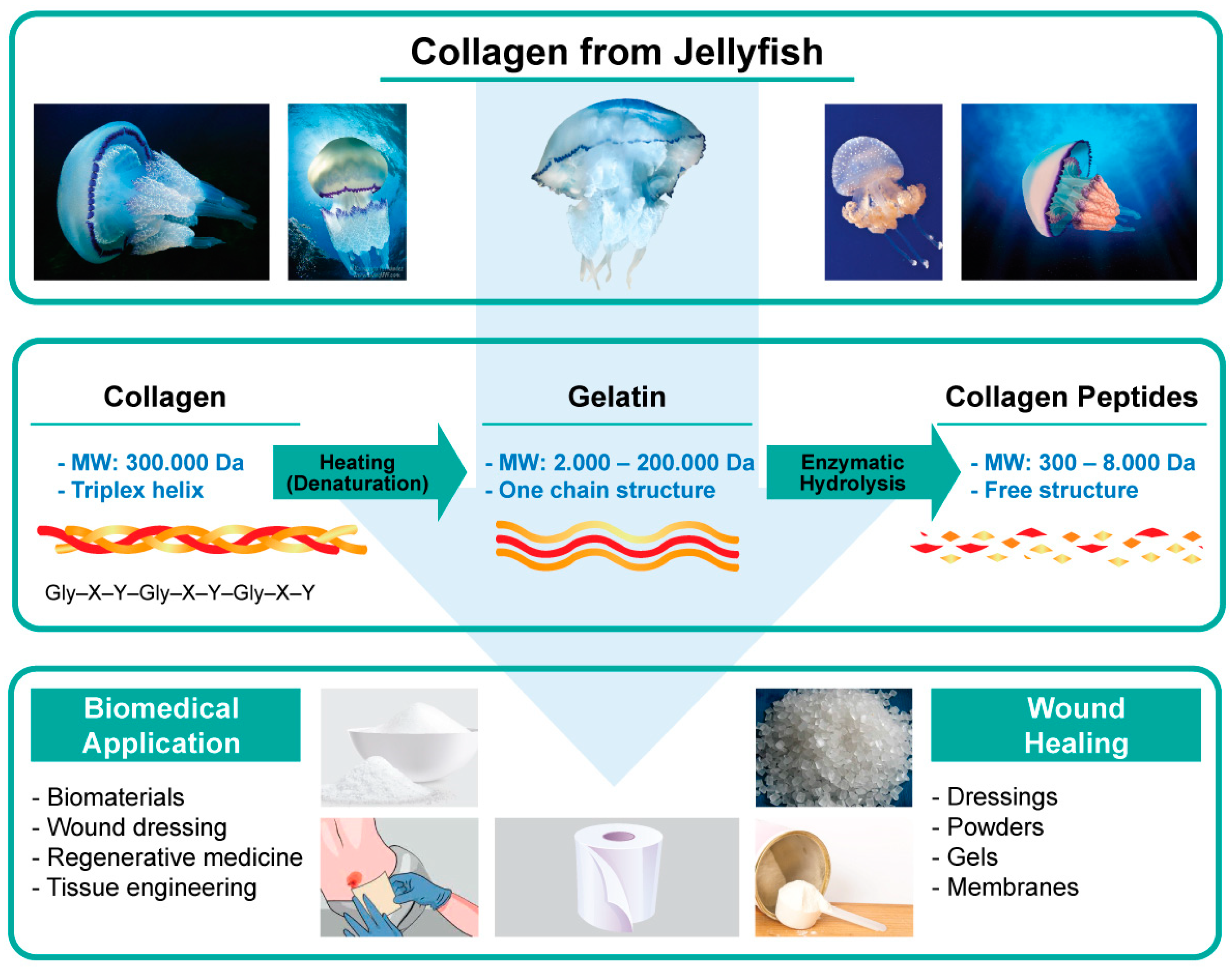
4.2.1. Collagen and Collagenic Peptides from Jellyfish
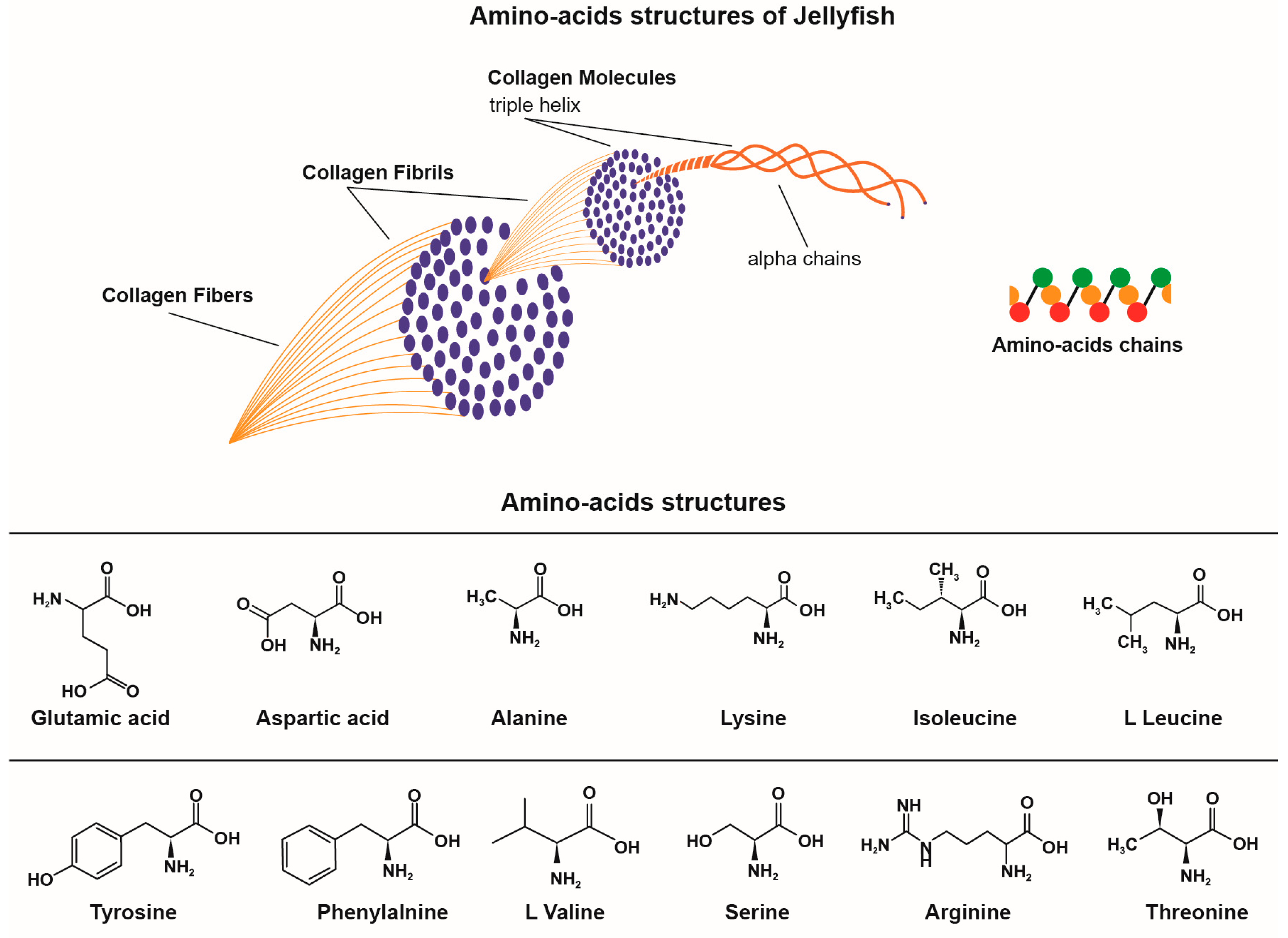
4.2.2. Amino Acids from Jellyfish
4.3. Biological Activities Useful in Wound Healing
5. Conclusions
Author Contributions
Funding
Institutional Review Board Statement
Informed Consent Statement
Data Availability Statement
Conflicts of Interest
Abbreviations
| FAO | Food and Agriculture Organization of The United Nations |
| JSPs | Jellyfish Polysaccharides |
| ECM | Extracellular Matrix |
| ROS | Reactive Oxygen Species |
| LPS | Lipopolysaccharides |
| GAG | Glycosaminoglycan |
| IL-1 β | Interleukin |
| TNF-α | Tumour Necrosis Factor Alpha |
| INF-γ | Interferon Gamma |
| TGF-α | Transforming Growth Factor-Alfa |
| TGF-β | Transforming Growth Factor-Beta |
| FGF | Fibroblast Growth Factor |
| PDGF | Platelet-Derived Growth Factor |
| VEGF | Vascular Endothelial Growth Factor |
| SDS-PAGE | Sodium Dodecyl Sulfate–Polyacrylamide Gel Electrophoresis |
| FTIR | Fourier-Transform Infrared Spectroscopy |
| Igs | Immunoglobulins |
| IgM | Immunoglobulin M |
| IgG | Immunoglobulin G |
| hPBL | Human Peripheral Blood Lymphocytes |
| RAW264.7 | Macrophage-Like, Abelson Leukaemia Virus-Transformed Cell Line |
References
- Wang, P.H.; Huang, B.S.; Horng, H.C.; Yeh, C.C.; Chen, Y.J. Wound healing. J. Chin. Med. Assoc. 2018, 81, 94–101. [Google Scholar] [CrossRef] [PubMed]
- Singh, S.; Young, A.; McNaught, C.-E. The physiology of wound healing. Surgery 2017, 35, 473–477. [Google Scholar] [CrossRef]
- de O Gonzalez, A.C.; Costa, T.F.; de A Andrade, Z.; Medrato, A.R.A.P. Wound healing—A literature review. An. Bras. Dermatol. 2016, 91, 614–620. [Google Scholar] [CrossRef] [Green Version]
- Frykberg, R.G.; Banks, J. Challenges in the treatment of chronic wounds. Adv. Wound Care 2015, 4, 560–582. [Google Scholar] [CrossRef] [PubMed] [Green Version]
- Childs, D.R.; Murthy, A.S. Overview of wound healing and management. Surg. Clin. N. Am. 2017, 97, 189–207. [Google Scholar] [CrossRef] [PubMed]
- Kiritsi, D.; Nyström, A. The role of TGFβ in wound healing pathologies. Mech. Ageing Dev. 2017, 172, 51–58. [Google Scholar] [CrossRef]
- Demidova-Rice, T.N.; Hamblin, M.R.; Herman, I.M. Acute and impaired wound healing: Pathophysiology and current methods for drug delivery, part 2: Role of growth factors in normal and pathological wound healing: Therapeutic potential and methods of delivery. Adv. Ski. Wound Care 2012, 25, 349–370. [Google Scholar] [CrossRef] [Green Version]
- Anderson, K.; Hamm, R.L. Factors that impair wound healing. J. Am. Coll. Clin. Wound Spec. 2012, 4, 84–91. [Google Scholar] [CrossRef] [Green Version]
- Schreml, S.; Szeimies, R.M.; Prantl, L.; Landthaler, M.; Babilas, P. Wound healing in the 21-st century. J. Am. Acad. Dermatol. 2010, 63, 866–881. [Google Scholar] [CrossRef] [PubMed]
- Galiano, R.D.; Tepper, O.M.; Pelo, C.R.; Bhatt, K.A.; Callaghan, M.; Bastidas, N. Topical vascular endothelial growth factor accelerates diabetic wound healing through increased angiogenesis and by mobilizing and recruiting bone marrow-derived cells. Am. J. Pathol. 2004, 164, 1935–1947. [Google Scholar] [CrossRef] [Green Version]
- Krishnan, S.; Pachippan, P. Imunomodulatory effects of the jelly fish venom C. quinquecirrha from vellar estuary, southeast coast of India. Int. J. Pharm. Pharm. Sci. 2013, 5, 60–62. [Google Scholar]
- Sorg, H.; Tilkorn, D.J.; Hager, S.; Hauser, J.; Mirastschijski, U. Skin wound healing: An update on the current knowledge and concepts. Eur. Surg. Res. 2017, 58, 81–94. [Google Scholar] [CrossRef]
- Eming, S.A.; Krieg, T.; Davidson, J.M. Inflammation in wound repair: Molecular and cellular mechanisms. J. Investig. Dermatol. 2007, 127, 514–525. [Google Scholar] [CrossRef] [Green Version]
- Velnar, T.V.; Ailey, T.B. The wound healing process: An overview of the cellular and molecular mechanisms. J. Int. Med. Res. 2009, 37, 1528–1542. [Google Scholar] [CrossRef]
- Okur, M.E.; Karantas, I.D.; Şenyiğit, Z.; Okur, N.U.; Siafaka, P.I. Recent trends on wound management: New therapeutic choices based on polymeric carriers. Asian J. Pharm. Sci. 2020, 15, 661–684. [Google Scholar] [CrossRef]
- Hoyer, B.; Bernhardt, A.; Lode, A.; Heinemann, S.; Sewing, J.; Klinger, M.; Notbohm, H.; Gelinsky, M. Jellyfish collagen scaffolds for cartilage tissue engineering. Acta Biomater. 2014, 10, 883–892. [Google Scholar] [CrossRef] [PubMed]
- Trinh, X.T.; Long, N.V.; Van Anh, L.T.; Nga, P.T.; Giang, N.N.; Chien, P.N.; Nam, S.Y.; Heo, C.Y. A Comprehensive Review of Natural Compounds for Wound Healing: Targeting Bioactivity Perspective. Int. J. Mol. Sci. 2022, 23, 9573. [Google Scholar] [CrossRef] [PubMed]
- Widdowson, P.J.; Picton, J.A.; Vinc, V.; Wright, C.J.; Mearns-Spragg, A. In vivo comparison of jellyfish and bovine collagen sponges as prototype medical devices. J. Biomed. Mater. Res. B Appl. Biomater. 2018, 106, 1524–1533. [Google Scholar] [CrossRef] [Green Version]
- Tajbakhsh, E.; Khamesipour, A.; Hosseini, S.R.; Kosari, N.; Shantiae, S.; Khamesipour, F. The effects of medicinal herbs and marine natural products on wound healing of cutaneous leishmaniasis: A systematic review. Microb. Pathogen. 2021, 161 Pt A, 105235. [Google Scholar] [CrossRef]
- Silva, T.H.; Moreira-Silva, J.; Marques, A.L.; Domingues, A.; Bayon, Y.; Reis, R.L. Marine origin collagens and its potential applications. Mar. Drugs 2014, 12, 5881–5901. [Google Scholar] [CrossRef] [Green Version]
- Sîrbu, R.; Zaharia, T.; Maximov, V.; Bechir, A.M.; Mariş, M.; Negreanu-Pîrjol, B.S.; Mariş, D.; Negreanu-Pîrjol, T.; Leca, M.; Cadar, E.M.; et al. Clean bio-technologies for obtaining new pharmaceutical formulations based on collagen gels and marine algae extracts for medical applications. JEPE 2010, 11, 654–665. [Google Scholar]
- Sewing, J.; Klinger, M.; Notbohm, H. Jellyfish collagen matrices conserve the chondrogenic phenotype in two- and three-dimensional collagen matrices. J. Tissue Eng. Regen. Med. 2017, 11, 916–925. [Google Scholar] [CrossRef] [PubMed]
- D’Ambra, I.; Merquiol, L. Jellyfish from Fisheries By-Catches as a Sustainable Source of High-Value Compounds with Biotechnological Applications. Mar. Drugs 2022, 20, 266. [Google Scholar] [CrossRef]
- Condon, R.H.; Graham, W.M.; Duarte, C.M.; Pitt, K.A.; Lucas, C.H.; Haddock, S.H.D.; Sutherland, K.R.; Robinson, K.L.; Dawson, M.N.; Decker, M.B.; et al. Questioning the rise of gelatinous zooplankton in the world’s oceans. Bioscience 2012, 62, 160–169. [Google Scholar] [CrossRef] [Green Version]
- Purcell, J.E.; Uye, S.I.; Lo, W.T. Anthropogenic causes of jellyfish blooms and their direct consequences for humans: A review. Mar. Ecol. Prog. Ser. 2007, 350, 153–174. [Google Scholar] [CrossRef]
- D’Ambra, I.; Lauritano, C. A Review of toxins from cnidaria. Mar. Drugs 2020, 18, 507. [Google Scholar] [CrossRef]
- Bosch-Belmara, M.; Milisenda, G.; Bassod, L.; Doylee, T.K.; Leone, A.; Pirainoa, S. Jellyfish Impacts on Marine Aquaculture and Fisheries. Rev. Fish. Sci. Aquat. 2021, 29, 242–259. [Google Scholar] [CrossRef]
- FAO. The of Mediterranean and Black Sea fisheries. In General Fisheries Commission for the Mediterranean; FAO: Rome, Italy, 2018; 172 state. [Google Scholar]
- FAO. Global Capture Production, Fishery Statistical Collections, Fisheries and Aquaculture Department. In Food and Agriculture Organization of the United Nations; FAO: Rome, Italy, 2021; Available online: http://www.fao.org/fishery/statistics/global-capture-production/en (accessed on 19 March 2023).
- Geahchan, S.; Baharlouei, P.; Rahman, A. Marine Collagen: A Promising Biomaterial for Wound Healing, Skin Anti-Aging and Bone Regeneration. Mar. Drugs 2022, 20, 61. [Google Scholar] [CrossRef]
- Mc Fadden, C.S.; Daly, M.R.; Brugler, M.R.; Cartwright, P.; Collins, A.G.; Dawson, M.N.; Fautin, D.G.; France, S.F.; Opresko, D.M.; Rodriguez, E.; et al. The Phylum Cnidaria: A Review of Phylogenetic Patterns and Diversity 300 Years after Linnaeus. Zootaxa 2007, 1668, 127–182. [Google Scholar] [CrossRef]
- Edelist, D.; Angel, D.L.; Canning-Clode, J.; Gueroun, S.K.M.; Aberle, N.; Javidpour, J.; Andrade, C. Jellyfishing in Europe: Current Status, Knowledge Gaps, and Future Directions towards a Sustainable Practice. Sustainability 2021, 13, 12445. [Google Scholar] [CrossRef]
- Dawson, M.N. Some implications of molecular phylogenetics for understanding biodiversity in jellyfishes, with emphasis on Scyphozoa. Develop. Hydrobiol. 2004, 177, 249–2160. [Google Scholar] [CrossRef] [Green Version]
- Bazi, C.C.; Pessatti, M.; Resgalla, C. Utilization of the jellyfish occurring in the bycatch for human consumption in the south of Brazil. Pan-Am. J. Aquat. Sci. 2019, 14, 13–23. Available online: http://panamjas.org/pdf_artigos/PANAMJAS_14(1)13-23.pdf (accessed on 19 March 2023).
- Brotz, L.; Pauly, D. Studying Jellyfish Fisheries: Towards Accurate National Catch Reports and Appropriate Methods for Stock Assessments. In Jellyfish: Ecology, Distribution Patterns and Human Interactions, 3rd ed.; Mariottini, G.L., Ed.; University of British Columbia: Vancouver, BC, Canada, 2017; pp. 313–329. [Google Scholar]
- Omori, A.M.; Kitamura, M. Taxonomic review of three Japanese species of edible jellyfish (Scyphozoa: Rhizostomeae). Plank. Biol. Ecol. 2004, 51, 36–51. Available online: http://www.plankton.jp/PBE/issue/vol51_1/vol51_1036.pdf (accessed on 19 March 2023).
- Omori, A.M.; Nakano, E. Jellyfish fisheries in southeast Asia. Hydrobiologia 2001, 451, 19–26. [Google Scholar] [CrossRef]
- Brotz, L.; Cheung, W.W.L.; Kleisner, K.; Pakhomov, E.; Pauly, D. Increasing jellyfish populations: Trends in Large Marine Ecosystems. Hydrobiologia 2012, 690, 3–20. [Google Scholar] [CrossRef] [Green Version]
- Kienberger, K.; Prieto, L. The jellyfish Rhizostoma luteum (Quoy & Gaimard, 1827): Not such a rare species after all. Mar. Biodiv. 2018, 48, 1455–1462. [Google Scholar] [CrossRef]
- Dong, J.; Wang, B.; Duan, Y.; Wang, A.; Li, Y.; Sun, M.; Chai, Y.; Liu, X.; Yu, X.; Guo, D.; et al. The Natural Ecology and Stock Enhancement of the Edible Jellyfish (Rhopilema esculentum Kishinouye, 1891) in the Liaodong Bay, Bohai Sea, China. Mar. Ecol.-Biot. Abiotic Interact 2018, 9, 753–823. [Google Scholar] [CrossRef] [Green Version]
- Kang, C.; Munawir, A.; Cha, M.; Sohn, E.T.; Lee, H.; Kim, J.S.; Yoon, W.D.; Lim, D.; Kim, E. Cytotoxicity and hemolytic activity of jellyfish Nemopilema nomurai (Scyphozoa: Rhizostomeae) venom. Comp. Biochem. Physiol. C. Toxicol. Pharmacol. 2009, 150, 85–90. [Google Scholar] [CrossRef] [PubMed]
- Brotz, L. Jellyfish Fisheries of the World. Ph.D. Thesis, University of British Columbia, Vancouver, BC, Canada, 2016; pp. 1–180. Available online: https://open.library.ubc.ca/media/stream/pdf/24/1.0340647/4 (accessed on 19 March 2023).
- Sumiyoshi, H.; Okamura, Y.; Kawaguchi, A.T.; Kubota, T.; Endo, H.; Yanagawa, T.; Yasuda, I.; Matsuki, Y.; Sachie Nakao, S.; Inagaki, Y. External administration of moon jellyfish collagen solution accelerates physiological wound healing and improves delayed wound closure in diabetic model mice. Regenerat. Ther. 2021, 18, 223–230. [Google Scholar] [CrossRef] [PubMed]
- Felician, F.F.; Yu, R.H.; Li, M.Z.; Li, C.J.; Chen, H.Q.; Jiang, Y.; Tang, T.; Qi, W.Y.; Xu, H.M. The wound healing potential of collagen peptides derived from the jellyfish Rhopilema Esculentum. Chin. J. Traumatol. 2019, 22, 12–20. [Google Scholar] [CrossRef]
- Gurtner, G.C.; Werner, S.; Barrandon, Y.; Longaker, M.T. Wound repair and regeneration. Nature 2008, 453, 314–321. [Google Scholar] [CrossRef] [PubMed]
- Han, G.; Ceilley, R. Chronic wound healing: A review of current management and treatments. Adv. Ther. 2017, 34, 599–610. [Google Scholar] [CrossRef] [PubMed] [Green Version]
- De Rinaldis, G.; Leone, A.; De Domenico, S.; Bosch-Belmar, M.; Slizyte, R.; Milisenda, G.; Santucci, A.; Albano, C.; Piraino, S. Biochemical Characterization of Cassiopea andromeda (Forsskål, 1775), Another Red Sea Jellyfish in the Western Mediterranean Sea. Mar. Drugs 2021, 19, 498. [Google Scholar] [CrossRef]
- Cheng, X.; Shao, Z.; Li, C.; Yu, L.; Raja, M.A.; Liu, C. Isolation, Characterization and Evaluation of Collagen from Jellyfish Rhopilema Esculentum Kishinouye for Use in Hemostatic Applications. PLoS ONE 2017, 12, e0169731. [Google Scholar] [CrossRef] [PubMed] [Green Version]
- Morishige, H.; Sugahara, T.; Nishimoto, S.; Muranaka, A.; Ohno, F.; Shiraishi, R.; Doi, M. Immunostimulatory effects of collagen from jellyfish in vivo. Cytotechnology 2011, 63, 481–492. [Google Scholar] [CrossRef] [PubMed] [Green Version]
- Mapoung, S.; Umsumarng, S.; Semmarath, W.; Arjsri, P.; Thippraphan, P.; Yodkeeree, S.; Limtrakul, P. Skin Wound-Healing Potential of Polysaccharides from Medicinal Mushroom Auricularia auricula-judae (Bull.). J. Fungi 2021, 7, 247. [Google Scholar] [CrossRef]
- Yu, H.; Liu, X.; Xing, R.; Liu, S.; Guo, Z.; Wang, P.B.; Li, C.-P.; Li, P.-C. In vitro determination of antioxidant activity of proteins from jellyfish Rhopilema esculentum. Food Chem. 2006, 95, 123–130. [Google Scholar] [CrossRef]
- Li, Q.M.; Wang, J.F.; Zha, X.Q.; Pan, L.H.; Zhang, H.L.; Luo, J.P. Structural characterization and immunomodulatory activity of a new polysaccharide from jellyfish. Carbohydr. Polym. 2017, 159, 188–194. [Google Scholar] [CrossRef]
- Shaw, T.J.; Martin, P. Wound repair at a glance. J. Cell Sci. 2009, 122, 3209–3213. [Google Scholar] [CrossRef] [Green Version]
- Barroso, A.F.N. Technological Advances in Cutaneous Wound Repair; Universidade del Lisboa: Lisboa, Portugal, 2019; Chapter 3; pp. 15–20. Available online: https://repositorio.ul.pt/bitstream/10451/43417/1/MICF_Andreia_Barroso.pdf (accessed on 19 March 2023).
- Öztürk, F.; Aylin, T.; Ermertcan, A.T. Wound healing: A new approach to the topical wound care. Cutan. Ocul. Toxicol. 2011, 30, 92–99. [Google Scholar] [CrossRef]
- Martin, P.; Nunan, R. Cellular and molecular mechanisms of repair in acute and chronic wound healing. Br. J. Dermatol. 2015, 173, 370–378. [Google Scholar] [CrossRef]
- Krzyszczyk, P.; Schloss, R.; Palmer, A.; Berthiaume, F. The role of macrophages in acute and chronic wound healing and interventions to promote pro-wound healing phenotypes. Front. Physiol. 2018, 9, 419. [Google Scholar] [CrossRef] [PubMed]
- Bowler, P.G.; Duerden, B.I. Wound microbiology and associated approaches to wound management. Clin. Microb. Rev. 2001, 14, 244–269. [Google Scholar] [CrossRef] [PubMed] [Green Version]
- Okur, M.E.; Karantas, I.D.; Okur, N.Ü.; Siafaka, P.I. Hypertension in 2017: Update in treatment and pharmaceutical innovations. Curr. Pharm. Des. 2017, 23, 6795–6814. [Google Scholar] [CrossRef] [PubMed]
- Okur, M.E.; Karantas, I.D.; Siafaka, P.I. Diabetes mellitus: A review on pathophysiology, current status of oral medications and future perspectives. Acta Pharm. Sci. 2017, 55, 61–82. [Google Scholar] [CrossRef]
- Broszczak, D.A.; Sydes, E.R.; Wallace, D.; Parker, T.J. Molecular aspects of wound healing and the rise of venous leg ulceration: Omics approaches to enhance anowledge and aid diagnostic discovery. Clin. Biochem. Rev. 2017, 38, 35–55. Available online: https://www.ncbi.nlm.nih.gov/pmc/articles/PMC5548371/ (accessed on 19 March 2023). [PubMed]
- Campos, A.C.; Groth, A.K.; Branco, A.B. Assessment and nutritional aspects of wound healing. Curr. Opin. Clin. Nutr. Metab. Care 2008, 11, 281–288. [Google Scholar] [CrossRef]
- Molnar, J.A.; Underdown, M.J.; Clark, W.A. Nutrition and chronic wounds. Adv. Wound Care 2014, 3, 663–681. [Google Scholar] [CrossRef]
- Edwards, R.; Harding, K.G. Bacteria and wound healing. Curr. Opin. Infect. Dis. 2004, 17, 91–96. [Google Scholar] [CrossRef]
- Ovington, L. Bacterial toxins and wound healing. Clin. Microb. Rev. 2003, 49, 8–12. Available online: https://pubmed.ncbi.nlm.nih.gov/12883157/ (accessed on 19 March 2023).
- Mangoni, M.L.; McDermott, A.M.; Zasloff, M. Antimicrobial peptides and wound healing: Biological and therapeutic considerations. Exp. Dermatol. 2016, 25, 167–173. [Google Scholar] [CrossRef] [PubMed] [Green Version]
- Russell, L. Understanding physiology of wound healing and how dressings help. Br. J. Nurs. 2000, 9, 10–21. [Google Scholar] [CrossRef] [PubMed]
- Hampton, S. The role of alginate dressings in wound healing. Diabet Foot. 2004, 7, 162–166. Available online: https://www.woundsme.com/uploads/resources/dotn/_master/2145/files/pdf/df7-4-162-7.pdf (accessed on 19 March 2023).
- Simões, D.; Miguel, S.P.; Ribeiro, M.P.; Coutinho, P.; Mendonça, A.G.; Correia, I.J. Recent advances on antimicrobial wound dressing: A review. Eur. J. Pharm. Biopharm. 2018, 127, 130–141. [Google Scholar] [CrossRef] [PubMed]
- Chattopadhyay, S.; Raines, R.T. Collagen-Based Biomaterials for Wound Healing. Biopolymers 2014, 101, 821–833. [Google Scholar] [CrossRef] [Green Version]
- Raposo, A.; Alasqah, I.; Alfheeaid, H.A.; Alsharari, Z.D.; Alturki, H.A.; Raheem, D. Jellyfish as Food: A Narrative Review. Foods 2022, 11, 2773. [Google Scholar] [CrossRef] [PubMed]
- Richardson, A.J.; Bakun, A.; Hays, G.C.; Gibbons, M. The jellyfish joyride: Causes, consequences and management responses to more gelatinous future. Trends Ecol. Evol. 2009, 24, 312–322. [Google Scholar] [CrossRef]
- Merquiol, L.; Romano, G.; Ianora, A.; D’Ambra, I. Biotechnological applications of scyphomedusae. Mar. Drugs 2019, 17, 604. [Google Scholar] [CrossRef] [Green Version]
- Stabili, L.; Rizzo, L.; Caprioli, R.; Leone, A.; Piraino, S. Jellyfish bioprospecting in the Mediterranean Sea: Antioxidant and lysozyme-like activities from Aurelia coerulea (Cnidaria, Scyphozoa) extracts. Mar. Drugs 2021, 19, 619. [Google Scholar] [CrossRef]
- Hsieh, Y.H.P.; Leong, F.M.; Rudloe, J. Jellyfish as food. Hydrobiologia 2001, 451, 11–17. [Google Scholar] [CrossRef]
- Torri, L.; Tuccillo, F.; Bonelli, S.; Piraino, S.; Leone. A. The attitudes of Italian consumers towards jellyfish as novel food. Food Qual. Prefer. 2020, 79, 103782. [Google Scholar] [CrossRef]
- Pitt, K.A.; Lucas, C.H.; Condon, R.H.; Duarte, C.M.; Stewart-Koster. Claims that anthropogenic stressors facilitate jellyfish blooms have been amplified beyond the available evidence: A systematic review. Front. Marin. Sci. 2018, 5, 451. [Google Scholar] [CrossRef] [Green Version]
- Sanz-Martin, M.; Pitt, K.A.; Condon, R.H.; Lucas, C.H.; de Santana, C.N.; Duarte, C.M. Flawed citation practices facilitate the unsubstantiated perception of a global trend toward increased jellyfish blooms. Glob. Ecol. Biogeogr. 2016, 25, 1039–1049. [Google Scholar] [CrossRef] [Green Version]
- Hays, G.C.; Doyle, T.K.; Houghton, J.D.R. A Paradigm Shift in the Trophic Importance of Jellyfish? Trends Ecol. Evol. 2018, 33, 874–884. [Google Scholar] [CrossRef] [PubMed] [Green Version]
- Richardson, A.J.; Gibbons, M. Are jellyfish increasing in response to ocean acidification? Limnol. Oceanogr. 2008, 53, 2040–2045. [Google Scholar] [CrossRef]
- Brodeur, R.D.; Link, J.S.; Smith, B.E.; Ford, E.M.; Kobayashi, D.R.; Jones, T.T. Ecological and economic consequences of ignoring jellyfish: A plea for increased monitoring of ecosystems. Fisheries 2016, 41, 630–637. [Google Scholar] [CrossRef]
- Boero, F.; Brotz, L.; Gibbons, M.; Piraino, S.; Zampardi, S. Impacts and effects of ocean warming on jellyfish. In: Laffoley, D., Baxter, J.M., Editors. Explaining ocean warming: Causes, scale, effects and consequences. J. Rev. Fish. Sci. Aquacult. 2016, 29, 213–237. [Google Scholar]
- Griffin, D.C.; Harrod, C.; Jonathan, D.R.; Houghton, J.D.; Capellini, I. Unravelling the macro-evolutionary ecology of fish—Jellyfish associations: Life in the ‘gingerbread house’. Proc. R. Soc. 2019, 286, 20182325. [Google Scholar] [CrossRef] [Green Version]
- Attrill, M.J.; Wright, J.; Edwards, M. Climate-related increases in jellyfish frequency suggest a more gelatinous future for the North Sea. Limnol. Oceanogr. 2007, 52, 480–485. [Google Scholar] [CrossRef]
- Milisenda, G.; Rosa, S.; Fuentes, V.; Boero, S.; Guglielmo, L.; Purcell, J.E.; Piraina, S.T. Jellyfish as Prey Frequency of Predation and Selective Foraging of Boops boops (Vertebrata, Actinopterygii) on the Mauve Stinger Pelagia noctiluca (Cnidaria, Scyphozoa). PLoS ONE 2014, 9, e94600. [Google Scholar] [CrossRef] [Green Version]
- Dong, Z.; Liu, D.; Keesing, J.K. Jellyfish blooms in China: Dominant species, causes and consequences. Mar. Pollut. Bull. 2010, 60, 954–963. [Google Scholar] [CrossRef]
- Arai, M.N. The potential importance of podocysts to the formation of scyphozoan blooms: A review. Hydrobiologia 2009, 616, 241–246. [Google Scholar] [CrossRef]
- Holst, S. Effects of climate warming on strobilation and ephyra production of North Sea scyphozoan jellyfish. Hydrobiologia 2012, 690, 127–140. [Google Scholar] [CrossRef]
- Helm, R.R. Evolution and development of scyphozoan jellyfish. Biol. Rev. 2018, 93, 1228–1250. [Google Scholar] [CrossRef] [PubMed] [Green Version]
- Collins, A.G. Phylogeny of Medusozoa and the evolution of cnidarian life cycles. J. Evol. Biol. 2002, 15, 418–432. [Google Scholar] [CrossRef] [Green Version]
- Cabrales-Arellano, P.; Islas-Flores, T.; Thome, P.E.; Villanueva, M.A. Indomethacin reproducibly induces metamorphosis in Cassiopea xamachana scyphistomae. PeerJ 2017, 5, e2979. [Google Scholar] [CrossRef] [PubMed] [Green Version]
- Kuniyoshi, H.; Okumura, I.; Kuroda, R.; Tsujita, N.; Arakawa, K.; Shoji, J.; Saito, T.; Osada, H. Indomethacin induction of metamorphosis from the asexual stage to sexual stage in the moon jellyfish, Aurelia aurita. Biosci. Biotech. Biochem. 2012, 76, 1397–1400. [Google Scholar] [CrossRef] [PubMed] [Green Version]
- Kroiher, M.; Siefker, B.; Berking, S. Induction of segmentation in polyps of Aurelia aurita (Scyphozoa, Cnidaria) into medusae and formation of mirror-image medusa anlagen. Int. J. Dev. Biol. 2000, 44, 485–490. Available online: https://pubmed.ncbi.nlm.nih.gov/11032183/ (accessed on 19 March 2023).
- Berking, S.; Czech, N.; Gerharz, M.; Herrmann, K.; Hoffmann, U.; Raifer, H.; Sekul, G.; Siefker, B.; Sommerei, A.; Vedder, F. A newly discovered oxidant defence system and its involvement in the development of Aurelia aurita (Scyphozoa, Cnidaria): Reactive oxygen species and elemental iodine control medusa formation. Int. J. Devel. Biol. 2005, 49, 969–976. [Google Scholar] [CrossRef] [PubMed] [Green Version]
- Schiariti, A.; Morandini, A.C.; Jarms, G.; Von Glehn Paes, R.; Franke, S.; Mianzan, H. Asexual reproduction strategies and blooming potential in Scyphozoa. Marin. Ecolog. Prog. Ser. 2014, 510, 241–253. [Google Scholar] [CrossRef] [Green Version]
- Martin-Abadal, M.; Ruiz-Frau, A.; Hinz, H.; Gonzalez-Cid, Y. Jellytoring: Real-time jellyfish monitoring based on deep learning object detection. Sensors 2020, 20, 1708. [Google Scholar] [CrossRef] [Green Version]
- Özdemir, S.; Erdem, E.; Birinci-Özdemir, Z. A preliminary study of bycatch of pelagic trawl fishery in the southern Black sea coast of Turkey: Moon jellyfish Aurelia aurita. Indian J. Geo-Mar. Sci. 2014, 43, 1832–1836. [Google Scholar]
- Leone, A.; Lecci, R.M.; Milisenda, G.; Piraino, S. Mediterranean jellyfish as novel food: Effects of thermal processing on antioxidant, phenolic, and protein contents. Eur. Food Res. Technol. 2019, 245, 1611–1627. [Google Scholar] [CrossRef] [Green Version]
- Yuferova, A.A. The impact of different drying modes of scyphozoan jellyfish Rhopilema esculentum and Aurelia aurita on the protein and carbohydrate components in their composition and the possibility of their use as dried prepared food. J. Food Process. Eng. 2017, 40, e12326. [Google Scholar] [CrossRef]
- Khong, N.M.; Yusoff, F.M.; Jamilah, B.; Basri, M.; Maznah, I.; Chan, K.W.; Nishikawa, J. Nutritional composition and total collagen content of three commercially important edible jellyfish. Food Chem. 2016, 196, 953–960. [Google Scholar] [CrossRef] [PubMed]
- Gueroun, S.K.M.; Torres, T.M.; Dos Santos, A.; Vasco-Rodrigues, N.; Canning-Clode, J.; Andrade, C. Catostylus tagi (Class: Scyphozoa, Order: Discomedusae, Suborder: Rhizostomida, Family: Catostylidae) life cycle and first insight into its ecology. PeerJ 2021, 9, e12056. [Google Scholar] [CrossRef]
- Morais, Z.B.; Pintão, A.M.; Costa, I.M.; Calejo, M.T.; Bandarra, N.M.; Abreu, P. Composition and in vitro antioxidant effects of jellyfish Catostylus tagi from Sado Estuary (SW Portugal). J. Aquat. Food Prod. Technol. 2009, 18, 90–107. [Google Scholar] [CrossRef]
- Morandini, A.C.; Da Silveira, F.L.; Jarms, G. The life cycle of Chrysaora lactea Eschscholtz, 1829 (Cnidaria, Scyphozoa) with notes on the scyphistoma stage of three other species. Hydrobiologia 2004, 530/531, 347–354. [Google Scholar] [CrossRef]
- Morandini, A.C.; Marques, A.C. Revision of the genus Chrysaora Péron and Lesueur, 1810 (Cnidaria: Scyphozoa). Zootaxa 2010, 2464, 1–97. [Google Scholar] [CrossRef] [Green Version]
- Ruiz, J.; Prieto, L.; Astorga, D. A model for temperature control of jellyfish (Cotylorhiza tuberculata) outbreaks: A causal analysis in a Mediterranean coastal lagoon. Ecol. Modell. 2012, 233, 59–69. [Google Scholar] [CrossRef] [Green Version]
- Riccio, G.; Martinez, K.A.; Martín, J.; Reyes, F.; D’Ambra, I.; Lauritano, C. Jellyfish as an Alternative Source of Bioactive Antiproliferative Compounds. Mar. Drugs 2022, 20, 350. [Google Scholar] [CrossRef]
- Helmholz, H.; Ruhnau, C.; Schütt, C.; Prange, A. Comparative study on the cell toxicity and enzymatic activity of two northern scyphozoan species Cyanea capillata (L.) and Cyanea lamarckii (Péron & Léslieur). Toxicon 2007, 50, 53–64. [Google Scholar] [CrossRef]
- Shiganova, T.A.; Mirzoyan, Z.A.; Studenikina, E.A.; Volovik, S.P.; Siokou-Frangou, I.; Zervoudaki, S.; Christou, E.D.; Skirta, A.Y.; Dumont, H. Population development of the invader ctenophore Mnemiopsis leidyi in the Black Sea and other seas of the Mediterranean basin. Mar. Biol. 2001, 139, 431–445. [Google Scholar] [CrossRef]
- Fuentes, V.L.; Angel, D.L.; Bayha, K.M.; Atienza, D.; Edelist, D.; Bordehore, C.; Gili, J.M.; Purcell, J.E. Blooms of the invasive ctenophore, Mnemiopsis leidyi, span the Mediterranean Sea in 2009. Hydrobiologia 2010, 212, 23–27. [Google Scholar] [CrossRef]
- Delpy, F.; Albouy-Boyer, S.; Pagano, M.; Thibault, D.; Blanchot, J.; Guilhaumon, F.; Molinero, J.C.; Bonnet, D. Identifying the drivers of abundance and size of the invasive ctenophore Mnemiopsis leidyi in Northwestern Mediterranean lagoons. Mar. Environ. Res. 2012, 119, 114–125. [Google Scholar] [CrossRef] [PubMed]
- Frazão, B.; Campos, A.; Osório, H.; Thomas, B.; Leandro, S.; Teixeira, A.; Vasconcelos, V.; Antunes, A. Analysis of Pelagia noctiluca proteome Reveals a Red Fluorescent Protein, a Zinc Metalloproteinase and a Peroxiredoxin. Protein. J. Chem. 2017, 36, 77–97. [Google Scholar] [CrossRef]
- Costa, R.; Capillo, G.; Albergamo, A.; Li Volsi, R.; Bartolomeo, G.; Bua, G.; Ferracane, A.; Savoca, S.; Gervasi, T.; Rando, R.A. Multi-screening Evaluation of the Nutritional and Nutraceutical Potential of the Mediterranean Jellyfish Pelagia noctiluca. Mar. Drugs 2019, 17, 172. [Google Scholar] [CrossRef] [PubMed] [Green Version]
- D’Ambra, I.; Malej, A. Scyphomedusae of the Mediterranean: State of the Art and Future Perspectives. Cent. Nerv. Syst. Agents Med. Chem. 2015, 15, 81–94. [Google Scholar] [CrossRef]
- Fleming, N.E.C.; Harrod, C.; Houghton, J.D.R. Identifying potentially harmful jellyfish blooms using shoreline surveys. Aquacult. Environ. Interact. 2013, 4, 263–272. [Google Scholar] [CrossRef] [Green Version]
- Wang, K. The use of untraditional sea food: The commercialization of Norwegian jellyfish, red sea cucumber and whelk. In SINTEF Report; SINTEF: Trondheim, Norway, 2007; 108p, Available online: https://openaccess.nhh.no/nhh-xmlui/bitstream/handle/11250/165171/R20_05.pdf?sequence=1 (accessed on 19 March 2023).
- Gueroun, S.K.M.; Yahia, O.K.D.; Deidun, A.; Fuentes, V.; Piraino, S.; Yahia, M.N.D. First record and potential trophic impact of Phyllorhiza punctata (Cnidaria: Scyphozoa) along the north Tunisian coast (South Western Mediterranean Sea). Ital. J. Zool. 2015, 82, 95–100. [Google Scholar] [CrossRef]
- Rizgalla, J.; Crocetta, F. First record of Phyllorhiza punctata von Lendenfeld, 1884 (Cnidaria: Scyphozoa: Rhizostomeae) in Libya through social media data mining. BioInvasions Rec. 2020, 9, 490–495. [Google Scholar] [CrossRef]
- Prieto, L.; Armani, A.; Macías, D. Recent strandings of the giant jellyfish Rhizostoma luteum Quoy and Gaimard, 1827 (Cnidaria: Scyphozoa: Rhizostomeae) on the Atlantic and Mediterranean coasts. Mar. Biol. 2013, 160, 3241–3247. [Google Scholar] [CrossRef] [Green Version]
- Holst, S.; Stotje, I.; Tiemann, H.; Jarms, G. Life cycle of the rhizostome jellyfish Rhizostoma octopus (L.) (Scyphozoa, Rhizostomeae), with studies on cnidocysts and statoliths. Mar. Biol. 2007, 151, 1695–1710. [Google Scholar] [CrossRef]
- Elliot, A.; Hobson, V.; Tang, K.W. Balancing fishery and conservation: A case study of the barrel jellyfish Rhizostoma octopus in South Wales. ICES. J. Mar. Sci. 2017, 74, 234–241. [Google Scholar] [CrossRef] [Green Version]
- Fuentes, V.L.; Straehler-Pohl, I.; Atienza, D.; Franco, I.; Tilves, U.; Gentile, M.; Acevedo, M.; Olariaga, A.; Gili, J.-M. Life cycle of the jellyfish Rhizostoma pulmo (Scyphozoa: Rhizostomeae) and its distribution, seasonality and inter-annual variability along the Catalan coast and the Mar Menor (Spain, NW Mediterranean). Marin. Biol. 2011, 158, 2247–2266. [Google Scholar] [CrossRef]
- Basso, L.; Papadia, P.; Rizzo, L.; Migoni, D.; Fanizzi, F.P.; Piraino, S. Trace metals do not accumulate over time in the edible Mediterranean jellyfish Rhizostoma pulmo (Cnidaria, Scyphozoa) from urban coastal waters. Water 2021, 13, 1410. [Google Scholar] [CrossRef]
- Stabili, L.; Rizzo, L.; Basso, L.; Marzano, M.; Fosso, B.; Pesole, G.; Piraino, S. The microbial community associated with Rhizostoma pulmo: Ecological significance and potential consequences for marine organisms and human health. Mar. Drugs 2020, 18, 437. [Google Scholar] [CrossRef] [PubMed]
- Edelist, D.; Guy-Haim, T.; Kuplik, Z.; Zuckerman, N.; Nemoy, P.; Angel, D.L. Phenological shift in swarming patterns of Rhopilema nomadica in the Eastern Mediterranean Sea. J. Plankton Res. 2020, 42, 211–219. [Google Scholar] [CrossRef]
- Balistreri, P.; Spiga, A.; Deidun, A.; Gueroun, S.K.; Yahia, M.N.D. Further spread of the venomous jellyfish Rhopilema nomadica Galil, Spannier & Ferguson, 1990 (Rhizostomeae, Rhizostomatidae) in the western Mediterranean. BioInvasions Rec. 2017, 6, 19–24. [Google Scholar] [CrossRef]
- Tawfik, M.M.; Eissa, N.; Althobaiti, F.; Fayad, E.; Abu Almaaty, A.H. Nomad Jellyfish Rhopilema nomadica Venom Induces Apoptotic Cell Death and Cell Cycle Arrest in Human Hepatocellular Carcinoma HepG2 Cells. Molecules 2021, 26, 5185. [Google Scholar] [CrossRef]
- Shen, S.; Chen, X.; Shen, Z.; Chen, H. Marine polysaccharides for wound dressing’s application: An overview. Pharmaceutics 2021, 13, 1666. [Google Scholar] [CrossRef]
- Doyle, T.K.; Houghton, J.D.; McDevitt, R.; Davenport, J.; Hays, G.C. The energy density of jellyfish: Estimates from bomb-calorimetry and proximate-composition. J. Exp. Mar. Biol. Ecol. 2007, 343, 239–252. [Google Scholar] [CrossRef]
- Abdullah, A.; Nurjanah, N.; Hidayat, T.; Aji, D.U. Fatty Acid Profile of Jellyfish (Aurelia aurita) as a Source Raw Material of Aquatic Result Rich Beneft. Int. J. Chem. Biomol. Sci. 2015, 1, 12–16. [Google Scholar]
- Solihat, S.H. Pemanfaatan Ubur-Ubur (Aurelia sp.) Sebagai Salah Satu Upaya Diverifikasi Pembuatan Kerupuk Ikan. Departemen Teknologi Hasil Perikanan, Fakultas Perikanan dan Ilmu Kelautan, Ed.; Institut Pertanian Bogor: Bogor, Indonesia, 2004; pp. 1–79. Available online: https://123dok.com/document/6qmegj5z-pemanfaatan-ubur-aurelia-salah-upaya-diverifikasi-pembuatan-kerupuk.html (accessed on 19 March 2023).
- Chen, L.; Ge, M.D.; Zhu, Y.J.; Song, Y.; Cheung, P.C.K.; Zhang, B.B.; Liu, L.M. Structure, bioactivity and applications of natural hyperbranched polysaccharides. Carbohydr. Polym. 2019, 223, 115076. [Google Scholar] [CrossRef]
- Zhang, H.L.; Cui, S.H.; Zha, X.Q.; Bansal, V.; Xue, L.; Li, X.L.; Hao, R.; Pan, L.H.; Luo, J.P. Jellyfish skin polysaccharides: Extraction and inhibitory activity on macrophage-derived foam cell formation. Carbohydr. Polym. 2014, 106, 393–402. [Google Scholar] [CrossRef]
- Cao, Y.; Gao, J.; Zhang, L.; Qin, N.; Zhu, B.; Xia, X. Jellyfish skin polysaccharides enhance intestinal barrier function and modulate the gut microbiota in mice with DSS-induced colitis. Food Funct. 2021, 12, 10121–10135. [Google Scholar] [CrossRef] [PubMed]
- Wakabayashi, K.; Sato, H.; Yoshie-Stark, Y.; Ogushi, M.; Tanaka, Y. Differences in the biochemical compositions of two dietary jellyfish species and their effects on the growth and survival of Ibacus novemdentatus phyllosomas. Aquac. Nutr. 2016, 22, 25–33. [Google Scholar] [CrossRef]
- Migone, C.; Scacciati, N.; Grassiri, B.; De Leo, M.; Braca, A.; Puppi, D.; Zambito, Y.; Piras, A.M. Jellyfish Polysaccharides for Wound Healing Applications. Int. J. Mol. Sci. 2022, 23, 11491. [Google Scholar] [CrossRef] [PubMed]
- Morgner, B.; Husmark, J.; Arvidsson, A.; Wiegand, C. Effect of a DACC-coated dressing on keratinocytes and fibroblasts in wound healing using an in vitro scratch model. J. Mater. Sci. Mater. Med. 2022, 33, 22. [Google Scholar] [CrossRef]
- Veeraperumal, S.; Qiu, H.M.; Zeng, S.S.; Yao, W.Z.; Wang, B.P.; Liu, Y.; Cheong, K.L. Polysaccharides from Gracilaria lemaneiformis promote the HaCaT keratinocytes wound healing by polarised and directional cell migration. Carbohydr. Polym. 2020, 241, 1110. [Google Scholar] [CrossRef]
- Zhang, X.; Shu, W.; Yu, Q.; Qu, W.; Wang, Y.; Li, R. Functional biomaterials for treatment of chronic wound. Front. Bioeng. Biotechnol. 2020, 8, 516. [Google Scholar] [CrossRef]
- Hochstein, A.O.; Bhatia, A. Collagen: Its role in wound healing. Podiatry Manag. 2014, 11, 103–110. Available online: https://podiatrym.com/pdf/2014/11/podmhochstein814webr2.pdf (accessed on 19 March 2023).
- Ricard-Blum, S. The Collagen Family. Cold Spring Harb. Perspect. Biol. 2011, 3, 1–19. [Google Scholar] [CrossRef] [PubMed] [Green Version]
- Ferreira, A.M.; Gentile, P.; Chiono, V.; Ciardelli, G. Collagen for bone tissue regeneration. Acta Biomater. 2012, 8, 3191–3200. [Google Scholar] [CrossRef] [PubMed]
- Mathew-Steiner, S.S.; Roy, S.; Sen, C.K. Collagen in Wound Healing. Bioengineering 2021, 8, 63. [Google Scholar] [CrossRef] [PubMed]
- Xue, M.; Jackson, C.J. Extracellular Matrix Reorganization during Wound Healing and Its Impact on Abnormal Scarring. Adv. Wound Care 2015, 4, 119–136. [Google Scholar] [CrossRef] [PubMed] [Green Version]
- Reinke, J.M.; Sorg, H. Wound repair and regeneration. Eur. Surg. Res. 2012, 49, 35–43. [Google Scholar] [CrossRef]
- Schultz, G.; Chin, G.; Moldawer, L.; Diegelmann, R. Principles of Wound Healing; University of Adelaide Press: Adelaide, Australia, 2011; p. 23. Available online: https://www.ncbi.nlm.nih.gov/books/NBK534261/ (accessed on 19 March 2023).
- Demidova-Rice, T.D.; Geevarghese, A.; Herman, I.M. Bioactive peptides derived from vascular endothelial cell extracellular matrices promote microvascular morphogenesis and wound healing in vitro. Wound Repair Regen. 2011, 19, 59–70. [Google Scholar] [CrossRef] [Green Version]
- Olczyk, P.; Mencner, Ł.; Komosinska-Vassev, K. The Role of the Extracellular Matrix Components in Cutaneous Wound Healing. Biomed. Res. Int. 2014, 2014, 747584. [Google Scholar] [CrossRef] [Green Version]
- Li, J.; Chen, J.; Kirsner, R. Pathophysiology of acute wound healing. Clin. Dermatol. 2007, 25, 9–18. [Google Scholar] [CrossRef]
- Nguyen, T.T.; Mobashery, S.; Chang, M. Roles of Matrix Metalloproteinases in Cutaneous Wound Healing. In Wound Healing—New Insights into Ancient Challenges; Alexandrescu, V.A., Ed.; IntechOpen: London, UK, 2016; pp. 37–71. [Google Scholar] [CrossRef] [Green Version]
- Son, D.G.; Yang, D.J.; Sun, J.S.; Kim, S.K.; Kang, N.; Jung Yun Kang, J.Y.; Choi, Y.-H.; Lee, J.H.; Moh, S.H.; Shin, D.M.; et al. A Novel Peptide, Nicotinyl–Isoleucine–Valine–Histidine (NA–IVH), Promotes Antioxidant Gene Expression and Wound Healing in HaCaT Cells. Mar. Drugs 2018, 16, 262. [Google Scholar] [CrossRef] [PubMed] [Green Version]
- Addad, S.; Exposito, J.-Y.; Faye, C.; Ricard-Blum, S.; Lethias, C. Isolation, Characterization and Biological Evaluation of Jellyfish Collagen for Use in Biomedical Applications. Mar. Drugs 2011, 9, 967–983. [Google Scholar] [CrossRef] [PubMed] [Green Version]
- Nagai, T.; Worawattanamateekul, W.; Suzuki, N.; Nakamura, T.; Ito, T.; Fujiki, K.; Nakao, M.; Yano, T. Isolation and characterization of collagen from rhizostomous jellyfish (Rhopilema asamushi). Food Chem. 2000, 70, 205–208. [Google Scholar] [CrossRef]
- Zhuang, Y.-L.; Sun, L.-P.; Zhao, X.; Hou, H.; Li, B.-F. Investigation of gelatin polypeptides of jellyfish (Rhopilema esculentum) for their antioxidant activity in vitro. Food Technol. Biotechnol. 2010, 48, 222–228. Available online: https://hrcak.srce.hr/file/81783 (accessed on 19 March 2023).
- Calejo, M.T.; Morais, Z.B.; Fernandes, A.I. Isolation and Biochemical Characterisation of a Novel Collagen from Catostylus tagi. J. Biomater. Sci. Polym. Ed. 2009, 20, 2073–2087. [Google Scholar] [CrossRef] [PubMed]
- Zhuang, Y.-L.; Sun, L.-P. Anti-Melanogenic Activities of Collagen Peptides from Jellyfish (Stomolophus meleagris). Adv. Mater. Res 2011, 343–344, 505–512. [Google Scholar] [CrossRef]
- Ding, J.F.; Li, Y.Y.; Xu, J.J.; Su, X.R.; Gao, X.; Yue, F.P. Study on effect of jellyfish collagen hydrolysate on anti-fatigue and anti-oxidation. Food Hydrocoll. 2011, 25, 1350–1353. [Google Scholar] [CrossRef]
- Barzideh, Z.; Latif, A.A.; Gan, C.-Y.; Benjakul, S.; Karim, A.A. Isolation and characterisation of collagen from the ribbon jellyfish (Chrysaora sp.). Int. J. Food Sci. Technol. 2014, 49, 1490–1499. [Google Scholar] [CrossRef]
- Li, R.; Yu, H.; Xue, W.; Yue, Y.; Liu, S.; Xing, R.; Li, P. Jellyfish venomics and venom gland transcriptomics analysis of Stomolophus meleagris to reveal the toxins associated with sting. J. Proteom. 2014, 106, 17–29. [Google Scholar] [CrossRef]
- Leone, A.; Lecci, R.M.; Durante, M.; Piraino, S. Extract from the Zooxanthellate Jellyfish Cotylorhiza tuberculata Modulates Gap Junction Intercellular Communication in Human Cell Cultures. Mar. Drugs 2013, 11, 1728–1762. [Google Scholar] [CrossRef] [Green Version]
- Leone, A.; Lecci, R.M.; Durante, D.; Meli, F.; Piraino, S. The Bright Side of Gelatinous Blooms: Nutraceutical Value and Antioxidant Properties of Three Mediterranean Jellyfish (Scyphozoa). Mar. Drugs 2015, 13, 4654–4681. [Google Scholar] [CrossRef] [Green Version]
- Coppola, D.; Oliviero, M.; Vitale, G.V.; Lauritano, C.; D’Ambra, I.; Iannace, S.; De Pascale, D. Marine Collagen from Alternative and Sustainable Sources: Extraction, Processing and Applications. Mar. Drugs 2020, 18, 214. [Google Scholar] [CrossRef] [Green Version]
- Lee, H.; Bae, S.K.; Kim, M.; Pyo, M.J.; Kim, M.; Yang, S.; Won, C.-K.; Yoon, W.D.; Han, C.H.; Kang, C. Anticancer Efect of Nemopilema nomurai Jellyfish Venom on HepG2 Cells and a Tumor Xenograft Animal Model. Evid. Based Complement. Altern. Med. 2017, 2017, 2752716. [Google Scholar] [CrossRef] [PubMed] [Green Version]
- Yu, H.; Li, R.; Liu, S.; Xing, R.E.; Chen, X.; Li, P. Amino acid composition and nutritional quality of gonad from jellyfish Rhopilema esculentum. Biomed. Prev. Nutr. 2014, 4, 399–402. [Google Scholar] [CrossRef]
- Li, J.; Li, Q.; Li, J. Peptides derived from Rhopilema esculentum hydrolysate exhibit angiotensin converting enzyme (ACE) inhibitory and antioxidant abilities. Molecules 2014, 19, 13587–13602. [Google Scholar] [CrossRef]
- Rastian, Z.; Pütz, S.; Wang, Y.; Kumar, S.; Fleissner, F.; Weidner, T.; Parekh, S. Type I Collagen from Jellyfish Catostylus mosaicus for Biomaterial Applications. ACS Biomater. Sci. Eng. 2018, 4, 2115–2125. [Google Scholar] [CrossRef] [PubMed]
- De Domenico, S.; De Rinaldis, G.; Paulmery, M.; Piraino, S.; Leone, A. Barrel Jellyfish (Rhizostoma pulmo) as Source of Antioxidant Peptides. Mar. Drugs 2019, 17, 134. [Google Scholar] [CrossRef] [Green Version]
- Ushida, K.; Rie Sato, R.; Momma, T.; Tanaka, S.; Kaneko, T.; Morishita, H. Jellyfish mucin (qniumucin) extracted with a modified protocol indicated its existence as a constituent of the extracellular matrix. Bioch. Bioph. Acta (BBA)–Gen. Subj. 2022, 1866, 130189. [Google Scholar] [CrossRef]
- Kogovšek, T.; Tinta, T.; Klun, K.; Malej, A. Jellyfish biochemical composition: Importance of standardised sample processing. Mar. Ecol. Prog. Ser. 2014, 510, 275–288. [Google Scholar] [CrossRef] [Green Version]
- Jafari, H.; Lista, A.; Mafosso Siekapen, M.; Ghaffari-Bohlouli, P.; Nie, L.; Alimoradi, H.; Shavandi, A. Fish Collagen: Extraction, Characterization, and Applications for Biomaterials Engineering. Polymers 2020, 12, 2230. [Google Scholar] [CrossRef]
- Straehler-Pohl, I.; Jarms, G. Back to the roots, Part 1—Early life cycle data of Rhopaliophora (Scyphozoa, Cubozoa and Staurozoa). Plankton Benthos Res. 2022, 17, 1–33. [Google Scholar] [CrossRef]
- Nishimoto, S.; Goto, Y.; Morishige, H.; Shiraishi, R.; Doi, M.; Akiyama, K.; Yamauchi, S.; Sugahara, T. Mode of Action of the Immunostimulatory Effect of Collagen from Jellyfish. Biosci. Biotech. Biochem. 2008, 72, 2806–2814. [Google Scholar] [CrossRef] [PubMed] [Green Version]
- Sugahara, T.; Ueno, M.; Goto, Y.; Shiraishi, R.; Doi, M.; Akiyama, K.; Yamauchi, S. Immunostimulation effect of the jellyfish collagen. Biosci. Biotech. Biochem. 2006, 70, 2131–2137. [Google Scholar] [CrossRef] [Green Version]
- Putra, A.B.N.; Nishi, K.; Shiraishi, R.; Doi, M.; Sugahara, T. Jellyfish Collagen Stimulates Maturation of Mouse Bone Marrow-derived Dendritic Cells. J. Funct. Foods 2015, 14, 308–317. [Google Scholar] [CrossRef]
- Rastogi, A.; Chakrabarty, D. Anticoagulant activity of barrel jellyfish Rhizostoma pulmo tentacle extract. Toxicon 2016, 116, 85–95. [Google Scholar] [CrossRef]
- Rastogi, A.; Sarkar, A.; Chakrabarty, D. Partial purification and identification of a metalloproteinase with anticoagulant activity from Rhizostoma pulmo (Barrel Jellyfish). Toxicon 2017, 132, 29–39. [Google Scholar] [CrossRef] [PubMed]
- Ayed, Y.; Sghaier, R.M.; Laouini, D.; Bacha, H. Evaluation of anti-proliferative and anti-inflammatory activities of Pelagia noctiluca venom in Lipopolysaccharide/Interferon-γ stimulated RAW264.7 macrophages. Biomed. Pharmacother. 2016, 84, 1986–1991. [Google Scholar] [CrossRef]
- Hwang, S.J.; Ahn, E.Y.; Park, Y.; Lee, H.J. An aqueous extract of Nomura’s jellyfish ameliorates inflammatory responses in lipopolysaccharide-stimulated RAW264. 7 cells and a zebrafish model of inflammation. Biomed. Pharmacother. 2018, 100, 583–589. [Google Scholar] [CrossRef]
- Stabili, L.; Rizzo, L.; Fanizzi, F.P.; Angilè, F.; Del Coco, L.; Girelli, C.R.; Lomartire, S.; Piraino, S.; Basso, L. The Jellyfish Rhizostoma pulmo (Cnidaria): Biochemical Composition of Ovaries and Antibacterial Lysozyme-like Activity of the Oocyte Lysate. Mar. Drugs 2019, 17, 17. [Google Scholar] [CrossRef] [Green Version]
- Parenteau-Bareil, R.; Gauvin, R.; Berthod, F. Collagen Based Biomaterials for Tissue Engineering Applications. Materials 2010, 3, 1863–1887. [Google Scholar] [CrossRef] [Green Version]
- Ramshaw, J.A.M.; Peng, Y.Y.; Glattauer, V.; Werkmeister, J.A. Collagens as biomaterials. J. Mater. Sci. Mater. Med. 2009, 20 (Suppl. 1), 3–8. [Google Scholar] [CrossRef]
- Wan, M.C.; Qin, W.; Lei, C.; Li, Q.H.; Meng, M.; Fang, M.; Song, W.; Chen, J.H.; Tay, F.; Niu, L.N. Biomaterials from the sea: Future building blocks for biomedical applications. Bioact. Mater. 2021, 6, 4255–4285. [Google Scholar] [CrossRef] [PubMed]
- Melat, C.; Sirbu, R.; Tomescu, A.; Popa, M.F.; Cadar, E. Comparative Studies on the Physico-chemical Characteristics of Bio-materials with Collagen from Calf and Fish Skins from Black Sea. Mater. Plast. 2019, 56, 179–185. [Google Scholar]
- Sirbu, R.; Mustafa, A.; Aneta Tomescu, S.; Stanciu, G.; Emin Cadar, E. Rheological and Microbiological Study on Bio-Composites with Marine Chitosan Polymers from Black Sea Stone Crabs used in Medical Therapy of Tissue Regeneration. Mater. Plast. 2019, 56, 148–155. [Google Scholar] [CrossRef]
- Prelipcean, A.M.; Iosageanu, A.; Gaspar-Pintiliescu, A.; Moldovan, L.; Craciunescu, O.; Negreanu-Pirjol, T.; Negreanu-Pirjol, B.; Mitran, R.A.; Marin, M.; D’Amora, U. Marine and Agro-Industrial By-Products Valorisation Intended for Topical Formulations in Wound Healing Applications. Materials 2022, 15, 3507. [Google Scholar] [CrossRef]
- Gaspar-Pintiliescu, A.; Anton, E.D.; Iosageanu, A.; Berger, D.; Matei, C.; Mitran, R.A.; Negreanu-Pirjol, T.; Craciunescu, O.; Moldovan, L. Enhanced Wound Healing Activity of Undenatured Type I Collagen Isolated from Discarded Skin of Black Sea Gilthead Bream (Sparus aurata) Conditioned as 3D Porous Dressing. Chem. Biodiversit. 2021, 18, 1–14. [Google Scholar] [CrossRef] [PubMed]
- Gaspar-Pintiliescu, A.; Stefan, L.M.; Anton, E.D.; Berger, D.; Matei, C.; Negreanu-Pirjol, T.; Moldovan, L. Physicochemical and Biological Properties of Gelatin Extracted from Marine Snail Rapana venosa. Marin. Drugs 2019, 17, 589. [Google Scholar] [CrossRef] [Green Version]
- Nudelman, R.; Alhmoud, H.; Delalat, B.; Fleicher, S.; Fine, E.; Guliakhmedova, T.; Elnathan, R.; Nyska, A.; Voelcker, N.H.; Gozin, M. Jellyfish-based smart wound dressing devices containing in situ synthesized antibacterial nanoparticles. Adv. Funct. Mater. 2019, 29, 1902783. [Google Scholar] [CrossRef]
- Pustlauk, W.; Paul, B.; Gelinsky, M.; Bernhardt, A. Jellyfish collagen and alginate: Combined marine materials for superior chondrogenesis of hMSC. Mater. Sci. Eng. 2016, 64, 190–198. [Google Scholar] [CrossRef]
- Ahmed, Z.; Powell, L.C.; Matin, N.; Mearns-Spragg, A.; Thornton, C.A.; Khan, I.M.; Francis, L.W. Jellyfish Collagen: A Biocompatible Collagen Source for 3D Scaffold Fabrication and Enhanced Chondrogenicity. Mar. Drugs 2021, 19, 405. [Google Scholar] [CrossRef]


| Species | Distribution in European Seas | Region | Biocompounds | Reference |
|---|---|---|---|---|
| Aurelia spp. | Baltic Sea; North Sea; Celtic Seas; Adriatic Sea; Gulf of Trieste; Bay of Biscay; Mediterranean Sea; Black Sea; Atlantic Ocean; Trondheimsfjorden | Turkey (jellyfish fishing); Iberia Peninsula; Macaronesia; Slovenia; Italy; Norway | JSPs; collagen peptides; Amino acids | [32,71,75,97,98,99,100] |
| Catostylus tagi | Bay of Biscay (Tagus estuary); Eastern North Atlantic | Iberian Peninsula; Macaronesia | Proteins; collagen peptides; amino acids | [32,71,101,102] |
| Chrysaora ssp. | Baltic Sea; North Sea; Celtic Seas; Bay of Biscay; Mediterranean Sea | Iberian Peninsula; Macaronesia | JSPs; proteins | [32,71,103,104] |
| Cotylorhiza tuberculata | Mediterranean Sea | Mar Menor; Spain; Italy | Collagen compounds | [32,71,100,105,106] |
| Cyanea capillata | Baltic Sea; North Sea; Celtic Seas; Bay of Biscay | Norway; Iberian Peninsula | Proteins; collagen | [32,107] |
| Cyanea lamarckii | Baltic Sea; North Sea; Celtic Seas; Bay of Biscay; Norvegian Sea; Trondheimsfjorden | Norway; Iberian Peninsula | Proteins | [32,107] |
| Mnemiopsis leidyi | Baltic Sea; North Sea; Mediterranean Sea; Black Sea; Adriatic Sea; Gulf of Trieste | Norway; Turkey; Slovenia | Collagen peptides | [108,109,110] |
| Pelagia noctiluca | Celtic Seas; Bay of Biscay and Mediterranean Sea; Black Sea; Atlantic Ocean Ionian Sea | Iberian Peninsula; Macaronesia; Italy | Collagen compounds; amino acids | [32,106,111,112,113,114] |
| Periphylla periphylla | North Sea Trondheimsfjorden | Norway | Collagen peptides | [32,115] |
| Phyllorhiza punctate | Mediterranean Sea; Black Sea; North Sea | Turkey; Norway | Collagen peptides | [32,116,117] |
| Rhizostoma luteum | Bay of Biscay; Atlantic Ocean | Iberian Peninsula | Collagen compounds | [32,118] |
| Rhizostoma octopus | Baltic; North; and Celtic Seas | Wales | Collagen compounds | [32,119,120] |
| Rhizostoma pulmo | Mediterranean Sea; Ionian Sea; Black Sea; Marmara Sea; Aegean Sea | Turkey; Slovenia; Italy | JSPs (GAG); collagen; amino acids | [32,98,106,121,122,123] |
| Rhopilema nomadica | Mediterranean Sea | Israel; to trade with China | JSPs; collagen compounds | [32,124,125,126] |
| Rhopilema esculentum | Mediterranean Sea; Atlantic Ocean | France | JSPs; collagen compounds | [32,44,99] |
| Jellyfish Species | Body Part | Protein (%) | Carbohydrates (%) | Lipid (%) | Moisture (%) | Ash (%) | Reference |
|---|---|---|---|---|---|---|---|
| Semaeostomeae | |||||||
| Aurelia aurita | Whole body | 3.49 -5.3 | 19.90 | 0.43 | - | 76.19 | [71,114] |
| Cyanea capillata | Whole body | 16.5 | 0.88 | 0.50 | 95.8 | 76.8 | [71,128] |
| Pelagia noctiluca | Whole body | 10.9–19.8 | 0.1–0.7 | 1.3–2.9 | - | - | [71,111,112] |
| Rhizostomeae | |||||||
| Acromitus hardenbergi | Umbrella | 21.38 | 17.66 | 0.38 | 98.40 | 48.42 | [71,100] |
| Oral arms | 33.69 | 6.02 | 1.08 | 97.93 | 31.10 | [71,100] | |
| Catostylus tagi | Oral arms | 0.43 | - | 0.05 | - | 1.82 | [71,75,100,101] |
| Umbrella | 0.18 | - | 0.02 | - | 1.88 | [71,75,100,101] | |
| Cotylorhiza tuberculata | Whole body | 2.2 | - | 12.3 | - | - | [71,105] |
| Rhizostoma octopus | Whole body | 12.8 | 0.83 | 0.32 | 96.1 | 83.4 | [71,120,128] |
| Rhizostomapolmo | Whole body | 4.67 | 13.54 | 9.2 | 67.33 | 3.26 | [71,122,123] |
| Stomolophus meleagris | Umbrella | 2.92 | - | <0.01 | 96.10 | 1.25 | [71,75] |
| Rhopilema hispidum | Umbrella | 19.95 | 18.20 | 0.46 | 97.80 | 57.15 | [71,100] |
| Rhopilema esculentum | Umbrella | 38.12 | 8.87 | 0.61 | 96.02 | 33.22 | [40,71,100] |
| Oral arms | 53.87 | 7.7 | 1.79 | 95.54 | 15.90 | [40,71,100] | |
| Chrysaora pacifica | Whole body | 7.53 | 22.71 | 0.72 | - | 69.05 | [71,100] |
| Species | Tissue Type | Collagen Content | References | ||
|---|---|---|---|---|---|
| Acid | Pepsin | ||||
| (mg/g DW) | (% DM) | (% WM) | |||
| Aurelia aurita | Whole body | 0.0079 | - | 0.01 | [73,151,161] |
| Cyanea nozakii Kishinouye | Bell | 13.0 | 5.5 | [101] | |
| Chrysaora sp. | Bell | - | 9–19 | [73,158,161] | |
| Pelagia noctiluca | Whole body | 0.074 | - | 0.07 | [73,151,161] |
| Catostylus tagi | Bell | 2.7 | [73,154,161] | ||
| Whole body | - | 4.5 | [73,154] | ||
| Cotylorhiza tuberculata | Oral Arms | 0.453 | - | 19.4 | [73,151,159] |
| Bell | 1.94 | <10 | 8.3–31.5 | [73,151,159] | |
| Rhizostoma pulmo | Oral Arms | 2.61–10.3 | - | 26–90 | [73,151,161,162] |
| Bell | 0.83–3.15 | <10 | [73,152,161,162] | ||
| Rhopilema asamushi | - | 35.2 | - | [73,152] | |
| Rhopilema esculentum | Mesoglea | 0.12 | - | 0.28 | [48,51,73,163,164] |
| Stomolophus meleagris | Mesoglea | 46.4 | - | [73,155,158] | |
| Nemopilema nomurai | Mesoglea | 2.2 | - | [73,151,162] | |
| Tissue | Aurelia aurita | Catostylus tagi | Pelagia noctiluca | Nemopilema nomurai | Stomolophus meleagris | Cotylorhiza tuberculata | Rhopilema esculentum | Rhizostoma pulmo |
|---|---|---|---|---|---|---|---|---|
| W | W | W | W | W | W | W | W | |
| Amino acids | ||||||||
| Hydroxiproline | - | 65 | - | - | 40 | 16.9 | - | - |
| Aspartic acid | 94 | 84 | 6.9 | 71 | 79 | 25 | 68 | 32 |
| Serine | 46 | 42 | 2.9 | 45 | 45 | 55 | 44 | 67 |
| Glutamic acid | 138 | 115 | 10.3 | 94 | 98 | 160 | 86 | 152 |
| Glycine | 145 | 269 | 13.5 | 344 | 309 | 59 | 268 | 53 |
| Histidine | 12 | - | 0.9 | 1 | 2 | 78 | 6 | 56 |
| Arginine | 69 | 62 | 5 | 57 | 52 | - | 77 | 20 |
| Threonine | 50 | 31 | 3.1 | 28 | 35 | 74 | 36 | 50 |
| Alanine | 67 | 101 | 4.1 | 77 | 82 | 43 | 109 | 39 |
| Proline | 104 | 78 | 4.1 | 79 | 82 | 51 | 72 | 39 |
| Cystine | 5 | 1 | - | - | - | - | 3 | 13 |
| Tyrosine | 29 | 4 | 1.8 | 3 | 6 | 70 | 18 | 76 |
| Valine | 36 | 24 | 3.1 | 24 | 35 | 59 | 38 | 49 |
| Methionine | 15 | 5 | - | 8 | 4 | 53 | 12 | 46 |
| Lysine | 68 | 29 | 4.9 | 24 | 38 | 61 | 51 | 69 |
| Isoleucine | 32 | 22 | 2.6 | 16 | 22 | 57 | 31 | 55 |
| Leucine | 44 | 31 | 3.6 | 27 | 34 | 74 | 42 | 91 |
| Phenylalnine | 44 | 6 | 2.1 | 8 | 10 | 80 | 30 | 93 |
| Hydroxylysine | - | 32 | - | 35 | 27 | - | - | - |
| Triptophan | - | - | - | - | - | - | - | - |
| Reference | [170] | [154] | [168] | [160] | [73] | [48] | [163] | [48] |
| Biological Activity | Jellyfish Species | Biological Active Compounds | Mechanism of Action | References |
|---|---|---|---|---|
| Immunomodulator activity | Nemopilema nomurai Kishinouye 1922 | Jellyfish collagen extracts | Stimulates production of immunoglobulins (Igs) and cytokines by human hybridoma cells and human peripheral blood lymphocytes. | [172] |
| Tumour necrosis factor-α (TNF-α), interferon (IFN-) and transforming growth factor (TGF)- are amplified in hPBL cells. | [171,173] | |||
| Chrysaora quinquecirrha | Jellyfish extract | Produces an increase in phagocytic cell activity. | [11] | |
| Anticoagulant activity | Rhizostoma pulmo | Tentacle extract | They demonstrate very strong fibrinogenolytic activity by cleaving the chains of the fibrinogen molecule. | [174,175] |
| Antihaemorrhagic activity | Rhopilema esculentum | Collagen extract | Haemostatic action of collagen fibres which can achieve a physical matrix by binding coagulation factors, rapidly forming a clot. | [48] |
| Anti-inflammatory activity | Rhopilema esculetum | Polysaccharides | Very good results achieved by decreasing pro-inflammatory cytokines TNF-α, IL/1 and IL/6 | [133] |
| Pelagia noctiluca | Aqueous jellyfish extract (polysaccharides) | Fractions from jellyfish venom inhibit NO generation in RAW 264.7 cells treated with interferon gamma (IFN-ɣ)/lipopolysaccharide. They found that the extracted fractions reduced NO generation by 80%. | [176] | |
| Nemopilema nomurai, Kishinouye 1922 | Aqueous protein extract | The aqueous extract of Nemopilema nomurai has been shown to be a therapeutic anti-inflammatory agent by inhibiting COX and iNOS expression through a blockade of signaling pathways that suppress macrophage activity. | [177] | |
| Oxidative anti-stress activity | Rhizostoma pulmo | Glycosaminoglycans (GAG) | RP-JSP exerted substantial protection against oxidative stress. | [135] |
| Antioxidant activity | Aurelia aurita Cotylorhizatuberculata Rhizostoma pulmo Rhopilema esculentum | Aqueous and hydroalcoholic extract | A remarkable antioxidant capacity was identified in the hydrolyzed protein fractions for all three species. Higher antioxidant activity is attributed to intrinsic protein components in C. tuberculata species compared to the other two species. | [160,166] |
| It has antioxidant and anti-obesity properties and helps to restore muscles. | [153,156] | |||
| Antibacterial activity | Rhizostoma pulmo | Aqueous extract from gonads | R. pulmo oocyte lysate exhibited increased lysozyme antibacterial activity on Micrococcus luteus microorganisms. A remarkable antibacterial activity was thus confirmed. | [178] |
| Jellyfish in the phylum Cnidaria can act as vectors for bacterial pathogens. | [123] | |||
| Tissue regeneration activity | Rhizostoma pulmo | Jellyfish extracts Glycosaminoglycans (GAGs) | They are used as wound-healing promoters, demonstrated by an in vitro scratch assay on murine fibroblast and human keratinocyte cell lines. Promotes both cell migration and proliferation. | [135] |
Disclaimer/Publisher’s Note: The statements, opinions and data contained in all publications are solely those of the individual author(s) and contributor(s) and not of MDPI and/or the editor(s). MDPI and/or the editor(s) disclaim responsibility for any injury to people or property resulting from any ideas, methods, instructions or products referred to in the content. |
© 2023 by the authors. Licensee MDPI, Basel, Switzerland. This article is an open access article distributed under the terms and conditions of the Creative Commons Attribution (CC BY) license (https://creativecommons.org/licenses/by/4.0/).
Share and Cite
Cadar, E.; Pesterau, A.-M.; Sirbu, R.; Negreanu-Pirjol, B.S.; Tomescu, C.L. Jellyfishes—Significant Marine Resources with Potential in the Wound-Healing Process: A Review. Mar. Drugs 2023, 21, 201. https://doi.org/10.3390/md21040201
Cadar E, Pesterau A-M, Sirbu R, Negreanu-Pirjol BS, Tomescu CL. Jellyfishes—Significant Marine Resources with Potential in the Wound-Healing Process: A Review. Marine Drugs. 2023; 21(4):201. https://doi.org/10.3390/md21040201
Chicago/Turabian StyleCadar, Emin, Ana-Maria Pesterau, Rodica Sirbu, Bogdan Stefan Negreanu-Pirjol, and Cezar Laurentiu Tomescu. 2023. "Jellyfishes—Significant Marine Resources with Potential in the Wound-Healing Process: A Review" Marine Drugs 21, no. 4: 201. https://doi.org/10.3390/md21040201