
New Eremophilane-Type Sesquiterpenes from the Marine Sediment-Derived Fungus Emericellopsis maritima BC17 and Their Cytotoxic and Antimicrobial Activities

 , , , , , , , , and
Abstract
:
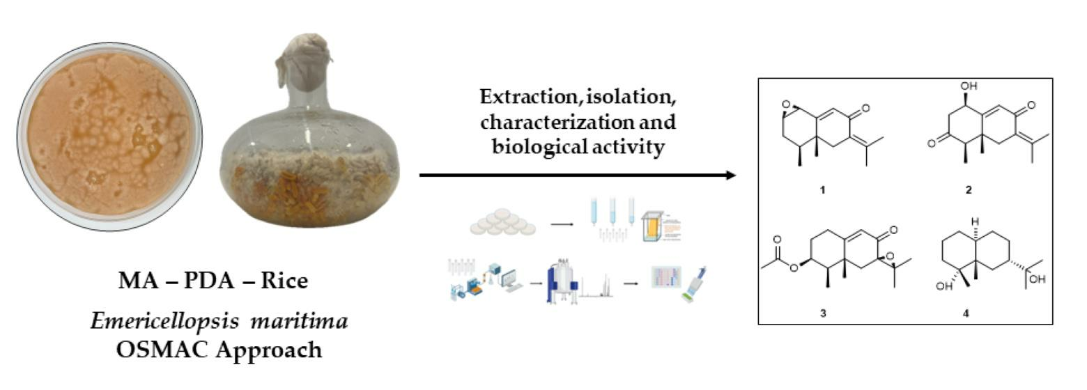
1. Introduction
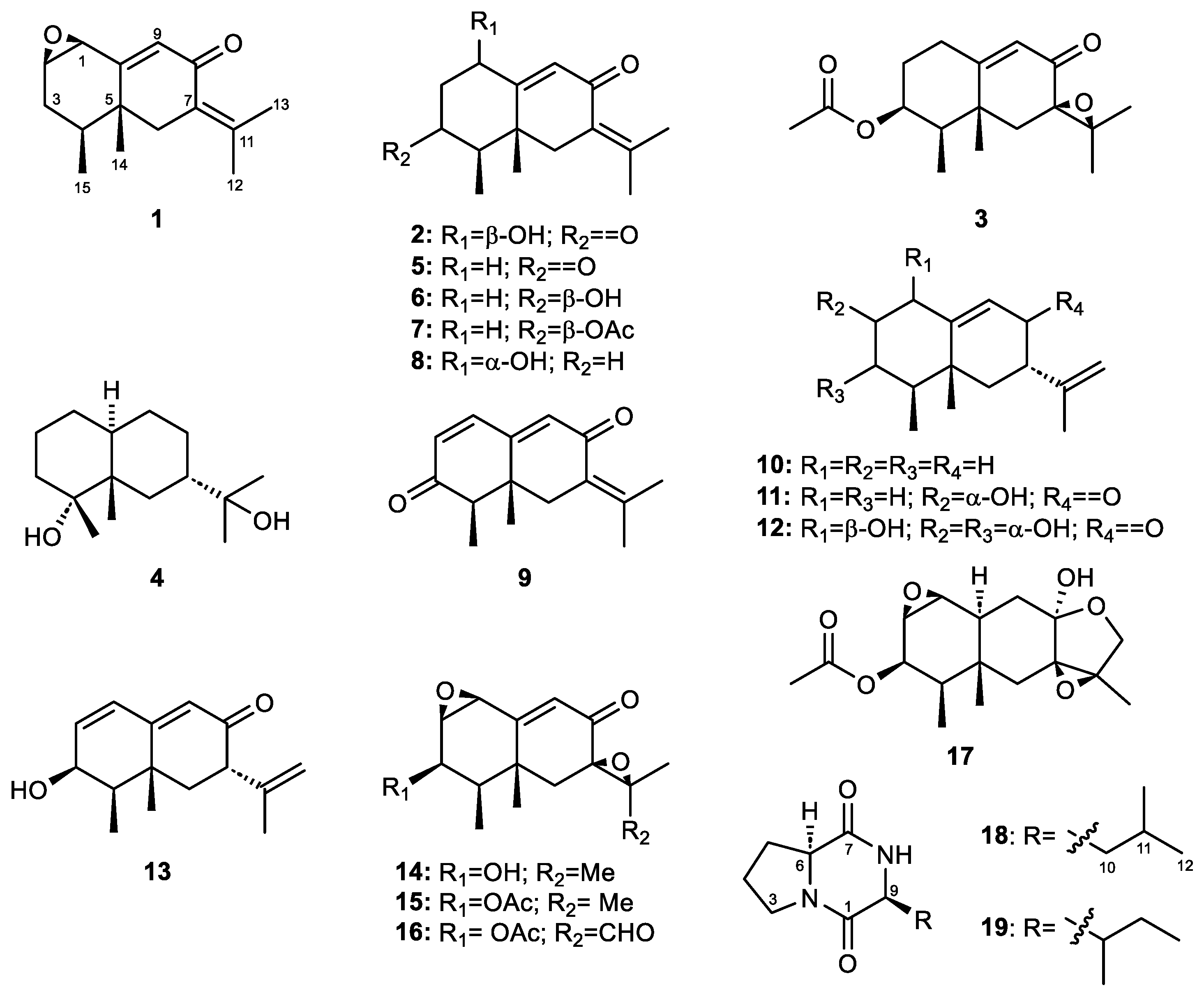
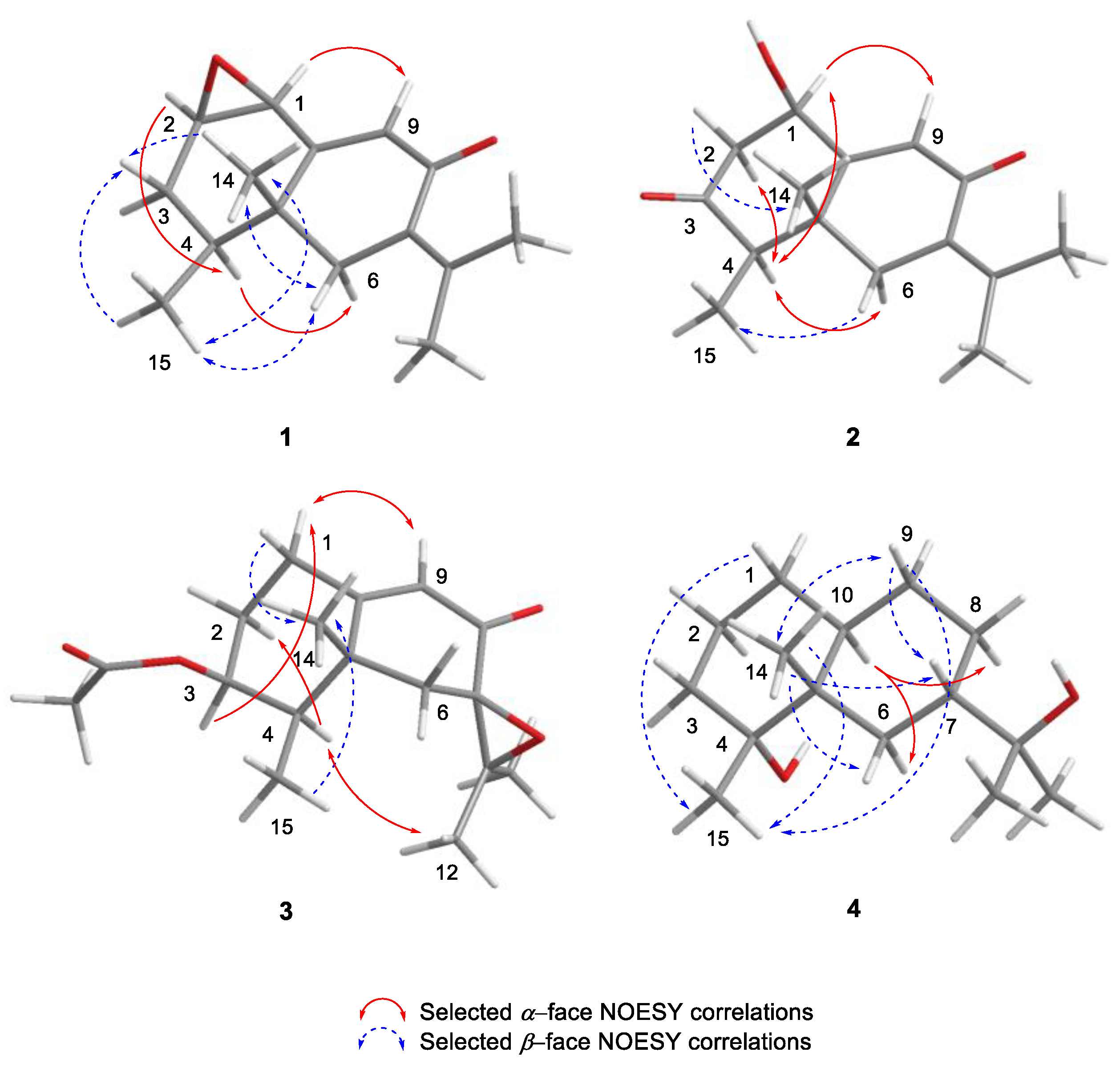
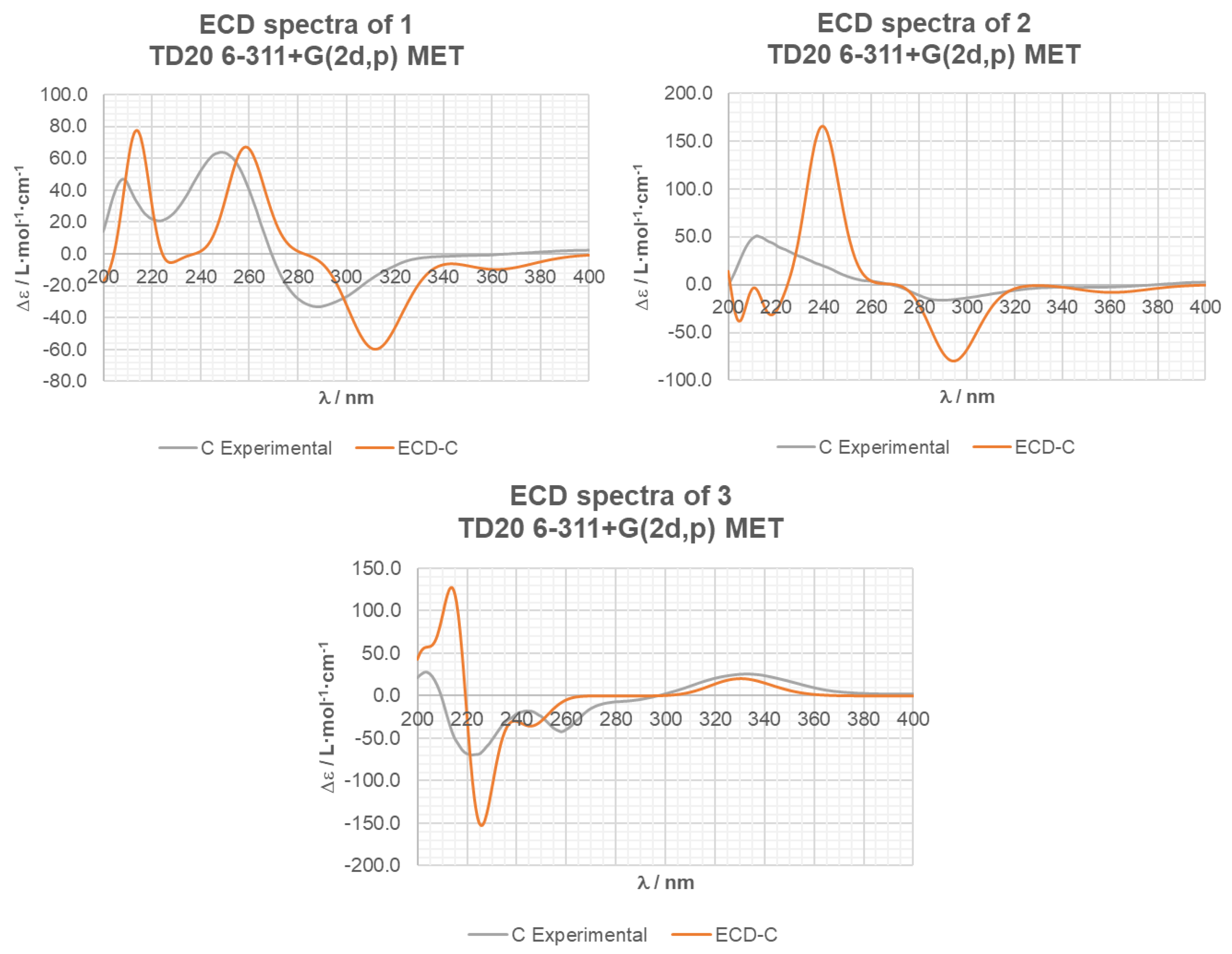
2. Results and Discussion
3. Materials and Methods
3.1. General Experimental Procedures
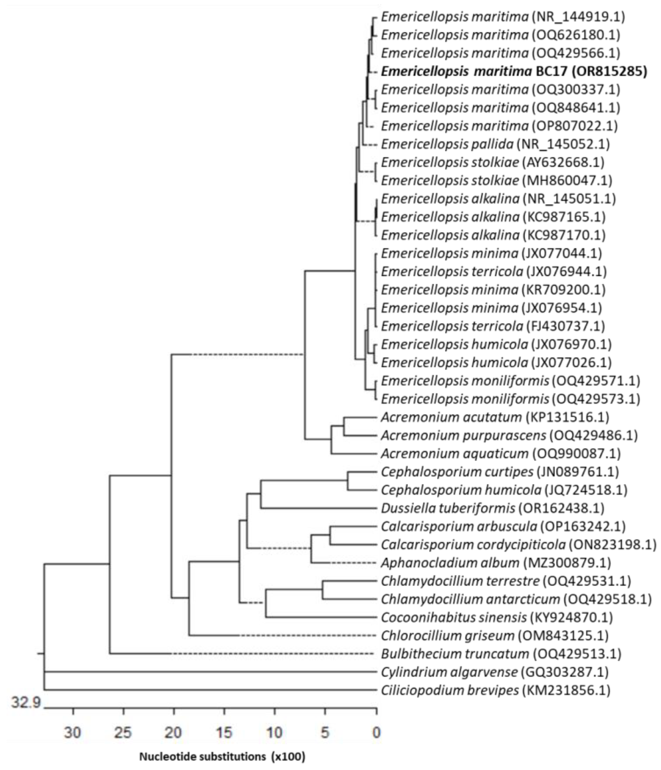
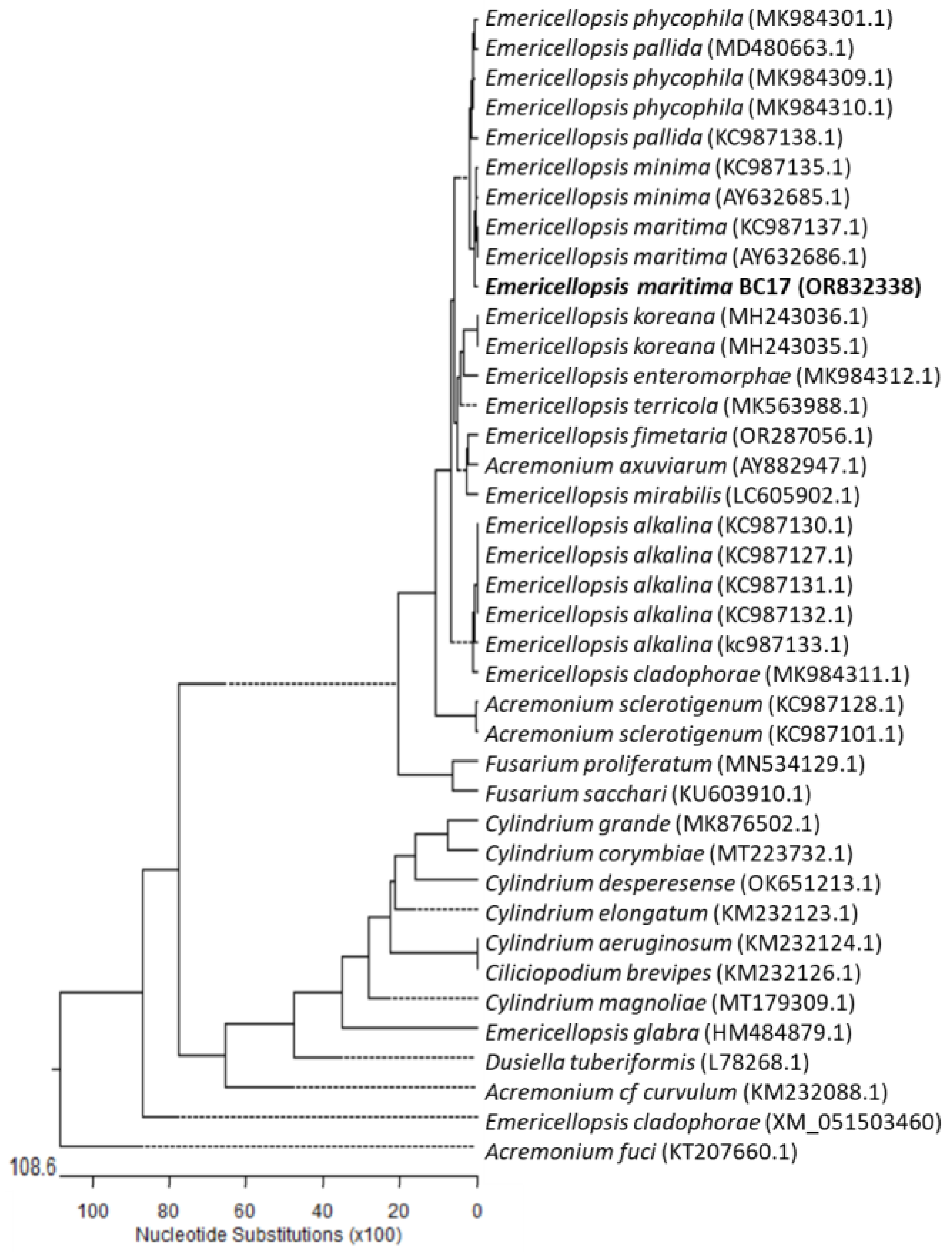
3.2. Fungal Material and Identification
3.3. OSMAC-Based Cultivation Procedures
3.4. Extraction, Isolation, and Characterization of Eremophilane-Type Sesquiterpenes
3.5. Mosher’s Esterification Reaction of Compound 2
3.6. Computational Details of ECD Calculations
3.7. In Vitro Antimicrobial Assay
3.8. In Vitro Antitumor Assay
4. Conclusions
Supplementary Materials
Author Contributions
Funding
Institutional Review Board Statement
Data Availability Statement
Conflicts of Interest
References
- Jin, L.; Quan, C.; Hou, X.; Fan, S. Potential Pharmacological Resources: Natural Bioactive Compounds from Marine-Derived Fungi. Mar. Drugs 2016, 14, 76. [Google Scholar] [CrossRef] [PubMed]
- Jensen, P.R.; Fenical, W. Marine Bacterial Diversity as a Resource for Novel Microbial Products. J. Ind. Microbiol. Biotechnol. 1996, 17, 346–351. [Google Scholar] [CrossRef]
- Romano, S.; Jackson, S.; Patry, S.; Dobson, A. Extending the “One Strain Many Compounds” (OSMAC) Principle to Marine Microorganisms. Mar. Drugs 2018, 16, 244. [Google Scholar] [CrossRef]
- Bode, H.B.; Bethe, B.; Höfs, R.; Zeeck, A. Big Effects from Small Changes: Possible Ways to Explore Nature’s Chemical Diversity. ChemBioChem 2002, 3, 619. [Google Scholar] [CrossRef]
- Pinedo-Rivilla, C.; Aleu, J.; Durán-Patrón, R. Cryptic Metabolites from Marine-Derived Microorganisms Using OSMAC and Epigenetic Approaches. Mar. Drugs 2022, 20, 84. [Google Scholar] [CrossRef]
- Liu, Y.; Li, X.-M.; Meng, L.-H.; Wang, B.-G. N-Formyllapatin A, a New N-Formylspiroquinazoline Derivative from the Marine-Derived Fungus Penicillium adametzioides AS-53. Phytochem. Lett. 2014, 10, 145–148. [Google Scholar] [CrossRef]
- Liu, Y.; Li, X.-M.; Meng, L.-H.; Jiang, W.-L.; Xu, G.-M.; Huang, C.-G.; Wang, B.-G. Bisthiodiketopiperazines and Acorane Sesquiterpenes Produced by the Marine-Derived Fungus Penicillium adametzioides AS-53 on Different Culture Media. J. Nat. Prod. 2015, 78, 1294–1299. [Google Scholar] [CrossRef]
- Qin, Y.; Zou, L.; Lei, X.; Su, J.; Yang, R.; Xie, W.; Li, W.; Chen, G. OSMAC Strategy Integrated with Molecular Networking Discovery Peniciacetals A−I, Nine New Meroterpenoids from the Mangrove-Derived Fungus Penicillium sp. HLLG-122. Bioorg. Chem. 2023, 130, 106271. [Google Scholar] [CrossRef]
- Perazzoli, G.; de los Reyes, C.; Pinedo-Rivilla, C.; Durán-Patrón, R.; Aleu, J.; Cabeza, L.; Melguizo, C.; Prados, J. Emericellopsis maritima and Purpureocillium lilacinum Marine Fungi as a Source of Functional Fractions with Antioxidant and Antitumor Potential in Colorectal Cancer: A Preliminary Study. J. Mar. Sci. Eng. 2023, 11, 2024. [Google Scholar] [CrossRef]
- Zuccaro, A.; Summerbell, R.C.; Gams, W.; Schroers, H.-J.; Mitchell, J.I. A New Acremonium Species Associated with Fucus spp., and Its Affinity with a Phylogenetically Distinct Marine Emericellopsis Clade. Stud. Mycol. 2004, 50, 283–297. [Google Scholar]
- Yamakawa, K.; Izuta, I.; Oka, H.; Sakaguchi, R. Total Synthesis of (±)-Isopetasol, (±)-3-Epiisopetasol, and (±)-Warburgiadion. Tetrahedron Lett. 1974, 15, 2187–2190. [Google Scholar] [CrossRef]
- Brooks, C.J.W.; Draffan, G.H. Sesquiterpenoids of Warburgia Species—I: Warburgin and Warburgiadione. Tetrahedron 1969, 25, 2865–2885. [Google Scholar] [CrossRef]
- Sumarah, M.W.; Puniani, E.; Sørensen, D.; Blackwell, B.A.; Miller, J.D. Secondary Metabolites from Anti-Insect Extracts of Endophytic Fungi Isolated from Picea rubens. Phytochemistry 2010, 71, 760–765. [Google Scholar] [CrossRef]
- Sørensen, D.; Raditsis, A.; Trimble, L.A.; Blackwell, B.A.; Sumarah, M.W.; Miller, J.D. Isolation and Structure Elucidation by LC-MS-SPE/NMR: PR Toxin- and Cuspidatol-Related Eremophilane Sesquiterpenes from Penicillium roqueforti. J. Nat. Prod. 2007, 70, 121–123. [Google Scholar] [CrossRef]
- Bohlmann, F.; Knoll, K.-H. Natürlich Vorkommende Terpen-Derivate, 200 Zwei Neue Eremophilan-Derivate Aus Senecio suaveolens. Liebigs Ann. der Chem. 1979, 1979, 470–472. [Google Scholar] [CrossRef]
- Brooks, C.J.W.; Draffan, G.H. The Constitution of Warburgiadione. Chem. Commun. 1966, 9, 701–703. [Google Scholar] [CrossRef]
- Cane, D.E.; Rawlings, B.J.; Yang, C.-C. Isolation of (−)-γ-Cadinene and Aristolochene from Aspergillus terreus. J. Antibiot. 1987, 40, 1331–1334. [Google Scholar] [CrossRef] [PubMed]
- Cane, D.E.; Salaski, E.J.; Prabhakaran, P.C. Preparation of (−)-Aristolochene from (+)-Valencene: Absolute Configuration of (+)-Aristolochene from Aspergillus terreus. Tetrahedron Lett. 1990, 31, 1943–1944. [Google Scholar] [CrossRef]
- Huang, Z.-Y.; Wu, Q.-Y.; Li, C.-X.; Yu, H.-L.; Xu, J.-H. Facile Production of (+)-Aristolochene and (+)-Bicyclogermacrene in Escherichia coli Using Newly Discovered Sesquiterpene Synthases from Penicillium expansum. J. Agric. Food Chem. 2022, 70, 5860–5868. [Google Scholar] [CrossRef] [PubMed]
- Daengrot, C.; Rukachaisirikul, V.; Tansakul, C.; Thongpanchang, T.; Phongpaichit, S.; Bowornwiriyapan, K.; Sakayaroj, J. Eremophilane Sesquiterpenes and Diphenyl Thioethers from the Soil Fungus Penicillium copticola PSU-RSPG138. J. Nat. Prod. 2015, 78, 615–622. [Google Scholar] [CrossRef] [PubMed]
- Zhou, Y.; Li, Y.-H.; Yu, H.-B.; Liu, X.-Y.; Lu, X.-L.; Jiao, B.-H. Furanone Derivative and Sesquiterpene from Antarctic Marine-Derived Fungus Penicillium sp. S-1-18. J. Asian Nat. Prod. Res. 2018, 20, 1108–1115. [Google Scholar] [CrossRef]
- Lin, A.; Wu, G.; Gu, Q.; Zhu, T.; Li, D. New Eremophilane-Type Sesquiterpenes from an Antarctic Deep-Sea Derived Fungus, Penicillium sp. PR19 N-1. Arch. Pharm. Res. 2014, 37, 839–844. [Google Scholar] [CrossRef] [PubMed]
- Moreau, S.; Cacan, M.; Lablache-Combier, A. Eremofortin C, a New Metabolite Obtained from Penicillium roqueforti Cultures and from Biotransformation of PR Toxin. J. Org. Chem. 1977, 42, 2632–2634. [Google Scholar] [CrossRef] [PubMed]
- Moreau, S.; Biguet, J.; Lablache-Combier, A.; Baert, F.; Foulon, M.; Delfosse, C. Structures et Stereochimie Des Sesquiterpenes de Penicillium roqueforti PR Toxine et Eremofortines A, B, C, D, E. Tetrahedron 1980, 36, 2989–2997. [Google Scholar] [CrossRef]
- Riclea, R.; Dickschat, J.S. Identification of Intermediates in the Biosynthesis of PR Toxin by Penicillium roqueforti. Angew. Chem. Int. Ed. 2015, 54, 12167–12170. [Google Scholar] [CrossRef] [PubMed]
- Adamczeski, M.; Reed, A.R.; Crews, P. New and Known Diketopiperazines from the Caribbean Sponge, Calyx cf. podatypa. J. Nat. Prod. 1995, 58, 201–208. [Google Scholar] [CrossRef] [PubMed]
- Bull, S.D.; Davies, S.G.; Parkin, R.M.; Sánchez-Sancho, F. The Biosynthetic Origin of Diketopiperazines Derived from d-Proline. J. Chem. Soc. Perkin Trans. 1 1998, 15, 2313–2320. [Google Scholar] [CrossRef]
- Fdhila, F.; Vázquez, V.; Sánchez, J.L.; Riguera, R. Diketopiperazines: Antibiotics Active against Vibrio anguillarum Isolated from Marine Bacteria Associated with Cultures of Pecten maximus. J. Nat. Prod. 2003, 66, 1299–1301. [Google Scholar] [CrossRef]
- Li, X.-C.; Ferreira, D.; Ding, Y. Determination of Absolute Configuration of Natural Products: Theoretical Calculation of Electronic Circular Dichroism as a Tool. Curr. Org. Chem. 2010, 14, 1678–1697. [Google Scholar] [CrossRef]
- Yuyama, K.T.; Fortkamp, D.; Abraham, W.-R. Eremophilane-Type Sesquiterpenes from Fungi and Their Medicinal Potential. Biol. Chem. 2018, 399, 13–28. [Google Scholar] [CrossRef]
- Rasmussen, R.R.; Rasmussen, P.H.; Larsen, T.O.; Bladt, T.T.; Binderup, M.L. In vitro Cytotoxicity of Fungi Spoiling Maize Silage. Food Chem. Toxicol. 2011, 49, 31–44. [Google Scholar] [CrossRef] [PubMed]
- Darsih, C.; Prachyawarakorn, V.; Wiyakrutta, S.; Mahidol, C.; Ruchirawat, S.; Kittakoop, P. Cytotoxic Metabolites from the Endophytic Fungus Penicillium chermesinum: Discovery of a Cysteine-Targeted Michael Acceptor as a Pharmacophore for Fragment-Based Drug Discovery, Bioconjugation and Click Reactions. RSC Adv. 2015, 5, 70595–70603. [Google Scholar] [CrossRef]
- Moulé, Y.; Moreau, S.; Bousquet, J.F. Relationships between the Chemical Structure and the Biological Properties of Some Eremophilane Compounds Related to PR Toxin. Chem. Biol. Interact. 1977, 17, 185–192. [Google Scholar] [CrossRef] [PubMed]
- Stewart, J.J.P. Optimization of Parameters for Semiempirical Methods V: Modification of NDDO Approximations and Application to 70 Elements. J. Mol. Model. 2007, 13, 1173–1213. [Google Scholar] [CrossRef] [PubMed]
- Frisch, M.J.; Trucks, G.W.; Schlegel, H.B.; Scuseria, G.E.; Robb, M.A.; Cheeseman, J.R.; Scalmani, G.; Barone, V.; Petersson, G.A.; Nakatsuji, H.; et al. Gaussian 16, Revision.01; Gaussian Inc.: Wallingford, CT, USA, 2016. [Google Scholar]
- Lee, C.; Yang, W.; Parr, R.G. Development of the Colle-Salvetti Correlation-Energy Formula into a Functional of the Electron Density. Phys. Rev. B 1988, 37, 785–789. [Google Scholar] [CrossRef] [PubMed]
- Becke, A.D. Density-functional Thermochemistry. III. The Role of Exact Exchange. J. Chem. Phys. 1993, 98, 5648–5652. [Google Scholar] [CrossRef]
- Bauernschmitt, R.; Ahlrichs, R. Treatment of Electronic Excitations within the Adiabatic Approximation of Time Dependent Density Functional Theory. Chem. Phys. Lett. 1996, 256, 454–464. [Google Scholar] [CrossRef]
- Casida, M.E.; Jamorski, C.; Casida, K.C.; Salahub, D.R. Molecular Excitation Energies to High-Lying Bound States from Time-Dependent Density-Functional Response Theory: Characterization and Correction of the Time-Dependent Local Density Approximation Ionization Threshold. J. Chem. Phys. 1998, 108, 4439–4449. [Google Scholar] [CrossRef]
- Cancès, E.; Mennucci, B.; Tomasi, J. A New Integral Equation Formalism for the Polarizable Continuum Model: Theoretical Background and Applications to Isotropic and Anisotropic Dielectrics. J. Chem. Phys. 1997, 107, 3032–3041. [Google Scholar] [CrossRef]
- Mennucci, B.; Cancès, E.; Tomasi, J. Evaluation of Solvent Effects in Isotropic and Anisotropic Dielectrics and in Ionic Solutions with a Unified Integral Equation Method: Theoretical Bases, Computational Implementation, and Numerical Applications. J. Phys. Chem. B 1997, 101, 10506–10517. [Google Scholar] [CrossRef]
- Tomasi, J.; Mennucci, B.; Cammi, R. Quantum Mechanical Continuum Solvation Models. Chem. Rev. 2005, 105, 2999–3093. [Google Scholar] [CrossRef]
- Zhang, L.; Ravipati, A.S.; Koyyalamudi, S.R.; Jeong, S.C.; Reddy, N.; Bartlett, J.; Smith, P.T.; de la Cruz, M.; Monteiro, M.C.; Melguizo, Á.; et al. Anti-Fungal and Anti-Bacterial Activities of Ethanol Extracts of Selected Traditional Chinese Medicinal Herbs. Asian Pac. J. Trop. Med. 2013, 6, 673–681. [Google Scholar] [CrossRef]


| 1 | 2 | 3 | 4 | |
|---|---|---|---|---|
| Position | δH, Mult (J in Hz) a | δH, Mult (J in Hz) a | δH, Mult (J in Hz) b | δH, Mult (J in Hz) c |
| 1α | 3.38, d (3.7) | 4.83, q (4.4) | 2.20, m | 1.39, dt (13.0, 6.6) |
| 1β | - | - | 2.73, td (13.5, 4.7) | 1.73, dtd (13.0, 3.3, 1.6) |
| 2α | 3.52, dd (3.7, 5.7) | 2.85, dd (15.2, 5.1) | 1.67, tt (14.2, 3.8) | 1.59, dt (13.5, 3.3) g |
| 2β | - | 2.66, dd (15.1, 4.8) | 2.19, ddt (14.2, 4.7, 2.3) | 1.55, dq (13.5, 4.3) g |
| 3α | 1.99, dt (15.5, 6.1) | - | 5.12, q (3.4) | 1.39, td (13.1, 4.3) |
| 3β | 1.80, dd (15.5, 12.6) | - | - | 1.08, td (13.1, 4.3) |
| 4α | 1.64, td (12.6, 6.1) | 2.53, q (6.6) | 1.89, qd (7.0, 3.4) | - |
| 6α | 2.06, br d (13.4) | 2.42, d (13.8) | 2.04, d (15.0) f | 1.16, td (13.1, 4.2) |
| 6β | 2.81, d (13.4) | 2.92, d (13.8) | 2.10, d (15.0) f | 1.43, dt (13.1, 3.1) |
| 7β | - | - | - | 1.35–1.30, m |
| 8α | - | - | - | 1.37–1.28, m |
| 8β | - | - | - | 1.61, m |
| 9α | 6.17, s | 6.09, d (0.9) | 5.94, s | 1.95, m 1.03, q (12.5) |
| 9β | ||||
| 10α | - | - | - | 1.22, m |
| 12 | 2.13, d (1.9) d | 2.16, d (2.1) e | 1.41, s | 1.17, s h |
| 13 | 1.84, d (1.9) d | 1.89, d (1.4) e | 1.31, s | 1.16, s h |
| 14β | 1.01, d (0.9) | 1.11, d (0.8) | 1.32, s | 0.88, s |
| 15β | 0.90, d (6.7) | 1.12, d (6.6) | 1.03, d (7.0) | 1.08, s |
| OCOMe | - | - | 2.12, s | - |
| 1 | 2 | 3 | 4 | |
|---|---|---|---|---|
| Position | δC, Type a | δC, Type a | δC, Type b | δC, Type c |
| 1 | 55.0, CH | 73.2, CH | 28.3, CH2 | 44.2, CH2 |
| 2 | 55.7, CH | 47.2, CH2 | 33.9, CH2 | 21.1, CH2 |
| 3 | 29.8, CH2 | 208.0, C | 72.5, CH | 42.3, CH2 |
| 4 | 39.1, CH | 52.6, CH | 42.7, CH | 73.1, C |
| 5 | 39.2, C | 43.5, C | 41.9, C | 35.6, C |
| 6 | 41.8, CH2 | 42.4, CH2 | 38.0, CH2 | 46.1, CH2 |
| 7 | 127.6, C | 126.6, C | 65.4, C | 51.3, CH |
| 8 | 190.6, C | 190.9, C | 195.0, C | 23.5, CH2 |
| 9 | 131.9, CH | 129.1, CH | 124.1, CH | 22.8, CH2 |
| 10 | 160.2, C | 161.9, C | 170.7, C | 55.7, CH |
| 11 | 145.3, C | 146.9, C | 64.8, C | 73.4, C |
| 12 | 23.0, d CH3 | 23.0, e CH3 | 21.3, CH3 | 27.5, f CH3 |
| 13 | 22.5, d CH3 | 22.7, e CH3 | 19.2, CH3 | 26.9, f CH3 |
| 14 | 16.0, CH3 | 20.2, CH3 | 23.8, CH3 | 19.2, CH3 |
| 15 | 14.6, CH3 | 7.4, CH3 | 12.2, CH3 | 22.5, CH3 |
| OCOMe | - | - | 170.3, C | - |
| OCOMe | - | - | 21.3, CH3 | - |
| MIC (µM) | ||||||||
|---|---|---|---|---|---|---|---|---|
| Compound | A. fumigatus ATCC46645 | C. albicans ATCC64124 | K. pneumonia ATCC700603 | E. coli ATCC25922 | MSSA ATCC29213 | MRSA MB5393 | A. baumannii ATCC19606 | P. aeruginosa PAO-1 |
| 1 | >552 | >552 | >552 | >552 | >552 | >552 | >552 | >552 |
| 3 | >438 | >438 | >438 | >438 | >438 | >438 | >438 | >438 |
| 5 | >414 | >414 | >414 | >414 | >414 | >414 | >414 | >414 |
| 6 | >410 | >410 | >410 | >410 | >410 | >410 | >410 | >410 |
| 8 | >410 | >410 | >410 | >410 | >410 | >410 | >410 | >410 |
| 9 | >556 | >556 | >556 | >556 | >556 | >556 | >556 | >556 |
| 10 | 471 | 471 | >471 | >471 | >471 | >471 | >471 | >471 |
| 11 | >410 | >410 | >410 | >410 | >410 | >410 | >410 | >410 |
| 13 | >414 | >414 | >414 | >414 | >414 | >414 | >414 | >414 |
| 14 | >121 | >121 | >121 | >121 | >121 | >121 | >121 | >121 |
| 15 | >262 | >262 | >261 | >261 | >261 | >261 | >261 | >261 |
| 16 | >300 | >300 | >300 | >300 | >300 | >300 | >300 | >300 |
| 17 | >296 | >296 | >296 | >296 | >296 | >296 | >296 | >296 |
| IC50 (µM) | |||||
|---|---|---|---|---|---|
| Compound | HepG2 | MCF7 | A549 | A2058 | Mia PaCa-2 |
| 1 | >172 | >172 | >172 | >172 | 159 (142–172) |
| 3 | >137 | >137 | >137 | >137 | >137 |
| 5 | >192 | >129 | >129 | >129 | >129 |
| 6 | >128 | >128 | >128 | >128 | >128 |
| 8 | >128 | >128 | >128 | >128 | >128 |
| 9 | >174 | >174 | >174 | >174 | 161 (139–187) |
| 10 | >147 | >147 | >147 | >147 | >147 |
| 11 | >128 | >128 | >128 | >128 | >128 |
| 13 | >129 | >129 | >129 | 125 (116–138) | >129 |
| 14 | >38 | >38 | >38 | >38 | >38 |
| 15 | >282 | >82 | >82 | >82 | >82 |
| 16 | 8.28 (7.88–8.72) | 5.53 (5.31–5.78) | 33.44 (31.56–35.00) | 3.75 (3.44–4.38) | 5.00 (4.69–5.31) |
| 17 | >93 | >93 | >93 | 74 (68–80) | >93 |
| Doxorubicin | 0.21 (0.19–0.23) | 0.21 (0.16–0.28) | 0.90 (0.70–1.00) | 0.10 (0.08–0.13) | 0.43 (0.36–0.50) |
Disclaimer/Publisher’s Note: The statements, opinions and data contained in all publications are solely those of the individual author(s) and contributor(s) and not of MDPI and/or the editor(s). MDPI and/or the editor(s) disclaim responsibility for any injury to people or property resulting from any ideas, methods, instructions or products referred to in the content. |
© 2023 by the authors. Licensee MDPI, Basel, Switzerland. This article is an open access article distributed under the terms and conditions of the Creative Commons Attribution (CC BY) license (https://creativecommons.org/licenses/by/4.0/).
Share and Cite
Virués-Segovia, J.R.; Millán, C.; Pinedo, C.; González-Rodríguez, V.E.; Papaspyrou, S.; Zorrilla, D.; Mackenzie, T.A.; Ramos, M.C.; de la Cruz, M.; Aleu, J.; et al. New Eremophilane-Type Sesquiterpenes from the Marine Sediment-Derived Fungus Emericellopsis maritima BC17 and Their Cytotoxic and Antimicrobial Activities. Mar. Drugs 2023, 21, 634. https://doi.org/10.3390/md21120634
Virués-Segovia JR, Millán C, Pinedo C, González-Rodríguez VE, Papaspyrou S, Zorrilla D, Mackenzie TA, Ramos MC, de la Cruz M, Aleu J, et al. New Eremophilane-Type Sesquiterpenes from the Marine Sediment-Derived Fungus Emericellopsis maritima BC17 and Their Cytotoxic and Antimicrobial Activities. Marine Drugs. 2023; 21(12):634. https://doi.org/10.3390/md21120634
Chicago/Turabian StyleVirués-Segovia, Jorge R., Carlos Millán, Cristina Pinedo, Victoria E. González-Rodríguez, Sokratis Papaspyrou, David Zorrilla, Thomas A. Mackenzie, María C. Ramos, Mercedes de la Cruz, Josefina Aleu, and et al. 2023. "New Eremophilane-Type Sesquiterpenes from the Marine Sediment-Derived Fungus Emericellopsis maritima BC17 and Their Cytotoxic and Antimicrobial Activities" Marine Drugs 21, no. 12: 634. https://doi.org/10.3390/md21120634