
Rhizoaspergillin A and Rhizoaspergillinol A, including a Unique Orsellinic Acid–Ribose–Pyridazinone-N-Oxide Hybrid, from the Mangrove Endophytic Fungus Aspergillus sp. A1E3

Abstract
:
1. Introduction
2. Results and Discussion
3. Materials and Methods
3.1. General Experimental Procedures
3.2. Fungus Material
3.3. Fermentation, Extraction and Isolation
3.4. Spectroscopic Data of Compounds
3.5. X-Ray Crystallographic Data of 4
3.6. Synthesis of the Derivative 4
3.7. Cell Culture and Cytotoxicity (MTT) Assay
3.8. Cell Cycle Analysis
4. Conclusions
Supplementary Materials
Author Contributions
Funding
Institutional Review Board Statement
Data Availability Statement
Acknowledgments
Conflicts of Interest
References
- Chintakunta, V.K.; Akella, V.; Vedula, M.S.; Mamnoor, P.K.; Mishra, P.; Casturi, S.R.; Vangoori, A.; Rajagopalan, R. 3-O-Substituted benzyl pyridazinone derivatives as COX inhibitors. Eur. J. Med. Chem. 2002, 37, 339–347. [Google Scholar] [CrossRef] [PubMed]
- Coelho, A.; Sotelo, E.; Novoa, H.; Peeters, O.M.; Blaton, N.; Raviña, E. Pyridazine derivatives. Part 38: Efficient Heck alkenylation at position 5 of the 6-phenyl-3(2H)-pyridazinone system. Tetrahedron Lett. 2004, 45, 3459–3463. [Google Scholar] [CrossRef]
- Gong, Y.; Barbay, J.K.; Dyatkin, A.B.; Miskowski, T.A.; Kimball, E.S.; Prouty, S.M.; Fisher, M.C.; Santulli, R.J.; Schneider, C.R.; Wallace, N.H.; et al. Synthesis and biological evaluation of novel pyridazinone-based α4 integrin receptor antagonists. J. Med. Chem. 2006, 49, 3402–3411. [Google Scholar] [CrossRef]
- Rathish, I.G.; Javed, K.; Bano, S.; Ahmad, S.; Alam, M.S.; Pillai, K.K. Synthesis and blood glucose lowering effect of novel pyridazinone substituted benzenesulfonylurea derivatives. Eur. J. Med. Chem. 2009, 44, 2673–2678. [Google Scholar] [CrossRef]
- Schoene, D.L.; Hoffmann, O.L. Maleic hydrazide, a unique growth regulant. Science 1949, 109, 588–589. [Google Scholar] [CrossRef]
- Shipp, J.L.; Wang, K.; Ferguson, G. Residual toxicity of avermectin b1 and pyridaben to eight commercially produced beneficial arthropod species used for control of greenhouse pests. Biol. Control 2000, 17, 125–131. [Google Scholar] [CrossRef]
- Navarro, A.; Bández, M.J.; Gómez, C.; Repetto, M.G.; Boveris, A. Effects of rotenone and pyridaben on complex I electron transfer and on mitochondrial nitric oxide synthase functional activity. J. Bioenerg. Biomembr. 2010, 42, 405–412. [Google Scholar] [CrossRef] [PubMed]
- Sugimoto, N.; Osakabe, M. Cross-resistance between cyenopyrafen and pyridaben in the twospotted spidermite Tetranychus urticae (Acari: Tetranychidae). Pest. Manag. Sci. 2014, 70, 1090–1096. [Google Scholar] [CrossRef]
- Basistyi, V.S.; Frederich, J.H. Pyridazine N-oxides as photoactivatable surrogates for reactive oxygen species. Org. Lett. 2022, 24, 1907–1912. [Google Scholar] [CrossRef]
- Chen, S.H.; Cai, R.L.; Liu, Z.M.; Cui, H.; She, Z.G. Secondary metabolites from mangrove-associated fungi: Source, chemistry and bioactivities. Nat. Prod. Rep. 2021, 39, 560–595. [Google Scholar] [CrossRef]
- Liu, X.; Fu, Y.; Zhou, Q.Q.; Wang, S.; Gao, L.; Lei, J.L.; Ke, A.B.; Li, Y.Y.; Zhang, X.X.; Huo, C.H.; et al. Aspergichromones A–E, five chromone derivatives with complicated polycyclic architecture from Aspergillus deflectus. Org. Lett. 2022, 24, 1610–1615. [Google Scholar] [CrossRef]
- Liu, L.; Duan, F.F.; Gao, Y.; Peng, X.G.; Chang, J.L.; Chen, J.; Ruan, H.L. Aspersteroids A–C, Three Rearranged Ergostane-type Steroids from Aspergillus ustus NRRL 275. Org. Lett. 2021, 23, 9620–9624. [Google Scholar] [CrossRef]
- Wang, L.; Yang, J.; Huang, J.P.; Li, J.; Luo, J.Y.; Yan, Y.J.; Huang, S.X. Bisaspochalasins A–C: Three cytochalasan homodimers with highly fused ring system from an endophytic Aspergillus flavipes. Org. Lett. 2020, 22, 7930–7935. [Google Scholar] [CrossRef]
- Liu, L.; Wang, L.; Bao, L.; Ren, J.W.; Basnet, B.B.; Liu, R.X.; He, L.W.; Han, J.J.; Yin, W.B.; Liu, H.W. Versicoamides F–H, prenylated indole alkaloids from Aspergillus tennesseensis. Org. Lett. 2017, 19, 942–945. [Google Scholar] [CrossRef] [PubMed]
- Kankanamge, S.; Khalil, Z.G.; Sritharan, T.; Capon, R.J. Noonindoles G–L: Indole diterpene glycosides from the Australian marine-derived fungus Aspergillus noonimiae CMB-M0339. J. Nat. Prod. 2023, 86, 508–516. [Google Scholar] [CrossRef]
- Neuhaus, G.F.; Loesgen, S. Antibacterial drimane sesquiterpenes from Aspergillus ustus. J. Nat. Prod. 2021, 84, 37–45. [Google Scholar] [CrossRef] [PubMed]
- Guo, Y.J.; Ding, L.; Ghidinelli, S.; Gotfredsen, C.H.; de la Cruz, M.; Mackenzie, T.A.; Ramos, M.C.; Sánchez, P.; Vicente, F.; Genilloud, O.; et al. Taxonomy driven discovery of polyketides from Aspergillus californicus. J. Nat. Prod. 2021, 84, 979–985. [Google Scholar] [CrossRef] [PubMed]
- Wei, M.S.; Huang, L.P.; Li, Q.; Qiao, X.Y.; Zhao, Z.M.; Yin, J.; Fu, A.M.; Guo, J.R.; Hao, X.C.; Gu, L.H.; et al. Spectasterols, aromatic ergosterols with 6/6/6/5/5, 6/6/6/6, and 6/6/6/5 ring systems from Aspergillus spectabilis. J. Nat. Prod. 2023, 86, 1385–1391. [Google Scholar] [CrossRef] [PubMed]
- Yang, W.C.; Chen, T.; Tan, Q.; Zang, Z.M.; Chen, Y.; Ou, Y.H.; Li, G.; Hu, D.; Wang, B.; Yao, H.L.; et al. Plasmodium-resistant indole diterpenoid biosynthesis gene cluster derived from Aspergillus oryzae was activated by exogenous P450 gene Ast B. J. Nat. Prod. 2023, 86, 1392–1401. [Google Scholar] [CrossRef] [PubMed]
- Holkers, J.S.E.; Kagal, S.A.; Mulheirn, L.J.; White, P.M. Some new metabolites of Aspergillus versicolor and a revised structure for averufin. Chem. Commun. 1966, 24, 911–913. [Google Scholar] [CrossRef]
- Castonguay, A. Synthesis of (±)-averufanin, noraverufanin and bis-deoxyaverufanin. Tetrahedron 1979, 35, 1557–1563. [Google Scholar] [CrossRef]
- Sakai, K.; Ohte, S.; Ohshiro, T.; Matsuda, D.; Masuma, R.; Rudel, L.L.; Tomoda, H. Selective inhibition of Acyl-CoA: Cholesterol acyltransferase 2 isozyme by flavasperone and sterigmatocystin from Aspergillus Species. J. Antibiot. 2008, 61, 568–572. [Google Scholar] [CrossRef]
- Shao, C.L.; Wang, C.Y.; Wei, M.Y.; Li, S.D.; She, Z.G.; Gu, Y.C.; Lin, Y.C. Structural and spectral assignments of six anthraquinone derivatives from the mangrove fungus (ZSUH-36). Magn. Reson. Chem. 2008, 46, 886–889. [Google Scholar] [CrossRef]
- Chen, M.; Shao, C.L.; Kong, C.J.; She, Z.G.; Wang, C.Y. A new anthraquinone derivative from a gorgonian-derived fungus Aspergillus sp. Chem. Nat. Compd. 2014, 50, 617–620. [Google Scholar] [CrossRef]
- Demirel1, D.; Ozkaya, F.C.; Ebrahim, W.; Sokullu, E.; Sahin, I.D. Aspergillus Carneus metabolite Averufanin induced cell cycle arrest and apoptotic cell death on cancer cell lines via inducing DNA damage. Sci. Rep. 2023, 13, 6460. [Google Scholar] [CrossRef]
- Zhu, F.; Lin, Y.C. Three xanthones from a marine-derived mangrove endophytic fungus. Chem. Nat. Compd. 2007, 43, 132–135. [Google Scholar] [CrossRef]
- Han, X.; Tang, X.L.; Luo, X.C.; Sun, C.X.; Liu, K.C.; Zhang, Y.; Li, P.L.; Li, G.Q. Isolation and identification of three new sterigmatocystin derivatives from the fungus Aspergillus versicolor guided by molecular networking approach. Chem. Biodivers. 2020, 17, e2000208. [Google Scholar] [CrossRef]
- Ebrahim, W.; El-Neketi, M.; Lewald, L.I.; Orfali, R.S.; Lin, W.; Rehberg, N.; Kalscheuer, R.; Daletos, G.; Proksch, P. Metabolites from the fungal endophyte Aspergillus austroafricanus in axenic culture and in fungal–bacterial mixed cultures. J. Nat. Prod. 2016, 79, 914–922. [Google Scholar] [CrossRef] [PubMed]
- Özkaya, F.C.; Ebrahim, W.; El-Neketi, M.; Tanrikul, T.T.; Kalscheuer, R.; Müller, W.E.G.; Guo, Z.Y.; Zou, K.; Liu, Z.; Proksch, P. Induction of new metabolites from sponge-associated fungus Aspergillus carneus by OSMAC approach. Fitoterapia 2018, 131, 9–14. [Google Scholar] [CrossRef] [PubMed]
- Rodríguez-Urra, A.B.; Jiménez, C.; Nieto, M.I.; Rodríguez, J.; Hayashi, H.; Ugalde, U. Signaling the induction of sporulation involves the interaction of two secondary metabolites in Aspergillus nidulans. ACS Chem. Biol. 2012, 7, 599–606. [Google Scholar] [CrossRef] [PubMed]
- Rasche, M.E.; White, R.H. Mechanism for the enzymatic formation of 4-(β-D-ribofuranosyl)aminobenzene 5’-phosphate during the biosynthesis of methanopterin. Biochemistry 1998, 37, 11343–11351. [Google Scholar] [CrossRef]
- White, R.H. The conversion of a phenol to an aniline occurs in the biochemical formation of the 1-(4-aminophenyl)-1-deoxy-D-ribitol moiety in methanopterin. Biochemistry 2011, 50, 6041–6052. [Google Scholar] [CrossRef] [PubMed]
- Xiang, Y.; Kotra, L.P.; Chu, C.K.; Schinazi, R.F. Synthesis and anti-HIV activities of 2’-deoxy-2’,2”-difluoro-β-L-ribofuranosyl-pyrimidine and -purine nucleosides. Bioorg. Med. Chem. Lett. 1995, 5, 743–748. [Google Scholar] [CrossRef]
- Maeba, I.; Suzuki, M.; Hara, O.; Takeuchi, T.; Iijimar, T.; Furukawa, H. C-Nucleosides. 6. Synthesis of 5-methoxy-5-(2,3,5-tri-O-benzoyl-.beta.-D-ribofuranosyl)furan-2(5H)-one and its ring transformation. J. Org. Chem. 1987, 52, 4521–4526. [Google Scholar] [CrossRef]
- Tomori, T.; Nagaoka, K.; Takeshita, L.; Shiozawa, T.; Miyatake, Y.; Masaki, Y.; Sekine, M.; Seio, K. Deoxynucleoside triphosphate containing pyridazin-3-one aglycon as a thymidine triphosphate substitute for primer extension and chain elongation by Klenow fragments. J. Org. Chem. 2018, 83, 8353–8363. [Google Scholar] [CrossRef] [PubMed]
- Rosato, R.R.; Almenara, J.A.; Grant, S. The histone deacetylase inhibitor MS-275 promotes differentiation or apoptosis in human leukemia cells through a process regulated by generation of reactive oxygen species and induction of p21CIP1/WAF11. Cancer Res. 2003, 63, 3637–3645. [Google Scholar]


| Position | 1 a | 4 b | ||
|---|---|---|---|---|
| δH, Multi. (J) | δC, Type | δH, Multi. (J) | δC, Type | |
| 1 | 163.3, C | 162.1, C | ||
| 2 | 5.78 d (8.0) | 102.6, CH | 5.79 d (8.0) | 103.4, CH |
| 3 | 7.96 d (8.0) | 140.7, CH | 7.39 d (8.0) | 139.4, CH |
| 4 | 151.1, C | 149.7, C | ||
| 5 | 5.96 d (6.4) | 87.8, CH | 6.06 d (6.3) | 87.8, CH |
| 6 | 4.46 dd (11.0, 5.8) | 72.0, CH | 5.47 t (6.3) | 72.6, CH |
| 7 | 5.45 dd (5.2, 2.8) | 74.0, CH | 5.65 t (4.9) | 72.0, CH |
| 8 | 4.26 dd (5.4, 2.8) | 83.0, CH | 4.56 m | 80.4, CH |
| 9 | 3.74 2H, br s | 61.2, CH2 | 4.39 dd (12.6, 2.8) 4.47 dd (12.6, 3.5) | 63.4, CH2 |
| 10 | 106.3, C | 108.6, C | ||
| 11 | 162.6, C | 164.9, C | ||
| 12 | 6.24 d (2.0) | 100.9, CH | 6.53 br s | 117.0, CH |
| 13 | 162.1, C | 156.2, C | ||
| 14 | 6.28 d (2.0) | 111.2, CH | 6.62 br s | 108.9, CH |
| 15 | 142.4, C | 142.9, C | ||
| 16 | 169.1, C | 170.1, C | ||
| 17 | 2.47 s | 23.1, CH3 | 2.66 s | 24.6, CH3 |
| 2’ | 11.40 s | 8.19 s | ||
| 18 | 176.2, C | |||
| 19 | 38.9, C | |||
| 20 | 1.35 s | 27.0, CH3 | ||
| 21 | 1.35 s | 27.0, CH3 | ||
| 22 | 1.35 s | 27.0, CH3 | ||
| 23 | 177.9, C | |||
| 24 | 39.3, C | |||
| 25 | 1.27 s | 27.3, CH3 | ||
| 26 | 1.27 s | 27.3, CH3 | ||
| 27 | 1.27 s | 27.3, CH3 | ||
| 28 | 177.3, C | |||
| 29 | 38.8, C | |||
| 30 | 1.09 s | 26.8, CH3 | ||
| 31 | 1.09 s | 26.8, CH3 | ||
| 32 | 1.09 s | 26.8, CH3 | ||
| 6-OH | 6.00 d (5.6) | |||
| 9-OH | 5.38 t (4.8) | |||
| 11-OH | 10.85 s | 11.08 s | ||
| 13-OH | 10.17 s | |||
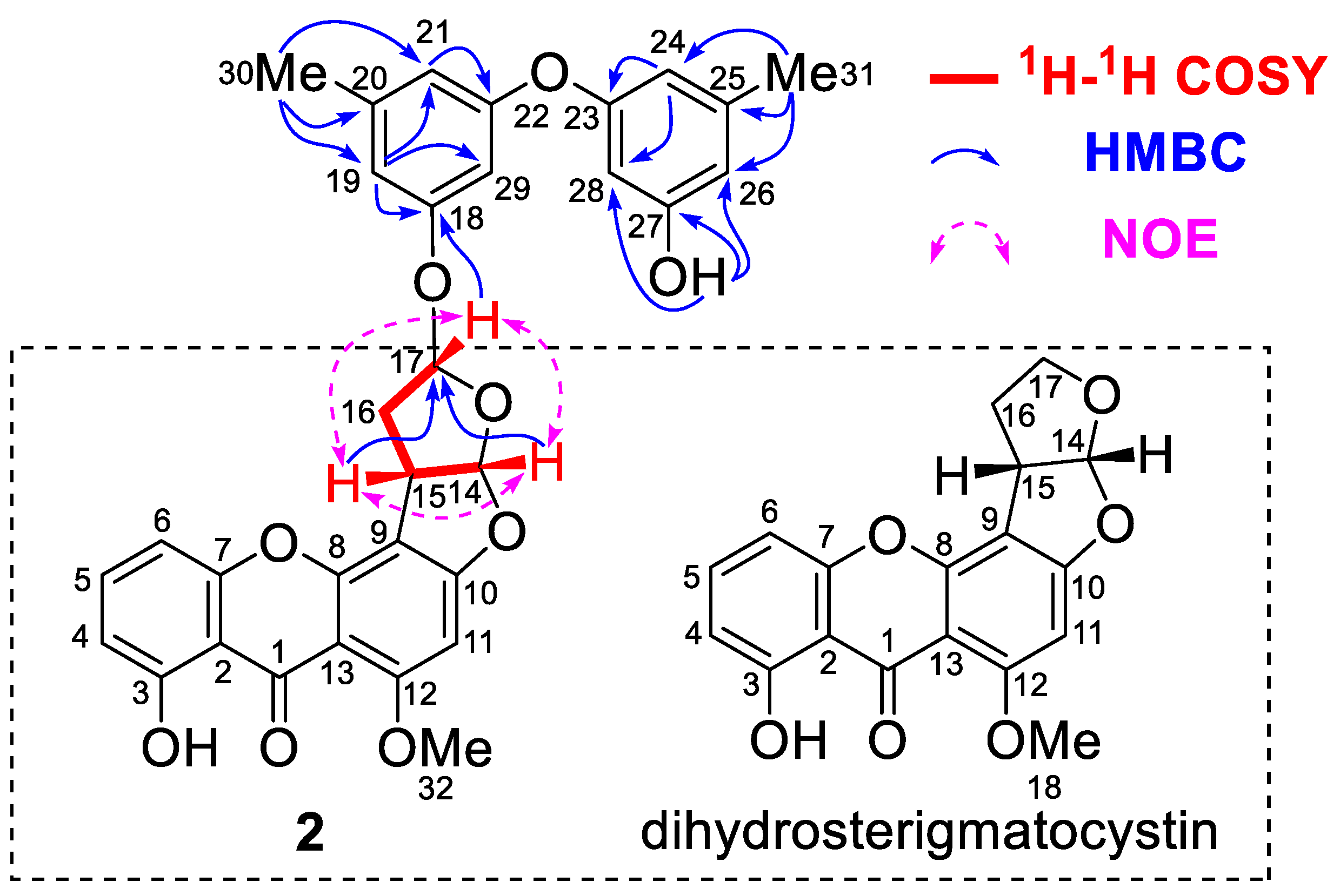
| Position | 2 (CDCl3) a | Dihydrosterigmatocystin (DMSO-d6) [26] | ||
|---|---|---|---|---|
| δH, Multi. (J) | δC, Type | δH, Multi. (J) | δC, Type | |
| 1 | 181.4, C | 180.0, C | ||
| 2 | 109.0, C | 108.1, C | ||
| 3 | 162.3, C | 161.3, C | ||
| 4 | 6.77 d (8.0) | 111.4, CH | 6.73 dd (8.5, 1.0) | 110.5, CH |
| 5 | 7.52 t (8.0) | 135.8, CH | 7.61 t (8.5) | 136.0, CH |
| 6 | 6.83 d (8.0) | 105.9, CH | 6.94 dd (8.5, 1.0) | 106.1, CH |
| 7 | 154.9, C | 154.4, C | ||
| 8 | 154.5, C | 153.8, C | ||
| 9 | 106.6, C | 105.4, C | ||
| 10 | 164.8, C | 165.8, C | ||
| 11 | 6.43 s | 90.6, CH | 6.60 s | 90.2, CH |
| 12 | 163.7, C | 162.9, C | ||
| 13 | 106.1, C | 104.8, C | ||
| 14 | 6.56 d (5.6) | 112.3, CH | 6.55 d (5.5) | 113.4, CH |
| 15 | 4.37 m | 42.5, CH | 4.25 m | 43.3, CH |
| 16 | 2.61 m, 2.73 m | 37.4, CH2 | 2.24 m, 2.45 m | 30.7, CH2 |
| 17 | 5.89 t (4.9) | 103.3, CH | 3.54 m, 4.10 m | 67.2, CH2 |
| 18 | 157.6, C | 56.5, OCH3 | ||
| 19 | 6.67 br s | 112.0, CH | ||
| 20 | 140.9, C | |||
| 21 | 6.51 br s | 114.0, CH | ||
| 22 | 157.8, C | |||
| 23 | 158.2, C | |||
| 24 | 6.400 d (2.0) | 111.4, CH | ||
| 25 | 141.0, C | |||
| 26 | 6.399 d (2.0) | 111.1, CH | ||
| 27 | 156.5, C | |||
| 28 | 6.29 t (2.0) | 103.3, CH | ||
| 29 | 6.56 d (2.0) | 105.1, CH | ||
| 30 | 2.30 s | 21.5, CH3 | ||
| 31 | 2.27 s | 21.7, CH3 | ||
| 32 | 4.01 s | 56.9, CH3 | ||
| 3-OH | 13.22 s | 13.38 s | ||
| 27-OH | 4.71 br s | |||
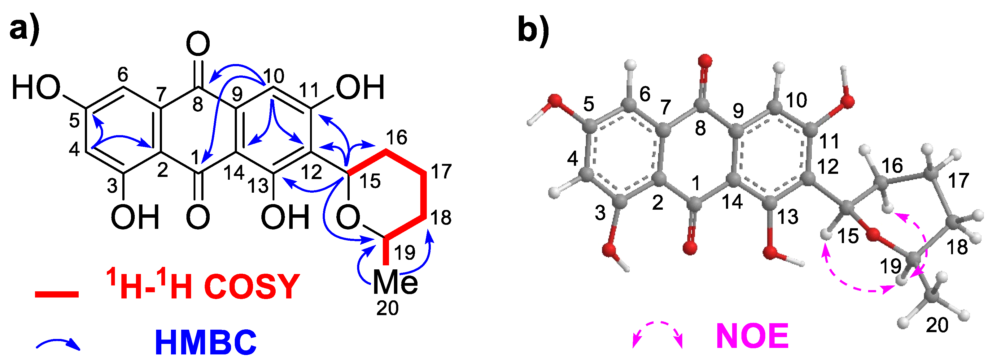
| Position | 3 a | |
|---|---|---|
| δH, multi. (J) | δC, type | |
| 1 | 188.5, C | |
| 2 | 108.4, C | |
| 3 | 165.5, C | |
| 4 | 6.51 d (1.2) | 108.0, CH |
| 5 | 164.3, C | |
| 6 | 7.03 br s | 109.0, CH |
| 7 | 134.7, C | |
| 8 | 181.0, C | |
| 9 | 133.1, C | |
| 10 | 7.00 s | 109.0, CH |
| 11 | 161.5, C | |
| 12 | 119.8, C | |
| 13 | 162.9, C | |
| 14 | 108.2, C | |
| 15 | 4.94 d (10.8) | 73.3, CH |
| 16 | 1.60 m, 1.93 m | 28.3, CH2 |
| 17 | 1.63 m, 1.87 m | 23.4, CH2 |
| 18 | 1.30 m, 1.65 m | 32.5, CH2 |
| 19 | 3.61 m | 74.6, CH |
| 20 | 1.18 d (6.0) | 21.9, CH3 |
| Compounds | IC50 (μΜ) ± SD | |||
|---|---|---|---|---|
| HepG2 | LLC | B16-F10 | MCF7 | |
| 1 | >50.0 | >50.0 | >50.0 | >50.0 |
| 2 | 8.83 ± 3.43 | 14.18 ± 3.84 | 15.12 ± 1.45 | >50.0 |
| 3 | 39.86 ± 1.27 | >50.0 | 48.63 ± 1.20 | >50.0 |
| MS-275 a | 1.01 ± 0.25 | 5.36 ± 1.05 | 4.00 ± 0.28 | 14.74 ± 0.44 |
Disclaimer/Publisher’s Note: The statements, opinions and data contained in all publications are solely those of the individual author(s) and contributor(s) and not of MDPI and/or the editor(s). MDPI and/or the editor(s) disclaim responsibility for any injury to people or property resulting from any ideas, methods, instructions or products referred to in the content. |
© 2023 by the authors. Licensee MDPI, Basel, Switzerland. This article is an open access article distributed under the terms and conditions of the Creative Commons Attribution (CC BY) license (https://creativecommons.org/licenses/by/4.0/).
Share and Cite
Wu, B.; Xu, C.; Chen, J.; Chen, G. Rhizoaspergillin A and Rhizoaspergillinol A, including a Unique Orsellinic Acid–Ribose–Pyridazinone-N-Oxide Hybrid, from the Mangrove Endophytic Fungus Aspergillus sp. A1E3. Mar. Drugs 2023, 21, 598. https://doi.org/10.3390/md21110598
Wu B, Xu C, Chen J, Chen G. Rhizoaspergillin A and Rhizoaspergillinol A, including a Unique Orsellinic Acid–Ribose–Pyridazinone-N-Oxide Hybrid, from the Mangrove Endophytic Fungus Aspergillus sp. A1E3. Marine Drugs. 2023; 21(11):598. https://doi.org/10.3390/md21110598
Chicago/Turabian StyleWu, Binbin, Chenglong Xu, Jianjun Chen, and Guangying Chen. 2023. "Rhizoaspergillin A and Rhizoaspergillinol A, including a Unique Orsellinic Acid–Ribose–Pyridazinone-N-Oxide Hybrid, from the Mangrove Endophytic Fungus Aspergillus sp. A1E3" Marine Drugs 21, no. 11: 598. https://doi.org/10.3390/md21110598