Article

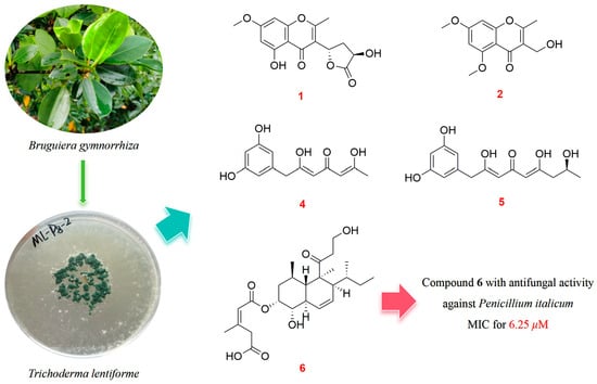
The Polyketides with Antimicrobial Activities from a Mangrove Endophytic Fungus Trichoderma lentiforme ML-P8-2

School of Chemistry, Sun Yat-sen University, Guangzhou 510006, China
*
Author to whom correspondence should be addressed.
Mar. Drugs 2023, 21(11), 566; https://doi.org/10.3390/md21110566
Submission received: 16 October 2023 / Revised: 26 October 2023 / Accepted: 27 October 2023 / Published: 28 October 2023
(This article belongs to the Special Issue Bioactive Secondary Metabolites of Marine Fungi 2.0)

Abstract

:
Five new polyketides, including two chromones (12), two phenyl derivatives (45), and a tandyukusin derivative (6), along with five known polyketides (3 and 710) were isolated from mangrove endophytic fungus Trichoderma lentiforme ML-P8-2. The planar structures of compounds were elucidated via detailed 1D, 2D NMR, and HR-ESI-MS analysis. ECD spectra, optical rotation values calculation, and alkali hydrolysis were applied in the determination of the absolute configuration of the new compounds. In bioassays, 6 and 9 exhibited promising antifungal activities against Penicillium italicum, with an MIC value of 6.25 μM for both compounds. Moreover, 3 displayed moderate AChE inhibitory activity with an IC50 value of 20.6 ± 0.3 μM.

Graphical Abstract

1. Introduction

Mangrove endophytic fungi are an important source to provide biologically active lead compounds due to their unique living environment. Increasing numbers of secondary metabolites from mangrove-associated fungi have been newly reported in recent decades [1,2]. Trichoderma species have been widely discovered in marine environments, including soil, decaying wood, and living plants in mangrove forests [3,4]. Over 450 metabolites have been structurally documented from the species of Trichoderma genus [4,5], wherein polyketides are considered a significant group of these metabolites. Lai et al. reported the isolation of two new chromone polyketides with a broad spectrum of antimicrobial activities from Trichoderma sp. JWM29-10-1 [6]. Yamada et al. documented six decalin polyketides named tandyukusins from Trichoderma harzianum OUPS-111D-4, some of which exhibited significant cytotoxicities against human cancer cell lines [7,8,9]. Polyketides have attracted extensive attention due to their diverse chemical structures and wide range of biological activities [3,4,10].
As part of our ongoing research for bioactive compounds from mangrove endophytic fungi [11,12,13], the study on chemical constituents of the ethyl acetate (EA) extract of the culture media of fungus Trichoderma lentiforme ML-P8-2 led to the isolation of ten polyketides (Figure 1), including five undescribed compounds (12 and 46) and five known compounds (3 and 710). The known compounds were identified as 5-hydroxy-3-(hydroxymethyl)-7-methoxy-2-methyl-4H-chromen-4-one (3) [14], trichoharzin (7) [15], tandyukisin D (8) [8], tandyukisin G (9) [6], and tandyukisin C (10) [8]. Antimicrobial, acetylcholinesterase (AChE) inhibitory, and cytotoxic activities of all isolated compounds were tested.
Herein, we report the detailed structural identification for the new compounds (12 and 46) and bioactivities results.

2. Results and Discussion

2.1. Structure Identification

Compound 1 was obtained as a light-yellow oil. Its molecular formula C15H14O7 with nine degrees of unsaturation was deduced by the ion peak of HR-ESI-MS m/z [M + Na]+ 329.0627 (calcd. for C15H14O7Na+, 329.0632). The 1H NMR spectrum showed two aromatic protons at δH 6.49 and 6.31, two oxymethines at δH 5.73 and 4.90, a methoxyl at δH 3.86, a methylene at δH 2.75 and 2.48, and a methyl at δH 2.52. The 13C NMR spectrum showed a conjugated carbonyl at δC 182.1, an ester carbonyl at δC 180.2, four oxygenated olefinic carbons at δC 168.0, 167.4, 163.2, and 159.0, two quaternary carbons at δC 117.8 and 105.1, two protonated olefinic carbons at δC 99.3 and 93.3, two oxymethines at δC 74.9 and 69.5, a methoxyl at δC 56.5, a methylene at δC 36.2, and a methyl at δC 18.1. The 1H–1H correlation spectroscopy (COSY) signal of H-6/H-8 and HMBC correlations from H-6 to C-4a, C-5, and C-7; H-8 to C-4a, C-7, and C-8a; H3-9 to C-7; and H3-10 to C-2 and C-3 revealed the presence of a 5-hydroxy-7-methoxy-2-methyl-4H-chromen-4-one moiety. Under the assistance of degrees of unsaturation, the 1H–1H COSY signals of H-3’/H-4’/H-5’ together with HMBC correlations from H-3’ to C-2’; H2-4’ to C-2’; H-5’ to C-2’ led to the identification of a 3’-hydroxy-2’-oxotetrahydrofuran moiety. And the above two moieties were connected via a C-3–C-5’ bond, according to HMBC correlations from H-5’ to C-2, C-3, C-4. Thus, the planar structure of 1 was established, as shown in Figure 2.
The calculated ECD curves of all four possible configurations and the experimental ECD spectrum of 1 are shown in Figure 3. Only the ECD curve of (3’R, 5’S)-1 was better matched with the experimental one, and the absolute configuration of 1 was assigned to be 3’R, 5’S. Finally, the structure of 1 was determined to be 5-hydroxy-3-((3’R, 5’S)-3’-hydroxy-2’-oxotetrahydrofuran-5’-yl)-7-methoxy-2-methyl-4H-chromen-4-one.
Compound 2 was isolated as a light-yellow oil. Its molecular formula C13H14O5 with seven degrees of unsaturation was deduced by the ion peak of HR-ESI-MS m/z [M + Na]+ 273.0734 (calcd. for C13H14O5Na+, 273.0733). The 1H NMR spectrum showed two aromatic protons at δH 6.59 and 6.50, an oxymethylene at δH 4.57, two methoxy signals at δH 3.91 and 3.90, and a methyl at δH 2.48. The 13C NMR spectrum showed a conjugated carbonyl at δC 178.2, four oxygenated olefinic carbons at δC 166.1, 165.5, 162.2, and 161.1, two quaternary carbons at δC 121.6 and 109.0, two protonated olefinic carbons at δC 97.1 and 93.9, two methoxys at δC 56.5 and 56.4, an oxymethylene at δC 55.5, and a methyl at δC 17.8. Comparing the 1D NMR data of 2 and 3, 2 was highly similar to 3 [14] but possessed one more methoxyl. The 2D NMR data (Figure 2), especially the HMBC correlation signal from H3-12 to C-5, indicated that 5-OH was replaced by 5-OCH3, determining the structure of 2, as 3-(hydroxymethyl)-5,7-dimethoxy-2-methyl-4H-chromen-4-one.
Compound 4 was acquired as a yellow oil. Its molecular formula C13H14O5 with seven degrees of unsaturation was deduced by the ion peak of HR-ESI-MS m/z [M + Na]+ 273.0734 (calcd. for C13H14O5Na+, 273.0733). The 1H NMR spectrum acquired in DMSO-d6 showed two exchangeable protons at δH 9.22, four aromatic protons at δH 6.12, 6.10, 6.07, and 6.04, a methylene adjacent to olefinic carbons at δH 3.65, and a methyl δH 2.20. The 13C NMR spectrum showed a conjugated carbonyl at δC 178.7, four oxygenated olefinic carbons at δC 167.6, 165.8, and 158.5 (2C), a quaternary carbon at δC 137.5, five protonated olefinic carbons at δC 113.2, 113.2, 106.9 (2C), and 101.2, a methylene adjacent to olefinic carbons at δC 38.7, and a methyl at δC 19.2. The HMBC correlation signals from H-3 to C-1, C-2, C-4, and C-5, and H-1 and H-5 to C-6 indicated the presence of a 2,4-dihydroxyphenyl moiety. HMBC correlations from H-9 to C-7, C-8, C-10, and C-11; H-11 to C-12; H3-13 to C-11 and C-12 constructed an 8,12-dihydroxyhepta-8,11-dien-10-one moiety. The two moieties connected with C-7 were determined via the analysis of HMBC correlations from H2-7 to C-1, C-5, and C-6. The NOESY correlations of H2-7/H-9 and H-11/H3-13 revealed that these protons were on the same side. Thus, the structure of 4 was established as (8Z,11Z)-7-(2,4-dihydroxyphenyl)-8,12-dihydroxyhepta-8,11-dien-10-one.
Compound 5 was isolated as a yellow oil. Its molecular formula C15H18O6 with seven degrees of unsaturation was deduced by the ion peak of HR-ESI-MS m/z [M − H2O + H]+ 277.1070 (calcd. for C15H17O5+, 277.1071). The 1H NMR spectrum acquired in DMSO-d6 showed three exchangeable protons at δH 9.22 (2H) and 4.80, four aromatic protons at δH 6.12, 6.10, 6.05, and 6.04, an oxymethine at δH 3.90, two methylenes at δH 3.64 and 2.52, and a methyl at δH 1.06. The 13C NMR spectrum showed a conjugated carbonyl at δC 178.7, four oxygenated olefinic carbons at δC 167.7, 166.9, and 158.5 (2C), a quaternary carbon at δC 137.5, five protonated olefinic carbons at δC 114.0, 113.3, 106.9 (2C), and 101.2, an oxymethine at δC 64.0, two methylenes at δC 42.6 and 38.8, and a methyl at δC 23.2. Compared with 4, compound 5 was identified to possess an iso-propanol group instead of a methyl connecting to C-12, which was verified via the analyses of 1H–1H COSY signals H2-13/H-14/H3-15 and HMBC correlations from 14-OH to C-13, C-14, and C-15; H2-13 to C-11 and C-12. The NOESY correlations H2-7/H-9 and H-11/H2-13 revealed that these protons were on the same side. Therefore, the planar structure of 5 was established, as shown in Figure 2.
To identify the configuration of the only chiral center of 5, C-14, experimental and calculated optical rotation values were obtained. The experimental optical rotation value [ α ] D 25 +23.5 (c 0.26, MeOH) was matched with the calculated one for 14S-5, [ α ] D 25 +29.0 (MeOH). Also, the calculated ECD spectrum of 14S-5 showed a good agreement with the experimental one, as shown in Figure 3. Accordingly, the structure of 5 was determined to be (14S,8Z,11Z)-7-(2,4-dihydroxyphenyl)-8,12,14-trihydroxynona-8,11-dien-10-one.
Compound 6 was isolated as a pale-yellow oil. Its molecular formula C25H38O7 with seven degrees of unsaturation was deduced by the ion peak of HR-ESI-MS m/z [M + Na]+ 473.2509 (calcd. for C25H38O7Na+, 473.2510). Analyses of the 1H and 13C NMR data aided with HSQC revealed the presence of three carbonyls, two quaternary carbons, ten methines, five methylenes, and five methyls (NMR data in Table 1). A comparison with the NMR data of tandyukusin D (8) [8] indicated that 6 and 8 shared the same core structure, eujavanicol A [16], and the main difference occurred on the side chain. With the key HMBC from H-2’ to C-4’ and C-6’; H2-4’ to C-3’, C-5’, and C-6’; H3-6’ to C-2’, C-3’, and C-4’; and NOESY correlation H-2’/H3-6’, the side chain was established as a (2’Z)-3’-methyl-2’-pentenedioic acid moiety. The 13C resonance of C-6’ at δC 26.2 (larger than 20 ppm) additionally supported the Z configuration of the double bond on the side chain [6,17]. The HMBC from H-8 to C-1’ attached the side chain at C-8 of the eujavanicol A fragment.
Further, the relative configuration of the eujavanicol A fragment was identified using the key NOESY correlations of H3-19 with H-6, H-10, and H-13 and of H-13 with H3-17, indicating these protons were on one face (Figure 4). The NOESY correlations of H-5 with H-9 and H3-18 suggested these protons were on the other face, and the decalin ring was trans. And 1H coupling constants J7α,8 = J7β,8 = J9,8 = 2.5 Hz showed that 9-OH was oriented cis to the esterified side chain at C-8 [7,8,9,15,16]. As for the configuration of C-14 at the sec-butyl group, the observed NOESY correlations in 6 were equal to those of 710 [6,8,15], which revealed that the rotation of the sec-butyl group in its pseudo-axial arrangement was restricted. Therefore, the relative configuration of C-14 was identified as R*. As a result, the relative configuration of 6 was determined as 4S*, 5S*, 6R*, 8R*, 9S*, 10R*, 13S*, and 14R*, in line with that in 710. To determine the absolute configuration of 6, treatments of 6 and tandyukusin D (8) with NaOH aqueous in MeOH were carried out, and the reactions resulted in the acquisition of eujavanicol A (Figure 4), which was identified according to its 1H, 13C NMR and HR-ESI-MS data (Figures S40–S42). Comparing the optical rotation values of products (eujavanicol A) from 6 and 8 with previous article [16] (respectively, for [ α ] D 25 +41.8, [ α ] D 25 +39.7, and [ α ] D 25 +49.9), it could be confirmed that the absolute configuration of 6 was 4S, 5S, 6R, 8R, 9S, 10R, 13S, and 14R. Finally, the structure of 6 was determined and named Tandyukisin J.

2.2. Antimicrobial Assays

The isolated compounds 110 along with the alkali hydrolysis treatment product eujavanicol A were evaluated for antibacterial activities against methicillin-resistant Staphylococcus aureus (MRSA), Staphylococcus aureus, Bacillus subtilis, Salmonella typhimurium, and Pseudomonas aeruginosa and for antifungal activities against Candida albicans and an agricultural plant pathogenic fungus Penicillium italicum. The results showed that the decalin derivatives (610 and eujavanicol A) exhibited promising inhibitory activities against the fungi, with a minimal inhibition concentration (MIC) value in the range of 6.25 to 50 μM (Table 2), wherein 6 and 9 showed significant antifungal activities against P. italicum, with an MIC value of 6.25 μM for both compounds. And the chromone derivative 3 displayed moderate inhibitory activity against C. albicans, with an MIC value of 25 μM.

2.3. AChE Inhibitory Activity Assays

The isolated compounds 110, along with the alkali hydrolysis treatment product eujavanicol A, were also evaluated for AChE inhibitory activities. The results showed that the chromone derivative 3 moderately inhibited AChE with IC50 values for 20.6 ± 0.3 μM, but the other tested compounds mostly exhibited weak inhibitory activities toward AChE (Table 3).

2.4. Cytotoxic Assays

The isolated compounds 110, along with the alkali hydrolysis treatment product eujavanicol A, were also tested for cytotoxicities against six human cancer cell lines, which were MDA-MB-435, MDA-MB-231, HCT116, A549, SNB19, and PC3. But, only compound 1 exhibited a weak cytotoxicity against A549 with an IC50 value of 47.2 ± 5.5 μM, and the other compounds were inactive to the tested cell lines (IC50 > 50 μM).

3. Experimental Section

3.1. General Experimental Procedures

The 1D and 2D NMR spectra were obtained on a Bruker Advance 400 MHz spectrometer (Billerica, MA, USA) at room temperature. HR-ESI-MS spectra were acquired from a Thermo Fisher LTQ-Orbitrap-LCMS spectrometer (Palo Alto, Santa Clara, CA, USA). Optical rotation values were measured on an MCP500 modular polarimeter (Anton Paar, North Ryde, Austria) at 25 °C. UV-Vis and ECD curves were achieved on an Applied Photophysics Chirascan spectropolarimeter (Surrey, UK). Semi-preparative HPLC was utilized on an Ultimate 3000 separation module combined with a DAD detector produced by Thermo Fisher, and a ChiralPak AY-H column (5 μm, 4.6 × 250 mm) was applied for separation at 22 °C. Organic solvent was evaporated using a vacuum pump with a Heidolph rotavapor.

3.2. Fungal Material

The fungus Trichoderma lentiforme ML-P8-2 was isolated from a fresh leaf of the mangrove plant Bruguiera gymnorrhiza, which was collected in July 2022 from Dong Zhai Gang National Nature Reserve in Hainan Province, China. The fungus strain was identified according to sequencing of the internal transcribed spacer, and the results of a BLAST search on National Center for Biotechnology Information (NCBI) revealed it was most similar (99%) to the sequence of Trichoderma lentiforme (compared to MK714910.1). The sequence data have been uploaded and deposited at GenBank with accession No. OR617437. And the fungus specimen was kept in our laboratory at −20 °C.

3.3. Fermentation, Extraction, and Isolation

The fungus ML-P8-2 was proliferated in potato dextrose broth (PDB) in 4 × 500 mL Erlenmeyer flasks at 28 °C for 4 days in a shaker and then cultured in 150 × 1 L Erlenmeyer flasks, each containing 60 mL of 0.3% saline and 60 g of rice. After fermentation for 28 days, the culture media were soaked with MeOH and extracted with EA three times after concentration. Then, the extracts were condensed under 45 °C with a vacuum pump to finally obtain a crude extract (61 g). The crude extract was separated using a silica gel column, eluting with a gradient of petroleum ether (PE)/EA from 1:0 to 0:1 to afford 7 fractions (Frs. 1–7).
Frs. 7 (10.6 g) was subjected to Sephadex LH-20 [dichloromethane (DCM)/MeOH, v/v, 1:1] to yield five subfractions (SFrs. 7.1–7.5). SFrs. 7.3 (5.4 g) was applied to silica gel column chromatography (CC) (DCM/MeOH, v/v, 20:1) to give a mixture of 1, 4, and 5 (24 mg). The mixture was isolated using reverse phase C18 silica gel column (40−60 μm, MeOH/H2O, v/v, 8:2) to yield 1 (5.1 mg), 4 (2.3 mg), and 5 (4.5 mg). Frs. 3 (13.2 g) was subjected to Sephadex LH-20 (DCM/MeOH, v/v, 1:1) to yield three subfractions (SFrs. 3.1–3.3). SFrs. 3.3 (4.6 g) was applied to silica gel CC (DCM/MeOH, v/v, 200:1) to give mixture of 2 and 3 (26 mg). The mixture was isolated utilizing HPLC with the ChiralPak AY-H column, respectively, at tR = 6.0 and 15.5 min (the gradient was n-hexane/2-propanol v/v, 9:1; flow rate: 1 mL/min) to give 2 (3.2 mg) and 3 (20.6 mg). Frs. 4 (15.5 g) was subjected to Sephadex LH-20 (DCM/MeOH, v/v, 1:1) to yield three subfractions (SFrs. 4.1–4.3). SFrs. 4.2 (6.0 g) was applied to silica gel CC (DCM/MeOH, v/v, 100:1) to give mixture of 6−10 (126 mg). The mixture was isolated utilizing HPLC with the ChiralPak AY-H column, respectively, at tR = 15.0, 18.5, 9.0, 12.5, and 4.5 min (the gradient was n-hexane/2-propanol v/v, 85:15; flow rate: 1 mL/min) to give 6 (10.2 mg), 7 (35.7 mg), 8 (34.3 mg) 9 (12.3 mg), and 10 (6.5 mg).
5-hydroxy-3-((3’R, 5’S)-3’-hydroxy-2’-oxotetrahydrofuran-5’-yl)-7-methoxy-2-methyl-4H-chromen-4-one (1): C15H14O7; Light-yellow oil; [ α ] D 25 +20.5 (c 0.35, MeOH); UV (MeOH) λmax (log ε): 206 (4.08), 249 (3.97), 258 (3.96), 291 (3.49) nm; HR-ESI-MS: m/z [M + Na]+ 329.0627 (calcd. for C15H14O7Na+, 329.0632); 1H and 13C NMR data (Table 4). Spectra in Figures S1–S8.
3-(hydroxymethyl)-5,7-dimethoxy-2-methyl-4H-chromen-4-one (2): C13H14O5; Light-yellow oil; UV (MeOH) λmax (log ε): 201 (3.81), 230 (3.98), 280 (3.51) nm; HR-ESI-MS: m/z [M + Na]+ 273.0734 (calcd. for C13H14O5Na+, 273.0733); 1H and 13C NMR data (Table 4). Spectra in Figures S9–S15.
(8Z,11Z)-7-(2,4-dihydroxyphenyl)-8,12-dihydroxyhepta-8,11-dien-10-one (4): C13H14O5; Yellow oil; UV (MeOH) λmax (log ε): 201 (4.12), 241 (3.74), 276 (3.04) nm; HR-ESI-MS: m/z [M + Na]+ 273.0734 (calcd. for C13H14O5Na+, 273.0733); 1H and 13C NMR data (Table 5). Spectra in Figures S16–S23.
(14S,8Z,11Z)-7-(2,4-dihydroxyphenyl)-8,12,14-trihydroxynona-8,11-dien-10-one (5): C15H18O6; Yellow oil; [ α ] D 25 +23.5 (c 0.26, MeOH); UV (MeOH) λmax (log ε): 201 (4.18), 246 (3.76), 277 (3.02) nm; HR-ESI-MS: m/z [M − H2O + H]+ 277.1070 (calcd. for C15H17O5+, 277.1071); 1H and 13C NMR data (Table 5). Spectra in Figures S24–S31.
Tandyukisin J (6): C25H38O7; Pale-yellow oil; [ α ] D 25 +17.6 (c 0.45, MeOH); UV (MeOH) λmax (log ε): 215 (3.82) nm; HR-ESI-MS: m/z [M + Na]+ 473.2509 (calcd. for C25H38O7Na+, 473.2510); 1H and 13C NMR data (Table 1). Spectra in Figures S32–S39.

3.4. Alkali-hydrolysis Treatments for Compounds 6 and 8

Compound 6 (3.0 mg, 6.7 μmol) in MeOH (200 μL) was stirred evenly for 5 min, and then 200 μL of aqueous NaOH (1.0 M) was added. The reaction mixture was stirred for 30 min at room temperature. After completion, the reaction mixture was extracted with EA thrice, and the organic layer was concentrated under reduced pressure to afford eujavanicol A (1.9 mg). In order to determine its absolute configuration and exclude potential differences in test conditions with the previous articles, the known compound 8 (5.0 mg) was also hydrolyzed to produce eujavanicol A (3.2 mg) following the same procedure.

3.5. ECD and Optical Rotation Computation Methods

Initial conformational analysis was carried out using the Merck molecular force field method with the Spartan 14’ software (Wavefunction Inc., Irvine, CA, USA). The conformation with a Boltzmann population larger than 1% was selected for optimization and calculation in MeOH at B3LYP/6-31+G(d,p) level with the density functional theory (DFT) executed via Gaussian 09 [18]. The ECD spectra and optical rotation values were extracted and generated via the SpecDis 1.6 software (University of Würzburg, Würzburg, Germany). The Gibbs free energy, Boltzmann population and Cartesian coordinates for low-energy conformers of 1 and 5 for calculation in Tables S1–S6.

3.6. Antimicrobial Assays

The compounds to be tested were dissolved individually in dimethyl sulfoxide (DMSO), and the antimicrobial activity assays were performed in 96-well plates via a serial dilution test in the range of 0.1–100 μM, according to the methods previously reported [11,19]. All measurements were conducted in triplicate. Ampicillin and ketoconazole were applied as positive controls for antibacterial and antifungal assays, respectively, and DMSO was utilized as a blank control.

3.7. AChE Inhibition Assays

Compounds 110 and eujavanicol A were evaluated for AChE inhibitory activity, following the same method previously described [12]. Donepezil hydrochloride was taken as a positive control. All measurements were conducted in triplicate from two independent experiments. The reported IC50 was the average value of two independent experiments.

3.8. Cytotoxic Assays

The cytotoxicities of compounds 110 and eujavanicol A on cells were assessed using MTT assay as described previously [13]. Six cell lines were used, MDA-MB-435 (breast cancer cells), MDA-MB-231 (breast cancer cells), HCT116 (colon cancer cells), A549 (lung cancer cells), SNB19 (glioma cells), and PC3 (prostate cancer cells), which were acquired from Fu Erbo Biotechnology Co., Ltd. (Guangzhou, China).

4. Conclusions

In summary, five new polyketides, including two chromones (12), two phenyl derivatives (45), and a tandyukusin derivative (6), along with five known polyketides (3 and 710), were isolated from mangrove endophytic fungus Trichoderma lentiforme ML-P8-2, and it is the first time to report secondary metabolites from this specific species. The planar structures of the isolated compounds were elucidated via detailed 1D, 2D NMR, and HR-ESI-MS analysis. ECD spectra, optical rotation values calculation, and alkali hydrolysis were applied in the determination of the absolute configuration of the new compounds. In bioassays, antimicrobial, AChE inhibitory, and cytotoxic activities tests for compounds 110, together with the alkali-hydrolysis treatment product eujavanicol A, were carried out. Compounds 6 and 9 exhibited promising antifungal activities against Penicillium italicum, with MIC, both for 6.25 μM. Moreover, 3 displayed moderate AChE inhibitory activity with an IC50 value of 20.6 ± 0.3 μM.

Supplementary Materials

The following supporting information can be downloaded at: https://www.mdpi.com/article/10.3390/md21110566/s1, the 1D, 2D NMR, HR-ESI-MS, and UV-vis spectra of compound 1 (Figures S1–S8), compound 2 (Figures S9–S15), compound 4 (Figures S16–S23), compound 5 (Figures S24–S31), and compound 6 (Figures S32–S39); the 1D, 2D NMR, and HR-ESI-MS spectra of the alkali hydrolysis treatment product eujavanicol A (Figures S40–S42); the Gibbs free energy, Boltzmann population and Cartesian coordinates for low-energy conformers of 1 and 5 for calculation (Tables S1–S6).

Author Contributions

Y.Y. performed the large-scale fermentation and isolated all compounds. Y.Y. and Q.T. carried out the structure identification and edited the manuscript. J.W. and T.C. carried out the biological assays. W.Y. participated in the experiments. Z.S. and B.W. designed and supervised this research. All authors have read and agreed to the published version of the manuscript.”

Funding

This research was generously funded by the Guangdong Marine Economy Development Special Project (GDNRC[2022]35 and GDNRC[2023]39), and the National Natural Science Foundation of China (U20A2001 and 42276114).

Institutional Review Board Statement

Not applicable.

Informed Consent Statement

Not applicable.

Data Availability Statement

Data are contained within this article and Supplementary Materials.

Acknowledgments

We acknowledge the South China Sea Institute of Oceanology for the generous support of ECD equipment.

Conflicts of Interest

The authors declare no conflict of interest.

References

  1. Xu, J. Bioactive natural products derived from mangrove-associated microbes. RSC Adv. 2015, 5, 841–892. [Google Scholar] [CrossRef]
  2. Chen, S.H.; Cai, R.L.; Liu, Z.M.; Cui, H.; She, Z.G. Secondary metabolites from mangrove-associated fungi: Source, chemistry and bioactivities. Nat. Prod. Rep. 2022, 39, 560–595. [Google Scholar] [CrossRef]
  3. Guo, R.; Li, G.; Zhang, Z.; Peng, X. Structures and biological activities of secondary metabolites from Trichoderma harzianum. Mar. Drugs 2022, 20, 701. [Google Scholar] [CrossRef]
  4. Guo, Q.; Shi, L.; Wang, X.; Li, D.; Yin, Z.; Zhang, J.; Ding, G.; Chen, L. Structures and biological activities of secondary metabolites from the Trichoderma genus (covering 2018–2022). J. Agric. Food Chem. 2023, 71, 13612–13632. [Google Scholar] [CrossRef] [PubMed]
  5. Su, D.Q.; Ding, L.J.; He, S. Marine-derived Trichoderma species as a promising source of bioactive secondary metabolites. Mini-Rev. Med. Chem. 2018, 18, 1702–1713. [Google Scholar] [CrossRef]
  6. Lai, C.; Chen, J.; Liu, J.; Tian, D.; Lan, D.; Liu, T.; Wu, B.; Bi, H.; Tang, J. New polyketides from a hydrothermal vent sediment fungus Trichoderma sp. JWM29-10-1 and their antimicrobial effects. Mar. Drugs 2022, 20, 720. [Google Scholar] [CrossRef]
  7. Yamada, T.; Mizutani, Y.; Umebayashi, Y.; Inno, N.; Kawashima, M.; Kikuchi, T.; Tanaka, R. Tandyukisin, a novel ketoaldehyde decalin derivative, produced by a marine sponge-derived Trichoderrna harzianum. Tetrahedron Lett. 2014, 55, 662–664. [Google Scholar] [CrossRef]
  8. Yamada, T.; Umebayashi, Y.; Kawashima, M.; Sugiura, Y.; Kikuchi, T.; Tanaka, R. Determination of the chemical structures of tandyukisins B-D, isolated from a marine sponge-derived fungus. Mar. Drugs 2015, 13, 3231–3240. [Google Scholar] [CrossRef] [PubMed]
  9. Suzue, M.; Kikuchi, T.; Tanaka, R.; Yamada, T. Tandyukisins E and F, novel cytotoxic decalin derivatives isolated from a marine sponge-derived fungus. Tetrahedron Lett. 2016, 57, 5070–5073. [Google Scholar] [CrossRef]
  10. Robertsen, H.L.; Musiol-Kroll, E.M. Actinomycete-derived polyketides as a source of antibiotics and lead structures for the development of new antimicrobial drugs. Antibiotics 2019, 8, 157. [Google Scholar] [CrossRef] [PubMed]
  11. Yin, Y.; Yang, W.; Chen, T.; Tan, Q.; Zou, G.; Zang, Z.; Li, J.; Wang, B.; She, Z. Cytosporones W and X: Two mutually converting epimers from a mangrove endophytic fungus Diaporthe sp. ZJHJYZ-1. ACS Omega 2023, 8, 26628–26634. [Google Scholar] [CrossRef] [PubMed]
  12. Xiao, Z.; Huang, H.; Shao, C.; Xia, X.; Ma, L.; Huang, X.; Lu, Y.; Lin, Y.; Long, Y.; She, Z. Asperterpenols A and B, new sesterterpenoids isolated from a mangrove endophytic fungus Aspergillus sp. 085242. Org. Lett. 2013, 15, 2522–2525. [Google Scholar] [CrossRef] [PubMed]
  13. Chen, Y.; Liu, Z.; Huang, Y.; Liu, L.; He, J.; Wang, L.; Yuan, J.; She, Z. Ascomylactams A–C, cytotoxic 12- or 13-membered-ring macrocyclic alkaloids isolated from the mangrove endophytic fungus Didymella sp. CYSK-4, and structure revisions of Phomapyrrolidones A and C. J. Nat. Prod. 2019, 82, 1752–1758. [Google Scholar] [CrossRef] [PubMed]
  14. Takenaka, Y.; Tanahashi, T.; Nagakura, N.; Hamada, N. 2,3-Dialkylchromones from mycobiont cultures of the lichen Graphis scripta. Heterocycles 2000, 53, 1589–1593. [Google Scholar]
  15. Kobayashi, M.; Uehara, H.; Matsunami, K.; Aoki, S.; Kitagawa, I. Trichoharzin, a new polyketide produced by the imperfect fungus Trichoderma harzianum separated from the marine sponge Micale cecilia. Tetrahedron Lett. 1993, 34, 7925–7928. [Google Scholar] [CrossRef]
  16. Nakadate, S.; Nozawa, K.; Horie, H.; Fujii, Y.; Nagai, M.; Hosoe, T.; Kawai, K.I.; Yaguchi, T.; Fukushima, K. Eujavanicols A-C, decalin derivatives from Eupenicillium javanicum. J. Nat. Prod. 2007, 70, 1510–1512. [Google Scholar] [CrossRef] [PubMed]
  17. Duh, C.Y.; Wang, S.K.; Chung, S.G.; Chou, G.C.; Dai, C.F. Cytotoxic cembrenolides and steroids from the formosan soft coral Sarcophyton crassocaule. J. Nat. Prod. 2000, 63, 1634–1637. [Google Scholar] [CrossRef] [PubMed]
  18. Frisch, M.J.; Trucks, G.W.; Schlegel, H.B.; Scuseria, G.E.; Robb, M.A.; Cheeseman, J.R.; Scalmani, G.; Barone, V.; Petersson, G.A.; Nakatsuji, H.; et al. Gaussian 09; Gaussian, Inc.: Wallingford, CT, USA, 2016. [Google Scholar]
  19. Pierce, C.G.; Uppuluri, P.; Tristan, A.R.; Wormley, F.L., Jr.; Mowat, E.; Ramage, G.; Lopez-Ribot, J.L. A simple and reproducible 96-well plate-based method for the formation of fungal biofilms and its application to antifungal susceptibility testing. Nat. Protoc. 2008, 3, 1494–1500. [Google Scholar] [CrossRef]
Figure 1. Structures of compounds 110.
Marinedrugs 21 00566 g001
Figure 2. Two-dimensional NMR correlation signals of new compounds 1, 2, 46.
Marinedrugs 21 00566 g002
Figure 3. (i) Experimental and calculated ECD spectra of 1; (ii) experimental and calculated ECD spectra of 5.
Marinedrugs 21 00566 g003
Figure 4. (i) alkali-hydrolysis reactions of compounds 6 and 8; (ii) key NOESY correlation signals of 6.
Marinedrugs 21 00566 g004
Table 1. 1H and 13C NMR data of 6 (acquired in CDCl3).
Position6Position6
δC, TypeδH (J in Hz)δC, TypeδH (J in Hz)
158.1, CH23.85, ddd (11.0, 6.0, 3.8)
3.93, ddd (11.0, 7.2, 3.4)
1437.3, CH1.14, m
241.3, CH22.69, ddd (18.9, 6.0, 3.4)
2.88, ddd (18.9, 7.2, 3.8)
1524.6, CH20.75, overlap
1.48, d (7.0)
3215.7, C 1612.7, CH30.77, d (4.2)
452.6, C 1719.4, CH30.95, d (6.7)
543.1, CH1.97, d (4.1)1822.4, CH30.60, d (5.8)
631.6, CH1.62, overlap1919.5, CH31.28, s
739.1, CH21.59, overlap
1.89, dd (12.1, 2.8)
1’167.5, C
873.6, CH5.30, m2’119.6, CH6.01, s
974.4, CH3.58, dd (10.9, 3.2)3’151.9, C
1040.4, CH2.12, d (10.9)4’39.8, CH23.63, d (14.6)
3.77, d (14.6)
11125.7, CH6.05, d (10.6)5’172.8, C
12124.1, CH5.72, ddd (10.6, 4.7, 2.6)6’26.2, CH32.06, s
1352.5, CH1.97, d (4.7)
Table 2. MIC for antibacterial and antifungal activities of compounds 110 and eujavanicol A.
MIC of Compounds/μM
12345678910Euj. A 1Amp. 2Ket. 3
MRSA>100>100>100>100>100>100>100>100>100>100>1000.25NT 4
S. aureus>100>100>100>100>100>100>100>10050>100>1000.25NT
B. subtilis>100>100>100>100>100>100>100>100>100>100>1000.25NT
S. typhimurium>100>100>100>100>100>100>10050>100>100>1000.25NT
P. aeruginosa>100>100>100>100>100>100>1005050>100>1000.13NT
C. albicans5010025>100>100255050252525NT0.13
P. italicum>100>100>100>100>1006.2512.512.56.255025NT1.56
1 Eujavanicol A; 2 Ampicillin, positive control toward bacteria; 3 Ketoconazole, positive control toward fungi; 4 Not tested.
Table 3. IC50 for AChE inhibitory activities of compounds 110 and eujavanicol A.
CompoundsIC50/μMCompoundsIC50/μM
138.6 ± 0.2738.3 ± 0.4
233.7 ± 0.4877.9 ± 1.7
320.6 ± 0.3943.6 ± 0.4
437.7 ± 0.61050.9 ± 0.5
551.3 ± 0.5Eujavanicol A32.4 ± 0.7
640.2 ± 0.7Donepezil
Hydrochloride 1
65.5 ± 1.5 (nM)
1 Positive control.
Table 4. 1H and 13C NMR data of 1 and 2 (acquired in CD3OD).
Position12
δC, TypeδH (J in Hz)δC, TypeδH (J in Hz)
2168.0, C 165.5, C
3117.8, C 121.6, C
4182.1, C 178.2, C
4a105.1, C 109.0, C
5163.2, C 162.2, C
699.3, CH6.32, d (2.2)97.1, CH6.50, d (2.3)
7167.4, C 166.1, C
893.3, CH6.49, d (2.2)93.9, CH6.59, d (2.3)
8a159.0, C 161.1, C
956.5, CH33.86, s56.4, CH33.91, s
1018.1, CH32.52, s17.8, CH32.48, s
11 55.5, CH24.57, s
12 56.5, CH33.90, s
2′180.2, C
3′69.5, CH4.90, dd (9.3, 5.9)
4′36.2, CH22.48, m
2.75, ddd (13.5, 9.3, 4.1)
5′74.9, CH5.73, dd (9.9, 4.1)
Table 5. 1H and 13C NMR data of 4 and 5 (acquired in DMSO-d6).
Position45
δC, TypeδH (J in Hz)δC, TypeδH (J in Hz)
1106.9, CH6.12, d (1.9)106.9, CH6.12, d (2.1)
2158.5, C 158.5, C
3101.2, CH6.10, d (1.9)101.2, CH6.10, d (2.1)
4158.5, C 158.5, C
5106.9, CH6.12, d (1.9)106.9, CH6.12, d (2.1)
6137.5, C 137.5, C
738.7, CH23.65, s38.8, CH23.64, s
8167.6, C 167.7, C
9113.2, CH6.04, d (2.3)113.3, CH6.04, d (2.3)
10178.7, C 178.7, C
11113.2, CH6.07, dd (2.3, 0.9)114.0, CH6.05, d (2.3)
12165.8, C 166.9, C
1319.2, CH32.20, d (0.9)42.6, CH22.52, d (3.7)
14 64.0, CH3.90, m
15 23.2, CH31.06, d (6.1)
2-OH 9.22, s 9.22, s
4-OH 9.22, s 9.22, s
14-OH 4.80, d (5.1)
Disclaimer/Publisher’s Note: The statements, opinions and data contained in all publications are solely those of the individual author(s) and contributor(s) and not of MDPI and/or the editor(s). MDPI and/or the editor(s) disclaim responsibility for any injury to people or property resulting from any ideas, methods, instructions or products referred to in the content.

