Article

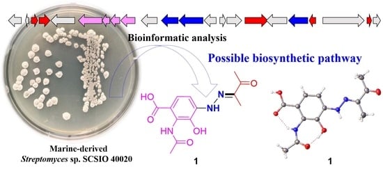
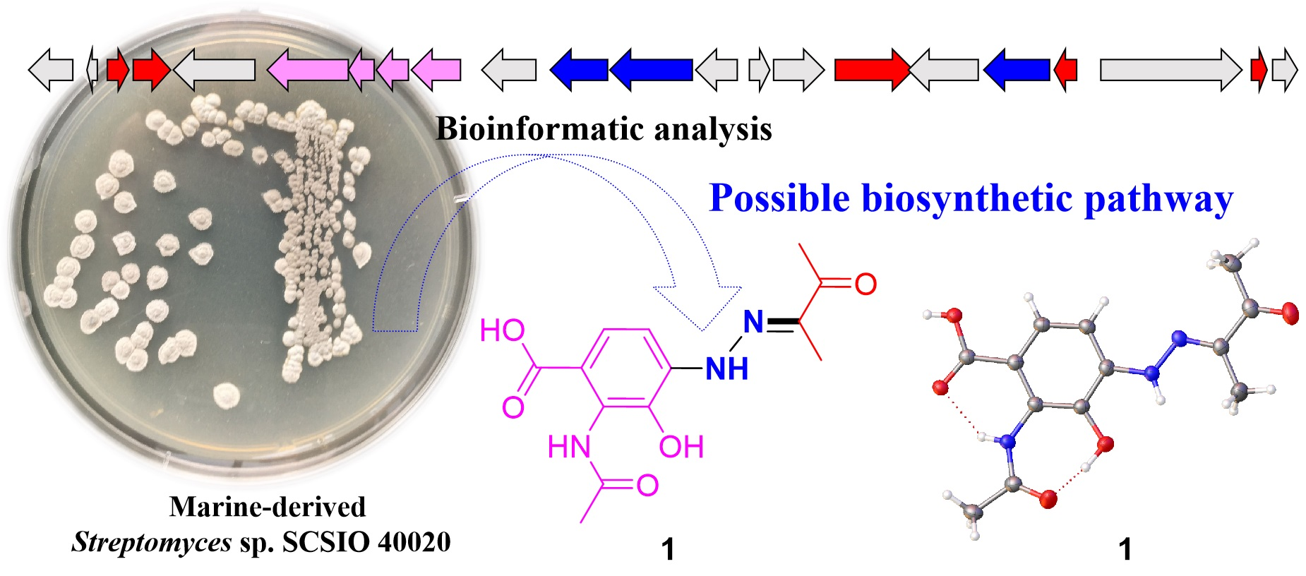
Two New Phenylhydrazone Derivatives from the Pearl River Estuary Sediment-Derived Streptomyces sp. SCSIO 40020

1
Key Laboratory of Tropical Marine Bio-Resources and Ecology, Guangdong Key Laboratory of Marine Materia Medica, South China Sea Institute of Oceanology, Innovation Academy for South China Sea Ecology and Environmental Engineering, Chinese Academy of Sciences, 164 West Xingang Road, Guangzhou 510301, China
2
State Key Laboratory of Applied Microbiology Southern China, Institute of Microbiology, Guangdong Academy of Sciences, 100 Central Xianlie Road, Guangzhou 510070, China
3
Southern Marine Science and Engineering Guangdong Laboratory (Guangzhou), No.1119, Haibin Road, Guangzhou 511458, China
4
University of Chinese Academy of Sciences, Beijing 100049, China
*
Authors to whom correspondence should be addressed.
Mar. Drugs 2022, 20(7), 449; https://doi.org/10.3390/md20070449
Submission received: 13 June 2022 / Revised: 5 July 2022 / Accepted: 7 July 2022 / Published: 9 July 2022

Abstract

:
Two new phenylhydrazone derivatives and one new alkaloid, penzonemycins A–B (12) and demethylmycemycin A (3), together with three known compounds including an alkaloid (4) and two sesquiterpenoids (56), were isolated from the Streptomyces sp. SCSIO 40020 obtained from the Pearl River Estuary sediment. Their structures and absolute configurations were assigned by 1D/2D NMR, mass spectroscopy and X-ray crystallography. Compound 1 was evaluated in four human cancer cell lines by the SRB method and displayed weak cytotoxicity in three cancer cell lines, with IC50 values that ranged from 30.44 to 61.92 µM, which were comparable to those of the positive control cisplatin. Bioinformatic analysis of the putative biosynthetic gene cluster indicated a Japp–Klingemann coupling reaction involved in the hydrazone formation of 1 and 2.

Graphical Abstract

1. Introduction

Hydrazones are a class of compounds with the characteristic structure R1R2C=NNR3R4. Hydrazones display various bioactivities, including antimicrobial, antitubercular, anti-HIV, anti-inflammatory, anticonvulsant, analgesic and antitumor properties [1]. The unique chemical properties and bioactivities of hydrazones have inspired organic chemists to develop numerous methods for the synthesis of their derivatives [2,3]. In contrast to the large number of synthetic hydrazones, only a handful of naturally occurring hydrazones are found [4,5,6,7,8,9] (Figure S1). To date, 38 natural hydrazones have been reported, including 21 from fungi [5,9], 13 from actinomycetes [4,6,7,8], and 4 from other resources [9] (Figure S1).
Marine-derived actinomycetes have been a prolific resource for novel natural product discovery [10,11]. Previously, we reported three novel hydrazone derivatives, pyrazolofluostatins A–C, which were isolated from South China Sea-derived Micromonospora rosaria SCSIO N160 [7]. In this study, we identified two novel phenylhydrazone derivatives, penzonemycins A–B (12), together with one new compound, dibenzoxazepinone demethylmycemycin A (3), and three known compounds (46) (Figure 1) from marine-derived Streptomyces sp. SCSIO 40020 [12]. Herein, we report the isolation and structural elucidation of penzonemycins A–B (12) and demethylmycemycin A (3), as well as a biological evaluation and a possible biosynthetic pathway of penzonemycins A–B (12).

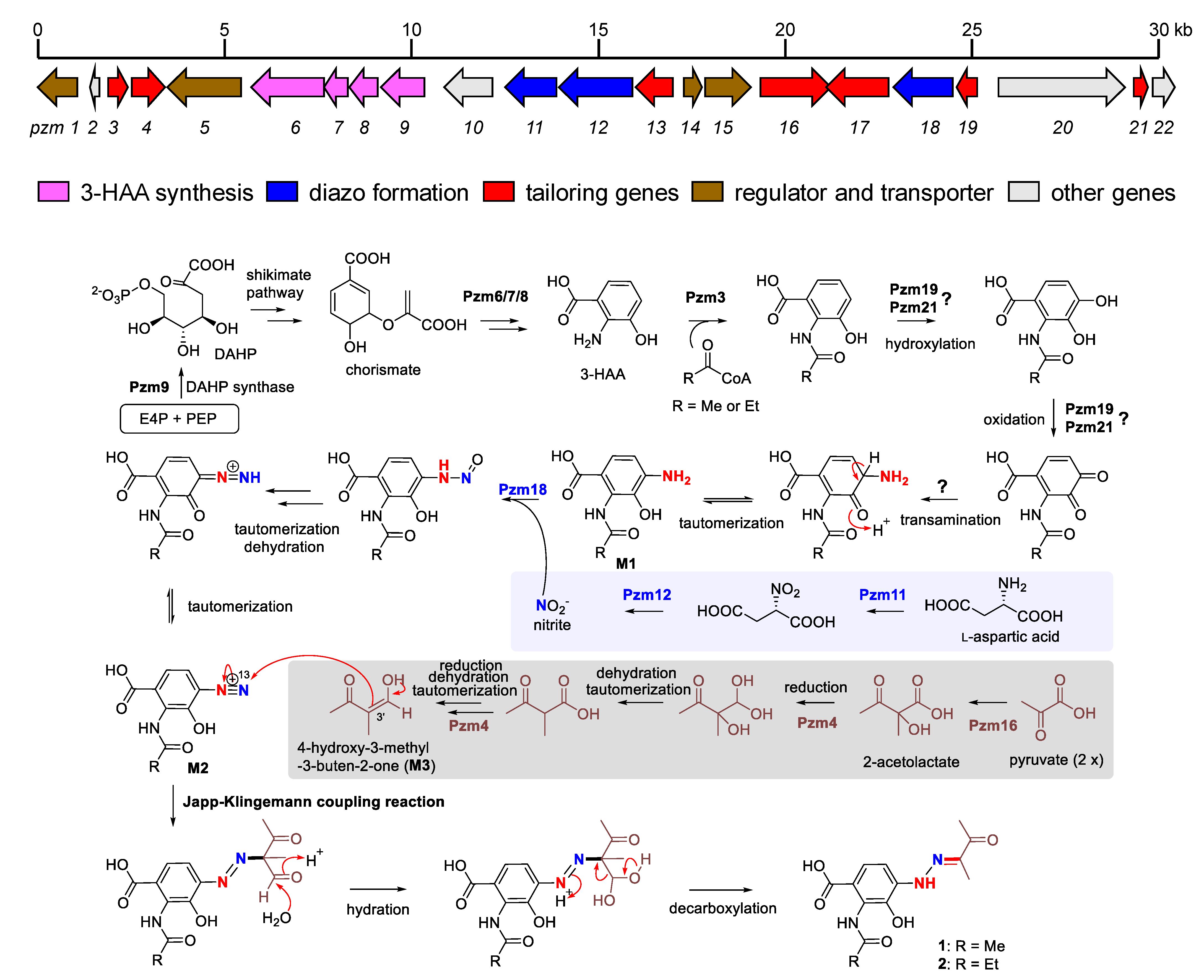
2. Results and Discussion

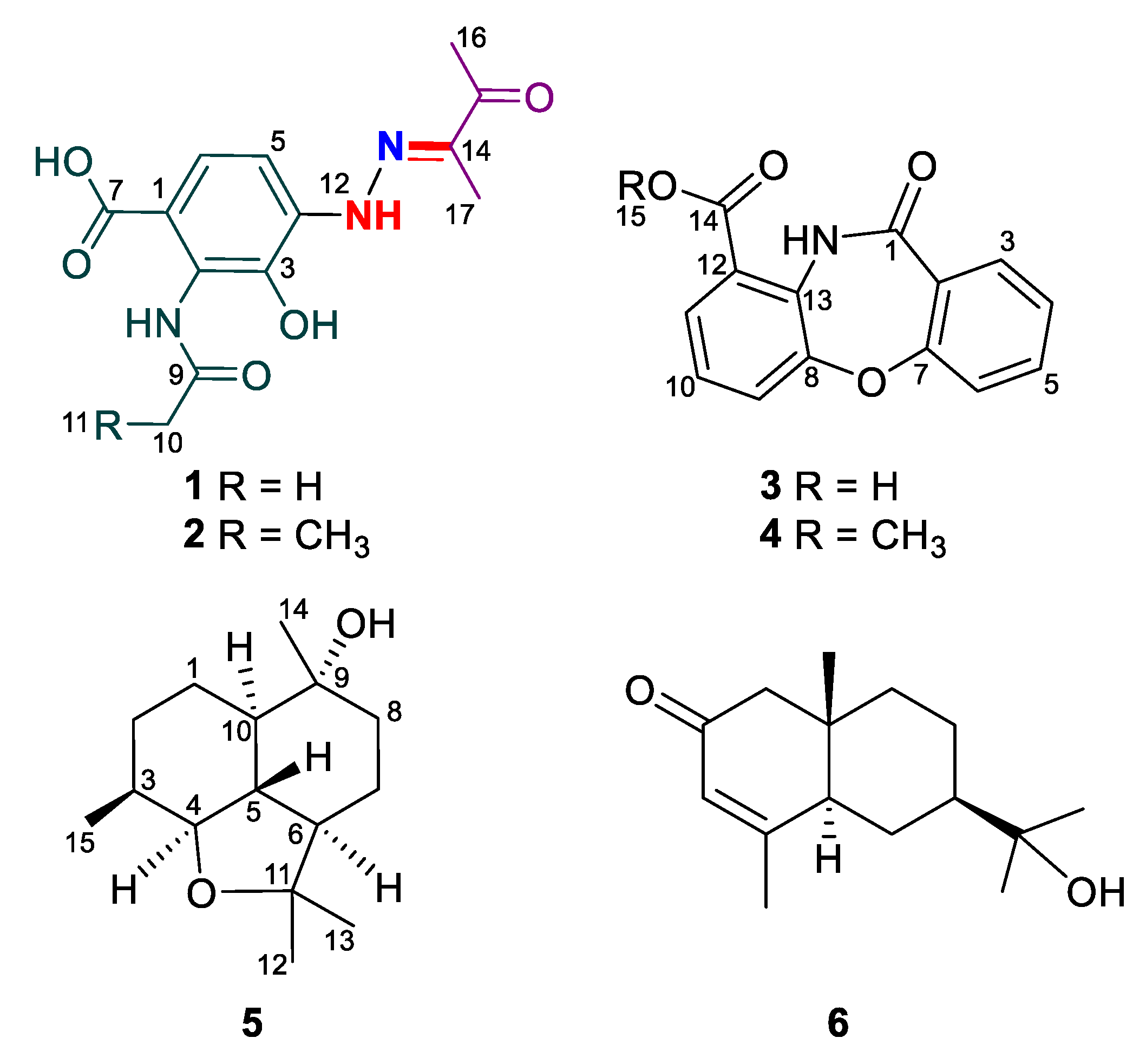
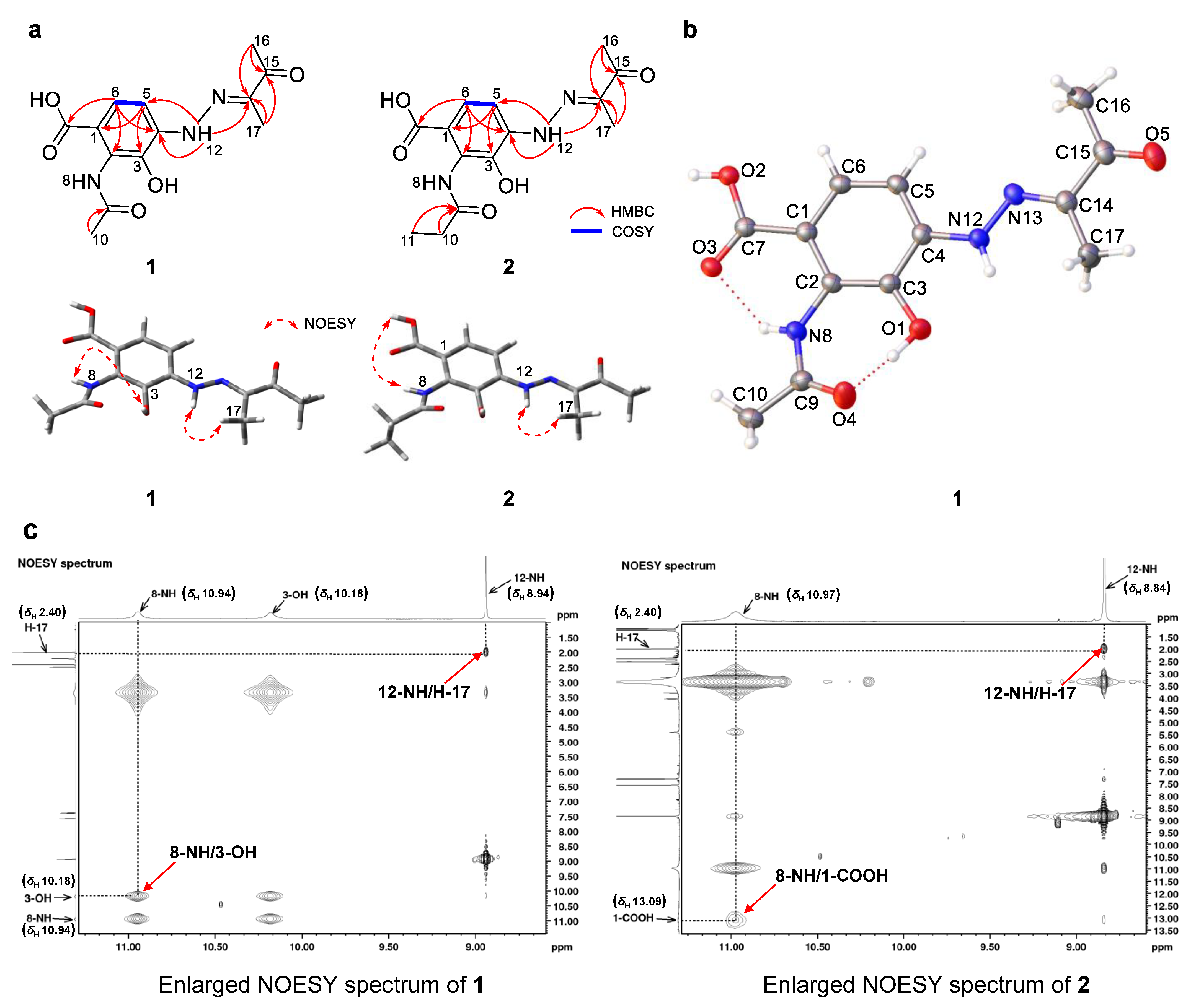
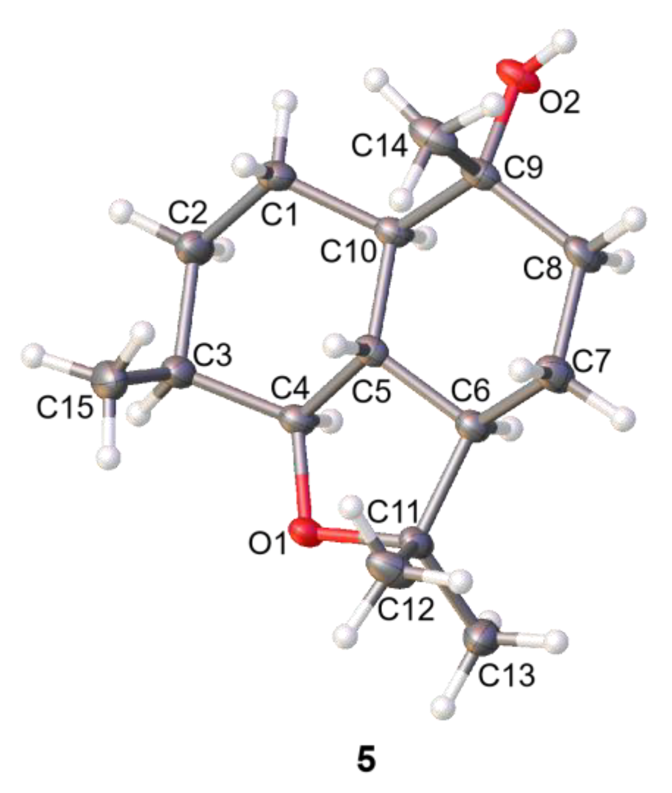
2.1. Structural Determination

Penzonemycin A (1) was obtained as yellow needles. The molecular formula of 1 was determined to be C13H15N3O5 based on the HRMS (ESI-TOF) m/z [M + H]+, calcd for 294.1084, found 294.1086, indicating 8 degrees of unsaturation. The IR absorptions at 3360 and 1676 cm−1 suggested the presence of OH and C=O functionalities. The 1H NMR spectrum of 1 (Table 1) showed three singlet methyl groups, δH 2.21 (3H, s), 2.00 (3H, s) and 2.40 (3H, s), two aromatic ortho proton doublets δH 7.37 (1H, d, J = 8.6 Hz) and 7.57 (1H, d, J = 8.6 Hz), and four exchangeable proton signals δH 13.00 (1H, br s, 1-COOH), 10.94 (1H, br s, NH-8), 10.18 (1H, br s, 3-OH) and 8.94 (1H, br s, NH-12). The 13C NMR spectrum of 1 (Table 1, Figure S3) displayed 13 carbon signals for a 1,2,3,4-tetra-substituted benzene ring, three methyl groups, δC 23.6 (C-10), 8.1 (C-16) and 24.2 (C-17), and 4 quaternary carbons, δC 168.6 (C-7), 171.3 (C-9), 144.2 (C-13) and 196.3 (C-14). The COSY correlations of H-5 and H-6 and the HMBC correlations from H-5 to C-1/C-3 and from H-6 to C-2/C-4 confirmed the presence of a tetra-substituted benzene ring unit (Figure S3). The key HMBC correlations (Figure 2 and Figure S3) from H-6 to C-7 (δC 168.6) indicated a carboxylic acid group linked to the tetra-substituted benzene ring at C-1. The HMBC correlations from H3-10 (δH 2.21) to C-9 (δC 171.3) and the chemical shift value of C-9 revealed an acetamide group, which was deduced to be located at C-2 of the tetra-benzene ring by the chemical shift value of C-2 (δC 127.5). The HMBC correlations from H3-16 (δH 2.00)/H3-17 (δH 2.40) to C-14 (δC 144.2)/C-15 (δC 196.3) and from NH-12 to C-14, together with the remaining two N atoms, established a rare butanone hydrazone unit, which was linked to the benzene ring at C-4 by HMBC correlations from NH-12 to C-3/C-5. The remaining hydroxyl group (δH 10.18, 3-OH) was placed at C-3 based on the NOESY correlations of 3-OH and NH-8 (Figure 2). Thus, the planar structure of 1 was established. The E configuration of the double bond Δ13,14 in 1 was assigned by the NOESY correlation of NH-12 and H3-17 (Figure 2). Fortunately, high-quality crystals of 1 were obtained, allowing the successful performance of a single-crystal X-ray diffraction experiment using Cu Ka radiation (CCDC 2131907), confirming the planar structure and absolute stereochemistry (EΔ13,14) of 1 (Figure 2, Table S4).
Penzonemycin B (2) was isolated as a yellowish solid. Its molecular formula was assigned as C14H17N3O5 by HRMS (ESI-TOF) m/z [M + H]+, calcd for 308.1241, found 308.1240, indicating 8 degrees of unsaturation. The IR spectrum of 2 displayed broad hydroxyl absorption at 3350 cm−1 and a carbonyl peak at 1676 cm−1. The 1H, 13C NMR data of 2 (Table 1, Figure S4) were highly similar to those of 1. The difference was that 2 had a propionamide moiety instead of the acetylamino unit in 1. The presence of the propionamide group in 2 was supported by the 1H-1H COSY correlation of H2-10 (δH 2.49) and H3-11 (δH 1.17) and HMBC correlations from H3-11 to C-9 (δC 174.5)/C-10 (Figure 2). The geometry of the double bond Δ13,14 in 2 was determined to be trans (E) by NOESY correlations of NH-12 and H3-17 (Figure 2).
Compound 3 was isolated as a white solid. The molecular formula of 3 was determined to be C14H9NO4 by HRMS (ESI-TOF) ([M + H]+, m/z 256.0616, calculated for 256.0604, Figure S5), indicating 11 degrees of unsaturation. The IR spectrum exhibited the presence of carbonyl (1716, 1633 cm−1) functional groups. The 1H NMR spectrum of 3 (Table 2, Figure S5) showed seven aromatic protons signals at δH 7.15 (1H, d, J = 8.3 Hz), 7.99 (1H, d, J = 8.6 Hz), 8.05 (1H, d, J = 7.8, 1.5 Hz), 8.12 (1H, d, J = 8.1 Hz), 7.11 (1H, d, J = 7.6 Hz), 7.57 (1H, d, J = 8.0, 1.7 Hz) and 7.58 (1H, d, J = 8.0 Hz). The 13C NMR spectrum of 3 (Table 2, Figure S5) displayed 14 carbon signals (Figure S5) for 7 methine carbons δC 115.4 (C-9), 117.4 (C-6), 120.1 (C-4), 125.4 (C-10), 127.4 (C-3), 127.5 (C-11) and 134.5 (C-5), 5 quaternary carbons signals δC 109.8 (C-2), 121.8 (C-12), 138.8 (C-13), 149.6 (C-8) and 158.4 (C-7) and 2 ester/amido carbonyl carbons signals δC 163.6 (C-1) and 165.6 (C-14). 1H and 13C NMR data of 3 (Table 2 and Figure S5) were highly similar to those of mycemycin A (4) [13]. Compound 3 was different from 4 by the absence of the methoxy group at C-14, which was supported by 14 mass units less compared to mycemycin A (4), as well as by a detailed analysis of 1D and 2D NMR spectra (Figure 3 and Figure S5). The COSY correlations of H-3 (δH 7.99)/H-4 (δH 7.11)/H-5 (δH 7.57)/H-6 (δH 7.15) and the HMBC correlations from H-4 to C-2/C-6, from H-3/H-5/H-6 to C-7, and from H-3 to C-1 established the salicylic acid fragment. The COSY correlations of H-9 (δH 8.12)/H-10 (δH 7.58)/H-11(δH 8.05) and the HMBC correlations from H-9/H-11 to C-13, from H-10 to C-12/C-8, and from H-11 to C-14 revealed a 3-hydroxyanthranilic acid unit. Given determined molecular formula as well as one less degree of unsaturation, two ring systems were connected by C-2-C-1-NH-C-13 and C-7-O-C-8. Thus, compound 3 was elucidated to be demethylmycemycin A.
Compounds 46 were identified as mycemycin A (4) [13], 9(R)-ganodermanol B (5) [14], and isopterocarpolone (6) [15], respectively. Their 1H and 13C NMR data (Figures S6–S8) were identical to those reported in the literature. In addition, we performed a crystallographic analysis of 5. For the first time, the absolute configuration of 5 was confirmed by the way of single-crystal X-ray diffraction analysis (Figure 4) using Cu Kα data (CCDC 2177578) with Flack parameter of 0.04(9) (Table S5).

2.2. Biosynthetic Implications

Penzonemycins A (1) and B (2) are new hydrazones harboring an −N−N=C− unit and a 3-hydroxyanthranilic acid (3-HAA) core. The whole genome of Streptomyces sp. SCSIO 40020 has been sequenced, revealing a putative gene cluster (pzm), spanning from pzm1 to pzm22 (Scheme 1, Table 3), that encodes the enzymatic machinery for the biosynthesis of 1 and 2. The 3-HAA core was proposed to be derived from a chorismate pathway, given the presence of the genes pzm9, pzm6, pzm7 and pzm8, which are predicted to encode 3-deoxy-D-arabino-heptulosonate-7-phosphate (DAHP) synthase, anthranilate synthase, isochorismatase and a dehydrogenase, respectively. Pzm9 catalyzes the condensation of D-erythrose-4-phosphate (E4P) and phosphoenolpyruvate (PEP) to form DAHP, the first intermediate in the shikimate pathway leading to chorismate [16,17]. The homologous proteins of Pzm6, Pzm7 and Pzm8 in the biosynthetic pathways of calcimycin and limazepine have been shown to be responsible for converting chorismate to 3-HAA (Scheme 1) [18,19]. Next, 3-HAA is putatively acylated by the N-acetyltransferase Pzm3 to afford an acetylated (or propionylated) product ready for two rounds of oxidations, likely catalyzed by the proposed oxidases Pzm19 and Pzm21. Subsequently, an unknown enzyme-catalyzed transamination occurs, followed by a tautomerization, to generate the putative intermediate M1 (Scheme 1). The proposed oxidative transamination reactions are similar to the recent reported reactions in the formation of 8-amino-flaviolin during the biosynthesis of naphthoquinone-based meroterpenoid [20]. Notably, the three enzymes, namely, the nitrosuccinate lyase Pzm11, the monooxygenase Pzm12 and the AMP-binding protein Pzm18, are predicted to be the orthologues of CreD, CreE and CreM, respectively, which are responsible for the diazo formation in cremeomycin biosynthesis, utilizing L-aspartic acid as a nitrogen source [21]. Therefore, Pzm11 and Pzm12 are postulated to generate nitrite from L-aspartic acid, which is subsequently incorporated to M1 by Pzm18, leading to the diazo group in M2 after dehydration and tautomerization, similar to the biosynthesis of cremeomycin and triacsin [21,22]. Recently, an electrophilic diazo-anthranilate has been reported to undergo nonenzymatic Japp−Klingemann coupling with a β-keto aldehyde-containing precursor to furnish the hydrazone group in the biosynthesis of tasikamides [4]. Interestingly, Pzm16 encodes an acetolactate synthase and putatively catalyzes the coupling of two pyruvates to form 2-acetolactate [23], which becomes 4-hydroxy-3-methyl-3-buten-2-one (M3) after two rounds of carboxyl reduction (probably catalyzed by the F420-dependent oxidoreductase Pzm4) and spontaneous dehydration (Scheme 1). Finally, the nonenzymatic Japp−Klingemann coupling reaction between M2 and M3 constructs the hydrazone moiety in 1 and 2 after subsequent hydration and decarboxylation (Scheme 1).

2.3. Biological Activities

Penzonemycin A (1) was evaluated for antitumor activities in four human cancer cell lines SF-268, MCF-7, A549 and HepG-2 by the SRB method. Compound 1 showed weak activity in these three cancer cell lines (IC50 30.44–61.92 µM) (Table S1). Penzonemycin A (1) was also assayed for its antibacterial activities, antifungal activities (Tables S2 and S3) and α-glucosidase inhibition activity. However, compound 1 showed no activities.

3. Materials and Methods

3.1. General Experimental Procedures

Optical rotations were measured using a 341 Polarimeter (Perkin-Elmer, Inc., Norwalk, CT, USA). UV spectra were measured on a U-2900 spectrophotometer (Hitachi, Tokyo, Japan). IR spectra were recorded on an Affinity-1 FT-IR spectrometer (Shimadzu, Tokyo, Japan). Melting points were measured using an SGW®X-5 micro melting point meter (Shanghai INESA Physico optiacal Instrument Co., Ltd., Shanghai, China). 1D and 2D NMR spectra were recorded on a Bruker AV-700 MHz NMR spectrometer (Bruker Biospin GmbH, Rheinstetten, Germany) with tetramethylsilane (TMS) as the internal standard. Mass spectrometric data were obtained on a quadrupole-time-of-flight mass spectrometer (Bruker Maxis 4G) for HRESIMS. Column chromatography was performed using silica gel (100–200 mesh, 300–400 mesh; Jiangyou Silica gel development, Inc., Yantai, China), Sephadex LH-20 (GE Healthcare Bio-Sciences AB, Uppsala, Sweden). HPLC was carried out while a reversed-phase column (Phenomenex Gemini C18, 250 mm × 4.6 mm, 5 μm; Phenomenex, Torrance, CA, USA) with UV detection at 254 nm and 360 nm. Semi-preparative HPLC was performed on a Hitachi HPLC station (Hitachi-L2130) with a Diode Array Detector (Hitachi L-2455) using a Phenomenex ODS column (250 mm × 10.0 mm, 5 μm; Phenomenex, Torrance, CA, USA) with UV detection at 360 nm.

3.2. Strain, Screening and Culture Methods

Streptomyces sp. SCSIO 40020 (original number LW701) was isolated from a marine sediment sample (E 114.0432°, N 22.0194°, Pearl River Estuary in China, at the depth of 28 m), and was identified by 16S rDNA sequence analysis (GenBank accession no. MW582618). The strain SCSIO 40020 was maintained in a 40% glycerol aqueous solution at −80 °C at the Research Center for Marine Microbiology Culture Collection Center of South China Sea Institute of Oceanology, Chinese Academy of Sciences. It was found that the strain SCSIO 40020 was best maintained on 38#-agar medium containing 3% sea salt for optimal growth and sporulation. A single colony was inoculated into 50 mL of four different media, including modifed-A1BFe+C [24] (soluble starch 1.0%, yeast extract 0.4%, tryptone 0.2%, CaCO3 0.2%, sea salts 3%, pH 7.2–7.4), ZM7 medium (glycerol 0.5%, arginine 0.1%, glucose 0.1%, K2HPO4 0.03%, MgSO4·7H2O 0.02%, NaCl 0.03%, vitamin B complex 0.5 mL, pH 7.2–7.4), AM6-4 [25] (glycerol 0.1%, bacterial peptone 0.5%, glycine 0.01%, alanine 0.01%, CaCO3 0.5%, sea salts 3%, pH 7.2–7.4) and modifed-ISP3 [25] (oat meal 1.5%, FeSO4 0.0001%, MnCl2 0.0001%, ZnSO4 0.0001%, sea salts 3%, pH 7.2–7.4), in 250 mL Erlenmeyer flasks and then incubated on a rotary shaker (200 rpm) at 28 °C for seven days. The culture broths were extracted with an equal volume of n-butanone, and the extracts were then monitored by HPLC-DAD. HPLC analyses were carried out using the following program: solvent system (solvent A, 10% acetonitrile in water supplemented with 0.08% formic acid; solvent B, 90% acetonitrile in water); 5% B to 100% B (linear gradient, 0–18 min), 100% B (18–23 min), 100% B to 5% B (23–27 min), 5% B (27–32 min); flow rate of 1 mL/min. A single colony was inoculated into 30 mL of modified A1BFe+C medium and incubated at 28 °C for 2–3 days. Then, 20 L of fermentation culture was prepared by inoculating 30 mL of the seed culture into a 1000 mL Erlenmeyer flask containing 200 mL of the modified ISP3 medium followed by cultivation on a rotary shaker (200 rpm) at 28 °C for 7 days.

3.3. Extraction, Isolation and Purification

The 20 L of culture broth of Streptomyces sp. SCSIO 40020 was centrifuged at 3900 rpm for 15 min at 25 °C. The mycelia were extracted three times, each with 2 L of acetone. The acetone extracts were concentrated under reduced pressure to afford an aqueous residue, which was extracted four times with an equal volume of n-butanone. The supernatants were extracted four times with an equal volume of n-butanone [12,24]. The butanone extracts were combined and concentrated under reduced pressure to afford the crude extracts (7.5 g). The crude extracts were subjected to column chromatography over silica gel, eluting with a gradient of CHCl3/MeOH mixtures i.e., 100/0, 95/5, 90/10, 80/20, 50/50 and 0/100 (v/v). We obtained five fractions (Fr.1–Fr.5). Then Fr.2 (3.50 g) was further purified via MPLC (Medium-Pressure Preparative Liquid Chromatography) with a reverse-phased C-18 column (14.5 cm × 2.5 cm i.d., 40–60 μm Agela Technologies, Torrance, CA, USA) by eluting with a linear gradient of H2O/MeOH (0–100%, 15 mL/min, 300 min), obtaining fractions Fr.2.1–Fr.2.10. Fractions Fr.2.5–Fr.2.6 (20 mg) were further purified by semi-preparative HPLC using a mobile phase of MeCN/H2O (50:50, v/v), which yielded compound 3 (1.8 mg) and compound 4 (1.5 mg). Fractions Fr.2.7–Fr.2.8 (200 mg) were further purified by semi-preparative HPLC using a mobile phase of MeCN/H2O (60:40, v/v), obtaining compound 1 (10.4 mg), compound 2 (2.2 mg), compound 5 (1.2 mg), and compound 6 (1.0 mg).

3.4. Physical and Chemical Properties of the New Compounds 13 and 5

Penzonemycin A (1): yellow needles; m.p. 205.8–209.3 °C; [α ] D 25 : −1.8 (c 0.11, MeCN); UV (MeCN) λmax (log ε): 358 (4.69) nm, 249 (4.50) nm, 210 (4.59) nm; IR νmax 3360, 2947, 2837, 2359, 2332, 1676 cm−1, 1H NMR (700 MHz, DMSO-d6) and 13C NMR (176 MHz, DMSO-d6), see Table 1; (+)-HRESIMS m/z [M + H]+ 294.1086 (calculated for C13H16N3O5, 294.1084), [M + Na]+ 316.0908 (calculated for C13H15N3NaO5, 316.0904).
Crystal data for penzonemycin A (1): C13H15N3O5 (M = 293.28 g/mol): monoclinic, space group P21/n (no. 14), a = 4.8385 (2) Å, b = 19.6128 (7) Å, c = 14.5170 (6) Å, β = 98.487 (4), V = 1362.53 (9) Å3, Z = 4, T = 103 (5) K, μ (Cu Kα) = 0.945 mm−1, Dcalc = 1.430 g/cm3, 6318 reflections measured (9.018° ≤ 2Θ ≤ 148.158°), 2631 unique (Rint = 0.0376, Rsigma = 0.0368) which were used in all calculations. The final R1 was 0.0576 (I > 2σ(I)), and wR2 was 0.1809 (all data).
Penzonemycin B (2): a yellowish solid; [α ] D 25 : −1.1 (c 0.19, MeCN); UV (MeCN) λmax (log ε): 358 (4.66) nm, 249 (4.48) nm, 210 (4.56) nm; IR νmax 3350, 2358, 1676, 1267, 667 cm−1; 1H and 13C NMR data, see Table 1; (+)-HRESIMS m/z [M + H]+ 308.1240 (calculated for C14H18N3O5, 308.1241), [M + Na]+ 330.1054 (calculated for C13H15N3NaO5, 330.1060).
Compound 3: a white solid; [α ] D 25 : +8.9 (c 0.20, MeCN); UV (CHCl3) λmax (log ε): 346 (4.07) nm, 335 (4.09) nm; 306 (3.92) nm; 264 (3.94) nm, 247 (3.89) nm; IR νmax 2953, 2914, 1716, 1633, 1548, 1489, 1303, 1259, 1244, 1192, 1159, 1060, 875, 786, 748, 717, 686 cm−1, 1H and 13C NMR data, see Table 2; (+)-HRESIMS m/z [M + H]+ 256.0616 (calculated for C14H18N3O5, 256.0604).
Crystal data for 9(R)-ganodermanol B (5): C15H26O2 (M = 238.36 g/mol): orthorhombic, space group P21/n (no. 19), a = 8.35172 (11) Å, b = 11.17809 (15) Å, c = 14.82535 (19) Å, V = 1384.04 (3) Å3, Z = 4, T = 99.8 (8) K, μ (Cu Kα) = 0.570 mm−1, Dcalc = 1.144 g/cm3, 7345 reflections measured (9.91° ≤ 2Θ ≤ 148.416°), 2727 unique (Rint = 0.0278, Rsigma = 0.0319) which were used in all calculations. The final R1 was 0.0355 (I > 2σ(I)), and wR2 was 0.0946 (all data).

3.5. X-Ray Crystallographic Analysis Data of Penzonemycin A (1) and 5

Single crystals of C13H15N3O5 (penzonemycin A 1) and C15H26O2 (9(R)-ganodermanol B 5) were obtained in MeOH solution. Their data were collected on an XtaLAB AFC12 (RINC): Kappa single diffractometer. The crystals were kept at 100.00 (10) K during data collection. Using Olex2 [26], their structures were solved with the ShelXT [27] structure solution program using Intrinsic Phasing and refined with the ShelXL [28] refinement package, using Least Squares minimization. Crystallographic data for 1 and 5 were deposited at the Cambridge Crystallographic Data Center (CCDC) with the deposition numbers 2,131,907 and 2,177,578, respectively.

3.6. Bioactivity Assays

3.6.1. Cytotoxic Activity Assay

The in vitro cytotoxic activities of penzonemycin A (1) were evaluated in four tumor cell lines, i.e., SF-268 (human glioma cell line), HepG-2 (human liver carcinoma cell line), MCF-7 (human breast adenocarcinoma cell line), and A549 (human lung adenocarcinoma cell) by the SRB method [24], according to a previously described protocol [29]. The cells were cultivated in RPMI 1640 medium [30]. Cells (180 μL) with a density of 3 × 10⁴ cells/mL were seeded onto 96-well plates and incubated for 24 h at 37 °C, 5% CO2. Subsequently, 20 μL of different concentrations of penzonemycin A (1), ranging from 0 to 100 µM in dimethyl sulfoxide (DMSO), was added to each plate well. An equal volume of DMSO was used as a negative control. After further incubation for 72 h, the cell monolayers were fixed with 50% (wt/v) trichloroacetic acid (50 μL) and then stained for 30 min with 0.4% (wt/v) SRB dissolved in 1% acetic acid. The unbound dye was removed by repeatedly washing with 1% acetic acid. The protein-bound dye was dissolved in 10 mM Tris-base solution (200 µL) for the determination of the optical density (OD) at 570 nm using a microplate reader. The cytotoxic compound cisplatin was used as a positive control. All data were obtained in triplicate and are presented as means ± S.D. IC50 values were calculated with the SigmaPlot 14.0 software using the non-linear curve-fitting method.

3.6.2. Antifungal Activity Assay

The antifungal activity of compound 1 was measured against three phytopathogenic fungi, i.e., Colletotrichum gloeosporioides Penz, Physalospora piricola Nose, Bipolaris sorokiniana, by the broth microdilution method [31]. Compound 1 was dissolved in DMSO at the final concentration of 2.56 mg mL−1. The indicator fungi strains were grown for 48 h on a rotary shaker at 28 °C. The cultures were diluted with sterilized medium to achieve a predetermined optical absorbance at 600 nm, then diluted 1000-fold before being added into 96-well microtiter plates. Three replicates of each compound were tested in dilution series, ranging from 128 to 0.25 μg mL−1. The lowest concentrations that completely inhibited the visible growth of the tested strains were recorded after 48 h of cultivation from two independent experiments. Nystatin was used as a positive control against the three phytopathogenic fungi.

3.6.3. Antibacterial Activity Assay

The antibacterial activity of compound 1 was assayed against Escherichia coli ATCC 25922, Acinetobacter baumannii ATCC 19606, Staphyloccocus aureus ATCC 29213, and Methicillin-resistant S. aureus (MRSA) ATCC 43300 by the broth microdilution method [31]. Compound 1 was dissolved in DMSO at the final concentration of 2.56 mg mL−1. The indicator strain was grown for 24 h on a rotary shaker at 37 °C. The cultures were diluted with sterilized medium to achieve a predetermined optical absorbance at 600 nm, then diluted 1000-fold before being placed into 96-well microtiter plates. Three replicates of each compound were tested in dilution series, ranging from 128 to 0.25 μg mL−1. The MIC values were recorded after 24 h of cultivation from two independent experiments. Ciprofloxacin and vancomycin were used as positive controls.

3.6.4. Alpha-glucosidase Inhibition Activity

A general inhibiting reaction assay contained 0.04 U/mL, 2.0 mM p-nitrophenyl-α-D-glucopyranoside solution (Shanghai Macklin Biochemical Co., Ltd., Shanghai, China) and a specific concentration of 1 and was carried out in PBS buffer (50 mM, pH 7.0), in a total volume of 100 μL. In detail, first, sample 1 was mixed with α-glucosidase and incubated at 37 °C for 10 min, and then, the reactions were initiated by adding the p-nitrophenyl-α-D-glucopyranoside solution and continued at 37 °C for another 15 min. After that, the absorbance at 405 nm was recorded on a microplate reader (Enspire, PerkinElmer, Singapore) within 30 min. In the assay, DMSO was used as a negative control, and acarbose (Shanghai Aladdin Biochemical Technology Co., Ltd., Shanghai, China) was used as a positive control. All treatments were performed in triplicate, and the IC50 value was determined by regression analysis [32].

3.7. Bioinformatics Analysis

The genomic DNA of Streptomyces sp. SCSIO 40020 was isolated according to a prior study [12]. The whole genome was sequenced and assembled by Nextomics Biosciences Co., Ltd. (Wuhan, China) using Oxford Nanopore GridION and canu. The putative biosynthetic gene clusters in the genome were predicted with antiSMASH 6.0 [33]. The deduced ORFs were analyzed using online 2ndFind software (https://biosyn.nih.go.jp/2ndfind/, accessed on 13 January 2022), and their functional predictions were accomplished with an online BLAST program (https://blast.ncbi.nlm.nih.gov/Blast.cgi, accessed on 13 January 2022). The DNA sequences of the penzonemycins gene cluster were deposited in GenBank, accession number ON345781.

4. Conclusions

In conclusion, two new phenylhydrazone derivatives and a new alkaloid, penzonemycins A–B (12) and demethylmycemycin A (3), together with three known compounds (46) were obtained from the Pearl River Estuary sediment-derived Streptomyces sp. SCSIO 40020. Penzonemycins feature a hydrazone unit (−N−N=C−) that is rare in natural products. Biosynthetically, a Japp−Klingemann reaction is proposed as a key reaction leading to the hydrazone moiety in 1 and 2, which is worth of further investigations.

Supplementary Materials

The following supporting information can be downloaded at: https://www.mdpi.com/article/10.3390/md20070449/s1, Table S1: Cytotoxicity of penzonemycin A (1). Table S2: Antifungal activity of penzonemycin A (1). Table S3: Antibacterial activity of penzonemycin A (1). Table S4: Crystal data and structure refinement for penzonemycin A (1). Table S5: Crystal data and structure refinement for 9(R)-ganodermanol B (5). Figure S1: Natural hydrazone-containing compounds reported in the literature. Figure S2: HPLC analysis of the metabolic profiles of Streptomyces sp. SCSIO 40020 cultured in different media. Figure S3: Spectral data for penzonemycin A (1). Figure S4: Spectral data for penzonemycin B (2). Figure S5: Spectral data for demethylmycemycins A (3). Figure S6: Spectral data for mycemycin A (4). Figure S7: Spectral data for 9(R)-ganodermanol B (5). Figure S8: Spectral data for isopterocarpolone (6).

Author Contributions

W.L. contributed to compounds isolation and structure elucidation. L.Z. analyzed the X-ray crystallographic diffraction data. Q.Z., Y.C. and W.Z. (Weimin Zhang) performed the cytotoxicity assays. W.L., L.M., H.Z., W.Z. (Wenjun Zhang) and C.Z. analyzed the data and wrote the manuscript. C.Z. supervised the study. All authors have read and agreed to the published version of the manuscript.

Funding

This work was supported in part by NSFC (42176127, 22193072, 21907098), Guangdong Basic and Applied Basic Research Foundation (2021A1515110284, 2021A1515010839), by the Science and Technology Planning Project of Guangzhou (202102020513), K. C. Wong Education Foundation (GJTD-2020-12), by the State Key Laboratory of Applied Microbiology, Southern China (Grant No. SKLAM004-2021).

Institutional Review Board Statement

Not applicable.

Informed Consent Statement

Not applicable.

Data Availability Statement

The data presented in this study are available on request from the corresponding author.

Acknowledgments

We appreciate the help of Xuan Ma in X-ray diffraction analyses and of Zhihui Xiao, Aijun Sun, Yun Zhang and Xiaohong Zheng in recording MS and NMR data. We are grateful to Zhengchao Wu for the collection of Pearl River Estuary sediment.

Conflicts of Interest

The authors declare no conflict of interest.

References

  1. Wang, Z.; Kang, D.; Chen, M.; Wu, G.; Feng, D.; Zhao, T.; Zhou, Z.; Huo, Z.; Jing, L.; Zuo, X.; et al. Design, synthesis, and antiviral evaluation of novel hydrazone-substituted thiophene [3,2-d]pyrimidine derivatives as potent human immunodeficiency virus-1 inhibitors. Chem. Biol. Drug Des. 2018, 92, 2009–2021. [Google Scholar] [CrossRef] [PubMed]
  2. Bolognino, I.; Giangregorio, N.; Tonazzi, A.; Martínez, A.L.; Altomare, C.D.; Loza, M.I.; Sablone, S.; Cellamare, S.; Catto, M. Synthesis and biological evaluation of dantrolene-like hydrazide and hydrazone analogues as multitarget agents for neurodegenerative diseases. ChemMedChem 2021, 16, 2807–2816. [Google Scholar] [CrossRef] [PubMed]
  3. Achagar, R.; Elmakssoudi, A.; Dakir, M.; Elamrani, A.; Zouheir, Y.; Zahouily, M.; Jamaleddine, J. A green and efficient protocol for the synthesis of phenylhydrazone derivatives catalyzed by nanostructured diphosphate Na2CaP2O7 and screening of their antibacterial activity. ChemistrySelect 2021, 6, 1366–1371. [Google Scholar] [CrossRef]
  4. Ma, G.; Candra, H.; Pang, L.; Xiong, J.; Ding, Y.; Tran, H.; Low, Z.; Ye, H.; Liu, M.; Zheng, J.; et al. Biosynthesis of tasikamides via pathway coupling and diazonium-mediated hydrazone formation. J. Am. Chem. Soc. 2022, 144, 1622–1633. [Google Scholar] [CrossRef] [PubMed]
  5. Liu, Y.; Fang, S.; Shi, Z.; Wang, B.; Li, X.; Ji, N. Phenylhydrazone and quinazoline derivatives from the cold-seep-derived fungus Penicillium oxalicum. Mar. Drugs 2021, 19, 10. [Google Scholar] [CrossRef] [PubMed]
  6. Cheng, P.; Xu, K.; Chen, Y.; Wang, T.; Chen, Y.; Yang, C.; Ma, S.; Liang, Y.; Ge, H.; Jiao, R. Cytotoxic aromatic polyketides from an insect derived Streptomyces sp. NA4286. Tetrahedron Lett. 2019, 60, 1706–1709. [Google Scholar] [CrossRef]
  7. Zhang, W.; Yang, C.; Huang, C.; Zhang, L.; Zhang, H.; Zhang, Q.; Yuan, C.; Zhu, Y.; Zhang, C. Pyrazolofluostatins A–C, pyrazole-fused benzo[a]fluorenes from south China sea-derived Micromonospora rosaria SCSIO N160. Org. Lett. 2017, 19, 592–595. [Google Scholar] [CrossRef]
  8. Matsuda, K.; Hasebe, F.; Shiwa, Y.; Kanesaki, Y.; Tomita, T.; Yoshikawa, H.; Shin-ya, K.; Kuzuyama, T.; Nishiyama, M. Genome mining of amino group carrier protein-mediated machinery: Discovery and biosynthetic characterization of a natural product with unique hydrazone unit. ACS Chem. Biol. 2017, 12, 124–131. [Google Scholar] [CrossRef]
  9. Blair, L.M.; Sperry, J. Natural products containing a nitrogen–nitrogen bond. J. Nat. Prod. 2013, 76, 794–812. [Google Scholar] [CrossRef]
  10. Ren, X.; Xie, X.; Chen, B.; Liu, L.; Jiang, C.; Qian, Q. Marine natural products: A potential source of anti-hepatocellular carcinoma drugs. J. Med. Chem. 2021, 64, 7879–7899. [Google Scholar] [CrossRef]
  11. Jagannathan, S.V.; Manemann, E.M.; Rowe, S.E.; Callender, M.C.; Soto, W. Marine actinomycetes, new sources of biotechnological products. Mar. Drugs 2021, 19, 365. [Google Scholar] [CrossRef]
  12. Zhai, S.; Liu, W.; Wu, Z.; Zhu, Y.; Zhang, W.; Zhang, C.; Ma, L. Identification of bafilomycins and heterologous expression of its biosynthetic gene cluster from the Pearl River Estuary sediment-derived Streptomyces sp. SCSIO 40020. Acta Microbiol. Sin. 2021, 61, 3991–4005. [Google Scholar]
  13. Liu, N.; Song, F.; Shang, F.; Huang, Y. Mycemycins A-E, new dibenzoxazepinones isolated from two different Streptomycetes. Mar. Drugs 2015, 13, 6247–6258. [Google Scholar] [CrossRef] [PubMed] [Green Version]
  14. Tan, Z.; Zhao, J.; Liu, J.; Zhang, M.; Chen, R.; Xie, K.; Dai, J. Sesquiterpenoids from the cultured mycelia of Ganoderma capense. Fitoterapia 2017, 118, 73–79. [Google Scholar] [CrossRef] [PubMed]
  15. Shaaban, K.A.; Singh, S.; Elshahawi, S.I.; Wang, X.; Ponomareva, L.V.; Sunkara, M.; Copley, G.C.; Hower, J.C.; Morris, A.J.; Kharel, M.K.; et al. The native production of the sesquiterpene isopterocarpolone by Streptomyces sp. RM-14-6. Nat. Prod. Res. 2014, 28, 337–339. [Google Scholar] [CrossRef] [PubMed] [Green Version]
  16. Cano-Prieto, C.; García-Salcedo, R.; Sánchez-Hidalgo, M.; Braña, A.; Fiedler, H.; Méndez, C.; Salas, J.; Olano, C. Genome mining of Streptomyces sp. Tü 6176: Characterization of the nataxazole biosynthesis pathway. Chembiochem 2015, 16, 1461–1473. [Google Scholar] [CrossRef]
  17. Ikeda, M. Towards bacterial strains overproducing L-tryptophan and other aromatics by metabolic engineering. Appl. Microbiol. Biotechnol. 2006, 69, 615–626. [Google Scholar] [CrossRef] [Green Version]
  18. Pavlikova, M.; Kamenik, Z.; Janata, J.; Kadlcik, S.; Kuzma, M.; Najmanova, L. Novel pathway of 3-hydroxyanthranilic acid formation in limazepine biosynthesis reveals evolutionary relation between phenazines and pyrrolobenzodiazepines. Sci. Rep. 2018, 8, 7810. [Google Scholar] [CrossRef]
  19. Gou, L.; Wu, Q.; Lin, S.; Li, X.; Liang, J.; Zhou, X.; An, D.; Deng, Z.; Wang, Z. Mutasynthesis of pyrrole spiroketal compound using calcimycin 3-hydroxy anthranilic acid biosynthetic mutant. Appl. Microbiol. Biotechnol. 2013, 97, 8183–8191. [Google Scholar] [CrossRef]
  20. Noguchi, T.; Isogai, S.; Terada, T.; Nishiyama, M.; Kuzuyama, T. Cryptic oxidative transamination of hydroxynaphthoquinone in natural product biosynthesis. J. Am. Chem. Soc. 2022, 144, 5435–5440. [Google Scholar] [CrossRef]
  21. Sugai, Y.; Katsuyama, Y.; Ohnishi, Y. A nitrous acid biosynthetic pathway for diazo group formation in bacteria. Nat. Chem. Biol. 2016, 12, 73–75. [Google Scholar] [CrossRef] [PubMed] [Green Version]
  22. Del Rio Flores, A.; Twigg, F.F.; Du, Y.; Cai, W.; Aguirre, D.Q.; Sato, M.; Dror, M.J.; Narayanamoorthy, M.; Geng, J.; Zill, N.A.; et al. Biosynthesis of triacsin featuring an N-hydroxytriazene pharmacophore. Nat. Chem. Biol. 2021, 17, 1305–1313. [Google Scholar] [CrossRef] [PubMed]
  23. Chipman, D.; Barak, Z.; Schloss, J.V. Biosynthesis of 2-aceto-2-hydroxy acids: Acetolactate synthases and acetohydroxyacid synthases. Biochim. Biophys. Acta. 1998, 1385, 401–419. [Google Scholar] [CrossRef]
  24. Liu, W.; Zhang, W.; Jin, H.; Zhang, Q.; Chen, Y.; Jiang, X.; Zhang, G.; Zhang, L.; Zhang, W.; She, Z.; et al. Genome mining of marine-derived Streptomyces sp. SCSIO 40010 leads to cytotoxic new polycyclic tetramate macrolactams. Mar. Drugs 2019, 17, 663. [Google Scholar] [CrossRef] [Green Version]
  25. Zhang, W.; Zhang, G.; Zhang, L.; Liu, W.; Jiang, X.; Jin, H.; Liu, Z.; Zhang, H.; Zhou, A.; Zhang, C. New polycyclic tetramate macrolactams from marine-derived Streptomyces sp. SCSIO 40060. Tetrahedron 2018, 74, 6839–6845. [Google Scholar] [CrossRef]
  26. Dolomanov, O.V.; Bourhis, L.J.; Gildea, R.J.; Howard, J.A.K.; Puschmann, H. OLEX2: A complete structure solution, refinement and analysis program. J. Appl. Crystallogr. 2009, 42, 339–341. [Google Scholar] [CrossRef]
  27. Sheldrick, G.M. SHELXT—Integrated space-group and crystal-structure determination. Acta Cryst. A Found Adv. 2015, 71, 3–8. [Google Scholar] [CrossRef] [Green Version]
  28. Sheldrick, G.M. Crystal structure refinement with SHELXL. Acta. Cryst. C Struct. Chem. 2015, 71, 3–8. [Google Scholar] [CrossRef]
  29. Skehan, P.; Storeng, R.; Scudiero, D.; Monks, A.; McMahon, J.; Vistica, D.; Warren, J.T.; Bokesch, H.; Kenney, S.; Boyd, M.R. New colorimetric cytotoxicity assay for anticancer-drug screening. J. Natl. Cancer. Inst. 1990, 82, 1107–1112. [Google Scholar] [CrossRef]
  30. Nakabayashi, H.; Taketa, K.; Miyano, K.; Yamane, T.; Sato, J. Growth of human hepatoma cells lines with differentiated functions in chemically defined medium. Cancer Res. 1982, 42, 3858–3863. [Google Scholar]
  31. Fang, Z.; Zhang, Q.; Zhang, L.; She, J.; Li, J.; Zhang, W.; Zhang, H.; Zhu, Y.; Zhang, C. Antifungal macrolides kongjuemycins from coral-associated rare actinomycete Pseudonocardia kongjuensis SCSIO 11457. Org. Lett. 2022, 24, 3482–3487. [Google Scholar] [CrossRef] [PubMed]
  32. Yang, C.; Huang, C.; Fang, C.; Zhang, L.; Chen, S.; Zhang, Q.; Zhang, C.; Zhang, W. Inactivation of flavoenzyme-encoding gene flsO1 in fluostatin biosynthesis leads to diversified angucyclinone derivatives. J. Org. Chem. 2021, 86, 11019–11028. [Google Scholar] [CrossRef] [PubMed]
  33. Blin, K.; Shaw, S.; Kloosterman, A.M.; Charlop-Powers, Z.; Van Wezel, G.P.; Medema, M.H.; Weber, T. AntiSMASH 6.0: Improving cluster detection and comparison capabilities. Nucleic Acids Res. 2021, 49, W29–W35. [Google Scholar] [CrossRef] [PubMed]
Figure 1. Chemical structures of compounds 16.
Marinedrugs 20 00449 g001
Figure 2. (a) Key HMBC, COSY and NOESY correlations for 1 and 2; (b) X-ray crystallographic structure of 1; (c) key NOESY correlations to support an hydroxyl group at C-3 and E configuration of the double bond Δ13,14 in 1 and 2.
Marinedrugs 20 00449 g002
Figure 3. Key HMBC and COSY correlations for compound 3.
Marinedrugs 20 00449 g003
Figure 4. X-ray crystallographic structure of compound 5.
Marinedrugs 20 00449 g004
Scheme 1. Plausible biosynthetic pathway of compounds 12.
Marinedrugs 20 00449 sch001
Table 1. 1H (700 MHz) and 13C NMR (176 MHz) data for 1 and 2 in DMSO-d6.
Position12
δC, TypeδH, Mult (J Hz)δC, TypeδH, Mult (J Hz)
1115.3, C 117.3, C
2127.5, C 128.0, C
3137.6, C 136.1, C
4137.0, C 136.4, C
5109.4, CH7.37, d (8.6)108.6, CH7.30, d (8.5)
6124.0, CH7.57, d (8.6)123.6, CH7.58, d (8.5)
7168.6, C 169.2, C
8-NH 10.94, br s 10.97, br s
9171.3, C 174.5, C
1023.6, CH32.21, s29.8, CH22.49, overlapping
11 9.8, CH31.17, t (7.6)
12-NH 8.94, s 8.84, s
14144.2, C 143.3, C
15196.3, C 196.2, C
168.1, CH32.00, s7.9, CH31.99, s
1724.2, CH32.40, s24.1, CH32.40, s
3-OH 10.18, br s n.d.
1-COOH 13.00, br s 13.09, br s
“n.d.”: not detection. Data were recorded on a Bruker Avance 700 MHz NMR spectrometer in DMSO-d6 with TMS as an internal standard. The signals were assigned with the aid of 1H-1H COSY, HSQC, and HMBC data.
Table 2. 1H (700 MHz) and 13C NMR (176 MHz) Data for 3 in DMSO-d6.
Position3
δC, TypeδH, Mult (J in Hz)
1163.6, C
2109.8, C
3127.4, CH7.99, dd (7.7, 0.8)
4120.1, CH7.11, t (7.6)
5134.5, CH7.57, t (8.0, 1.7)
6117.4, CH7.15, d (8.3)
7158.4, C
8149.6, C
9115.4, CH8.12, d (8.1)
10125.4, CH7.58, t (8.0)
11127.5, CH8.05, dd (7.8, 1.5)
12121.8, C
13138.8, C
14165.6, C
Data were recorded on a Bruker Avance 700 MHz NMR spectrometer in DMSO-d6, with TMS as an internal standard. The signals were assigned with the aid of 1H-1H COSY, HSQC, and HMBC data.
Table 3. List of genes in the penzonemycin (pzm) biosynthetic gene cluster and their proposed functions. The DNA sequence of the pzm gene cluster has been deposited in GenBank (accession: ON345781).
GeneSize
(aa)
Annotation and Closest Homologue (Source, Accession No.)% Identity/
Similarity
pzm1353LysR family transcriptional regulator (Streptomyces halstedii LGO-A4, MBV7672918)94/94
pzm283hypothetical protein (Streptomyces halstedii LGO-A4, MBV7672919)100/100
pzm3171N-acetyltransferase (Streptomyces halstedii LGO-A4, MBV7672920)98/98
pzm4298F420-dependent oxidoreductase (Streptomyces halstedii LGO-A4, MBV7672921)99/99
pzm5653AfsR/SARP family transcriptional regulator (Streptomyces sp. SM18, AWL43113)99/99
pzm6650anthranilate synthase (Streptomyces halstedii LGO-A4, MBV7672922)99/99
pzm7207isochorismatase (Streptomyces halstedii LGO-A4, MBV7672923)99/100
pzm82562,3-dihydro-2,3-dihydroxybenzoate dehydrogenase (Streptomyces halstedii LGO-A4, MBV7672924)98/99
pzm93933-deoxy-d-arabino-heptulosonate 7-phosphate (DAHP) synthase(Streptomyces halstedii LGO-A4, MBV7672925)99/98
pzm10432transposase (Streptomyces halstedii LGO-A4, MBV7672926)99/99
pzm11459nitrosuccinate lyase, CreD homolog(Streptomyces cremeus NBRC 12760, BAU09301)67/73
pzm12659monooxygenase, CreE homolog (Streptomyces cremeus NBRC 12760, BAU09302)62/70
pzm13334quinone reductase (Streptomyces halstedii LGO-A4, MBV7672929)99/99
pzm14161Rrf2 family transcriptional regulator (Streptomyces halstedii LGO-A4, MBV7672930)100/100
pzm15406major facilitator superfamily (MFS) transporter (Streptomyces halstedii LGO-A4, MBV7672931)100/100
pzm16622acetolactate synthase (Streptomyces halstedii LGO-A4, MBV7672932)99/99
pzm17560Acyl-CoA ligase (Streptomyces halstedii LGO-A4, MBV7672933)99/99
pzm18527AMP-binding protein, CreM homolog (Streptomyces cremeus NBRC 12760, BAU09310)48/61
pzm19180SRPBCC family protein (Streptomyces halstedii LGO-A4, MBV7672935)99/99
pzm201128discoidin domain-containing protein (Streptomyces halstedii LGO-A4, MBV7672936)99/99
pzm21126vicinal oxygen chelate (VOC) family protein (Streptomyces halstedii LGO-A4, MBV7672937)100/100
pzm22198RNA 2′,3′-cyclic phosphodiesterase (Streptomyces halstedii LGO-A4, MBV7672938)99/100
Publisher’s Note: MDPI stays neutral with regard to jurisdictional claims in published maps and institutional affiliations.

