Article

Youssoufenes A2 and A3, Antibiotic Dimeric Cinnamoyl Lipids from the ΔdtlA Mutant of a Marine-Derived Streptomyces Strain

1
Key Laboratory of Marine Drugs, Ministry of Education, School of Medicine and Pharmacy, Ocean University of China, Qingdao 266003, China
2
Laboratory for Marine Drugs and Bioproducts of Qingdao National Laboratory for Marine Science and Technology, Qingdao 266237, China
*
Authors to whom correspondence should be addressed.
These authors contributed equally to this work.
Mar. Drugs 2022, 20(6), 394; https://doi.org/10.3390/md20060394
Submission received: 23 March 2022 / Revised: 17 May 2022 / Accepted: 20 May 2022 / Published: 15 June 2022
(This article belongs to the Special Issue Bioactive Compounds from Marine Streptomyces)

Abstract

:
Two new dimeric cinnamoyl lipids (CL) featuring with an unusual dearomatic carbon-bridge, named youssoufenes A2 (1) and A3 (2), were isolated from the ΔdtlA mutant strain of marine-derived Streptomyces youssoufiensis OUC6819. Structures of the isolated compounds were elucidated based on extensive MS and NMR spectroscopic analyses, and their absolute configurations were determined by combination of the long-range NOE-based 1H-1H distance measurements and ECD calculations. Compounds 1 and 2 exhibited moderate growth inhibition against multi-drug-resistant Enterococcus faecalis CCARM 5172 with an MIC value of 22.2 μM.

1. Introduction

The ortho-substituted cinnamoyl lipids (CL) comprise a small class of secondary metabolites, which are attractive due to their broad bioactive properties, including antibacterial [1], antifungal [2], antitumor [3], antiangiogenic [4] and antituberculosis activities [5,6]. To date, only a small number of CL-containing compounds have been discovered [1,3,7,8,9,10]. Youssoufenes are a series of cryptic compounds which were activated by disruption of aminotransferase family gene dtlA in marine-derived Streptomyces youssoufiensis OUC6819 [1]. Youssoufenes B1–B4 represent a typical ortho-substituted CL skeleton; while youssoufene A1 comprises unique dearomatic carbon-bridged CL dimers [1]. Interestingly, the antibacterial activity of youssoufene A1 against multi-drug-resistant (MDR) Enterococcus faecalis was increased 4-fold compared to its monomer [1]. It attracted our interest in the dimeric CL, potentially as a novel drug scaffold. Thus, to discover new dimeric youssoufene analogs, an LC-MS-directed isolation was conducted towards the ΔdtlA mutant strain, and two new compounds (1 and 2) were obtained (Figure 1). Herein, we describe the isolation, structural elucidation, as well as biological evaluation of these compounds.

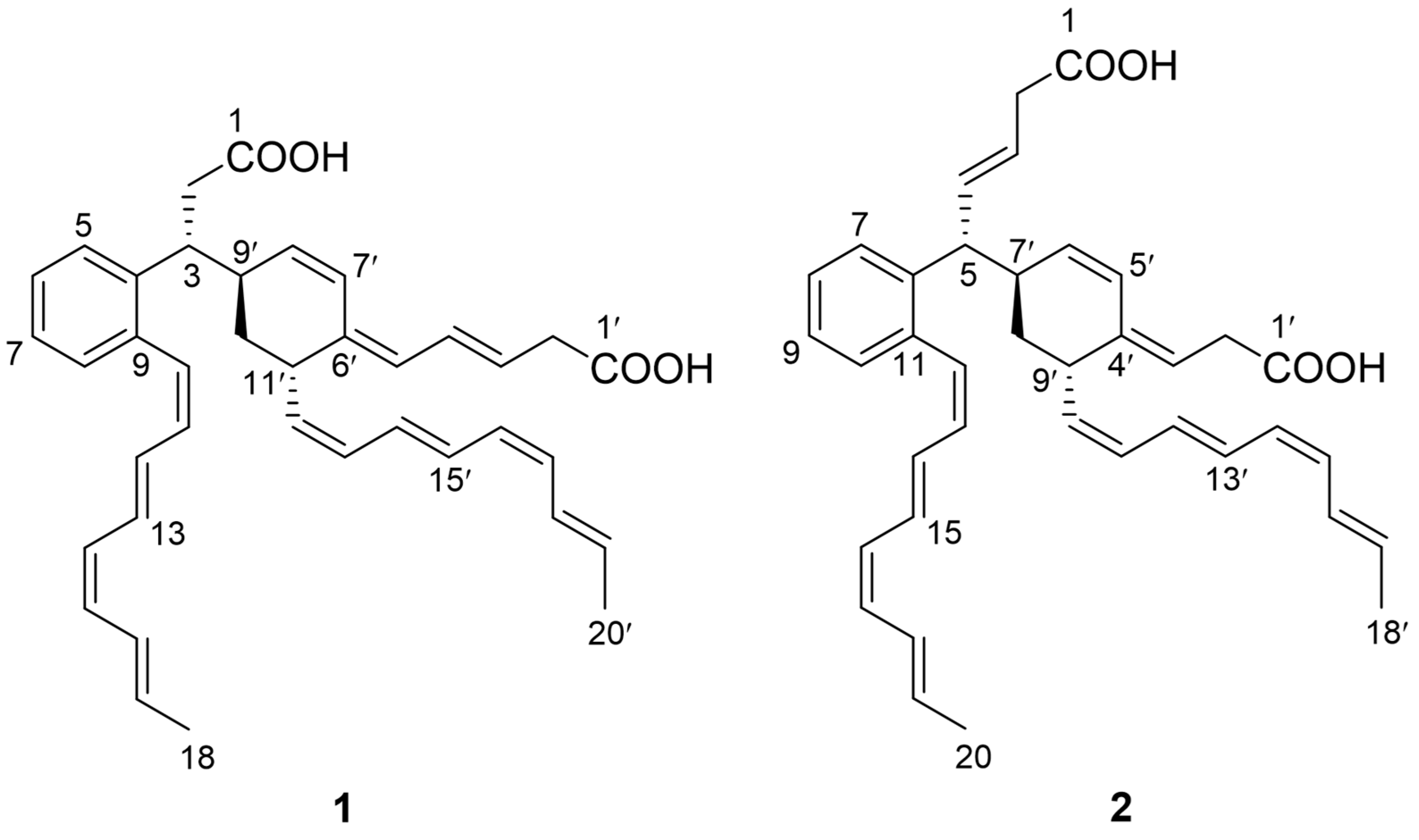
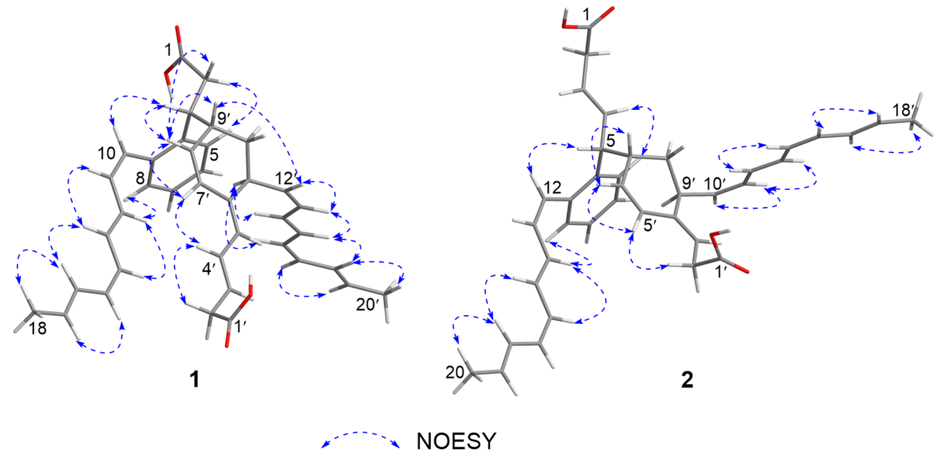
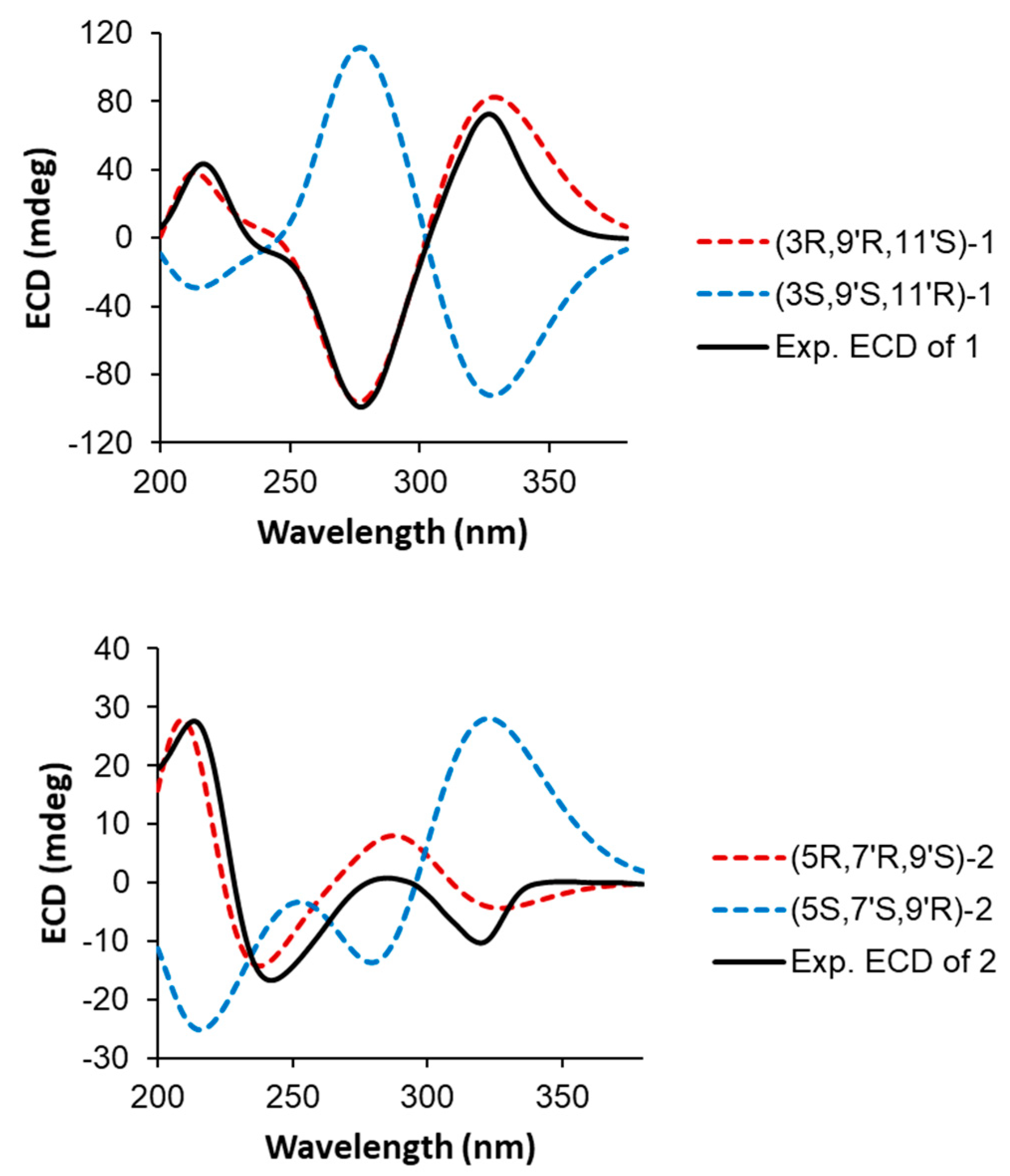
2. Results and Discussion

The ΔdtlA mutant strain, which was constructed in our previous study [1], was cultured for 50 mL, and the culture broth was extracted with EtOAc followed by HPLC-HRESIMS analysis (Figure S1). Except for youssoufene A1, two minor peaks (m/z 563 [M + H]+]) with similar UV-spectra were observed (Figures S1 and S2), which were proposed to be new dimeric youssoufene analogs. Then, large-scale fermentation (50 L) of the ΔdtlA mutant was conducted and afforded compounds 1 and 2.
Compound 1 was isolated as a yellow amorphous solid. The molecular formula of 1 was established as C38H42O4 on the basis of the HRESIMS data ([M + H]+ at m/z 563.3163, calcd 563.3161), indicating the presence of two additional olefinic carbons compared to youssoufene A1 [1]. The structure of 1 was determined by the NMR data collected in CD3OD. In the COSY spectrum of 1, two methylated olefinic 1H spin systems (H-10~H-18 and H-12’~H-20’) (Figure 2) were observed, suggesting the presence of two terminal methyl-octyltetraene chains. The HMBC correlations (Figure 2) from H-5 (δH 7.37) to C-7 (δC 125.6) and C-9 (δC 137.4), from H-8 (δH 7.19) to C-4 (δC 141.2) and C-6 (δC 126.9), and from H-10 (δH 6.83) to C-4, C-8 (δC 130.1) and C-9 revealed the existence of an 4,9-ortho-substituted aromatic ring with a methyl-octyltetraene chain at C-9. The COSY correlations between the methylene protons H2-2 (δH 2.75, 2.62) and the methine proton H-3 (δH 3.62) (Figure 2), together with the HMBC correlation (Figure 2) from H-5 to C-3 (δC 41.5), confirmed the C-2/C-3 fragment to be connected to the aromatic ring at C-4. The 1H spin systems of H2-2’ (δH 3.03)~H-5’ (δH 5.70) and H-7’ (δH 6.60)~H-20’ (δH 1.85) in COSY (Figure 2), together with the COSY correlation of H-3/H-9’ (δH 2.68), and the HMBC correlations (Figure 2) from H-7’, H-10’ (δH 1.62, 1.58) and H-12’ (δH 5.24) to the olefinic quaternary carbon C-6’ (δC 135.3), from H-7’ to C-9’ (δC 38.4) and C-11’ (δC 37.1), and from H-9’ to C-2 (δC 38.9), revealed that a 6’,9’,11’-tri-substituted cyclohexene moiety with a methylated octyltetraene chain at C-11’ was connected to C-3. However, unexpectedly, the carboxyl carbons (C-1 and C-1’) were not detected in the NMR spectra of 1 recorded in CD3OD. Then, we obtained the NMR data of 1 in DMSO-d6, from which the presence of carboxyl carbon C-1’ was confirmed by the HMBC correlation from H-2’ (δH 2.80) to C-1’ (δC 174.7) (Figure 2, Table S1), while C-1 was not detected, even in DMSO-d6. Based on these assigned substructures, together with the HRESIMS data, 1 was determined to be composed of two CL units with terminal carboxyl groups, which formed a dearomatic carbon-bridged dimeric CL skeleton composed of youssoufenes B1 and B3 (or serpentene).
The NOE correlations of H-10/H-11, H-11/H-13, H-12/H-14, H-13/H-16, H-16/H-18, H-11’/H-14’, H-12’/H-13’, H-13’/H-15’, H-17’/H-19′ and H-18′/H-20′ (Figure 3), together with the vicinal coupling constant values (3JH,H) of 3J10,11 (11.3 Hz), 3J14,15 (10.8 Hz), 3J12′,13′ (10.3 Hz) and 3J16′,17′ (10.8 Hz) (Table 1) revealed both of the methylated octyltetraene chains in 1 share the same double-bond geometries with youssoufene A1 [1], which were determined as 10-Z, 12-E, 14-Z, 16-E, 12′-Z, 14′-E, 16′-Z and 18′-E, respectively. The geometries of 3′-E, 5′-E and 7′-Z were determined by combination of the NOEs of H-4′/H-7′, H-5′/H-11′ and H-7′/H-8′ (Figure 3), and the 3JH,H values of 3J3′,4′ (14.8 Hz) and 3J7′,8′ (10.2 Hz). Moreover, the NOE correlation of H-9′/H-12′ suggested anti-configuration between H-9′ and H-11′, and the NOEs of H-8′/H-9′ and H-8′/H-3 supported syn-configuration between H-3 and H-9′.
The absolute configurations of C-3, C-9′ and C-11′ in 1 were determined by ECD calculations performed on the CAM-B3LYP/6-31G(d) level of theory with Gaussian 09. The conformers of (3R,9′R,11′S)-1 or (3S,9′S,11′R)-1 used in the calculations were selected from the conformers built with SYBYL-X 2.0, in which distances between each atom supported the NOESY data. The calculated ECD curve of (3R,9′R,11′S)-1 was in good agreement with the experimental ECD data (Figure 4). Thus, compound 1 was finally identified as a new dimeric CL, named youssoufene A2. The 1H and 13C NMR chemical shift values of 1 are listed in Table 1.
Compound 2 was isolated as a yellow amorphous solid. The molecular formula of 2 was established as C38H42O4 on the basis of the HRESIMS data ([M + H]+ at m/z 563.3167, calcd 563.3161), indicative of an isomer of youssoufene A2. Compound 2 shares similar NMR data with those of youssoufenes A1 and A2. The difference between 2 and youssoufene A1 was the additional C-3 (δC 125.3)/C-4 (δC 134.1) double bond in 2, which showed HMBC correlations to H-5 (δH 3.58). Thus, compound 2 was determined to comprise a dimeric CL skeleton with dearomatic youssoufene B1 connected to youssoufene B3/serpentene unit at C-5. By combination of the NOE correlations (Figure 3) and 3JH,H values (Table 1), the double-bond geometries in 2 were determined to be 3-E, 12-Z, 14-E, 16-Z, 18-E, 3′-E, 5′-Z, 10′-Z, 12′-E, 14′-Z and 16′-E, respectively. The absolute configurations of C-5, C-7′ and C-9′ in 1 were determined by ECD calculations performed on the CAM-B3LYP/6-31G(d) level of theory with Gaussian 09. The calculated ECD curve of (5R,7′R,9′S)-2 was in good agreement with the experimental ECD data of 2 (Figure 4). Thus, compound 2 was identified as a new dimeric CL, named youssoufene A3. The 1H and 13C NMR chemical shift values of 2 are listed in Table 1.
In the antibacterial assay, both youssoufenes A2 (1) and A3 (2) showed growth inhibition against multi-drug-resistant Enterococcus faecalis CCARM 5172 with an MIC value of 22.2 μM (Table S2), but not active against Staphylococcus aureus CCARM 3090 or Escherichia coli CCARM 1009. These results were comparable to that of youssoufene A1, which displayed over 4-fold-increased activity compared to youssoufenes B1–B4 [1]. This result demonstrated that the dimeric CL skeleton endows the youssoufene A-type with notably enhanced antibacterial activities compared to their monomeric B-type structures. While we have demonstrated that monomeric youssoufenes B1-B4 are assembled via an unusual type II polyketide synthetase pathway [11], the diaromatic dimerization to afford youssoufene A-type structures remains unclear.

3. Materials and Methods

3.1. General Experimental Procedures

Optical rotations were recorded with a JASCO P-1020 digital polarimeter (JASCO, Tokyo, Japan). UV spectra in MeOH were recorded on a PerkinElmer Lambda 35 (PerkinElmer, Waltham, MA, USA). Experimental ECD spectra in MeOH were recorded on a JASCO J-815 spectrometer (JASCO, Tokyo, Japan). IR spectra were measured on a Nicolet NEXUE470 FTIR (Thermo, Waltham, MA, USA). Then, 1D (1H and 13C) and 2D (COSY, HSQC, HMBC and NOESY) NMR spectra were recorded on a Bruker Avance III 600 spectrometer (Bruker, Billerica, MA, USA). Chemical shifts were reported with reference to the respective solvent peaks and residual solvent peaks (δH 3.31 and δC 49.0 for CD3OD; δH 2.50 and δC 39.5 for DMSO-d6). LC-MS experiments were performed on Agilent 1260 HPLC (Agilent, Santa Clara, CA, USA) system coupled with a Q-TOF Ultima Global GAA076 mass spectrometer (Waters, Milford, MA, USA). Preparative HPLC was performed on a Hitachi Chromaster System (Hitachi, Tokyo, Japan).

3.2. LC-MS-Based Production Analyses of the ΔdtlA Mutant Strain of S. youssoufiensis OUC6819

The ΔdtlA mutant strain was fermented for 50 mL in the medium (1% soluble starch, 2% glucose, 0.4% corn syrup, 1% yeast extract, 0.3% beef extract, 0.05% MgSO4·7H2O, 0.05% KH2PO4, 0.2% CaCO3, and 3.3% sea salt, pH = 7.0) at 30 °C, 220 rpm for 7 days. The fermentation supernatant was extracted twice with an equal volume of EtOAc. The resulting EtOAc extract was subjected to HPLC-HRESIMS analysis, eluting with a linear gradient of 20–100% B/A (phase B: 100% ACN + 0.1% HCOOH; phase A: H2O + 0.1% HCOOH; flow rate: 0.2 mL/min; wavelength: 300 nm) using an Agilent Eclipse Plus C18 (100 × 2.1 mm, 3.5 μm) (Agilent, Santa Clara, CA, USA) column to trace the youssoufene A analogs.

3.3. Fermentation, Extraction and Isolation of the Compounds

A total of 50 L of fermentation culture of the ΔdtlA mutant strain were prepared and extracted with EtOAc. The EtOAc extract (8.5 g) was partitioned between 90% MeOH and n-hexane to yield two residues. The aqueous MeOH layer (7.1 g) was applied to a reversed-phase (C18) open column (100 × 30 mm) chromatography to give 13 fractions (Fr.1–13) by eluting with gradient from 20% to 100% MeOH. The Fr. 9 (50.4 mg) was further subjected to semipreparative HPLC using a YMC ODS-A column (250 × 10 mm, 5 μm) eluting with 70% ACN to afford compounds 1 (1.1 mg, tR 51.9 min) and 2 (1.2 mg, tR 55.1 min).
Youssoufene A2 (1): yellow amorphous solid; [α]D25 +5.88 (c 0.08, MeOH); UV (MeOH) λmax (log ε) 208 (4.16), 238 (0.65), 280 (4.15), 299 (4.11), 305 (4.13), 314 (4.07), 320 (4.08) nm (Figure S2); ECD (c 0.5, MeOH) λmaxε) 216 (+14.83), 277 (−33.69), 326 (+24.77) nm (Figure S3); IR (KBr) νmax 3844.6, 3738.0, 3651.0, 3206.4, 1747.2, 1652.6, 1521.1, 1160.1, 1064.2, 980.3, 878.1, 712.6, 608.2, 510.3, 435.5 (Figure S4); 1H and 13C NMR data, see Table 1; HRESIMS m/z 563.3163 [M + H]+ (calcd for C38H43O4, 563.3161).
Youssoufene A3 (2): yellow amorphous solid; [α]D25 −42.38 (c 0.08, MeOH); UV (MeOH) λmax (log ε) 207 (4.18), 211 (4.14), 212 (4.15), 227 (4.03), 244 (4.07), 263 (3.98), 292 (4.32), 296 (4.31), 304 (4.45), 312 (4.35), 319 (4.42) nm (Figure S2); ECD (c 0.5, MeOH) λmaxε) 213 (+9.39), 242 (−5.64), 285 (0.29), 319 (−3.47), 353 (+0.07) nm (Figure S17); IR (KBr) νmax 3852.0, 3742.4, 3612.0, 3134.0, 1745.7, 1648.8, 1520.6, 1174.1, 1063.6, 979.3, 883.5, 739.7, 650.9, 569.9, 463.9 cm−1 (Figure S18); 1H and 13C NMR data, see Table 1; HRESIMS m/z 563.3167 [M + H]+ (calcd for C38H43O4, 563.3161).

3.4. Computational Methods

Conformational searches were run by the “Random search” procedure implemented in the SYBYL-X 2.0 program (Certara, Princeton, NJ, USA) using the Molecular Merck force field (MMFF94). Among the generated conformers of each compound, the conformers that well supported the NOESY data were selected and were subjected to geometry optimization with DFT calculations at the TZVP/6-31G(d) level using the Gaussian 09 program (Gaussian, Inc., Pittsburgh, PA, USA). The TD calculations were performed on each optimized conformer using the long-range-corrected hybrid CAM-B3LYP. The number of excited states per molecule was 50. Solvent effects were considered by using the polarizable continuum model (PCM) for MeOH. The ECD spectra were generated by the program GaussView 5.0 (Gaussian, Inc., Pittsburgh, PA, USA).

3.5. Antibacterial Activity Assay

The antibacterial activity of compounds 1 and 2 was evaluated using the MIC (minimum inhibitory concentration) assay. The multi-drug-resistant Enterococcus faecalis CCARM 5172, Staphylococcus aureus CCARM 3090 and Escherichia coli CCARM 1009 strains were purchased from Culture Collection of Antimicrobial Resistant Microbes (Seoul Women’s University, Seoul, Korea). The strain was grown overnight at 37 °C in LB medium and diluted with LB broth to 106 cfu/mL. Then, 10 μL of the compound solutions with different concentrations in MeOH were dispensed into 190 μL of the cell suspension in the 96-well plates. LB broth was used as a blank. MeOH was used as a negative control; ciprofloxacin and tetracycline were used as positive controls. The bacterial growth was measured after 18 h of incubation at 37 °C on a microplate reader at a wavelength of 600 nm. Each assay was performed in triplicate.

4. Conclusions

In summary, two new dimeric cinnamoyl lipids youssoufenes A2 (1) and A3 (2), which feature with a unique dearomatic carbon-bridge, were isolated from the ΔdtlA mutant strain of marine-derived S. youssoufiensis OUC6819. Compounds 1 and 2 exhibited growth inhibition against multi-drug-resistant E. faecalis CCARM 5172 (MIC = 22.2 μM), which was comparable to the positive controls, and meanwhile, notably higher than its monomeric form. These results indicated that yousoufene A-type could be an interesting new chemical scaffold for the development of next-generation antibacterial drugs.

Supplementary Materials

The following supporting information can be downloaded at: https://www.mdpi.com/article/10.3390/md20060394/s1, Figures S1–S25: the spectroscopic data (HPLC-HRESIMS, NMR, UV and ECD) of compounds 1 and 2; Table S1: NMR data of compound 1 in DMSO-d6; Table S2: antibacterial activity of compounds 1 and 2; Table S3: bacteria and plasmids; Tables S4–S7: the coordinates of the selected conformers for ECD calculations. Refs. [12,13] are listed in Table S3.

Author Contributions

J.L. contributed to fermentation, compound isolation, antibacterial activity assay, and manuscript preparation; H.L. contributed to structure elucidation, manuscript writing and editing, and project supervision; Z.L. involved in ECD calculation; T.L. involved in construction of the mutant strain; F.X. involved in manuscript review; W.L. contributed to manuscript review and editing, project administration, and funding acquisition. All authors have read and agreed to the published version of the manuscript.

Funding

This research was funded by the National Natural Science Foundation of China (NSFC) under grants U1706206, 32070054, 82073720, 81991525, and 31570032, the National Key R&D Program of China (2019YFC0312501), and the Foundation of Guangdong Key Laboratory of Marine Materia Medica (LMM2020-3).

Institutional Review Board Statement

Not applicable.

Informed Consent Statement

Not applicable.

Data Availability Statement

The data presented in this study are available on request from the corresponding author.

Conflicts of Interest

The authors declare no conflict of interest.

References

  1. Li, H.; Liu, J.; Deng, Z.; Li, T.; Liu, Z.; Che, Q.; Li, W. Genetic Manipulation of an Aminotransferase Family Gene dtlA Activates Youssoufenes in Marine-Derived Streptomyces youssoufiensis. Org. Lett. 2020, 22, 729–733, Correction in Org. Lett. 2020, 22, 7773. [Google Scholar] [CrossRef] [PubMed]
  2. Bae, M.; Kim, H.; Moon, K.; Nam, S.-J.; Shin, J.; Oh, K.-B.; Oh, D.-C. Mohangamides A and B, New Dilactone-Tethered Pseudo-dimeric Peptides Inhibiting Candida albicans Isocitrate Lyase. Org. Lett. 2015, 17, 712–715. [Google Scholar] [CrossRef] [PubMed]
  3. Shi, J.; Liu, C.; Zhang, B.; Guo, W.; Zhu, J.; Chang, C.-Y.; Zhao, E.; Jiao, R.; Tan, R.; Ge, H. Genome Mining and Biosynthesis of Kitacinnamycins as a STING Activator. Chem. Sci. 2019, 10, 4839–4846. [Google Scholar] [CrossRef] [PubMed] [Green Version]
  4. Bae, M.; Oh, J.; Bae, E.; Oh, J.; Hur, J.; Suh, Y.-G.; Lee, S.; Shin, J.; Oh, D.-C. WS9326H, an Antiangiogenic Pyrazolone-Bearing Peptide from an Intertidal Mudflat Actinomycete. Org. Lett. 2018, 20, 1999–2002. [Google Scholar] [CrossRef] [PubMed]
  5. Liu, Q.; Liu, Z.; Sun, C.; Shao, M.; Ma, J.; Wei, X.; Zhang, T.; Li, W.; Ju, J. Discovery and Biosynthesis of Atrovimycin, an Antitubercular and Antifungal Cyclodepsipeptide Featuring Vicinal-Dihydroxylated Cinnamic Acyl Chain. Org. Lett. 2019, 21, 2634–2638. [Google Scholar] [CrossRef] [PubMed]
  6. Sun, C.; Yang, Z.; Zhang, C.; Liu, Z.; He, J.; Liu, Q.; Zhang, T.; Ju, J.; Ma, J. Genome Mining of Streptomyces atratus SCSIO ZH16: Discovery of Atratumycin and Identification of its Biosynthetic Gene Cluster. Org. Lett. 2019, 21, 1453–1457. [Google Scholar] [CrossRef] [PubMed]
  7. Ritzau, M.; Drautz, H.; Zähner, H.; Zeeck, A. Serpentene, a Novel Polyene Carboxylic Acid from Streptomyces. Liebigs An. Chem. 1993, 266, 433–435. [Google Scholar] [CrossRef]
  8. Ohlendorf, B.; Schulz, D.; Beese, P.; Erhard, A.; Schmaljohann, R.; Imhoff, J. Diacidene, a Polyene Dicarboxylic Acid from a Micromonospora Isolate from the German Wadden Sea. Naturforsch C J. Biosci. 2012, 67, 445–450. [Google Scholar] [CrossRef] [PubMed] [Green Version]
  9. Mehnaz, S.; Saleem, R.; Yameen, B.; Pianet, I.; Schnakenburg, G.; Pietraszkiewicz, H.; Valeriote, F.; Josten, M.; Sahl, H.-G.; Franzblau, S.; et al. Lahorenoic Acids A–C, Ortho-Dialkyl-Substituted Aromatic Acids from the Biocontrol Strain Pseudomonas aurantiaca PB-St2. J. Nat. Prod. 2013, 76, 135–141. [Google Scholar] [CrossRef] [PubMed]
  10. Pohle, S.; Appelt, C.; Roux, M.; Fiedler, H.-P.; Süssmuth, R. Biosynthetic Gene Cluster of the Non-Ribosomally Synthesized Cyclodepsipeptide Skyllamycin: Deciphering Unprecedented Ways of Unusual Hydroxylation Reactions. J. Am. Chem. Soc. 2011, 133, 6194–6205. [Google Scholar] [CrossRef]
  11. Deng, Z.; Liu, J.; Li, T.; Li, H.; Liu, Z.; Dong, Y.; Li, W. An Unusual Type II Polyketide Synthase System Involved in Cinnamoyl Lipid Biosynthesis. Angew. Chem. 2021, 60, 153–158. [Google Scholar] [CrossRef]
  12. Che, Q.; Zhu, T.; Qi, X.; Mándi, A.; Kurtán, T.; Mo, X.; Li, D. Hybrid Isoprenoids from a Reeds Rhizosphere Soil Derived Actinomycete Streptomyces sp. CHQ-64. Org. Lett. 2012, 14, 3438–3441. [Google Scholar]
  13. Yao, T.; Liu, J.; Liu, Z.; Li, T.; Li, H.; Che, Q.; Zhu, T.; Li, D.; Li, W. Genome mining of cyclodipeptide synthases unravels unusual tRNA-dependent diketopiperazine-terpene biosynthetic machinery. Nat. Commun. 2018, 9, 4091. [Google Scholar]
Figure 1. Structures of compounds 1 and 2.
Marinedrugs 20 00394 g001
Figure 2. COSY and key HMBC correlations of 1 and 2.
Marinedrugs 20 00394 g002
Figure 3. Key NOESY correlations of 1 and 2.
Marinedrugs 20 00394 g003
Figure 4. Experimental and calculated ECD spectra of 1 and 2.
Marinedrugs 20 00394 g004
Table 1. 1H (600 MHz) and 13C (150 MHz) NMR data of 1 and 2 in CD3OD a.
12
PositionδC, typeδH (J in Hz)PositionδC, typeδH (J in Hz)
1b-1b-
238.9, CH22.75, 2.62, m2b, CH22.95, m
341.5, CH3.62, m3125.3, CH5.66, m
4141.2, C-4134.1, CH5.74, m
5126.9, CH7.37, d (7.8)549.6, CH3.58, m
6126.9, CH7.27, t (7.5)6141.4, C-
7125.6, CH7.22, t (7.2)7127.1, CH7.39, d (7.6)
8130.1, CH7.19, d (7.4)8127.2, CH7.29, td (7.0, 0.9)
9137.4, C-9125.3, CH7.21, m
10129.9, CH6.83, d (11.3)10129.9, CH7.19, m
11130.8, CH6.43, m11136.5, C-
12129.3, CH6.42, m12129.3, CH6.60, m
13129.9, CH6.83, t (13.0)13130.9, CH6.46, m
14127.1, CH5.84, t (10.8)14129.1, CH6.33, m
15129.6, CH5.98, t (10.8)15129.8, CH6.85, dd (14.2, 11.9)
16127.2, CH6.64, m16127.0, CH5.83, m
17130.3, CH5.79, m17129.5, CH5.97, m
1817.3, CH31.85, d (6.7)18127.2, CH6.63, m
1′a-19130.2, CH5.78, m
2′40.5, CH23.03, m2017.3, CH31.84, d (6.7)
3′127.7, CH5.73, m1′a-
4′127.4, CH6.48, dd (14.8, 11.9)2′35.1, CH23.03, m
5′125.3, CH5.70, m3′120.1, CH5.41, m
6′135.3, C-4′136.5, C-
7′123.9, CH6.60, d (10.2)5′123.1, CH6.33, m
8′131.2, CH5.71, t (10.9)6′131.7, CH5.49, d (10.6, 9.1)
9′38.4, CH2.68, m7′38.3, CH2.71, m
10′32.7, CH21.62, m
1.58, m
8′33.6, CH21.74, m
1.63, m
11′37.1 CH3.19, m9′37.5, CH3.48, m
12′133.7, CH5.24, t (10.3)10′134.1, CH5.41, m
13′128.4, CH6.05, t (10.3)11′128.0, CH6.08, t (11.0)
14′128.1, CH6.30, dd (14.1, 12.0)12′127.9, CH6.49, m
15′128.5, CH6.70, dd (14.6, 10.8)13′128.4, CH6.71, m
16′127.1, CH5.96, m14′127.2, CH5.97, m
17′129.7, CH5.98, t (10.8)15′130.2, CH5.97, m
18′127.2, CH6.64, m16′127.2, CH6.63, m
19′130.5, CH5.79, m17′130.7, CH5.78, m
20′17.3, CH31.85, d (6.7)18′17.3, CH31.84, d (6.7)
a 13C chemical shifts were obtained by combination of 13C NMR, HSQC and HMBC analysis; b not detected.
Publisher’s Note: MDPI stays neutral with regard to jurisdictional claims in published maps and institutional affiliations.

