Article

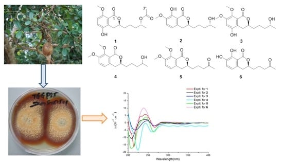
Talaromarins A–F: Six New Isocoumarins from Mangrove-Derived Fungus Talaromyces flavus TGGP35

1
Key Laboratory of Tropical Medicinal Resource Chemistry of Ministry of Education, College of Chemistry and Chemical Engineering, Hainan Normal University, Haikou 571158, China
2
Key Laboratory of Tropical Medicinal Plant Chemistry of Hainan Province, Haikou 571158, China
3
Key Laboratory of Advanced Materials of Tropical Island Resources of Ministry of Education, School of Chemical Engineering and Technology, Hainan University, Haikou 570228, China
*
Authors to whom correspondence should be addressed.
These authors contributed equally to this work.
Mar. Drugs 2022, 20(6), 361; https://doi.org/10.3390/md20060361
Submission received: 22 April 2022 / Revised: 24 May 2022 / Accepted: 24 May 2022 / Published: 27 May 2022
(This article belongs to the Special Issue Bio-Active Products from Mangrove Ecosystems)

Abstract

:
Six new isocoumarin derivative talaromarins A-F (16), along with 17 known analogues (723), were isolated from the mangrove-derived fungus Talaromyces flavus (Eurotiales: Trichocomaceae) TGGP35. Their structures were identified by detailed IR, UV, 1D/2D NMR and HR-ESI-MS spectra. The absolute configurations of new compounds were determined by the modified Mosher’s method and a comparison of their CD spectra with dihydroisocoumarins described in the literature. The antioxidant, antibacterial, anti-phytopathogenic and inhibitory activity against α-glucosidase of all the isolated compounds were tested. Compounds 611, 1719 and 2122 showed similar or better antioxidant activity than the IC50 values ranging from 0.009 to 0.27 mM, compared with the positive control trolox (IC50 = 0.29 mM). Compounds 10, 18, 21 and 23 exhibited strong inhibitory activities against α-glucosidase with IC50 values ranging from 0.10 to 0.62 mM, while the positive control acarbose had an IC50 value of 0.5 mM. All compounds showed no antibacterial or anti-phytopathogenic activity at the concentrations of 50 μg/mL and 1 mg/mL, respectively. These results indicated that isocoumarins will be useful to developing antioxidants and as diabetes control agents.

Graphical Abstract

1. Introduction

Marine fungi, particularly the mangrove-derived fungi, have proven to be a prolific source of structurally novel and biologically active secondary metabolites, which increasingly attracted the attention of both pharmaceutical and natural product chemists [1,2,3]. Up to now, more than 1400 new secondary metabolites produced by mangrove-derived fungi have been reported, including polyketides, alkaloids, terpenes and so on, and more than 40% of secondary metabolites displayed cytotoxic, antibacterial and insecticidal activities etc. [3,4,5,6,7,8]. Among them, isocoumarins are lactone-type derivatives derived from the polyketide pathway [9,10], and they possess a wide range of pharmacological activities such as antibacterial 7-hydroxyoospolactone and parapholactone [11], anti-inflammatory (±)-prunomarin A [12], cytotoxic lunatinin [13], insecticidal peniciisocoumarins A and B, antiplasmodial monocerin [14], antioxidant and α-glucosidase inhibitory penicimarin N [15], brine shrimp (Artemia salina) lethal and broad-spectrum antimicrobial penicimarins G–K [16,17], penicoffrazins B and C [18].
The genus of Talaromyces has been studied and applied as a biocontrol agent, as a rich source of chitinolytic enzymes and producer of secondary metabolites [19,20,21]. Different classes of bioactive secondary metabolites have been found from the genus of Talaromyces [19,20,21], including some bioactive isocoumarins, such as selective antimigratory talarolactone A [22], α-glucosidase inhibitory sescandelin B and 3,4-dimethyl-6,8-dihydroxyisocoumarin [23], antibacterial tratenopyrone [24], antibacteria and antifungi (-)-8-hydroxy-3-(4-hydroxypentyl)-3,4-dihydroisocoumarin [25]. These results indicated that the genus of Talaromyces can be used for the control of pathogenic bacteria, phytopathogenic microorganisms, diabetes control agents and so on [19,26].
As part of our continuing exploration of structurally novel and biologically interesting secondary metabolites from mangrove-derived fungi [27,28,29,30,31,32,33], a fungus Talaromyces flavus TGGP35 obtained from the medicinal mangrove plant Acanthus ilicifolius attracted our attention, because of the EtOAc extract of T. flavus TGGP35 showed potent antioxidant activity. A chemical investigation of this fungus led to the identification of six new isocoumarins talaromarins A-F (16) and 17 known analogues (723) (Figure 1). Herein, we report the isolation, structure identification, and bioactivities of these compounds.

2. Results and Discussion

Compound 1 was isolated as a colorless oil. The molecular formula of 1 was established as C17H22O6 (seven degrees of unsaturation) by its HR-ESI-MS spectrum at m/z 323.1475 [M + H]+ (calcd for C17H23O6, 323.1472). The IR spectrum showed the presence of a hydroxyl group (3432 cm−1) and an aromatic ring (1743, 1618 and 1388 cm−1) in 1. The 1H-NMR data (Table 1) displayed a pair of ortho coupled aromatic protons at δH 7.00 (d, J = 8.8 Hz) and 6.78 (d, J = 8.8 Hz), one methoxyl group at δH 3.88 (s), two oxygenated methine groups at δH 4.90 (m) and 4.34 (m), four methylene groups at δH [3.08 (m) and 2.64 (m)], 1.85 (m), 1.57 (m) and 1.52 (m), one methyl group at δH 1.22 (d, J = 6.4 Hz). The combination of 13C NMR and DEPT data (Table 2) exhibited 17 carbon resonances, including two ester carbonyls at δC (171.1 and 163.1), six aromatic carbons at δC (155.5, 145.3, 128.4, 121.2, 114.6 and 111.5), one methoxyl group at δC (56.6), two oxygenated methine groups at δC (77.4 and 70.9), four methylene groups at δC (35.7, 34.7, 28.0 and 21.0), two methyl groups at δC (21.6 and 20.1). The above 1D NMR spectroscopic data indicated that 1 had an isocoumarin skeleton structure, and 1 is similar to penicimarin G (12) [16]. The major difference was the presence of an acetoxy group at [(δC 171.1 (C), 21.6 (CH3) and δH 2.04, (s)] in 1, indicating that the hydroxyl group in 12 was replaced by an acetoxy group in 1. The 1H-1H COSY correlations of CH2(2′)–CH2(3′)−CH(4′)−CH3(5′), combined with the HMBC correlations from H-5′ to C-4′/C-3′ (Figure 2), confirmed the acetoxy group connected at C-4′ in 1, and the planar structure of 1 was determined (Figure 1).
The absolute configurations of C-3 and C-4′ in 1 were determined by chemical hydrolysis, modified Mosher’s method and a comparison of CD spectra with dihydroisocoumarins described in the literature [28,34]. The major hydrolysis product (1a) of 1 was obtained with K2CO3 and anhydrous ethanol at 28 °C for 1.5 h (Figure 3), and 1a showed the same planar structure with 12 [16]. The modified Mosher’s method was used to determine the configuration of C-4′ for 1a. The differences in 1H NMR chemical shifts of 1a between (S)- and (R)-MTPA esters (Δδ = δSδR) were calculated to assign the absolute configuration of C-4′ to be R (Figure 4), the same as 12 [16]. The negative cotton effect at 266 nm suggested the R configuration at C-3 (Figure 5), by comparison with data for dihydroisocoumarins described in the literature [34]. Thus, the structure of 1 was determined and named talaromarin A.
Compound 2 was isolated as a white powder. The molecular formula was deduced to be C15H20O5 on the basis of HR-ESI-MS spectrum, implying six degrees of unsaturation. According to the IR spectrum, the hydroxyl group (3414 cm−1) and aromatic rings (1668, 1619, 1586, 1502 and 1442 cm−1) were observed. The 1H and 13C NMR spectroscopic data (Table 1 and Table 2) revealed that 2 also belonged to the isocoumarin class and had a similar structural relationship to penicimarin M [17], except for the presence of one oxygenated methine at [δH 3.80 (m), δC 67.9 (CH)] for C-4′, and the absence of a carbonyl group at δC 208.4 (C) in 2. The above results suggested a carbonyl group in penicimarin M was replaced by an oxygenated methine group in 2. Furthermore, the 1H-1H COSY correlations of H-5′ to H-4′ and H-3′ to H-2′/H-4′, and the HMBC correlations from the methyl H-5′ to C-3′/C-4′ established the oxygenated methine at C-4′ (Figure 2). The absolute configuration of C-4′ was determined as R by Mosher’s method [28] (Figure 4). The negative cotton effect at 265 nm suggested the R configuration at C-3 (Figure 5), by comparison with data for dihydroisocoumarins described in the literature [34]. Thus, the absolute configuration of 2 was established as 3R,4′R and named talaromarin B.
Compound 3 was isolated as a yellow oil, with the molecular formula C16H22O6 (six degrees of unsaturation) by the HR-ESI-MS spectrum. The IR spectrum indicated that 3 had hydroxyl group (3415 cm−1) and aromatic ring (1638, 1618 and 1384 cm−1). The 1H, 13C NMR data (Table 1 and Table 2) and HR-ESI-MS data revealed that 3 closely resembled those of 2, the main differences were the presence of a methoxyl group at [δH 3.89 (s), δC 61.8 (CH3)] in 3, and an aromatic proton signal at δH 6.62 (d, J = 8.0 Hz) was absented in 3. Moreover, the chemical shift of C-5 at δC (117.1) in 2 was downfield-shifted to δC (147.7) in 3. The HMBC correlations from 8-OCH3 to C-8, 7-OCH3 to C-7 and H-6 to C-8/C-7/C-4a (Figure 2), indicated the additional methoxyl group was attached to C-8 and the hydroxyl group was connected to C-5 (Figure 2). The absolute configurations of C-3 and C-4′ were determined to be the same R by comparison with CD data described in the literature [34] and Mosher’s method [28] (Figure 4 and Figure 5). Thus, the structure of 3 was determined and named talaromarin C.
Compound 4 was isolated as a yellow oil, and the molecular formula was established as C16H22O5 (six degrees of unsaturation) on the basis of its HR-ESI-MS spectrum. The IR spectrum of 4 displayed absorption bands for hydroxyl (3475 cm−1), carbonyl (1706 cm−1) and aromatic (1637 and 1617 cm−1) groups. The 1H and 13C NMR spectroscopic data (Table 1 and Table 2) suggested that 4 was very similar to those of 2, the only difference was the presence of a methoxyl group at [δH 3.96 (s) and δC 61.7 (CH3)] in 4. The location of the methoxyl groups at C-7 and C-8 were established by HMBC correlations from 7-OCH3 to C-7, 8-OCH3 to C-8, H-6 to C-8/C-4a and H-5 to C-7/C-8a/C-4 (Figure 2). The 1H-1H COSY, HMQC, and HMBC spectra established the complete assignment for 4 (Figure 2). Mosher’s method was tried to determine the absolute configuration of C-4′ in 4 [28]; unfortunately, the reaction failed. The negative cotton effect at 259 nm suggested the R configuration at C-3 (Figure 5) [34]. Thus, the structure of 4 was determined and named talaromarin D.
Compound 5 was isolated as a colorless oil and had the molecular formula of C16H20O5 as determined by HR-ESI-MS and NMR data, requiring seven degrees of unsaturation. The presence of an aromatic ring (1638, 1617 cm−1) was observed in the IR spectrum. The 1H and 13C NMR data (Table 1 and Table 2) of 5 were structurally similar to those of 4, except for the presence of a ketone carbonyl carbon at δC 208.7 (C) and the absence of one oxygenated methine carbon at [δC 68.1 (CH), δH 3.83 (m)] at C-4′ in 5, indicating that the oxygenated methine group in 4 was replaced by a carbonyl group for C-4′ in 5. The HMBC correlations from H-3′ to C-1′, H-5′ to C-4′/C-3′ further confirmed 5 with a carbonyl unit at C-4′ (Figure 2). The whole structure was further determined by the 2D NMR spectra (Figure 2). The absolute configuration of C-3 was determined as R by CD spectra (Figure 5) [34], and 5 was named talaromarin E.
Compound 6 was isolated as a yellow oil. The molecular formula of 6 was established as C14H16O5 (seven degrees of unsaturation) on the basis of its HR-ESI-MS data. The IR spectrum of 6 showed the hydroxyl group at 3461 and 3407 cm−1 and the aromatic rings at 1736 and 1671 cm−1. The 1H and 13C NMR spectroscopic data (Table 1 and Table 2) revealed that 6 was an isocoumarin derivative, with a similar structural relationship to peniciisocoumarin D (18), the obvious difference was that 6 lacked a methoxy group at C-8. The methoxy group (8-OMe) in 18 was replaced by a hydroxy group (8-OH) in 6, which was supported by the appearance of a hydrogen-bonded hydroxyl group at δH 11.00 (s). The HMBC correlations from the hydroxyl group 8-OH to C-8a/C-8/C-7 further confirmed the 8-OH was connected at C-8 (δC 149.1) (Figure 2). The 1H-1H COSY, HMQC, and HMBC spectra determined the complete assignment for 6 (Figure 2). The absolute configuration of C-3 was determined as R by CD spectroscopy (Figure 5) [34] and 6 was named talaromarin F.
By comparing physical and spectroscopic data with the literature, the 17 known homologues were identified as (R)-3-(3-hydroxypropyl)-8-hydroxy-3,4-dihydroisocoumarin (7) [35], peniciisocoumarin C (8) [28], 7-hydroxy-3-(3-hydroxypropyl)-8-methoxyisochroman-1-one (9) [34], 5,6-dihydroxy-3-(4-hydroxypentyl)-isochroman-1-one (10) [23], peniciisocoumarin F (11) [28], penicimarin G (12) [16], penicimarin C (13) [36], peniciisocoumarin A (14) [28], peniciisocoumarin B (15) [28], aspergillumarin A (16) [37], penicimarin H (17) [16], peniciisocoumarin D (18) [28], peniciisocoumarin G (19) [28], peniciisocoumarin E (20) [28], penicimarin N (21) [15], 6,8-dihydroxy-3-(2-hydroxypropyl)-7-methyl-1H-isochromen-1-one (22) [38] and pestalotiorin (23) [39].
The plausible biosynthetic pathways of compounds 123 were also proposed (Scheme 1). Isocoumarins were originated from the acetate-malonate or the polyketide synthase (PKS) pathway [10]. Peniciisocoumarin C (8), penicimarin C (13) and peniciisocoumarin A (14) would be biosynthesized from malonyl-CoA and acetyl-CoA and can be considered as intermediates which would be transformed to other isolated compounds from the fungus TGGP35. Compound 8 would be transformed to 7, 9 and 2023 by condensation, aromatization, esterification, dihydroxylation, methoxylation reaction and so on. Compound 13 would be transformed to 10 by dehydroxylation, demethoxylation and dehydroxylation reaction. Compounds 16, 1015 and 1719 would be deduced from 14 with dihydroxylation, methoxylation, methylation, hydroxylation, esterification, acetylation, Baeyer–Viliger oxidation reaction, etc.
The antioxidant activities of compounds 123 were evaluated. Compounds 611, 1719 and 22 exhibited potent antioxidant activity with the IC50 values ranging from 0.009 to 0.27 mM, while the positive control trolox was IC50 = 0.29 mM (Table 3).
The preliminary structure–activity relationship of the isolated isocoumarins was discussed. The substitution site, orientation of hydroxyl and methoxy groups on the skeleton of isocoumarins, and the substitution of different groups by side chain C-4′ can affect their antioxidant activity. Compound 2 which possesses a hydroxyl group on C-8, showed better antioxidant activity than that of 3 and 4, indicating that the chelated hydroxyl group at C-8 is important in enhancing antioxidant activity. Compound 6 possesses two hydroxyl groups at C-7 and C-8, which showed higher antioxidant activity than 16 (only one hydroxyl group on C-8), indicating that the hydroxyl group at C-7 is an important antioxidant activity site. Compounds 17 and 18 possess a ketone group at C-4′, which showed higher antioxidant activity than 12 and 13, which have an oxygenated methine at C-4′, suggesting that the substitution of different groups by side chain at C-4′ can affect antioxidant activity. Compound 22 possesses a hydroxyl group at C-8, showed higher antioxidant activity than 23, suggesting that the chelated hydroxyl group at C-8 is important in enhancing antioxidant activity. Furthermore, compounds 7, 8, 19 and 21 show antioxidant activities, which may be due to the existence of a chelated hydroxyl group.
Compounds 10, 18, 21, and 23 showed strong inhibitory activity against α-glucosidase with the IC50 values of 0.10, 0.38, 0.62 and 0.54 mM, respectively (the positive control acarbose with the IC50 value of 0.5 mM).
All compounds were tested for their antibacterial activities against Staphylococcus. aureus, Escherichia coli, S. epidermidis and Pseudomonas aeruginosa; however, all compounds showed no antibacterial activity at the concentration of 50 μg/mL. All compounds showed no biological activity against five phytopathogens (Colletotrichum asianum, C. acutatum, Fusarium oxysporum, Pyricularia oryzae and Curvularia australiensis) at the concentration of 1 mg/mL.

3. Materials and Methods

3.1. General Experimental Procedures

Optical rotations were measured on a JASCO P-1020 digital polarimeter (JASCO, Tokyo, Japan). IR spectra were recorded on a Thermo Nicolet 6700 (using KBr disks) spectrophotometer. UV spectra were measured on a PERSEE TU-1990 spectrophotometer. CD spectra were recorded on a Mos-450 spectrometer and 1D and 2D NMR spectra were recorded on a Bruker AV spectrometer (400 MHZ for 1H and 100 MHZ for 13C) and a JNM-ECZS spectrometer (600 HMZ for 1H and 125 MHZ for 13C). HR-ESI-MS spectra were obtained on a Q-TOF Ultima Global GAA076 LC mass spectrometer. ESI-MS spectra were recorded on a MAT-95-MS mass spectrometer. HPLC were used for the Agilent 1100 prep-HPLC system with an Agilent C18 analytical (9.4 × 250 mm, 5 μm) HPLC column. Silical gel (200−300 mesh, Qingdao Marine Chemical Factory, Qingdao, China) were used for column chromatography (CC). Sephadex LH-20 gel column (GE Healthcare, Bio-Sciences Corp, Piscataway, NJ, USA) were used for CC. Biological activities were tested on an ultra-clean workbench (Suzhou Sujing Company, Suzhou, China) and the biological activities’ results were tested with a full wavelength multifunctional microplate reader (Bio-Tek Instruments, Winooski, VT, USA). Methanol, ethyl acetate, petroleum ether, chloroform, dimethyl sulfoxide and other conventional chemical reagents were used in the experimental operation (Guangzhou Xilong Chemical Reagent Factory, Guangzhou, China).

3.2. Fungal Materials

The fungus TGGP35 was isolated from the stem of the mangrove plant Acanthus ilicifolius, which were collected in the Dongzhai Port, Haikou, Hainan Province in August, 2015. The fungus was identified according to its morphological characteristics and a molecular biological protocol by 18S rRNA amplification and sequencing of the ITS region. The sequence data have been submitted to GeneBank, with accession number MT071116, and the fungal strain was identified as Talaromyces flavus (Eurotiales: Trichocomaceae). The strains have been stored in the Key Laboratory of Tropical Medicinal Resources Chemistry of the Ministry of Education, School of Chemistry and Chemical Engineering, Hainan Normal University (PDA medium, stored at 4 °C).

3.3. Fermentation, Extraction and Isolation

The seed culture was prepared in potato liquid medium (6 g sea salt and 10 g peptone in 2 L of potato infusion, in 1 L × 4 erlenmeyer flasks each containing 500 mL seed medium), and incubated on a rotary shaker (170 rpm) for 4 days at 28 °C. In total, 20 mL seed culture was then transferred into 1 L erlenmeyer flasks with solid rice medium with a total of 100 bottles of fermentation (each flask contained 60 mL rice, 0.6 g sea salt and 1.0 g peptone) at 28 °C for 4 weeks. The whole rice fermented material was extracted three times with EtOAc, and then concentrated in vacuo to yield crude extracts (120.8 g). The total crude extracts were subjected to silica gel column chromatography (CC) eluted with petroleum ether/EtOAc (v/v, gradient 100:0–0:100) and EtOAc/MeOH (v/v, gradient 100:0–70:30) to generate fifteen fractions (Fr. A-Fr. L). Fr. F (20.5 g) was fractionated by silica gel CC (200-300 mesh) using a gradient elution of petroleum ether/EtOAc system (7:1–0:1) to obtain ten fractions (Fr. F1-Fr. F10) by the TLC analysis. Fr. F2 (10.3 g) was subjected to Sephadex LH-20 (Petroleum ether-CHCl3-MeOH, 2:1:1, v/v), and then to semi-preparative HPLC (MeOH-H2O, 70:30, v/v) to give compounds 1 (4.2 mg), 2 (5.5 mg), 3 (3.8 mg), 5 (4.5 mg) and 7 (4.7 mg). Subfraction Fr. F3 was further separated by semi-preparative HPLC (MeOH-H2O, 60:40, v/v) for four subfractions Fr. F3a-3d. Compounds 6 (4.7 mg), 8 (5.5 mg), 9 (5.8 mg), and 10 (3.5 mg) were isolated from subfraction Fr. F3a. Compounds 11 (3.7 mg), 14 (4.7 mg), 16 (5.5 mg), 17 (5.8 mg) and 19 (3.5 mg) for subfraction Fr. F3b. Compounds 12 (6.3 mg), 13 (3.2 mg) and 15 (5.7 mg) for subfraction Fr. F3c, and compounds 18 (6.8 mg), 19 (6.2 mg) and 21 (3.6 mg) for subfraction Fr. F3d. Fr. F4 (5.4 g) were subjected to Sephadex LH-20 (CHCl3-MeOH, 1:1, v/v), then used for semi-preparative HPLC (MeOH-H2O, 65:35, v/v) to give compound 20 (3.0 mg). Fr. F5 (2.4 g) was subjected to Sephadex LH-20 (CHCl3-MeOH, 1:1, v/v), then used for semi-preparative HPLC (MeOH-H2O, 35:65, v/v) to give compounds 22 (3.4 mg) and 23 (2.7 mg).
Talaromarin A (1): yellow oil; [α]25D −21.4 (c 0.10, MeOH); UV (MeOH) λmax (log ε) 330, 221 nm; IR (KBr) νmax 3432, 1716, 1618, 1383 cm−1; CD (c 0.05, MeOH) λmaxε) 243 (+5.68), 266 (−2.89) nm; 1H and 13C NMR data see Table 1 and Table 2; HR-ESI-MS m/z: 323.1475 [M + H]+, (C17H23O6, calcd. for 323.1472).
Talaromarin B (2): white powder; [α]25D −23.6 (c 0.10, MeOH); UV (MeOH) λmax (log ε) 330, 255, 223 nm; IR (KBr) νmax 3414, 1668, 1619, 1586, 1502, 1442 cm−1; CD (c 0.05, MeOH) λmaxε) 244 (+2.27), 265 (−2.17) nm; 1H and 13C NMR data see Table 1 and Table 2; HR-ESI-MS m/z: 279.1240 [M − H], (C15H19O5, calcd. for 279.1239).
TalaromarinC (3): yellow oil; [α]25D −22.0 (c 0.10, MeOH); UV (MeOH) λmax (log ε) 317, 215 nm; IR (KBr) νmax 3415, 1638, 1618, 1384 cm−1; CD (c 0.05, MeOH) λmaxε) 248 (+1.36), 266 (−2.01) nm; 1H and 13C NMR data see Table 1 and Table 2; HR-ESI-MS m/z: 311.1485 [M + H]+, (C16H23O6, calcd. for 311.1482).
Talaromarin D (4): yellow oil; [α]25D −21.8 (c 0.10, MeOH); UV (MeOH) λmax (log ε) 313, 222 nm; IR (KBr) νmax 3475, 1706, 1637, 1617 cm−1; CD (c 0.05, MeOH) λmaxε) 243 (−0.31), 259 (−6.25) nm; 1H and 13C NMR data see Table 1 and Table 2; HR-ESI-MS m/z: 295.1527 [M + H]+, (C16H23O5, calcd. for 295.1524).
Talaromarin E (5): colorless oil; [α]25D −24.4 (c 0.10, MeOH); UV (MeOH) λmax (log ε) 314, 219 nm; IR (KBr) νmax 1638,1617 cm−1; CD (c 0.05, MeOH) λmaxε) 245 (+3.93), 264 (−11.89) nm; 1H and 13C NMR data see Table 1 and Table 2; HR-ESI-MS m/z: 293.1370 [M + H]+, (C16H21O5, calcd. for 293.1374).
Talaromarin F (6): yellow oil; [α]25D −21.2 (c 0.10, MeOH); UV (MeOH) λmax (log ε) 328, 254, 223 nm; IR (KBr) νmax 3481, 3407, 1736, 1671 cm−1; CD (c 0.05, MeOH) λmaxε) 238 (+10.12), 267 (−2.07) nm; 1H and 13C NMR data see Table 1 and Table 2; HR-ESI-MS m/z: 265.1050 [M + H]+, (C14H17O5, calcd. for 265.1053).

3.4. Preparations of the (S)- and (R)-MTPA Esters of Compounds 1, 2 and 3

Compound 1 was hydrolyzed in anhydrous ethanol solution for 90 min with potassium carbonate in an equivalent ratio of 1:2; the mixed product after hydrolysis was purified by semi-preparative HPLC (MeOH-H2O, 70:30, v/v) to obtain 1a. The preparation of (S)- and (R)-MTPA ester derivatives of 1a, 2 and 3 was performed as described previously [28].
Hydrolysate of 1 (1a): 1H NMR (CDCl3, 400 MHz): δH 6.99 (1H, d, J = 8.8 Hz H-6), 6.79 (1H, d, J = 8.8 Hz, H-7), 4.26 (1H, m, H-3), 3.83 (1H, m, H-4′), 3.10 and 2.63 (2H, m, H-4), 1.90 (2H, m, H-1′), 1.70 (2H, m, H-2′), 1.50 (2H, m, H-3′), 1.21 (3H, d, J = 6.0 Hz, H-5′); 13C NMR (CDCl3, 100 MHz): δC 163.3, 159.4, 151.5, 145.1, 135.0, 128.5, 121.2, 111.4, 68.2, 56.6, 39.1, 34.9, 28.0, 23.8, 21.4; ESI-MS m/z 281.1 [M + H]+.
(S)-MTPA ester of 1a: 1H NMR (CDCl3, 600 MHz): δH 7.29 (1H, d, J = 9.0 Hz, H-6), 6.94 (1H, d, J = 9.0 Hz, H-7), 5.12 (1H, m, H-4′), 4.21 (1H, m, H-3), 2.64 and 2.49 (2H, m, H-4), 1.86 (2H, m, H-1′), 1.67 (2H, m, H-2′), 1.55 (2H, m, H-3′), 1.25 (3H, d, J = 6.6 Hz, H-5′); ESI-MS m/z 758.2 [M + Na]+.
(R)-MTPA ester of 1a: 1H NMR (CDCl3, 600 MHz): δH 7.31 (1H, d, J = 9.0 Hz, H-6), 6.94 (1H, d, J = 9.0 Hz, H-7), 5.11 (1H, m, H-4′), 4.09 (1H, m, H-3), 2.62 and 2.41 (2H, m, H-4), 1.83 (2H, m, H-1′), 1.64 (2H, m, H-2′), 1.53 (2H, m, H-3′), 1.33 (3H, d, J = 6.0 Hz, H-5′); ESI-MS m/z 758.2 [M + Na]+.
(S)-MTPA ester of 2: 1H NMR (CDCl3, 600 MHz): δH 7.01 (1H, d, J = 7.8 Hz, H-5), 6.63 (1H, d, J = 7.8 Hz, H-6), 5.17 (1H, m, H-4′), 4.52 (1H, m, H-3), 2.82 (2H, m, H-4), 1.88 (2H, m, H-1′), 1.62 (2H, m, H-2′), 1.50 (2H, m, H-3′), 1.29 (3H, d, J = 6.0 Hz, H-5′); ESI-MS m/z 758.3 [M + Na]+.
(R)-MTPA ester of 2: 1H NMR (CDCl3, 600 MHz): δH 7.01 (1H, d, J = 8.4 Hz, H-5), 6.62 (1H, d, J = 8.4 Hz, H-6), 5.16 (1H, m, H-4′), 4.42 (1H, m, H-3), 2.76 (2H, m, H-4), 1.81 (2H, m, H-1′), 1.59 (2H, m, H-2′), 1.46 (2H, m, H-3′), 1.37 (3H, d, J = 6.0 Hz, H-5′); ESI-MS m/z 774.3 [M + K]+.
(S)-MTPA ester of 3: 1H NMR (CDCl3, 600 MHz): δH 6.88 (1H, s, H-6), 5.14 (1H, m, H-4′), 4.21 (1H, m, H-3), 2.43 (2H, m, H-4), 1.72 (2H, m, H-1′), 1.52 (2H, m, H-2′), 1.43 (2H, m, H-3′), 1.35 (3H, d, J = 9.4 Hz, H-5′); ESI-MS m/z 766.6 [M + Na]+.
(R)-MTPA ester of 3: 1H NMR (CDCl3, 600 MHz): δH 6.86 (1H, s, H-6), 5.13 (1H, m, H-4′), 4.07 (1H, m, H-3), 2.33 (2H, m, H-4), 1.65 (2H, m, H-1′), 1.51 (2H, m, H-2′), 1.42 (2H, m, H-3′), 1.28 (3H, d, J = 9.4 Hz, H-5′); ESI-MS m/z 766.0 [M + Na]+.

3.5. Biological Assays

3.5.1. Antioxidant Activity

The antioxidant activity assay was based on the reported methods [15]. The assay was performed on a 96-well microplate, the reaction was initiated by adding 10 µL of sample solution to 200 µL of ABTS working solution. All test group gradients (including positive control) were of 2.0, 1.0, 0.5, 0.25 mg/mL, respectively. PBS buffer was used as the blank control, DMSO as the negative control, and trolox as the positive control (IC50 = 0.29 mM). The antioxidant effect was evaluated by a full wavelength multifunctional microplate reader measurement at 734 nm. The inhibition rate of each sample was calculated according to the following formula: inhibition rate = [(Ablank − Acompound)/Ablank] × 100%. Finally, the SPSS software was used to calculate the IC50 value.

3.5.2. Antibacterial Activity

All compounds were determined against four pathogenic bacteria: Staphylococcus aureus (ATCC 25923), Escherichia coli (ATCC 25922), S. epidermidis (ATCC 17749) and Pseudomonas aeruginosa (ATCC 17749). The concentration value of the test group and positive control was 1mg/mL by the microplate assay method [28]. The antibacterial effect was evaluated by a full wavelength multifunctional microplate reader measurement at 630 nm; the broth medium containing pathogenic bacteria was used as the blank group and DMSO as the negative control, ciprofloxacin was used as the positive control. The positive control ciprofloxacin showed antibacterial activities against four pathogenic bacteria S. aureus, E. coil, S. epidermidis and P. aeruginosa with the MIC values of 0.097, 0.78, 0.195 and 0.78 μg/mL, respectively.

3.5.3. Anti-Phytopathogenic Activity

All compounds were tested against five plant pathogens (Colletotrichum asianum, C. acutatum, Fusarium oxysporum, Pyricularia oryzae and Curvularia australiensis) by disk method [40]. DMSO was used as a negative control, carbendazim as a positive control. The concentration values of all test groups, negative control and positive control were 1 mg/mL; the anti-phytopathogenic results were recorded on a vernier caliper.

3.5.4. Inhibitory Activity against α-Glucosidase

The α-glucosidase inhibitory activity of the tested compounds was determined using the method in [17], with modifications for carrying it out in 96-well plates. The initial concentration of all test samples (including positive control and negative control) was 1 mg/mL, the optimized method was a mixture of 0.1 mM potassium phosphate buffer (pH = 6.8, 0.5 mL) and 10 mg/L α-glucosidase (100 µL), the testing sample (0.5 mL) was incubated at 37 °C for 5 min, and the 2.5 mM (4-nitrophenyl-β-D-glucopyranoside) PNPG (0.5 mL) was added, followed by mixing. The reaction was carried out at 37 °C for 15 min and then stopped by adding 0.2 M solution of Na2CO3 (0.75 mL). The inhibitory activity against α-Glucosidase was evaluated by a full wavelength multifunctional microplate reader measurement at 405 nm. Finally, inhibition rate = [(Acontrol − Acompound)/Acontrol] × 100%. The SPSS software was used to calculate the IC50 value. DMSO was used as the negative control and acarbose was used as the positive control (IC50 = 0.5 mM).

4. Conclusions

In summary, 23 secondary metabolites, including six new isocoumarin derivative talaromarins A-F (16) and 17 known analogues (723) were obtained from the mangrove-derived fungus Talaromyces flavus TGGP35. Compounds 611, 1719 and 22 exerted similar or better antioxidant activity than the positive control trolox (IC50 = 0.29 mM), with IC50 values ranging from 0.009 to 0.27 mM. Compounds 10, 18, 21 and 23 exhibited strong inhibitory activities against α-glucosidase with IC50 values ranging from 0.10 to 0.62 mM, while the IC50 value of positive control acarbose was 0.5 mM. All compounds showed no antibacterial or anti-phytopathogenic activity at the concentrations of 50 μg/mL and 1 mg/mL, respectively. Their plausible biosynthetic pathway and structure–activity relationships were also explored. Therefore, these findings demonstrate the potential of these active compounds as lead compounds for developing antioxidants and as diabetes control agents.

Supplementary Materials

The following supporting information can be downloaded at: https://www.mdpi.com/article/10.3390/md20060361/s1, D, 2D NMR and MS data of new compounds 16.

Author Contributions

J.C. and X.-C.Z. performed the experiments for the isolation, structure elucidation and prepared the manuscript; W.-N.Z. and B.W. contributed to the antioxidant activity and antimicrobial evaluation; Y.-P.L. contributed to part of the structure determination; J.L., M.-J.C. and G.-Y.L. organized documents and performed basic experiments; G.-L.H. and G.-Y.C. contributed the optimization of fermentation; C.-J.Z. and J.X. supervised the research work and revised the manuscript. All authors have read and agreed to the published version of the manuscript.

Funding

This work was supported by Key Science and Technology Program of Hainan Province (No. ZDKJ202008), the National Natural Science Foundation of China (Nos. 32160108, 41866005, 82160675 and 81973229), Key Research and Development Program of Hainan Province (No. ZDYF2021SHFZ270 and ZDYF2021SHFZ108), Hainan Provincial Natural Science Foundation of China (No. 220RC593), the Innovation Platform for Academicians of Hainan Province Specific Research Fund of The Innovation Platform for Academicians of Hainan Province (No. YSPTZX202030).

Institutional Review Board Statement

Not applicable.

Informed Consent Statement

Not applicable.

Data Availability Statement

The data presented in this study are available in this article and Supplementary Material.

Conflicts of Interest

The authors declare no competing financial interest.

References

  1. Carroll, A.R.; Copp, B.R.; Davis, R.A.; Keyzers, R.A.; Prinsep, M.R. Marine natural products. Nat. Prod. Rep. 2022, 18, 1R–49R. [Google Scholar] [CrossRef] [PubMed]
  2. Xu, W.F.; Wu, N.N.; Wu, Y.W.; Qi, Y.X.; Wei, M.Y.; Pineda, L.M.; Ng, M.G.; Spadafora, C.; Zheng, J.Y.; Lu, L.; et al. Structure modifcation, antialgal, antiplasmodial, and toxic evaluations of a series of new marine-derived 14-membered resorcylic acid lactone derivatives. Mar. Life Sci. Technol. 2022, 4, 88–97. [Google Scholar] [CrossRef]
  3. Chen, S.H.; Cai, R.L.; Liu, Z.M.; Gui, H.; She, Z.G. Secondary metabolites from mangrove-associated fungi: Source, chemistry and bioactivities. Nat. Prod. Rep. 2022, 39, 560–595. [Google Scholar] [CrossRef] [PubMed]
  4. Jiang, H.M.; Cai, R.L.; Zang, Z.M.; Yang, W.C.; Wang, B.; Zhu, G.; Yuan, J.; She, Z.G. Azaphilone derivatives with anti-inflammatory activity from the mangrove endophytic fungus Penicillium sclerotiorum ZJHJJ-18. Bioorg. Chem. 2022, 122, 105721. [Google Scholar] [CrossRef]
  5. Wu, Q.; Chang, Y.M.; Che, Q.; Li, D.H.; Zhang, G.J.; Zhu, T.J. Citreobenzofurans D-F and phomenones A-B: Five novel sesquiterpenoids from the mangrove-derived fungus Penicillium sp. HDN13-494. Mar. Drugs 2022, 20, 137. [Google Scholar] [CrossRef]
  6. Qin, X.Y.; Huang, J.G.; Zhou, D.X.; Zhang, W.X.; Zhang, Y.J.; Li, J.; Yang, R.Y.; Huang, X.S. Polyketide derivatives, guhypoxylonols A-D from a mangrove endophytic fungus Aspergillus sp. GXNU-Y45 that inhibit nitric oxide production. Mar. Drugs 2022, 20, 5. [Google Scholar] [CrossRef]
  7. Su, J.H.; Wang, M.Q.; Li, Y.Z.; Lin, Y.S.; Gu, J.Y.; Zhu, L.P.; Yang, W.Q.; Jiang, S.Q.; Zhao, Z.X.; Sun, Z.H. Rare cytochalasans isolated from the mangrove endophytic fungus Xylaria arbuscular. Fitoterapia 2022, 157, 105124. [Google Scholar] [CrossRef]
  8. Zeng, W.N.; Huang, G.L.; Cai, J.; Zheng, C.J. Secondary metabolites and bioactivities of Penicillium sp. sourced from mangrove from 2007 to 2020. Chin. J. Org. Chem. 2021, 41, 4255–4278. [Google Scholar] [CrossRef]
  9. Saeed, A. Isocoumarins, miraculous natural products blessed with diverse pharmacological activities. Eur. J. Med. Chem. 2016, 116, 290–317. [Google Scholar] [CrossRef]
  10. Noor, A.O.; Almasri, D.M.; Bagalagel, A.A.; Abdallah, H.M.; Mohamed, S.G.A.; Mohamed, G.A.; Ibrahim, S.R.M. Naturally occurring isocoumarins derivatives from endophytic fungi: Sources, isolation, structural characterization, biosynthesis, and biological activities. Molecules 2020, 25, 395. [Google Scholar] [CrossRef] [Green Version]
  11. Xu, X.L.; Li, J.P.; Zhang, K.; Wei, S.Z.; Lin, R.; Polyak, S.W.; Yang, N.; Song, F.H. New isocoumarin analogues from the marine-derived fungus Paraphoma sp. CUGBMF180003. Mar. Drugs 2021, 19, 313. [Google Scholar] [CrossRef] [PubMed]
  12. Zhang, X.Q.; Lu, Z.H.; Xia, G.R.; Song, W.M.; Guo, Z.Y.; Proksch, P. (+)-/(−)-Prunomarin A and (+)-pestalactone B, three new isocoumarin derivatives from the endophytic fungus Phomopsis prunorum. Tetrahedron. Lett. 2021, 75, 153205. [Google Scholar] [CrossRef]
  13. Ran, Y.Q.; Lan, W.J.; Qiu, Y.; Guo, Q.; Feng, G.K.; Deng, R.; Zhu, X.F.; Li, H.J.; Dong, J. Monarubins A-C from the marine shellfish-associated fungus Monascus ruber BB5. Mar. Drugs 2020, 18, 100. [Google Scholar] [CrossRef] [PubMed] [Green Version]
  14. Coronado, L.; Zhang, X.Q.; Dorta, D.; Escala, N.; Pineda, L.M.; Ng, M.G.; Olmo, E.D.; Wang, C.Y.; Gu, Y.C.; Shao, C.L.; et al. Semisynthesis, antiplasmodial activity, and mechanism of action studies of isocoumarin derivatives. J. Nat. Prod. 2021, 84, 1434–1441. [Google Scholar] [CrossRef] [PubMed]
  15. Zeng, W.N.; Cai, J.; Wang, B.; Chen, L.Y.; Pan, C.X.; Chen, S.J.; Huang, G.L.; Zheng, C.J. A new bioactive isocoumarin from the mangrove-derived fungus Penicillium sp. TGM112. J. Asian Nat. Prod. Res. 2021, 1–6. [Google Scholar] [CrossRef]
  16. Huang, G.L.; Zhou, X.M.; Bai, M.; Liu, Y.X.; Zhao, Y.L.; Luo, Y.P.; Niu, Y.Y.; Zheng, C.J.; Chen, G.Y. Dihydroisocoumarins from the mangrove-derived fungus Penicillium citrinum. Mar. Drugs 2016, 14, 177. [Google Scholar] [CrossRef] [Green Version]
  17. Mei, R.Q.; Wang, B.; Zeng, W.N.; Huang, G.L.; Chen, G.Y.; Zheng, C.J. Bioactive isocoumarins isolated from a mangrove-derived fungus Penicillium sp. MGP11. Nat. Prod. Res. 2022, 36, 1260–1265. [Google Scholar] [CrossRef]
  18. Cao, J.; Li, X.M.; Li, X.; Li, H.L.; Meng, L.H.; Wang, B.G. New lactone and isocoumarin derivatives from the marine mangrove-derived endophytic fungus Penicillium coffeae MA-314. Phytochem. Lett. 2019, 32, 1–5. [Google Scholar] [CrossRef]
  19. Proksa, B. Talaromyces flavus and its metabolites. Chem. Pap. 2010, 64, 696–714. [Google Scholar] [CrossRef]
  20. Nicoletti, R.; Trincone, A. Bioactive compounds produced by strains of Penicillium and Talaromyces of marine origin. Mar. Drugs 2016, 14, 37. [Google Scholar] [CrossRef] [Green Version]
  21. Nicoletti, R.; Salvatore, M.M.; Andolfi, A. Secondary metabolites of mangrove-associated strains of Talaromyces. Mar. Drugs 2018, 16, 12. [Google Scholar] [CrossRef] [PubMed] [Green Version]
  22. Yuan, W.H.; Teng, M.T.; Yun, Y.F.; Jiang, N.; Ma, L.; Sun, S.S.; Yuan, B.; Tang, J.; Wu, Q.Y.; Li, Q.; et al. Talarolactone A, an isocoumarin derivative fused with dihydrothiophene with selective antimigratory activity from the endolichenic fungus Talaromyces sp. J. Nat. Prod. 2020, 83, 1716–1720. [Google Scholar] [CrossRef] [PubMed]
  23. Chen, S.H.; Liu, Y.Y.; Liu, Z.M.; Cai, R.L.; Lu, Y.J.; Huang, X.S.; She, Z.G. Isocoumarins and benzofurans from the mangrove endophytic fungus Talaromyces amestolkiae possess α-glucosidase inhibitory and antibacterial activities. RSC Adv. 2016, 6, 26412–26420. [Google Scholar] [CrossRef]
  24. Buttachon, S.; May, Z.; War, W.; Dethoup, T.; Gales, L.; Pereira, J.A.; Silva, A.M.S.; Kijjoa, A. Secondary metabolites from the culture of the marine sponge-associated fungi Talaromyces tratensis and Sporidesmium circinophorum. Planta Med. 2016, 82, 888–896. [Google Scholar] [CrossRef] [PubMed] [Green Version]
  25. Miao, F.; Yang, R.; Chen, D.D.; Wang, Y.; Qin, B.F.; Yang, X.J.; Zhou, L. Isolation, identification and antimicrobial activities of two secondary metabolites of Talaromyces verruculosus. Molecules 2012, 17, 14091–14098. [Google Scholar] [CrossRef] [Green Version]
  26. Duo-Chuan, L.I.; Chen, S.; Jing, L.U. Purification and partial characterization of two chitinases from the mycoparasitic fungus Talaromyces flavus. Mycopathologia 2005, 159, 223–229. [Google Scholar] [CrossRef]
  27. Bai, M.; Zheng, C.J.; Chen, G. Austins-type meroterpenoids from a mangrove-derived Penicillium sp. J. Nat. Prod. 2021, 84, 2104–2110. [Google Scholar] [CrossRef]
  28. Bai, M.; Zheng, C.J.; Huang, G.L.; Mei, R.Q.; Wang, B.; Luo, Y.P.; Zheng, C.; Niu, Z.G.; Chen, G.Y. Bioactive meroterpenoids and isocoumarins from the mangrove-derived fungus Penicillium sp. TGM112. J. Nat. Prod. 2019, 82, 1155–1164. [Google Scholar] [CrossRef]
  29. Liao, H.X.; Shao, T.M.; Mei, R.Q.; Huang, G.L.; Zhou, X.M.; Zheng, C.J.; Wang, C.Y. Bioactive secondary metabolites from the culture of the mangrove-derived fungus Daldinia eschscholtzii HJ004. Mar. Drugs 2019, 17, 710. [Google Scholar] [CrossRef] [Green Version]
  30. Bai, M.; Zheng, C.J.; Nong, X.H.; Zhou, X.M.; Luo, Y.P.; Chen, G.Y. Four new insecticidal xanthene derivatives from the mangrove-derived fungus Penicillium sp. JY246. Mar. Drugs 2019, 17, 649. [Google Scholar] [CrossRef] [Green Version]
  31. Bai, M.; Huang, G.L.; Mei, R.Q.; Wang, B.; Luo, Y.P.; Nong, X.H.; Chen, G.Y.; Zheng, C.J. Bioactive lactones from the mangrove-derived fungus Penicillium sp. TGM112. Mar. Drugs 2019, 17, 433. [Google Scholar] [CrossRef] [PubMed] [Green Version]
  32. Liao, H.X.; Zheng, C.J.; Huang, G.L.; Mei, R.Q.; Nong, X.H.; Shao, T.M.; Chen, G.; Wang, C.Y. Bioactive polyketide derivatives from the mangrove-derived fungus Daldinia eschscholtzii HJ004. J. Nat. Prod. 2019, 82, 2211–2219. [Google Scholar] [CrossRef] [PubMed]
  33. Zheng, C.J.; Bai, M.; Zhou, X.M.; Huang, G.L.; Shao, T.M.; Luo, Y.P.; Niu, Y.Y.; Chen, G.Y.; Han, C.R. Penicilindoles A-C, cytotoxic indole diterpenes from the mangrove-derived fungus Eupenicillium sp. HJ002. J. Nat. Prod. 2018, 81, 1045–1049. [Google Scholar] [CrossRef] [PubMed]
  34. Yan, Z.Y.; Wen, S.; Ding, M.; Guo, H.X.; Huang, G.Y.; Zhu, X.T.; Huang, J.Y.; She, Z.G.; Long, Y.H. The purification, characterization, and biological activity of new polyketides from mangrove-derived endophytic fungus Epicoccum nigrum SCNU-F0002. Mar. Drugs 2019, 17, 414. [Google Scholar] [CrossRef] [Green Version]
  35. Sun, J.; Zhu, Z.X.; Song, Y.L.; Ren, Y.; Dong, D.; Zheng, J.; Liu, T.; Zhao, Y.F.; Tu, P.F.; Li, J. Anti-neuroinflammatory constituents from the fungus Penicillium purpurogenum MHZ 111. Nat. Prod. Res. 2017, 31, 562–567. [Google Scholar] [CrossRef]
  36. Qi, J.; Shao, C.L.; Li, Z.Y.; Gan, L.S.; Fu, X.M.; Bian, W.T.; Zhao, H.Y.; Wang, C.Y. Isocoumarin derivatives and benzofurans from a sponge-derived Penicillium sp. fungus. J. Nat. Prod. 2013, 76, 571–579. [Google Scholar] [CrossRef]
  37. Li, S.D.; Wei, M.Y.; Chen, G.Y.; Lin, Y.C. Two new dihydroisocoumarins from the endophytic fungus Aspergillus sp. collected from the South China Sea. Chem. Nat. Compd. 2012, 48, 371–373. [Google Scholar] [CrossRef]
  38. Zin, W.W.M.; Buttachon, S.; Dethoup, T.; Pereira, J.A.; Gales, L.; Inacio, A.; Costa, P.M.; Lee, M.; Sekeroglu, N.; Silva, A.M.S.; et al. Antibacterial and antibiofilm activities of the metabolites isolated from the culture of the mangrove-derived endophytic fungus Eurotium chevalieri KUFA 0006. Phytochemistry 2017, 141, 86–97. [Google Scholar] [CrossRef]
  39. Arunpanichlert, J.; Rukachaisirikul, V.; Phongpaichit, S.; Supaphon, O.; Sakayaroj, J. Meroterpenoid, isocoumarin, and phenol derivatives from the seagrass-derived fungus Pestalotiopsis sp. PSU-ES194. Tetrahedron 2015, 71, 882–888. [Google Scholar] [CrossRef]
  40. He, J.; Yang, M.S.; Wang, W.X.; Li, Z.H.; Elkhateeb, W.A.M.; Wen, T.C.; Ai, H.L.; Feng, T. Anti-phytopathogenic sesquiterpenoid-xanthone adducts from potato endophytic fungus Bipolaris eleusines. RSC Adv. 2019, 9, 128–131. [Google Scholar] [CrossRef] [Green Version]
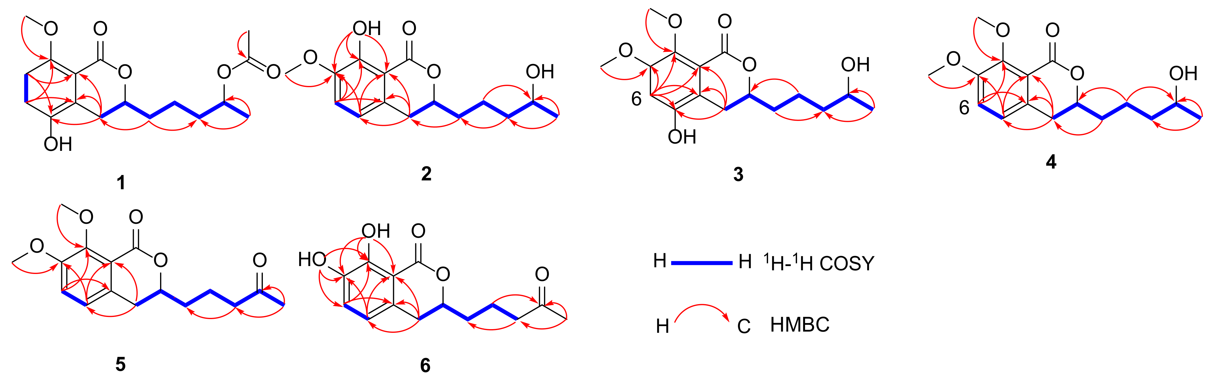
Figure 1. The structures of compounds 123.
Marinedrugs 20 00361 g001
Figure 2. 1H-1H COSY and key HMBC correlations for compounds 16.
Marinedrugs 20 00361 g002
Figure 3. Reaction route of hydrolysis for compound 1.
Marinedrugs 20 00361 g003
Figure 4. Δδ (=δSδR) values for (S)- and (R)-MTPA esters of compounds 13.
Marinedrugs 20 00361 g004
Figure 5. The experimental CD spectra of compounds 16.
Marinedrugs 20 00361 g005
Scheme 1. Plausible biosynthetic pathways of compounds 123.
Marinedrugs 20 00361 sch001
Table 1. 1H NMR spectroscopic data (400/600 MHz) (δ in ppm, J in Hz) for 16 in CDCl3.
Position123456
34.34, m4.54, m4.35, m4.36, m4.37, m4.58, m
42.64, dd (16.4, 11.6)
3.08, dd (16.4, 2.8)
2.86, m2.56, m
2.98, m
2.82, m2.82, m2.87, m
5 6.62, d (8.0) 6.91, d (8.0)6.91, d (8.4)6.61, d (8.0)
67.00, d (8.8)6.98, d (8.4)6.67, s7.06, d (8.0)7.06, d (8.0)7.07, d (8.0)
76.78, d (8.8)
7-OH 5.53, s
7-OMe 3.87, s3.84, s3.88, s3.88, s
8-OMe3.88, s 3.89, s3.96, s3.95, s
8-OH 11.18, s 11.00, s
1′1.85, m1.87, m1.85, m1.88, m1.82, m1.78, m
2′1.57, m1.58, m1.60, m1.58, m1.78, m1.80, m
3′1.52, m1.47, m1.50, m1.51, m2.52, t (6.4)2.53, m
4′4.90, m3.80, m3.82, m3.83, m
5′1.22, d (6.4)1.19, d (6.4)1.21, d (6.0)1.21, d (4.0)2.16, s2.15, m
7′2.04, s
Table 2. 13C NMR spectroscopic data (100/150 MHz) for 16 in CDCl3.
Position123456
1163.1, C170.4, C162.7, C162.6, C162.5, C170.2, C
377.4, CH80.4, CH78.3, CH78.5, CH78.3, CH80.6, CH
428.0, CH232.4, CH227.4, CH233.8, CH233.7, CH232.2, CH2
4a128.4, C130.1, C119.9, C132.3, C132.2, C129.7, C
5145.3, C117.1, CH147.7, CH122.4, CH122.4, CH117.9, CH
6121.2, CH117.5, CH106.1, C117.5, CH117.4, CH120.8, CH
7111.5, CH147.4, C153.1, C153.0, C153.0, C143.9, C
8155.5, C152.5, C142.9, C151.6, C151.5, C149.1, C
8a114.6, C108.6, C117.7, C119.6, C119.5, C108.4, C
7-OMe 56.4, CH356.6, CH356.5, CH356.5, CH3
8-OMe56.6, CH3 61.8, CH361.7, CH361.7, CH3
1′34.7, CH234.8, CH234.9, CH234.8, CH234.0, CH234.1, CH2
2′21.0, CH221.3, CH221.5, CH221.5, CH219.4, CH219.2, CH2
3′35.7, CH238.9, CH239.1, CH239.1, CH243.2, CH243.0, CH2
4′70.9, CH67.9, CH68.2, CH68.1, CH208.7, C208.6, C
5′20.1, CH323.7, CH323.8, CH323.8, CH330.1, CH330.1, CH3
6′171.1, C
7′21.6, CH3
Table 3. Antioxidant activity for compounds 2, 611, 1719, 21 and 22.
Compound2678910111718192122Trolox a
IC50 (mM)28.390.140.170.130.100.110.120.120.160.1520.660.0090.29
a Trolox was used as a positive control.
Publisher’s Note: MDPI stays neutral with regard to jurisdictional claims in published maps and institutional affiliations.

