Article

Protective Effects of Marine Alkaloid Neolamellarin A Derivatives against Glutamate Induced PC12 Cell Apoptosis

1
Key Laboratory of Marine Drugs, Ministry of Education, School of Medicine and Pharmacy, Ocean University of China, Qingdao 266003, China
2
Marine Biomedical Research Institute of Qiangdao, Qingdao 266237, China
3
Laboratory for Marine Drugs and Bioproducts, Qingdao National Laboratory for Marine Science and Technology, Qingdao 266237, China
*
Authors to whom correspondence should be addressed.
These authors contributed equally to this work.
Mar. Drugs 2022, 20(4), 262; https://doi.org/10.3390/md20040262
Submission received: 21 March 2022 / Revised: 6 April 2022 / Accepted: 11 April 2022 / Published: 12 April 2022

Abstract

:
Marine alkaloids obtained from sponges possess a variety of biological activities and potential medicinal value. The pyrrole-derived lamellarin-like alkaloids, especially their permethyl derivatives, show low cytotoxicity and potent MDR reversing activity. Neolamellarin A is a novel lamellarin-like alkaloid which was extracted from marine animal sponges. We reported the synthetic method of permethylated Neolamellarin A and its derivatives by a convergent strategy in 2015. In 2018, we reported the synthesis and the neuroprotective activity in PC12 cells of 3,4-bisaryl-N-alkylated permethylated Neolamellarin A derivatives. In this report, another series of 15 different 3,4-bisaryl-N-acylated permethylated Neolamellarin A derivatives were synthesized, and the outstanding protective effects of these compounds against glutamate induced PC12 cell apoptosis were presented and discussed. These Neolamellarin A derivatives which possessed low cytotoxicity and superior neuroprotective activity may have the potential to be developed into antagonists against glutamate induced nerve cell apoptosis.

1. Introduction

The excitatory amino acid glutamate, which is one of the most important neurotransmitters in the central nervous system, has been indicated to be involved in rapid synaptic transmission, neuroplasticity, learning, and memory [1]. However, excessive release of glutamate can cause neurotoxicity, and further lead to acute conditions including epileptic seizures and chronic neurodegenerative disorders such as Alzheimer’s disease [2]. To date, two mechanisms have been reported for glutamate-mediated neurotoxicity. One is through competitive inhibition of cystine uptake, leading to oxidative stress [3,4,5], and the other is mediated by several types of excitatory amino acid receptors, such as N-methyl-D-aspartate (NMDA)-subtype glutamate receptor, resulting in a massive influx of extracellular Ca2+ [6,7].
Lamellarins, a type of marine alkaloids was first isolated from the prosobranch mollusc Lamellaria sp. in 1985 [8]. Now, more than 70 different lamellarins and related natural pyrrole-derived alkaloids have been reported. These lamellarins which were derived from 2-amino-3-(3′,4′-dihydroxyphenyl) propionic acid (DOPA), have a variety of promising bioactivities, such as inhibition of HIV-1 integrase and MCV topoisomerase, multidrug resistance reversal (MDR) and antitumor activity [3,4,9,10]. The structural modifications and bioactivity studies of lamellarins have been reported by several groups [11,12,13]. We found that those 3,4-diarylpyrrole-derived alkaloids which showed superior multi-drug resistance (MDR) reversal even at noncytotoxic concentrations by inhibition of P-glycoprotein (P-gp)-mediated drug efflux possessed the characteristics of high lipophilicity and low cytotoxicity [5,6,7].
Neolamellarin A (Figure 1), a 3,4-bisaryl-pyrrole structural marine alkaloid was isolated from sponge Dendrilla nigra in 2007 [14] and was synthesized firstly in 2009 by Arafeh and Ullah [15]. Our group has studied permethylated Neolamellarin A and its derivatives for several years. In 2015, the synthesis of permethylated Neolamellarin A and its derivatives by a convergent strategy were reported [16], and then in 2018, we reported the synthesis and the neuroprotective activity on PC12 cells of 3,4-bisaryl-N-alkylated permethylated Neolamellarin A analogues [17]. In this paper, the synthesis and neuroprotective activity studies on PC12 cell line of 15 novel 3,4-bisaryl-N-acylated permethylated Neolamellarin A derivatives were discussed.

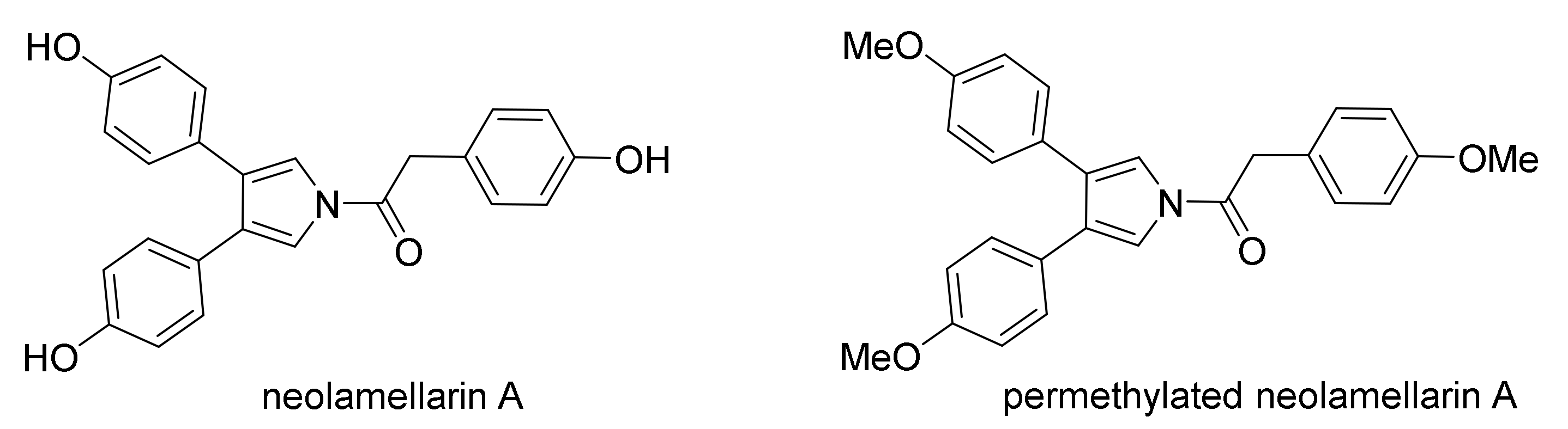
2. Results and Discussion
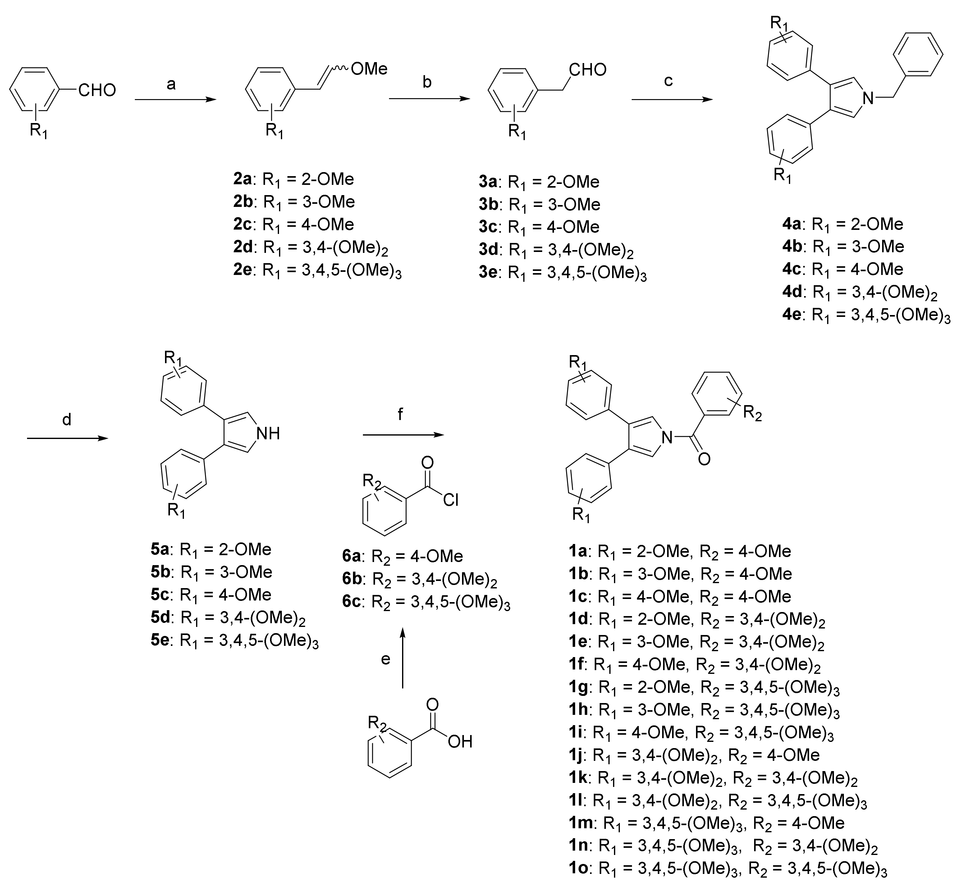
2.1. Chemistry

There should be two strategies to synthesize these 3,4-bisaryl-N-acylated permethylated Neolamellarin A derivatives. One strategy is directly building the 3,4-bisaryl-N-acylated pyrrole ring by one-pot process, and the other is acylation between 3,4-bisaryl-1H-pyrrole core and phenylacetyl chloride. The latter strategy was proved to be efficient in our previous study, and the synthesis of 3,4-bisaryl-1H-pyrrole is the critical step. We have reported the synthesis of the 3,4-bisaryl-1H-pyrrole through Suzuki cross-coupling reaction between 3,4-diiodinated N-trisisopropylsilyl pyrrole and aryl boronic acid ester [16]. But the long route, high cost and low yield of this method bring difficulties in building compound libraries. Hence, in this study we first synthesized 3,4-bisaryl-1-benzyl pyrrole through one-pot AgOAc-mediated method, and then debenzylation was performed to obtain the key 3,4-bisaryl-1H-pyrrole intermediates.
The one-pot AgOAc-mediated synthetic method of building 3,4-bisaryl-1-benzyl pyrrole was studied in our previous report [17]. The debenzylation of 3,4-bisaryl-1-benzyl pyrroles 4a4e was failed in the Pd/C, H2 and MeOH system (with or without catalyst) or CF3COOH, catalytic amount of TfOH at the reflux condition. Finally, the use of t-BuOK as a base in THF/DMSO, and oxidization by O2 at room temperature for 1 h gave the 3,4-bisaryl-1H pyrroles 5a5e at the yields of 85–95% [18]. The acylation of 5a5e with fresh acid chloride 6a6c was achieved by using n-BuLi as a base in THF at 30 °C for 10 h to produce 1a1o at the yields of 40–50% (Scheme 1).

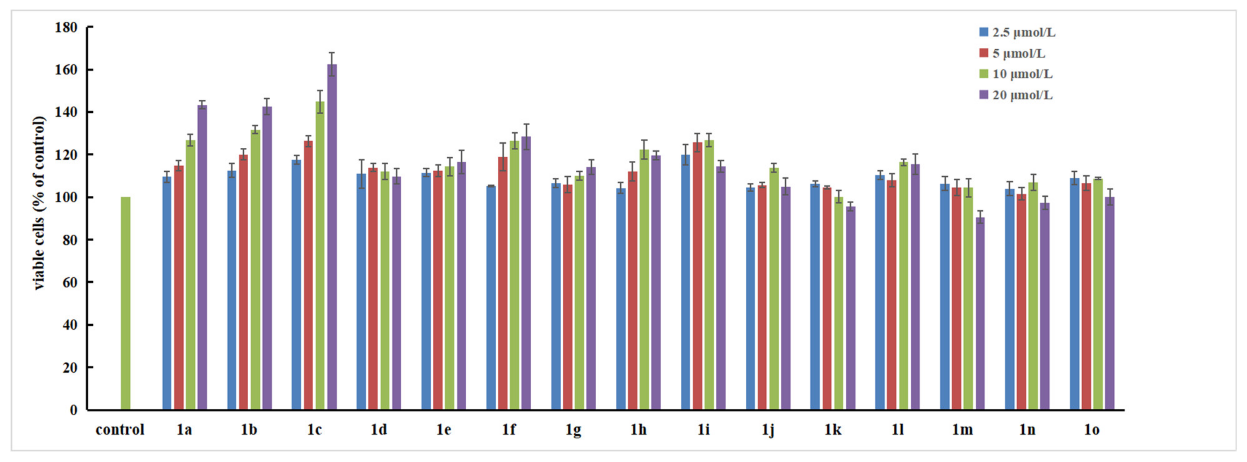
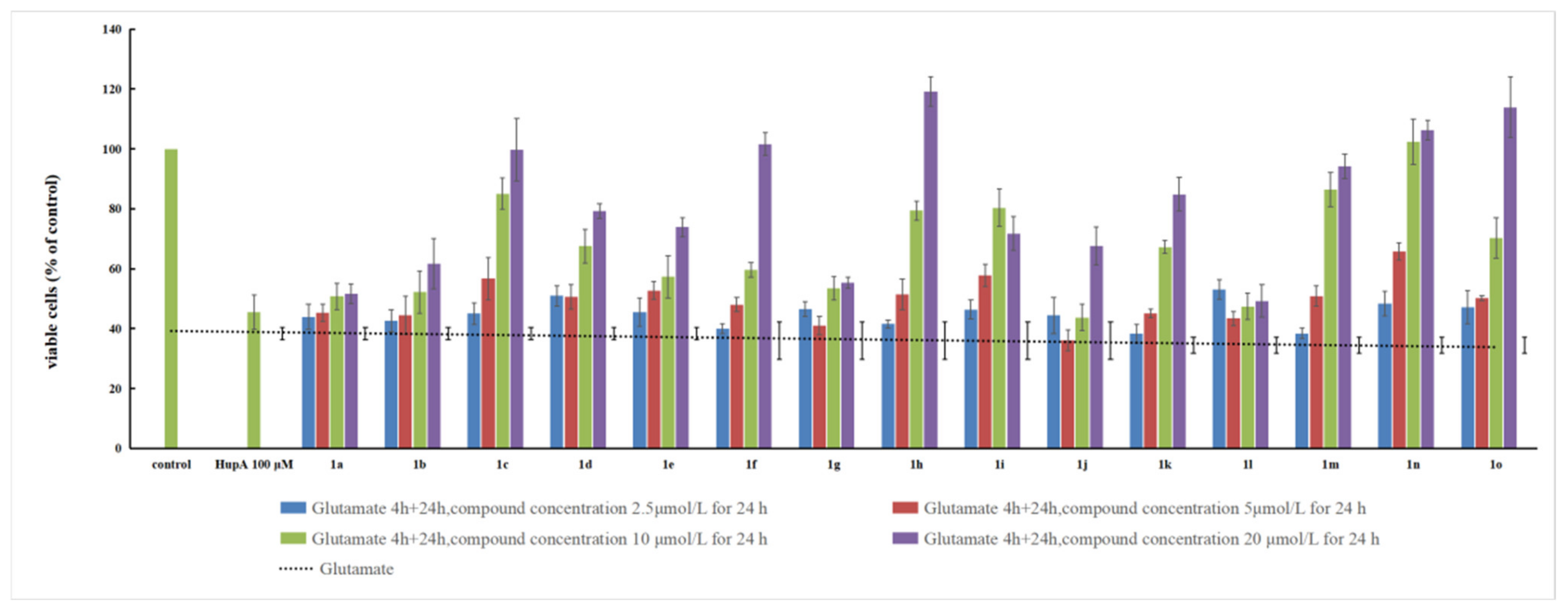
2.2. 3,4-Bisaryl-N-Acylated Permethylated Neolamellarin A Derivatives as Antagonists against Glutamate-Induced PC12 Cell Death

PC12 cells are derived from rat adrenal medulla pheochromocytoma, which are widely used as an in vitro model for the neuronal apoptosis and differentiation research due to the differentiated PC12 cells induced by nerve growth factor (NGF) with the typical characteristic of the neurons in morphology and function [19,20,21]. The reports have showed that high concentration of glutamate induces PC12 cell death and different types of compounds could protect PC12 cells from glutamate-induced damage, such as sesquiterpenoids, neolignan glycosides, YC-1, and xanthoceraside [22,23,24,25,26]. Here, we used the PC12 cell model of glutamate-induced damage to evaluate the neuroprotective effect of these novel Neolamellarin A derivatives 1a1o.
Firstly, the cytotoxicity of the permethylated Neolamellarin A derivatives 1a1o on PC12 cell line at concentrations of 2.5, 5, 10, and 20 μM was evaluated, and the results showed that the cell viability was above 80% even at the concentration of 20 μM, indicating that the derivatives had low cytotoxicity on PC12 cells (Figure 2). Especially the compound 1a1c exhibited superior proliferation activities. The data were as follows: 1a (2.5 μM 110.0%, 5.0 μM 114.9%, 10.0 μM 126.7%, 20.0 μM 143.3%), 1b (2.5 μM 112.5%, 5.0 μM 120.0%, 10.0 μM 131.6%, 20.0 μM 142.5%) and 1c (2.5 μM 117.5%, 5.0 μM 126.4%, 10.0 μM 144.9%, 20.0 μM 162.5%). The mechanisms of the compounds 1a1c that promote the proliferative activity of PC12 cells need to be further investigated and explore whether they can be further developed. The low cytotoxicity of these compounds on PC12 cells laid a foundation for further neuroprotective effect evaluation on glutamate-induced PC12 cell death.
We constructed the glutamate-induced PC12 cell death model by the method of the previous report [27]. Firstly, PC12 cells were inoculated into 96-well plates at a density of 5000 cells/well, and cultivated for 24 h. Cells were then treated with 8 mM glutamate, and cell viability was detected after 4 h. The results showed that the survival rate of PC12 cells decreased significantly (35% ± 3%, relative to the untreated control group). Then, we used this model to evaluate the neuroprotective activity of these permethylated Neolamellarin A derivatives 1a1o and Huperzine-A (HupA, 100 μM) was used as positive control. Compounds 1a1o were added to the glutamate damaged PC12 cells at a final concentration of 2.5, 5, 10 and 20 μM, respectively, and continually cultured for another 24 h. Cell viability was measured by MTT assay. The results in Figure 3 show that all of the 15 derivatives can effectively inhibit the apoptosis of PC12 cells (induced by glutamate-induced damage) in a concentration dependent manner. With the increase of concentration, the neuroprotective activity on PC12 cells increased. In particular, when the concentration of compounds 1c, 1f, 1h, 1n, and 1o reached 20 μM, the cell viability achieved almost 100%.

3. Materials and Methods

3.1. Chemical Synthesis

3.1.1. General

Tetrahydrofuran (THF) was distillated with Na in the presence of benzophenone under argon atmosphere. Dichloromethane was distillated by molecular sieve. Dimethyl sulfoxide (DMSO) was distillated with t-BuOK. All other materials and solvents were obtained from commercial sources and used without further purification. Thin-layer chromatography (TLC) was performed on precoated E. Merck silica-gel 60 F254 plates. Column chromatography was performed on silica gel (200–300 mesh, Qingdao, China). Melting points were determined on a Mitamura-Riken micro-hot stage without correction. 1H and 13C NMR spectra were recorded on the Broker AVANCE NEO and Agilent DD2 500 with 500 or 600 MHz for proton (1H NMR) and 125 or 150 MHz for carbon (13C NMR) with tetramethylsilane (Me4Si) as internal standard, respectively. The chemical shifts (δ) were expressed in parts per million (ppm) downfield, and the coupling constant (J) values were described as hertz. High-resolution (ESI) MS spectra were recorded using a Q TOF-2 Micromass spectrometer.
The general procedures for the synthesis of important intermediates 4a4e, 5a5e and 6a6c can be seen in Supplementary Materials.

3.1.2. General Procedure for the Synthesis of 1a1o

n-BuLi (1.2 mL, 3.0 mmol, 2 mol L-1 in hexane) was added dropwise to a solution of 3,4-bisaryl-1H-pyrrole 5a5e (2 mmol) in anhydrous THF (10 mL) at −78 °C under argon atmosphere and stirred for 0.5 h. Then 6a6c in anhydrous THF (10 mL) was added into the reaction mixture and warmed up to room temperature whereafter. The solution continued to stirred at 30 °C for 10 h and diluted with saturated aqueous NaHCO3 (20 mL). The mixture was extracted with EtOAc (20 × 3 mL) and washed with brine and dried with MgSO4, then concentrated by vacuum evaporation to give a residue of compounds 1a1o, which was purified by flash chromatography on silica gel (petroleum ether: ethyl acetate = 1:1).

(3,4-Bis(2-methoxyphenyl)-1H-pyrrol-1-yl)(4-methoxyphenyl)methanone (1a)

White solid 125 mg, yield 30.3%. m.p. 145–146 °C. 1H NMR (600 MHz, CDCl3) δ 7.87–7.84 (d, J = 8.8 Hz, 2H), 7.51 (s, 2H), 7.21–7.19 (m, 2H), 7.16–7.15 (dd, J = 7.1, 1.6Hz, 2H), 7.01–7.00 (d, J = 9.3 Hz, 2H), 6.87–6.85 (t, J = 7.2 Hz, 2H), 6.82–6.81 (d, J = 7.7 Hz, 2H), 3.89 (s, 3H, OCH3), 3.45 (s, 6H, 2OCH3). 13C NMR (150 MHz, CDCl3) δ 166.86 (C), 162.92 (C), 156.62 (2C), 132.06 (2CH), 130.41 (2CH), 128.10 (2CH), 125.46 (C), 125.38 (2C), 124.43 (2C), 120.53 (2CH), 120.39 (2CH), 113.87 (2CH), 110.86 (2CH), 55.63 (CH3), 55.12 (2CH3). HRMS (ESI) m/z: calcd for M+ C26H24NO4, 414.1700; found, M+ 414.1701.

(3,4-Bis(3-methoxyphenyl)-1H-pyrrol-1-yl)(4-methoxyphenyl)methanone (1b)

White solid 134 mg, yield 32.4%. m.p. 132–134 °C. 1H NMR (600 MHz, CDCl3), δ 7.85–7.83 (d, J = 8.6 Hz, 2H), 7.42 (s, 2H), 7.21–7.18 (t, J = 7.7 Hz, 2H), 7.03–7.02 (d, J = 8.6Hz, 2H), 6.88–6.86 (d, J = 7.9 Hz, 2H), 6.83–6.80 (m, 4H), 3.91 (s, 3H, OCH3), 3.69 (s, 6H, 2OCH3). 13C NMR (150 MHz, CDCl3) δ 166.96 (C), 163.24 (C), 159.49 (2C), 135.43 (2C), 132.14 (2CH), 129.36 (2CH), 127.94 (2C), 124.89 (C), 121.22 (2CH), 120.04 (2CH), 114.06 (2CH), 113.99 (2CH), 112.80 (2CH), 55.68 (CH3), 55.23 (2CH3). HRMS (ESI) m/z: calcd for M+ C26H24NO4, 414.1700; found, M+ 414.1702.

(3,4-Bis(4-methoxyphenyl)-1H-pyrrol-1-yl)(4-methoxyphenyl)methanone (1c)

White solid 154 mg, yield 37.3%. m.p. 120–121 °C. 1H NMR (600 MHz, CDCl3) δ 7.84–7.83 (d, J = 8.8 Hz, 2H), 7.35 (s, 2H), 7.20–7.19 (d, J = 8.8 Hz, 4H), 7.02–7.01 (d, J = 8.8Hz, 2H), 6.84–6.82 (d, J = 8.8 Hz, 4H), 3.90(s, 3H), 3.80(s, 6H). 13C NMR (150 MHz, CDCl3) δ 166.93 (C), 163.10 (C), 158.69 (2C), 132.02 (2CH), 129.76 (4CH), 127.77 (C), 126.65 (2C), 125.17 (2C), 119.35 (2CH), 113.99 (2CH), 113.84 (4CH), 55.65 (CH3), 55.33 (2CH3). HRMS (ESI) m/z: calcd for M+ C26H24NO4, 414.1700; found, M+ 414.1704.

(3,4-Bis(2-methoxyphenyl)-1H-pyrrol-1-yl)(3,4-dimethoxyphenyl)methanone (1d)

White solid 162 mg, yield 36.2%. m.p. 152–154 °C. 1H NMR (600 MHz, CDCl3) δ (ppm) 7.53 (s, 2H), 7.51–7.49 (dd, J = 8.5, 2.0 Hz, 1H), 7.41 (d, J = 1.6 Hz, 1H), 7.21–7.19 (t, J = 8.5 Hz, 2H), 7.16–7.14 (dd, J = 7.7, 1.7 Hz, 2H), 6.96–6.94 (d, J = 8.3 Hz, 1H), 6.86 (t, J = 7.4 Hz, 2H), 6.83–6.81 (d, J = 7.7 Hz, 2H), 3.97 (s, 3H), 3.95 (s, 3H), 3.45 (s, 6H). 13C NMR (150 MHz, CDCl3) δ (ppm) 166.85 (C), 156.61 (2C), 152.59 (C), 148.98 (C), 130.41 (2CH), 128.13 (2CH), 125.54 (C), 125.45 (2C), 124.39 (2C), 123.94 (2CH), 120.57 (2CH), 120.41 (2CH), 112.68 (CH), 110.87 (CH), 110.31 (CH), 56.21 (2CH3), 55.14 (CH3), 55.10 (CH3). HRMS (ESI) m/z: calcd for M+ C27H26NO5, 444.1805; found, M+ 444.1807.

(3,4-Bis(3-methoxyphenyl)-1H-pyrrol-1-yl)(3,4-dimethoxyphenyl)methanone (1e)

White solid 157 mg, yield 35.4%. m.p. 154–156 °C. 1H NMR (600 MHz, CDCl3) δ (ppm) 7.48–7.46 (dd, J = 8.3, 1.7 Hz, 1H), 7.44 (s, 2H), 7.41 (d, J = 1.6 Hz, 1H), 7.21–7.18 (t, J = 7.7 Hz, 2H), 6.97–6.96 (d, J = 8.2 Hz, 1H), 6.88–6.87 (d, J = 7.7 Hz, 2H), 6.82–6.79 (m, 4H), 3.98 (s, 3H), 3.96 (s, 3H), 3.69 (s, 6H). 13C NMR (150 MHz, CDCl3) δ (ppm) 166.93 (C), 159.50 (2C), 152.94 (C), 149.20 (C), 135.39 (2C), 129.36 (2CH), 128.00 (2C), 124.99 (C), 124.01 (CH), 121.20 (2CH), 120.09(2CH), 114.02 (2CH), 112.82 (2CH), 112.71 (CH), 110.33 (CH), 56.25 (2CH3), 55.21 (2CH3). HRMS (ESI) m/z: calcd for M+ C27H26NO5, 444.1805; found, M+ 444.1803.

(3,4-Bis(4-methoxyphenyl)-1H-pyrrol-1-yl)(3,4-dimethoxyphenyl)methanone (1f)

White solid 145 mg, yield 32.7%. m.p. 156–158 °C. 1H NMR (600 MHz, CDCl3) δ (ppm) 7.47–7.45 (dd, J = 8.5, 1.9 Hz, 1H), 7.40–7.39 (d, J = 2.2 Hz, 1H), 7.36 (s, 2H), 7.19–7.18 (d, J = 8.8 Hz, 4H), 6.97–6.95 (d, J = 8.8 Hz, 1H), 6.83–6.82 (d, J = 8.8 Hz, 4H), 3.97 (s, 3H), 3.96 (s, 3H), 3.80 (s, 6H). 13C NMR (150 MHz, CDCl3) δ (ppm) 166.91 (C), 158.71 (2C), 152.79 (C), 149.13 (C), 129.75 (4CH), 127.82 (2C), 126.60 (CH), 125.26 (C), 123.91 (2C), 119.39 (2CH), 113.85 (4CH), 112.68 (CH), 110.30 (CH), 56.23 (2CH3), 55.32 (2CH3). HRMS (ESI) m/z: calcd for M+ C27H26NO5, 444.1805; found, M+ 444.1812.

(3,4-Bis(2-methoxyphenyl)-1H-pyrrol-1-yl)(3,4,5-trimethoxyphenyl)methanone (1g)

White solid 187 mg, yield 37.5%. m.p. 180–182 °C. 1H NMR (600 MHz, CDCl3) δ (ppm) 7.52 (s, 2H), 7.22–7.19 (td, J = 8.1, 1.6 Hz, 2H), 7.14–7.13 (dd, J = 7.6, 1.6 Hz, 2H), 7.09 (s, 2H), 6.87–6.85 (t, J = 7.8 Hz, 2H), 6.83–6.82 (d, J = 8.2 Hz, 2H), 3.94 (s, 3H), 3.92 (s, 6H), 3.45 (s, 6H). 13C NMR (150 MHz, CDCl3) δ (ppm) 166.17 (C), 155.87 (2C), 152.45 (2C), 140.78 (C), 129.68 (2CH), 127.64 (C), 127.51 (2CH), 125.07 (2C), 123.52 (2C), 119.72 (4CH), 110.16 (2CH), 106.43 (2CH), 55.83 (CH3), 54.40 (4CH3). HRMS (ESI) m/z: calcd for M+ C28H28NO6, 474.1911; found, M+ 474.1911.

(3,4-Bis(3-methoxyphenyl)-1H-pyrrol-1-yl)(3,4,5-trimethoxyphenyl)methanone (1h)

White solid 174 mg, yield 36.7%. m.p. 140–142 °C. 1H NMR (500 MHz, CDCl3) δ (ppm) 7.44 (s, 2H), 7.21–7.19 (m, 2H), 7.07 (s, 2H), 6.87–6.85 (d, J = 7.6 Hz, 2H), 6.81–6.80 (m, 4H), 3.95 (s, 3H), 3.93 (s, 6H), 3.69 (s, 6H). 13C NMR (125 MHz, CDCl3) δ (ppm) 166.86 (C), 159.35 (2C), 153.10 (2C), 141.70 (C), 135.07 (2C), 129.26 (2CH), 128.19 (2C), 127.65 (C), 121.02 (2CH), 119.80 (2CH), 113.89 (2CH), 112.72 (2CH), 107.05 (2CH), 61.00 (CH3), 56.40 (2CH3), 55.06 (2CH3). HRMS (ESI) m/z: calcd for M+ C28H28NO6, 474.1911; found, M+ 474.1908.

(3,4-Bis(4-methoxyphenyl)-1H-pyrrol-1-yl)(3,4,5-trimethoxyphenyl)methanone (1i)

White solid 178 mg, yield 37.6%. m.p. 163–164 °C. 1H NMR (600 MHz, CDCl3) δ (ppm) 7.36 (s, 2H), 7.19–7.17 (d, J = 8.8 Hz, 4H), 7.06 (s, 2H), 6.84–6.83 (d, J = 8.3 Hz, 4H), 3.95 (s, 3H), 3.92 (s, 6H), 3.80 (s, 6H). 13C NMR (150 MHz, CDCl3) δ (ppm) 166.24 (C), 158.07 (2C), 152.50 (2C), 141.02 (C), 129.03 (4CH), 127.46 (2C), 127.35 (C), 125.72 (2C), 118.53 (2CH), 113.16 (4CH), 106.47 (2CH), 55.84 (CH3), 54.60 (4CH3). HRMS (ESI) m/z: calcd for M+ C28H28NO6, 474.1911; found, M+ 474.1906.

(3,4-Bis(3,4-dimethoxyphenyl)-1H-pyrrol-1-yl)(4-methoxyphenyl)methanone (1j)

White solid 186 mg, yield 39.3%. 1H NMR (500 MHz, CDCl3) δ (ppm) 7.84–7.83 (d, J = 8.6 Hz, 2H), 7.38 (s, 2H), 7.03–7.01 (d, J = 8.6 Hz, 2H), 6.86–6.85 (d, J = 8.3 Hz, 2H), 6.81–6.78 (m, 4H), 3.90 (s, 3H), 3.87 (s, 6H), 3.69 (s, 6H). 13C NMR (125 MHz, CDCl3) δ (ppm) 166.75 (C), 163.01 (C), 148.44 (2C), 147.97 (2C), 131.92 (2CH), 127.72 (2C), 126.70 (2C), 124.90 (C), 120.82 (2CH), 119.19 (2CH), 113.87 (2CH), 111.99 (2CH), 110.95 (2CH), 55.83 (2CH3), 55.67 (2CH3), 55.51 (CH3). HRMS (ESI) m/z: calcd for M+ C28H28NO6, 474.1911; found, M+ 474.1901.

(3,4-Bis(3,4-dimethoxyphenyl)-1H-pyrrol-1-yl)(3,4-dimethoxyphenyl)methanone (1k)

White solid 198 mg, yield 39.4%. 1H NMR (500 MHz, CDCl3) δ (ppm) 7.47–7.45 (d, J = 8.2 Hz, 1H), 7.39 (s, 3H), 6.97–6.95 (d, J = 8.3 Hz, 1H), 6.85–6.77 (m, 6H), 3.97–3.95 (d, J = 7.5 Hz, 6H), 3.87 (s, 6H), 3.69 (s, 6H). 13C NMR (125 MHz, CDCl3) δ (ppm)166.75 (CO), 152.67 (C), 148.96 (C), 148.39 (2C), 147.95 (2C), 127.74 (2C), 126.61 (2C), 124.93 (C), 123.76 (2CH), 120.77 (2CH), 119.22 (CH), 112.47 (CH), 111.92 (2CH), 110.90 (2CH), 110.11 (CH), 56.09 (2CH3), 55.80 (2CH3), 55.63 (2CH3). HRMS (ESI) m/z: calcd for M+ C29H30NO7, 504.2017; found, M+ 504.2003.

(3,4-Bis(3,4-dimethoxyphenyl)-1H-pyrrol-1-yl)(3,4,5-trimethoxyphenyl)methano-ne (1l)

White solid 213 mg, yield 39.9%. 1H NMR (500 MHz, CDCl3) δ (ppm) 7.40 (s, 2H), 7.06 (s, 2H), 6.85–6.80 (m, 4H), 6.76 (s, 2H), 3.94 (s, 3H), 3.92 (s, 6H), 3.87 (s, 6H), 3.69 (s, 6H). 13C NMR (125 MHz, CDCl3) δ (ppm) 166.81 (CO), 153.07 (2C), 148.43 (2C), 148.05 (C), 141.61 (2C), 128.10 (C), 127.77 (2C), 126.45 (2C), 120.78 (2CH), 119.11 (2CH), 111.94 (2CH), 110.93 (2CH), 107.02 (2CH), 61.01 (CH3), 56.39 (2CH3), 55.82 (2CH3), 55.65 (2CH3). HRMS (ESI) m/z: calcd for M+ C30H32NO8, 534.2122; found, M+ 534.2116.

(3,4-Bis(3,4,5-trimethoxyphenyl)-1H-pyrrol-1-yl)(4-methoxyphenyl)methanone (1m)

White solid 209 mg, yield 39.1%. 1H NMR (500 MHz, CDCl3) δ (ppm) 7.84–7.83 (d, J = 7.6 Hz, 2H), 7.43 (s, 2H), 7.04–7.02 (d, J = 7.6 Hz, 2H), 6.49 (s, 4H), 3.90 (s, 3H), 3.83 (s, 6H), 3.70 (s, 12H). 13C NMR (125 MHz, CDCl3) δ (ppm) 166.75 (CO), 163.14 (C), 152.86 (4C), 136.96 (2C), 131.96 (2CH), 129.38 (2C), 127.81 (2C), 124.63 (C), 119.37 (2CH), 113.94 (2CH), 105.79 (4CH), 60.89 (2CH3), 55.95 (4CH3), 55.54 (CH3). HRMS (ESI) m/z: calcd for M+ C30H32NO8, 534.2122; found, M+ 534.2113.

(3,4-Bis(3,4,5-trimethoxyphenyl)-1H-pyrrol-1-yl)(3,4-dimethoxyphenyl)methanone (1n)

White solid 223 mg, yield 39.5%. 1H NMR (500 MHz, CDCl3) δ 7.47–7.45 (dd, J = 8.3, 1.9 Hz, 1H), 7.44 (s, 2H), 7.41–7.40 (d, J = 1.9 Hz, 1H), 6.98–6.96 (d, J = 8.4 Hz, 1H), 6.50 (s, 4H), 3.98 (s, 3H), 3.96 (s, 3H), 3.83 (s, 6H), 3.70 (s, 12H). 13C NMR (125 MHz, CDCl3) δ 166.72 (CO), 152.92 (4C), 152.90(C), 149.13 (C), 137.13 (C), 129.35 (2C), 127.89 (2C), 124.79 (CH), 123.81 (2C), 119.43 (2CH), 112.62 (CH), 110.22 (CH), 105.92 (4CH), 60.89 (2CH3), 56.14(CH3), 56.13(CH3), 55.98 (4CH3).

(3,4-Bis(3,4,5-trimethoxyphenyl)-1H-pyrrol-1-yl)(3,4,5-trimethoxyphenyl)metha-None (1o)

White solid 245 mg, yield 42.2%. 1H NMR (500 MHz, CDCl3) δ (ppm) 7.43 (s, 2H), 7.06 (s, 2H), 6.47 (s, 4H), 3.94 (s, 3H), 3.91 (s, 6H), 3.82 (s, 6H), 3.69 (s, 12H). 13C NMR (125 MHz, CDCl3) δ (ppm) 166.82 (CO), 153.14 (2C), 152.91 (4C), 141.79 (C), 137.09 (2C), 129.16 (2C), 128.22 (2C), 127.54 (C), 119.30 (2CH), 107.09 (2CH), 105.81 (4CH), 61.04 (CH3), 60.92 (2CH3), 56.41 (2CH3), 55.95 (4CH3). HRMS (ESI) m/z: calcd for M+ C32H36NO10, 594.2334; found, M+ 594.2322.

3.2. Bioactivity Study

3.2.1. Cell Culture

Rat PC12 cells (adrenal gland; pheochromocytoma) were obtained from Cell Bank of Chinese Academy of Sciences (Shanghai, China). PC12 cells were cultured in Dulbecco’s Modified Eagle’s medium (high glucose, Gibco, San Diego, CA, USA) supplemented with 10% fetal bovine serum (FBS) (Gibco, San Diego, CA, USA), 100 U mL−1 penicillin and 100 μg mL−1 streptomycin in a 95% humidified atmosphere with 5% CO2 at 37 °C. Cells were passaged with trypsin (0.025% EDTA) every 3 days with the subcultivation ratio of 1:6. Cell used was within 5–20 passages.

3.2.2. Cell Viability Assay

The derivatives were first dissolved in DMSO (Solarbio, CN) and then diluted to the test concentration with complete growth media for cellular viability assay.
For cellular viability assay, a 96-wells plate was cultured with 5000 cells well−1 in 100 μL complete cell culture medium for 24 h. Then, 100 μL complete medium containing serial concentrations of 2.5, 5, 10, and 20 μM of each compound was added to each well and continued to culture for another 24 h, cell viability was measured by MTT assay as described [28].
For the neuroprotective assay, PC12 cells were cultured into 96-well plates at a density of 5000 cells/well for 24 h. After treating with glutamate of 8 mM for 4 h, the PC12 cells were co-incubated with neolamellarin A derivatives (1a1o) at final concentrations of 2.5, 5, 10, and 20 μM for 24 h. Percentage of viable cells were detected. Cell viability was measured by MTT assay.

4. Conclusions

Neurotoxicity caused by glutamate widely exists in almost all nervous system diseases, including cerebral hemorrhage (aneurysms, hypertensive encephalorrhagia, etc.), cerebral ischemia, stroke, brain trauma, brain tumors and some neurodegenerative diseases, such as amyotrophic lateral sclerosis, Parkinson’s disease, chronic progressive chorea, etc. In this study, we synthesized and evaluated the protective effects of 15 different 3,4-bisaryl-N-acylated permethylated Neolamellarin A derivatives against glutamate induced PC12 cell apoptosis. The results showed that all of these compounds were effectively against glutamate-induced PC12 cell death. Combined with the previous study results, both the 3,4-bisaryl-N-acylated and 3,4-bisaryl-N-alkylated permethylated Neolamellarin A derivatives showed outstanding neuroprotective activity, and the number and location of methoxy groups had no significant effect on neuroprotective activity. Therefore, we speculate that the skeleton structure of permethylated Neolamellarin A is the key to neuroprotective activity on PC12 cell of these compounds.

Supplementary Materials

The following supporting information can be downloaded at: https://www.mdpi.com/article/10.3390/md20040262/s1. Table S1: The cytotoxicity of the permethylated Neolamellarin A derivatives 1a1o at gradient concentrations on PC12 cell line; Table S2: The neuroprotective activity of the permethylated Neolamellarin A derivatives 1a1o at gradient concentrations on PC12 cell death induced by glutamate; General procedures for the synthesis of important intermediates 4a4e, 5a5e and 6a6c; Figures S1–S73: Copies of 1H and 13C NMR spectra of compounds 4a4e, 5a5e and 1a1o.

Author Contributions

T.J. and R.Y. conceived and designed the experiments; K.Z. performed the chemical experiments; X.G. and X.Z. performed the biological experiments; L.L. analyzed the data; R.Y. and K.Z. wrote the paper. All authors have read and agreed to the published version of the manuscript.

Funding

This research was funded by the Natural Science Foundation of China (Grant No. 82003583 and 82073759), the Fund of Greater Bay Area Institute of Precision Medicine (Guangzhou) (No. IPM2021C009), National Science and Technology Major Project for Significant New Drugs Development (2018ZX09735004).

Institutional Review Board Statement

Not applicable.

Informed Consent Statement

Not applicable.

Data Availability Statement

Data are included in the article.

Acknowledgments

We sincerely appreciate the invaluable efforts of the entire research team during the research work.

Conflicts of Interest

The authors declare no conflict of interest.

References

  1. Choi, D.W. Glutamate neurotoxicity and diseases of the nervous system. Neuron 1988, 1, 623–634. [Google Scholar] [CrossRef]
  2. Kanno, H.; Kawakami, Z.; Mizoguchi, K.; Ikarashi, Y.; Kase, Y. Yokukansan, a Kampo Medicine, Protects PC12 Cells from Glutamate-Induced Death by Augmenting Gene Expression of Cystine/Glutamate Antiporter System Xc. PLoS ONE 2014, 9, e116275. [Google Scholar] [CrossRef] [PubMed]
  3. Fan, A.; Lin, W.; Jia, Y. Recent progress in the research on lamellarins and related pyrrole-derived alkaloids from marine organisms. J. Chin. Pharm. Sci. 2011, 20, 425–441. [Google Scholar] [CrossRef]
  4. Jiang, L.; Yin, R.; Wang, X.; Dai, J.; Li, J.; Jiang, T.; Yu, R. Design and synthesis of neolamellarin a derivatives targeting heat shock protein 90. Eur. J. Med. Chem. 2017, 135, 24–33. [Google Scholar] [CrossRef]
  5. Plisson, F.; Huang, X.-C.; Zhang, H.; Khalil, Z.; Capon, R.J. Lamellarins as Inhibitors of P-Glycoprotein-Mediated Multidrug Resistance in a Human Colon Cancer Cell Line. Chem. Asian J. 2012, 7, 1616–1623. [Google Scholar] [CrossRef]
  6. Quesada, A.M.R.; Gravalos, M.D.G.; Puentes, J.L.F. Polyaromatic alkaloids from marine invertebrates as cytotoxic compounds and inhibitors of multidrug resistance caused by P-glycoprotein. Br. J. Cancer 1996, 74, 677–682. [Google Scholar] [CrossRef] [Green Version]
  7. Zhang, P.Y.; Wong, I.L.K.; Yan, C.S.W.; Zhang, X.Y.; Jiang, T.; Chow, L.M.C.; Wan, S.B. Design and Syntheses of Permethyl Ningalin B Analogues: Potent Multidrug Resistance (MDR) Reversal Agents of Cancer Cells. J. Med. Chem. 2010, 53, 5108–5120. [Google Scholar] [CrossRef]
  8. Andersen, R.J.; Faulkner, D.J.; He, C.H.; Van Duyne, G.D.; Clardy, J. Metabolites of the marine prosobranch mollusk Lamellaria sp. J. Am. Chem. Soc. 1985, 107, 5492–5495. [Google Scholar] [CrossRef]
  9. Bailly, C. Lamellarins: A tribe of bioactive marine natural products. In Outstanding Marine Molecules; La Barre, S., Kornprobst, J.-M., Eds.; Wiley-VCH: Weinheim, Germany, 2014; pp. 377–386. [Google Scholar]
  10. Fan, H.; Peng, J.; Hamann, M.T.; Hu, J.-F. Lamellarins and Related Pyrrole-Derived Alkaloids from Marine Organisms. Chem. Rev. 2008, 108, 264–287. [Google Scholar] [CrossRef] [Green Version]
  11. Klumthong, K.; Chalermsub, P.; Sopha, P.; Ruchirawat, S.; Ploypradith, P. An Expeditious Modular Hybrid Strategy for the Diversity-Oriented Synthesis of Lamellarins/Azalamellarins with Anticancer Cytotoxicity. J. Org. Chem. 2021, 86, 14883–14902. [Google Scholar] [CrossRef]
  12. Sopha, P.; Phutubtim, N.; Chantrathonkul, B.; Ploypradith, P.; Ruchirawat, S.; Chittchang, M. Roles of autophagy in relation to mitochondrial stress responses of HeLa cells to lamellarin cytotoxicity. Toxicology 2021, 462, 152963. [Google Scholar] [CrossRef]
  13. Zheng, L.; Gao, T.; Ge, Z.; Ma, Z.; Xu, J.; Ding, W.; Shen, L. Design, Synthesis and Structure-Activity Relationship Studies of Glycosylated Derivatives of Marine Natural Product Lamellarin D. Eur. J. Med. Chem. 2021, 214, 113226. [Google Scholar] [CrossRef]
  14. Liu, R.; Liu, Y.; Zhou, Y.-D.; Nagle, D.G. Molecular-Targeted Antitumor Agents. 15. Neolamellarins from the Marine Sponge Dendrilla nigra Inhibit Hypoxia-Inducible Factor-1 Activation and Secreted Vascular Endothelial Growth Factor Production in Breast Tumor Cells. J. Nat. Prod. 2007, 70, 1741–1745. [Google Scholar] [CrossRef] [Green Version]
  15. Arafeh, K.M.; Ullah, N. Synthesis of Neolamellarin A, an Inhibitor of Hypoxia-Inducible Factor-1. Nat. Prod. Commun. 2009, 4, 925–926. [Google Scholar] [CrossRef] [Green Version]
  16. Yin, R.; Jiang, L.; Wan, S.; Jiang, T. Efficient syntheses of permethylated derivatives of neolamellarin A, a pyrrolic marine natural product. J. Ocean Univ. China 2015, 14, 329–334. [Google Scholar] [CrossRef]
  17. Zhang, M.; Yin, R.; Zhang, Y.; Hao, C.; Zhang, L.; Jiang, T. Synthesis and Neuroprotective activity of Neolamellarin A analogues. J. Ocean. Univ. China 2018, 17, 967–972. [Google Scholar] [CrossRef]
  18. Nedolya, N.A.; Tarasova, O.A.; Albanov, A.I.; Trofimov, B.A. Structural reorganization of (allyl-, benzyl-, and propargylsulfanyl)-substituted 2-aza-1,3,5-trienes in t-BuOK/THF/DMSO: Access to rare functionalized 2-thiazolines. Tetrahedron Lett. 2014, 55, 2495–2498. [Google Scholar] [CrossRef]
  19. Greene, L. Nerve growth factor prevents the death and stimulates the neuronal differentiation of clonal PC12 pheochromocytoma cells in serum-free medium. J. Cell Biol. 1978, 78, 747–755. [Google Scholar] [CrossRef] [Green Version]
  20. Ishima, T.; Nishimura, T.; Iyo, M.; Hashimoto, K. Potentiation of nerve growth factor-induced neurite outgrowth in PC12 cells by donepezil: Role of sigma-1 receptors and IP3 receptors. Prog. Neuro Psychopharmacol. Biol. Psychiatry 2008, 32, 1656–1659. [Google Scholar] [CrossRef]
  21. Rukenstein, A.; Rydel, R.; Greene, L. Multiple agents rescue PC12 cells from serum-free cell death by translation- and transcription-independent mechanisms. J. Neurosci. 1991, 11, 2552–2563. [Google Scholar] [CrossRef]
  22. Kawakami, Z.; Kanno, H.; Ikarashi, Y.; Kase, Y. Yokukansan, a kampo medicine, protects against glutamate cytotoxicity due to oxidative stress in PC12 cells. J. Ethnopharmacol. 2011, 134, 74–81. [Google Scholar] [CrossRef]
  23. Xu, J.; Zhu, H.-L.; Zhang, J.; Du, T.; Guo, E.-Y.; Liu, W.-Y.; Luo, J.-G.; Ye, F.; Feng, F.; Qu, W. Sesquiterpenoids from Chloranthus anhuiensis with Neuroprotective Effects in PC12 Cells. J. Nat. Prod. 2018, 81, 1391–1398. [Google Scholar] [CrossRef]
  24. Gan, M.; Zhang, Y.; Lin, S.; Liu, M.; Song, W.; Zi, J.; Yang, Y.; Fan, X.; Shi, J.; Hu, J.; et al. Glycosides from the Root of Iodes cirrhosa. J. Nat. Prod. 2008, 71, 647–654. [Google Scholar] [CrossRef]
  25. Yang, X.; Wang, Y.; Luo, J.; Liu, S.; Yang, Z. Protective Effects of YC-1 Against Glutamate Induced PC12 Cell Apoptosis. Cell. Mol. Neurobiol. 2011, 31, 303–311. [Google Scholar] [CrossRef]
  26. Zhu, L.; Yang, L.; Zhao, X.; Liu, D.; Guo, X.; Liu, P.; Chi, T.; Ji, X.; Zou, L. Xanthoceraside modulates NR2B-containing NMDA receptors at synapses and rescues learning-memory deficits in APP/PS1 transgenic mice. Psychopharmacology 2018, 235, 337–349. [Google Scholar] [CrossRef]
  27. Hao, C.; Gao, L.; Zhang, Y.; Wang, W.; Yu, G.; Guan, H.; Zhang, L.; Li, C. Acetylated chitosan oligosaccharides act as antagonists against glutamate-induced PC12 cell death via Bcl-2/Bax signal pathway. Mar. Drugs 2015, 13, 1267–1289. [Google Scholar] [CrossRef] [Green Version]
  28. Mosmann, T. Rapid colorimetric assay for cellular growth and survival: Application to proliferation and cytotoxicity assays. J. Immunol. Methods 1983, 65, 55–63. [Google Scholar] [CrossRef]
Figure 1. The structures of Neolamellarin A and permethylated Neolamellarin A.
Marinedrugs 20 00262 g001
Scheme 1. Synthesis of permethylated Neolamellarin A derivatives 1a1o. Reagents and conditions: (a) Ph3P+CH2OCH3Cl, t-BuOK, THF, 0 °C then r.t., 4 h; (b) TAF, H2O, CH2Cl2, r.t.,18 h; (c) benzylamine, AgOAc, NaOAc, THF, 60 °C, 8 h; (d) t-BuOK, DMSO, THF, O2, r.t., 2 h; (e) chloroacetyl chloride, CH2Cl2, r.t., 4 h; (f) n-BuLi, THF, −78 °C, then 30 °C, 15 h.
Marinedrugs 20 00262 sch001
Figure 2. Percentage of viable PC12 cells after 48 h of exposure to the permethylated Neolamellarin A derivatives 1a1o at concentrations of 2.5, 5, 10, and 20 μM compared to the compound-free control (100% viability). The derivatives were first dissolved in DMSO and then diluted to the test concentration with complete growth media. Each value was calculated from three independent experiments. The data were shown as mean ± SD deviation. The cell viability at concentrations of 2.5, 5, 10, and 20 μM were presented in Supplementary Table S1.
Marinedrugs 20 00262 g002
Figure 3. Neolamellarin A derivatives against glutamate induced PC12 cell apoptosis. After treating with glutamate of 8 mM for 4 h, the PC12 cells were co-incubated with neolamellarin A derivatives (1a1o) at final concentrations of 2.5, 5, 10, and 20 μM for 24 h. Percentage of viable cells were detected. The compound-free control group was added with equal volume of DMSO. Huperzine-A (HupA, 100 μM) was used as positive control. The derivatives were first dissolved in DMSO and then diluted to the test concentration with complete growth media. Each value was calculated from three independent experiments. The data were shown as mean ± SD deviation. The cell viability at concentrations of 2.5, 5, 10, and 20 μM were presented in Supplementary Table S2.
Marinedrugs 20 00262 g003
Publisher’s Note: MDPI stays neutral with regard to jurisdictional claims in published maps and institutional affiliations.

