Review

Conus regius-Derived Conotoxins: Novel Therapeutic Opportunities from a Marine Organism

by
Francesco Margiotta
1,
Laura Micheli
1,
Clara Ciampi
1,
Carla Ghelardini
1,
J. Michael McIntosh
2,3,4 and
Lorenzo Di Cesare Mannelli
1,*
1
Department of Neuroscience, Psychology, Drug Research and Child Health—NEUROFARBA, Pharmacology and Toxicology Section, University of Florence, 50139 Florence, Italy
2
George E. Wohlen Veterans Affairs Medical Center, Salt Lake City, UT 84148, USA
3
Department of Psychiatry, University of Utah, Salt Lake City, UT 84108, USA
4
School of Biological Sciences University of Utah, Salt Lake City, UT 84112, USA
*
Author to whom correspondence should be addressed.
Mar. Drugs 2022, 20(12), 773; https://doi.org/10.3390/md20120773
Submission received: 7 November 2022 / Revised: 3 December 2022 / Accepted: 6 December 2022 / Published: 10 December 2022
(This article belongs to the Special Issue Conotoxin and Conotoxin Analogues: A Pharmacy Cabinet under the Sea)

Abstract

:
Conus regius is a marine venomous mollusk of the Conus genus that captures its prey by injecting a rich cocktail of bioactive disulfide bond rich peptides called conotoxins. These peptides selectively target a broad range of ion channels, membrane receptors, transporters, and enzymes, making them valuable pharmacological tools and potential drug leads. C. regius-derived conotoxins are particularly attractive due to their marked potency and selectivity against specific nicotinic acetylcholine receptor subtypes, whose signalling is involved in pain, cognitive disorders, drug addiction, and cancer. However, the species-specific differences in sensitivity and the low stability and bioavailability of these conotoxins limit their clinical development as novel therapeutic agents for these disorders. Here, we give an overview of the main pharmacological features of the C. regius-derived conotoxins described so far, focusing on the molecular mechanisms underlying their potential therapeutic effects. Additionally, we describe adoptable chemical engineering solutions to improve their pharmacological properties for future potential clinical translation.

1. Introduction

Cone snail venom was first studied in the 1960s in order to understand the pharmacological basis for fatal envenomation resulting from cone snail stings [1,2,3]. These studies showed Conus venom to be a complex mixture of neuroactive peptides termed conotoxins. Since then, interest in these peptides has continued to grow for their possibilities both as natural drug leads for treatment of human diseases and as pharmacological probes for the characterization of several receptors. Today, the genus Conus comprises more than 750 species and each of them produces venom containing hundreds to thousands conotoxins [4]. In this review, we focused our attention on Conus regius venom, because it harbours a variety of conotoxins with powerful pharmacological properties. As each species of the Conus genus, C. regius exploits its venom to selectively capture prey, immobilizing it through molecules affecting the nervous system [5,6,7]. To date, the characterized peptides from C. regius consists essentially of α-conotoxins and mini-M conotoxins. While the activities of the latter are still unknown, α-conotoxins target nicotinic acetylcholine receptors (nAChRs), including α9α10, α3β2, α3β4, and α7 subtypes [8,9]. Since the signalling of these receptor subtypes is involved in various illnesses, C. regius-derived conotoxins are currently under investigation to better explore their potential uses, mainly in the treatment of pain, but also for cognitive disorders, drug addiction, and cancer. We report here the pharmacological characteristics of the most important C. regius-derived conotoxins described so far. Furthermore, we describe the strategies used to improve their activity, selectivity and stability for future, possible, therapeutic applications.

2. Conus regius and Its Venom Composition

C. regius, or the royal cone, is a venomous mollusk that harpoons its prey. These cone snails specialize on amphinomid polychaetes, or “fireworms”, that have painful stinging bristles, as their primary prey. C. regius is one of the most widespread and common species of the Caribbean region. These predators live in shallow to moderately deep waters, and are found in Florida and south through the Antilles and the Central American coast to central Brazil. C. regius is part of a subgenus of species known as Stephanoconus; these cones have strong tubercles on the spire of their shell leading to being referred to as regal in appearance [10].
Venom composition of C. regius was analyzed for the first time by Vianna Braga et al., in order to investigate the correlation between feeding behavior and intra-specific differences in venom. High performance liquid chromatography (HPLC) did not reveal a significant pattern of variation in the venom composition of different samples. Interestingly, although 104 peaks were detected, only one was selected for characterization based on its homogeneity throughout the different populations, its estimated solubility, its relative abundance in the profile, and its molecular weight. De novo sequencing showed a novel peptide belonging to the I- gene superfamily of conotoxins and having the XI framework (C-C-CC-CC-C-C), named rg11a [7]. Unfortunately, there have been no further studies about its biological activities. Thus, by describing the first conotoxin from C. regius, the authors have laid the groundwork for starting to study the biologically active toxins derived from this marine species.
Subsequently, other new conotoxins (reg1a–f, RgIA, RegIIA, RgIB and mini-M conotoxins) from C. regius were discovered using HPLC fractionation, PCR-based methods, and RNA sequencing [11,12,13,14,15]. As discussed below, some of these newly discovered conotoxins were used in their natural form or chemically synthesized. In addition, several synthetic derivatives were generated in order to improve the pharmacological characteristics of the peptides.

3. Conotoxins

Conotoxins are small disulfide-rich peptides that have different targets, including ion channels, G protein-coupled receptors (GPCRs), transporters, and enzymes [16,17]. Generally, conotoxins can be divided into several subfamilies, depending on their signal peptide sequence, cysteine framework pattern, and biological target [18]. The comparison of signal peptide sequences allowed definition of 30 groups, the gene superfamilies, that share higher sequence similarity. These groups are represented by Latin letters, such as A, D, I, J, M, O, P, S, T, and others [19]. Cysteine residues, which stabilize the secondary structure of such peptides [20], are highly conserved and useful to categorize conotoxins into 32 cysteine frameworks families, represented by Roman numbers (I–XXX, XXXII, and XXXIII) (Table 1) [4,19,21].
Furthermore, Greek letters are used to divide conotoxins in 12 pharmacological families (α, γ, δ, ε, ι, κ, μ, ρ, σ, τ, χ, ω), according to their targets [9,21,22,23]. However, the classification system is well described and analyzed in ConoServer (Institute of Molecular Bioscience IMB, Brisbane, Australia), a database specializing in the sequence and structures of conopeptides (http://www.conoserver.org/index.php, accessed on 2 November 2022) [19,22,24].

4. α-Conotoxins and Their Targets
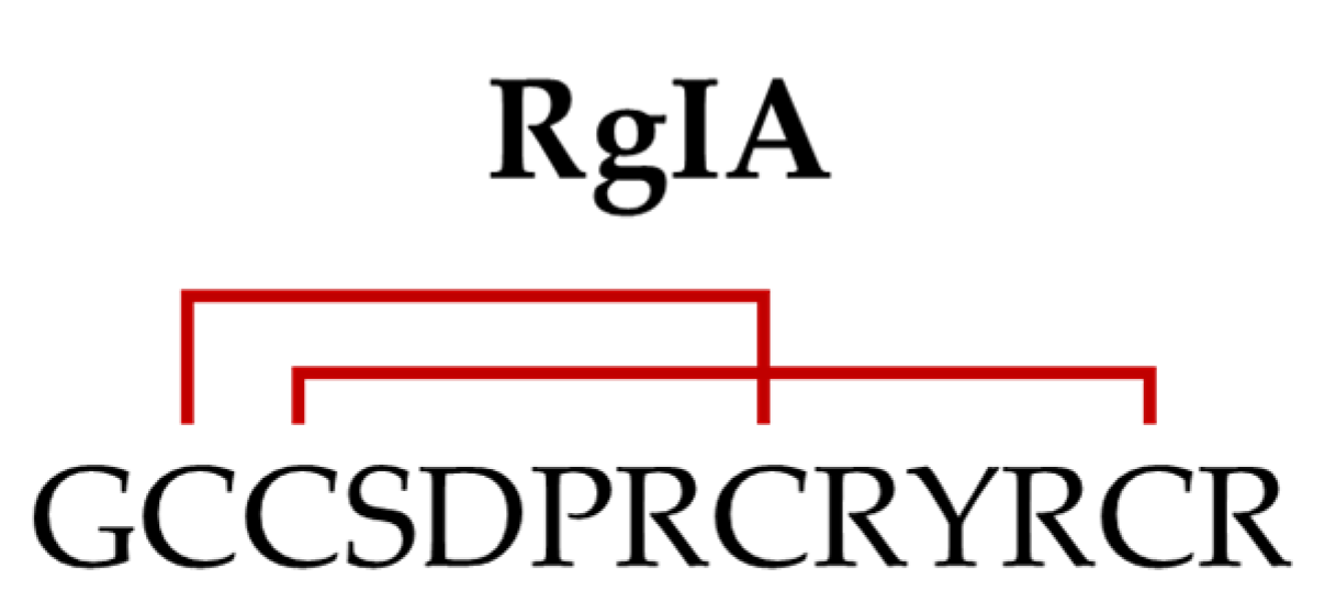
α-Conotoxins are the largest group of characterized conotoxins, due to their abundance and pharmacological profile. Normally, α-conotoxins act as antagonists of neuronal and/or muscular nAChRs [8,9]. nAChRs are pentameric ligand-gated ion channels composed of α (α1–α10) and non-α (β1–β4, γ, δ, and ε) subunits. Each receptor is composed of five individual subunits, giving rise to an extensive range of combinatorial arrangements [25]. The α1, β1, δ, ε, and γ subunits form the nAChR subtypes (adult and fetal) found at the neuromuscular junction. The remaining α and β subunits assemble in various combinations to form numerous and distinct receptor subtypes, mainly, but not exclusively, expressed by nervous system cells [26]. nAChRs are actively being investigated as targets for pain and inflammation treatments [26].
α-Conotoxins activities for nAChRs are based on constituent amino acid residues and, particularly, intramolecular interaction among the cysteines. The most common cysteine framework of α-conotoxins is framework I, with four cysteines (C) arranged in the C1C2-Xn-C3-Ym-C4 pattern, where Xn is a loop of amino acids with n= 3–4 and Ym is a loop of amino acids with m = 3–8 [4]. The number and nature of the amino acids in these loops classify α-conotoxins in 3/5, 4/3, 4/4, 4/6, 4/7, and 4/8 subtypes, each of which has a different binding and selectivity towards the nAChRs subtypes [8,27]. Furthermore, depending on the disulfide bond connectivity, three possible isomers can be formed, namely globular (C1-C3, C2-C4), ribbon (C1-C4, C2-C3) and bead (C1-C2, C3-C4) [4,9]. Other less common cysteine patterns of α-conotoxins are the II, III, IV, VII, XIV, XX, and XXIV frameworks (Table 1) [4].
Reg1a–f were the first α4/3-conotoxins isolated from C. regius by HPLC (Figure 1) [12]. Sequences of reg1a,b,e,f are unusual as they are post-translationally hydroxylated on proline residues to produce hydroxyprolines (Hyp or O): reg1f has two Hyp, whereas reg1a,b,e have only one Hyp. Reg1b,c have the same sequence, but reg1c is not hydroxylated at Pro6. Reg1c,d only differ in residue 11 (Gln in reg1c vs. Glu in reg1d) [12]. Overall, reg1a–e have significant sequence homology, while reg1f has no homology. Hydroxylation enhances the polarity and hydrogen-bonding capabilities to these conotoxins and probably defines their mode of binding to nAChRs [12]. However, no biological activities of these conotoxins have been described, except for reg1e. Reg1e (50 nM) caused a 40% inhibition of acetylcholine-evoked currents in hybrid human (h) α9/rat (r) α10 (hα9/rα10) nAChRs [28]. Moreover, it inhibited voltage-gated calcium channel (VGCC) in rat dorsal root ganglia (DRG) neurons via γ-aminobutyric acid type B receptor (GABABR) activation (IC50 20.5 nM) [28]. Interestingly, RgIA, which has similar biological activities (see below), is supposed to be the precursor sequence of reg1e [11,28,29].

5. RgIA

α-RgIA (or RgIA) is the most documented α-conotoxin described in C. regius. RgIA was identified using a PCR-based discovery method, which later allowed its synthesis using standard Fmoc chemistry [11]. This peptide is characterized by the cysteine framework I (CC-C-C) and displays a globular disulfide connectivity (Figure 2) [30].
More specifically, the four amino acids in the first loop and the three in the second loop place it in the α4/3 subfamily of conotoxins [11]. RgIA was first tested as an antagonist on Xenopus oocyte expressing rα9α10, rα7, rα2β2, rα2β4, rα3β2, rα3β4, rα4β2, rα4β4, and rα6β2β3 nAChRs, showing a 1000–2000-fold greater potency at inhibiting the rα9α10 subtype compared to the others. Then, its high specificity for rα9α10 nAChR was also confirmed on the native channel expressed by inner hair cells [11]. The high affinity block of rα9α10 nAChRs by RgIA has been attributed, in part, to residues Asp5, Pro6; Arg7 in loop I and Arg9 in loop II [31].
The α9α10 nAChR subtype, which is critical for mediating synaptic transmission from the medial olivocochlear to the cochlear hair cells, has also been implicated in a series of pathological conditions, including neuropathic pain [26,32,33,34,35,36], tumor proliferation [37,38,39], and immune-mediated disorders [40,41,42,43,44]. Considering the high affinity of RgIA towards this receptor subtype [11,45], its discovery led to the possibility of studying the role of α9α10 nAChRs in several diseases.

5.1. Analgesic and Disease-Modifying Effects of RgIA

RgIA is reported as a potent pain-relieving compound in rat models of neuropathy. A single intramuscular injection of RgIA (0.02 and 0.2 nmol) produced an acute, dose-dependent, antihyperalgesic effect in a chronic constriction injury (CCI) rat model. Repeated daily injections of 0.2 nmol of RgIA, for 5 days, resulted in a sustained analgesic effect and decreased inflammation at the sciatic nerve [46]. Daily administration of RgIA (2 and 10 nmol) for 7 and 14 days provided acute antinociceptive effects on both days in CCI rats, with long-lasting effects evident only by day 14 [33]. Additionally, treatment with RgIA attenuated the degree of inflammation in the sciatic nerve and DRG, reducing edema and infiltrate. Both dosages of RgIA also significantly decreased the density of glial cells in the spinal cord and prevented morphological derangements in DRG [33]. The same α-conotoxin doses also showed pain-relieving and neuroprotective properties in a rat model of oxaliplatin-induced neuropathy [35]. The repeated administration (2 and 10 nmol, intramuscularly) reduced the oxaliplatin-induced neuropathic pain hyperalgesia and allodynia, and DRG damage. In the spinal cord, the numerical increase of astrocyte cell density present in oxaliplatin-treated rats was partially prevented by RgIA treatment. Nevertheless, the administration of the α-conotoxin was able, per se, to elicit a numerical increase and a morphological activation of microglia and astrocytes in specific brain areas [35], suggesting that RgIA may modulate glial cells in order to promote neurorestoration and reduce pain. Supporting this evidence, the administration of RgIA (2 nmol) for two weeks in oxaliplatin-treated rats elicited similar antinociceptive effects, avoiding DRG morphological alterations [47] These observations were consistent with a progressive disease-modifying effects in neuropathic context by RgIA. The presence of α9α10 nAChRs in immune cells [42,48,49,50] has led to the hypothesis that the long-term effects on neuropathic pain produced by RgIA might be due to the modulation of immune cells infiltration and the consequent release of inflammatory mediators into the site of injury, which may sensitize nerves to nociceptive stimuli [51,52].
However, RgIA was also proposed to exert its analgesic effects by modulating the N-type VGCC CaV2.2 [53,54,55,56]. This inhibition is mediated by the activation of GABABR, and block also occurs in DRG neurons of α9 knockout (KO) mice [54], suggesting that the α9α10 subtype is not involved in the modulation of the calcium channels. Inhibition of N-type channels, in turn, decreases neurotransmitter release and synaptic transmission between DRG neurons and second-order neurons in the spinal cord and thus impairs the transmission of nociceptive signals to the brain [57]. This is the mechanism of action of another well-known Conus-derived compound, ω-conotoxin MVIIA, an FDA approved drug known as Prialt (Elan Pharmaceuticals, Dublin, Ireland), for the treatment of chronic pain [58,59].
RgIA (0.1 μM) was shown to inhibit VGCC currents by 40–50% in >75% of DRG neurons isolated from either mice or rats [54,55], an effect that was prevented by competitive GABABR antagonists [55] or the knockdown of the GABABR by siRNA [56]. Consequently, GABABR seems to be necessary for the inhibition of currents by the conotoxin. RgIA (10 μM) does not displace the binding of the antagonist [3 H]-CGP54626 to human GABABR transiently transfected in HEK293T [60], suggesting a non-competitive mechanism. The GABABR also functionally couples to the G protein coupled inwardly rectifying potassium (GIRK) channels to attenuate nociceptive transmission [61]. Recently, it has been shown that RgIA (1 μM) potentiates inwardly rectifying potassium currents in HEK293T cells heterologously expressing human GABABR coupled to GIRK1/2 channels [62], albeit it failed to elicit the same effects in Xenopus oocytes in a previous study [60]. In support of the latter evidence, Wright et al. [63] demonstrated that RgIA (0.1–1 μM) also had an insignificant effect on VGCC currents (inhibition >10% in <20% of rat DRG neurons), showing contrasting data to those obtained by Callaghan et al. [55]. Moreover, no correlation was found between the responses induced by RgIA and baclofen, a GABABR agonist. By activating the presynaptic GABABR in DRG neurons, baclofen inhibits excitatory post-synaptic currents (EPSCs) and prevents the release of glutamate, which blocks pain transmission between the primary nociceptors and second-order neurons in the dorsal horn of the spinal cord [34,64]. α-Conotoxins are charged peptides, so they may not cross the blood–brain barrier and reach spinal neuron synapses, suggesting that the VGCC mechanisms are not involved in RgIA-induced analgesia [34,60,63]. These findings are consistent with a study that demonstrated no inhibition of EPSCs in the dorsal horn neurons by Vc1.1, another peptide of the α-conotoxin family [64].
There are several conflicting results that make it difficult to determine the molecular mechanism by which α-conotoxins exert their analgesic effect. Many studies suggested that RgIA and some other α-conotoxins do not relieve pain by the activation of GABABR, reinforcing the idea of the involvement of α9α10 nAChRs [34,36,60,63,64]. As mentioned above, RgIA and other α9α10 nAChRs antagonists have been associated with disease-modifying effects in neuropathic pain conditions [9,33,34,35,36,60]. Known GABABR agonists, such as baclofen, did not show similar properties. RgIA4 (see below for its amino acid composition), a RgIA derivative that lacks GABABR activity, maintains the capacity to prevent the development of neuropathic pain [36,65,66]. Moreover, RgIA, which induces calcium transients in murine granulocytes, decreased reactive oxygen species production [42,44] and increased the production of the anti-inflammatory interleukin-10 in these cells. Even in this case, the α9α10 nAChR mechanism is more suitable than the GABABR mechanism for explaining such regulation, given that the inhibition of VGCC currents mediated by GABABR might result in lowering intracellular calcium, not in its increase [42]. Finally, evidence from α9 subunit KO mice also supports a role for α9α10 nAChRs in pain [36,67,68].

5.2. Anti-Colitis Effects of RgIA

In addition to analgesic and disease-modifying activities in animal models of pain, AlSharari et al. reported for the first time the anti-inflammatory effects of RgIA in the dextran sodium sulfate (DSS) experimental mice colitis model. Briefly, the lower doses (0.02 and 0.1 nmol) of the RgIA treatment in DSS-treated mice were inactive, whereas the higher dose (0.2 nmol) reversed the disease activity index score, loss of body weight, total histological damage score, as well as the colonic increase of the TNF-α concentration compared to the control group. Moreover, 0.2 nmol of RgIA significantly prevented the colon length shortening in DSS-treated mice [40]. The block of α9α10 nAChRs on gut immune cells as an anti-inflammatory signal was suggested as a pharmacodynamic mechanism.

5.3. Anticancer Effects of RgIA

As mentioned above, nAChRs also have a major role in tumorigenesis and cancer progression [37,38,39]. The expression of α9 and/or α10 subunits has been reported in several cancer cell lines, human tumors, and immune cells that could promote cancer-related inflammation [42,48,49,50,69,70,71,72]. On these bases, α9α10 nAChR blockers were studied as antitumor agents. Intraperitoneal injections of 0.1 nmol/kg of RgIA led to changes of a degenerative nature of both cancer cells and leukocytes infiltrating the tumor in Ehrlich carcinoma (EC)-bearing mice [43]. Interestingly, the α-conotoxin (1 nmol/kg) also enhanced the antitumor activity of splenocytes and increased the survival rate of EC-bearing mice by impairing tumor growth [73]. In vitro, RgIA increased the cytotoxic effects of the lipoxygenase pathway inhibitors nordihydroguaiaretic acid (24 h of treatment) and baicalein (48 h of treatment) on EC cells, which display an increased activity of the arachidonic acid cascade [74]. By contrast, a recent report showed that while baicalein exerted a significant antiproliferative and cytotoxic activities against C6 glioma cells, RgIA enhanced the proliferation of these cells [75]. No additional studies on the use of RgIA as an anticancer agent are reported. RgIA may have contrasting effects on cell proliferation, depending on the concerned cell line. Better anticancer activity was reported for synthetic derivatives of RgIA, as described below for RgIA4 [71].

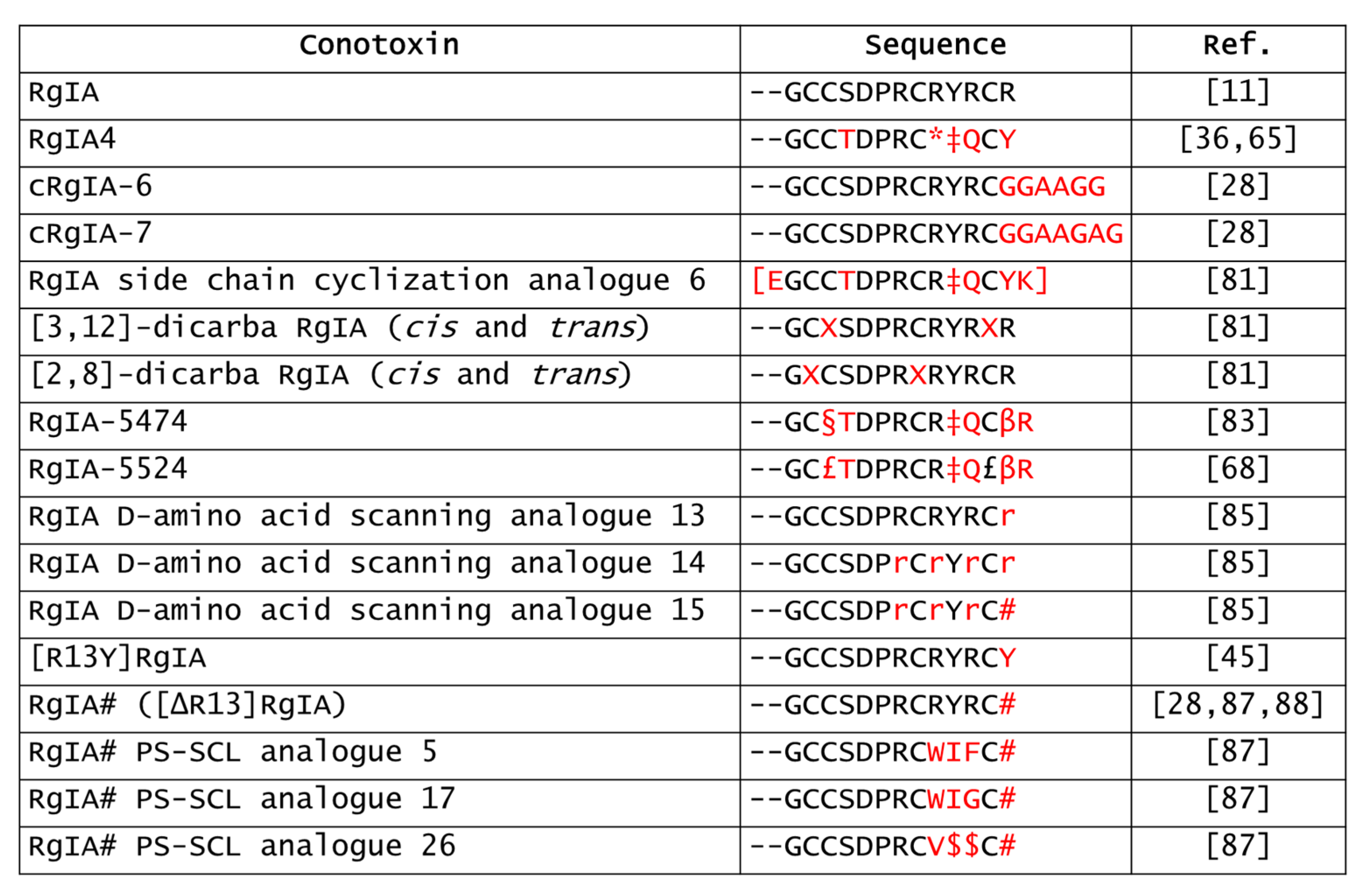
5.4. Derivatives of RgIA
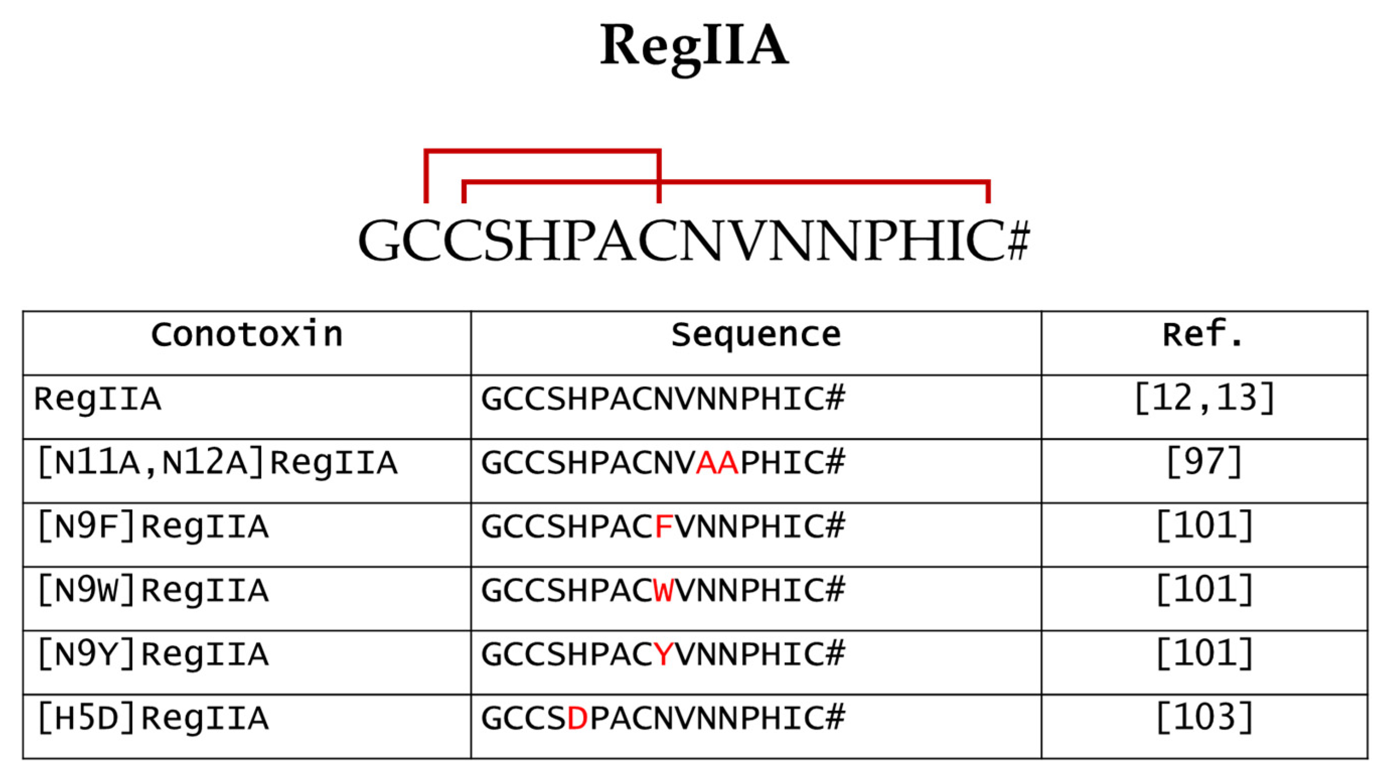
Owing to its high specificity towards α9α10 nAChRs, RgIA also provided a promising lead for developing novel ligands that selectively target these receptor subtypes. Unfortunately, several α-conotoxins, including RgIA, were initially tested in rodent nAChRs-expressing systems or native receptors as well as preclinical pain studies were most often carried out on rodent models. The crystal structure of hα9 nAChR extracellular domain with RgIA revealed that RgIA also interacted with hα9α10 nAChRs, involving its Asp5-Pro6-Arg7 triad of loop I and Arg11 of loop II [76]. However, there is a 300-fold difference (IC50 rat 1.5 nM vs. IC50 human 490 nM) of RgIA potency between the rat and human receptors, due to the residue at position 56 (Ile in humans vs. Thr in rats) of the α9 subunit [77,78]. These species differences likely led to the failure of the clinical development of a potential hα9α10 antagonist based on Vc1.1 [79]. Hence, the derivatives of native α-conotoxins are needed to improve specificity and potency against hα9-containing nAChRs together with pharmacokinetics features (Figure 3).
RgIA4 is the most reported RgIA-analogue in the literature data. This peptide has 5 of the 13 total amino acids modified but shows an increased potency and selectivity towards the hα9α10 subtype, retaining a high affinity for both rat and human receptors (IC50 rat 0.9 nM vs. IC50 human 1.5 nM) [36]. With a 1200-fold lesser potency than that for α9α10, RgIA4 inhibits the hα7 subtype (IC50 1.8 μM). Interestingly, RgIA4 lacks activity on GABABR, allowing the dissection of the involvement of α9α10 vs. GABABR in pain relief [36]. Daily subcutaneous injections of RgIA4 (0.128, 16 and 80 μg/kg) prevented cold allodynia and mechanical hypersensitivity in oxaliplatin-treated rats, after only one week of treatment [36]. The same treatment with 40 μg/kg of RgIA4 reversed oxaliplatin-induced cold allodynia in mice but only after 3 weeks of treatment. Interestingly, the effects lasted up to 3 weeks post-treatment, indicating a disease-modifying effect like that of the native peptide [65]. Long-lasting effects have also been reported in a paclitaxel-induced neuropathy rat model, where animals subcutaneously injected with RgIA4 (80 μg/kg) showed a significant reduction of mechanical allodynia by day 12 post-treatment [66]. These data support the effects of RgIA4 in multiple chemotherapy-induced neuropathy models. As anticipated above, RgIA4 (1 μM) completely abolished the proliferation of A549 adenocarcinoma cell line induced by 48 h treatment with 100 nM nicotine, blocking the nicotine-induced activation of Akt and ERK [71].
However, RgIA4 is not an ideal drug candidate because of its high in vivo degradation and poor serum stability [21]. Synthetic cyclization, with appropriately sized linkers between the N- and the C-termini, could be useful to overcome this issue. In fact, in a previous study, the insertion of a linker of 6 or 7 amino acids in the native RgIA (cRgIA-6 and cRgIA-7) caused an improvement in stability compared to the linear peptide, while maintaining a high potency towards the α9α10 nAChR [28]. By contrast, incorporating smaller linkers does not make significant improvements, suggesting that they are likely to force the N- and the C-termini together, putting a strain on the peptide conformation [28]. Similarly, the side chain cyclization of RgIA4 (analogue 6) led to an increased serum stability over linear RgIA4, while exhibiting a similar affinity with RgIA4 at the hα9α10 nAChR and analgesic effects in oxaliplatin-treated rats [80]. Structurally, the lactam linkage introduced in RgIA4 stabilized the globular conformation and suppressed disulfide scrambling, leading to stability improvements.
Disulfide bond replacement with a dicarba unsaturated bridge is a further strategy to enhance the serum stability, as well as to modulate the specificity of conotoxins [81,82]. Additionally, [3,12]-dicarba RgIA analogues retain inhibition at the α9α10 nAChR but lack GABABR activity, whereas [2,8]-dicarba analogues display reverse target selectivity, suggesting that the C1-C3 bridge is important for inhibiting the α9α10 nAChR and the C2-C4 bridge for GABABR modulation [81]. Molecular dynamics simulations suggested that substitution at Cys2 and Cys8 abolishes the RgIA activity at α9α10, mainly by the reduction of contacts between RgIA-Tyr10 and α9-Arg138 and between RgIA-Arg9 and α10-Trp81 [81]. The importance of the C1-C3 bridge on the activity of RgIA has also been demonstrated by two recent works. The substitution of the Cys3 with the Cys surrogate L-penicillamine, together with amino acids replacements and addition of L-Arg in position 14, resulted in RgIA-5474, a peptide with a 9000-fold increased potency towards the hα9α10 nAChR (IC50 0.05 nM) and improved serum stability compared to RgIA [83]. To date, RgIA-5474 is one of the most potent RgIA-derivatives against hα9α10 nAChR. A fluorescently tagged derivative of this peptide has been synthesized using click chemistry and can be used to visualize native α9α10 nAChRs [84]. Furthermore, the replacement of the C2-C4 bridge with unreducible methylene thioacetal and the same above-mentioned amino acids modifications led to the generation of RgIA-5524, a further compound with highly selectivity for hα9α10 nAChR (IC50 0.9 nM), reduced serum degradation and pain-relieving effects in oxaliplatin-treated mice (40 μg/kg) [68]. Hence, disulfide loop modifications could be helpful to improve biophysical properties of RgIA, by stabilizing its globular structure and avoiding disulfide scrambling.
In addition to the disulfide scrambling that makes its structure unstable, RgIA is more susceptible to proteolysis than other conotoxins due to its arginine-rich loop II and Tyr10, which provide cleavage sites for proteases. Ren et al. provided the solution by generating three RgIA D-amino acid scanning analogues (called peptides 13, 14, and 15) that are more resistant in serum and intestinal fluid than native RgIA [85]. D-amino acids are less prevalent in nature, thus they are unlikely to be susceptible to enzymatic degradation. Notably, peptide 15 displayed a two-fold increase in the inhibition of hα9α10 nAChR, whereas peptide 13 had a two-fold more potency against rα9α10 nAChR, compared to RgIA [85]. Peptide 13 retained its strong affinity because it contained D-enantiomer of Arg at the C-terminal, which is not involved in binding to α9α10 [31,85]. All the other synthesized D-enantiomer analogues showed decreased potency at the α9α10 receptor, indicating that amino acid chirality is important for the activity of these α-conotoxins. Separately, it was recently found that the substitution of Arg13 with Tyr ([R13Y]RgIA) significantly improved the potency of RgIA for hα9α10 nAChRs by 240-fold, nullifying the difference between rats and humans [45]. Thus, scan strategies are useful in revealing the critical amino acid involved in the peptide-receptor interaction. Positional-scanning synthetic combinatorial libraries (PS-SCL) consist of a mixture of peptides where one or more of the positions are individually fixed at a specific amino acid, while the remaining positions are comprised of an equimolar mixture of amino acids [86]. PS-SCL of RgIA# ([∆R13]RgIA) based on positions 9, 10, and 11 provided 10,648 possible conotoxin sequences, but, in the end, only three lead compounds (5, 17, and 26) displayed increased antinociception compared to the native peptide when intraperitoneally injected in mice (10 mg/kg) [87].
Finally, a further way to enhance the activity of α-conotoxins is dimerization. PEG9-dimeric RgIA# resulted in increased inhibitions at hα7 (IC50 dimeric RgIA# 63.1 nM vs. IC50 RgIA# >1000 nM) and hα9α10 (IC50 dimeric RgIA# 38.5 nM vs. IC50 RgIA# 248.7 nM), by 17- and 7-fold, respectively, compared to RgIA# [88]. Considering the role of these nAChRs in the development of cancer [71], dimeric RgIA# could be a useful pharmacological tool in cancer research. The PEG-linker also allows the conotoxin to simultaneously bind two adjacent binding sites, and further elongation of the linker should not significantly affect the activity of the dimer. Indeed, the PEG6-dimeric RgIA# showed comparable potency to the PEG9-dimeric RgIA# [88].
Figure 3. Sequences of the RgIA analogues reported in this review. *, citrulline; ‡, 3-Iodo-Tyrosine; [], side chain cyclization; X, positional sequence insertion of L-allylglycine residues prior to ring-closing olefin metathesis cyclization; §, L-penicilammine; β, 3-homo tyrosine; £, methylene thioacetal replacement; #, amidated C-terminal; r, D-arginine; $, norleucine [11,28,36,45,65,68,81,83,85,87,88].
Marinedrugs 20 00773 g003
Overall, chemical modifications of α-conotoxins may not only be helpful to improve their pharmacokinetic and pharmacodynamic features, but also to elucidate the molecular mechanisms underlying their activities, in order to facilitate their use as drug leads.

6. RegIIA

RegIIA was isolated from C. regius venom using a HPLC-based strategy and its amino acid sequence was obtained by Edman degradation [12,13]. Limited amounts of the natural peptide precluded further investigations, thus RegIIA was later chemically synthesized [13]. It belongs to the α4/7 subclass of α-conotoxins having the conserved cysteine framework I (CC-C-C). Due to its C1-C3 and C2-C4 disulfide connectivity (Figure 4), RegIIA exhibits a classical globular structure [13].
The signal sequence of RegIIA places it in the A-superfamily of conotoxins. RegIIA potently inhibited acetylcholine-evoked currents of rα3β2 and rα3β4, and hα7 (IC50 values of 33, 97, and 103 nM, respectively), whereas it did not inhibit rα4β2, rα9α10, and muscle nAChRs (IC50 > 1000 nM) [13]. For these reasons, despite the poor selectivity, RegIIA is considered one of the most potent antagonists of α3β4 nAChRs.
Neuronal nAChRs play an important role in the central and peripheral nervous systems, where they are functionally involved in several physiological and pathophysiological processes [89]. For example, the α3β4 nAChR has been shown to be involved in lung cancer, drug abuse, and nicotine addiction, whereas the α3β2 subtype is related to disorders associated with specific areas, such as the medial habenula, cerebellum, spinal cord, retina, and autonomic ganglia [90,91,92,93]. However, α3β4 and α3β2 subtypes have also been detected in non-neuronal cells, such as human vascular endothelial cells, epithelial cells, immune cells, keratinocytes, fibroblasts, and glial cells [94,95,96], where they modulate a number of biological processes, such as cell proliferation, migration, differentiation, survival, and gene expression [95,96]. Therefore, α3β4 and α3β2 subtypes could be promising therapeutic targets, making attractive pharmacologically active substances modulating these nAChR subtypes.
To date, however, the effect of RegIIA on nAChRs-related diseases has not yet been evaluated in any preclinical studies, suggesting that improvements in selectivity are needed to make it a potential lead compound. Below, we reported some of the chemical modifications to enhance the selectivity and sensitivity of RegIIA (Figure 4).

6.1. Strategies to Improve RegIIA Selectivity towards Specific nAChRs Subtypes

Although native RegIIA is a potent inhibitor of the α3β4, it cannot be considered a selective pharmacological probe. Alanine scan mutagenesis approach revealed the double alanine-analogue [N11A,N12A]RegIIA inhibited the rα3β4 nAChR subtype with a seven-fold less potency than native RegIIA but, at the rα3β2 and hα7 nAChR subtypes, the potency decreased by 1000- and 360-fold, respectively, thus indicating an increase in the selectivity for the rα3β4 subtype [97]. Concerning human nAChR subtypes, RegIIA exhibited a higher selectivity towards hα3β4 than hα3β2 [98,99]. Receptor mutagenesis sustained the importance of loop D residue 59 and loop E residue 113 in nAChRs as key residues for RegIIA affinity. Specifically, two mutations, [T59K]α3β2 and [S113R]α3β2, strongly enhanced the α-conotoxin affinity compared with wild-type α3β2, whereas opposite point mutations in α3β4 exerted the contrary effect [98]. Moreover, a recent study showed that oligosaccharide chains on hα3β4 nAChR are crucial for interaction with RegIIA and that His14 plays an important role for the structure-activity relationship of this conotoxin [100].
Since specific RegIIA analogues for α3β2 have not yet been developed, a computational-based scanning approach has shown that introducing an aromatic residue to position 9 of RegIIA ([N9F], [N9W], and [N9Y]RegIIA) led to an increased potency against hα3β2 compared to hα3β4, identifying in [N9Y]RegIIA the most selective inhibitor [101]. In this way, it would be possible to study the role of the α3β2 subtype in nAChR-related diseases.

6.2. Species-Specific Differences in RegIIA Sensitivity
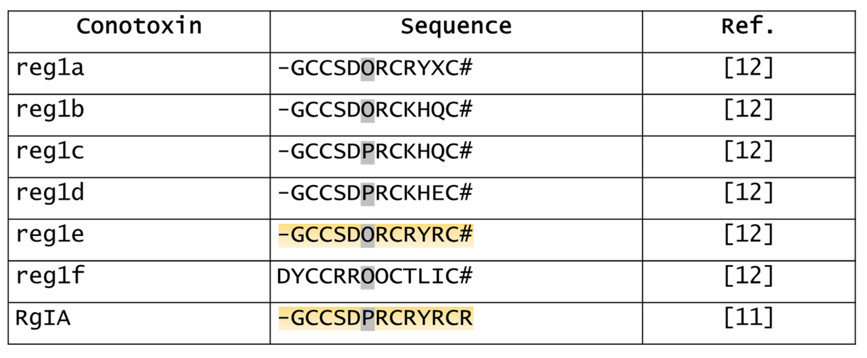
As mentioned above, the characterization of α-conotoxins in heterologous expression systems using mainly rat-cloned nAChR subunits is an obstacle to the effective development of nAChR subtype-selective α-conotoxins as pharmacological tools with therapeutic potential. In fact, conotoxins are likely to exhibit a different potency profile against the nAChRs subtypes of different species.
RegIIA, characterized as a competitive antagonist of rα3β2 and rα3β4 nAChRs, is 70-fold less potent at the homologous hα3β2 subtype [99]. This change was even more prominent for [N11A,N12A]RegIIA, which was inactive at the hα3β2 subtype but retained micromolar potency at rα3β2 (IC50 9.9 µM) [97]. At α3β4, neither RegIIA nor the analogue, showed species-specific differences in sensitivity, suggesting that the determinants for the species difference would reside in the β2 subunit [97,99]. Surprisingly, when the non-homologous residues in the rat β2 subunit were exchanged to the human counterpart, a non-significant loss in sensitivity to RegIIA was observed. Receptor mutagenesis and molecular dynamics studies indicated that this difference can be mainly attributed to a single amino acid residue in position 198 on the rat α3 nAChR subunit. Consequently, a single exchange of residue Gln198 in the rat α3 subunit led to a loss of potency consistent with those of hα3β2 nAChRs. This residue exchange causes structural changes at the agonist binding site, reducing the interaction with RegIIA residues [99].
Another species-specific difference concerns the α7 nAChRs, which play an important role in cancer development [71] and in cognitive disorders, such as Alzheimer’s disease and schizophrenia [102]. As mentioned above, RegIIA is also an antagonist of the hα7 subtype [13], although it inhibits the rα7 subtype five-fold more powerfully [103]. The aspartate analogue [H5D]RegIIA strongly increased to 130-fold the difference in this species selectivity, which has been shown to be influenced by the residue in position 185 on the receptor [103].
Hence, it can be understood that mutagenesis studies coupled with molecular dynamics simulations can lead to the customized design of conotoxin analogues, which may be useful in increasing their specificity not only towards a specific nAChR subtype of the receptor, but also towards a species-specific subtype.
Figure 4. Sequences of RegIIA (with indication of the two disulfide bonds) and the RegIIA analogues reported in this review. #, amidated C-terminal [12,13,97,101,103].
Marinedrugs 20 00773 g004

7. RgIB

α-RgIB (or RgIB) is another 4/7 α-conotoxin characterized by cysteine framework I (CC-C-C) (Figure 5) [14]. This conotoxin was isolated from the C. regius venom through HPLC fractionation and purification. To date, neither the features of its disulfide connectivity nor to which gene superfamily it belongs are known. The unique report in the literature data showed two biological activities by RgIB. First, intracranial injections of RgIB (1 nmol) aroused hyperactive behavior in mice, while lower doses (0.1 and 0.5 nmol) caused breathing difficulty. Second, RgIB (10 μM) induced an irreversible blockage of carbamylcholine-induced currents by 40% in nAChRs-expressing cells (PC12 cells), acting very likely on α3β4 and α3β4α5 subtypes [14].
Without any other evidence about this α-conotoxin, it is currently not possible to establish with certainty its specific targets and related pharmacological effects. Further studies are needed to better characterize the pharmacological and structural properties of RgIB.

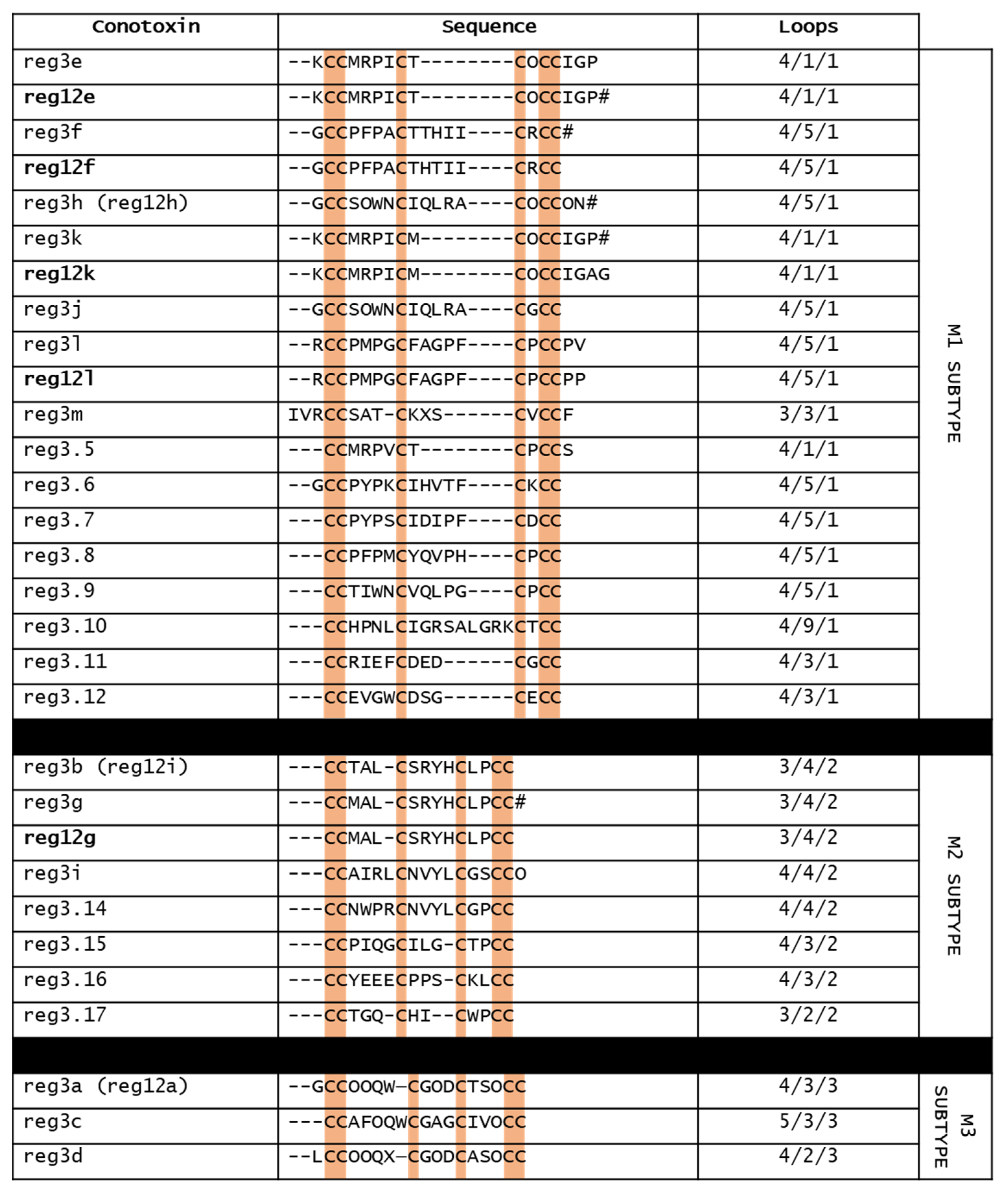
8. Reg3b and Mini-M Conotoxins

Conotoxins of the M-superfamily have been found in the venom of all Conus species [104]. Normally, M-superfamily conotoxins display a cysteine framework III (CC−C−C−CC), with a wide variability in the cysteine pattern and size of the three loop regions. Furthermore, compared to framework I of α-conotoxins, M-conotoxins having the framework III are constrained by three disulfide bonds. Within this gene superfamily, there are five M-subtypes (M1–M5), based on the number of amino acids in the third loop. M1–M3 conotoxins are grouped in “mini-M” (<22 residues), whereas M4 and M5 belong to the “maxi-M” group (>22 residues) [104,105]. While the latter are the better-known M-superfamily conotoxins, unfortunately little is known about mini-M conotoxins. Figure 6 shows the sequences of the mini-M conotoxins found in the venom of C. regius.
Reg12a,e–i,k,l were the first mini-M conotoxins isolated by HPLC and sequenced from the venom of C. regius [12]. These conotoxins did not have sequence homology and did not share the same post-translational modification pattern. Concerning their loop size, they were characterized by four variants (3/4/2, 4/1/1, 4/3/3, and 4/5/1), suggesting that their targets might be diverse. Reg12e,f,h,k,l belonged to M1 subtype, reg 12g,i to the M2 subtype, and reg12a to the M3 subtype [12]. More recently, further mini-M conotoxins have been found in C. regius venom duct by transcriptomic (reg3.5–12 and reg3.14–17) and peptidomic analysis (reg3a–m) [15]. Of these, only reg3a–m have been isolated by HPLC, because they are expressed in larger quantities.
These conotoxins exhibited variability in loop sizes, post-translational modifications, disulfide connectivities and three-dimensional folds, which are determinant for the final shape of the peptidic scaffolds and their activities. Based on loop sizes, seven of the isolated reg3 conotoxins belonged to the M1 group, three to the M2 group, and three to the M3 group [15]. Three-dimensional structure analysis was performed only on reg3b, the most abundant mini-M conotoxins of C. regius. Reg3b has 3/4/2 loop sizes and form three disulfide bonds between the first and sixth, second and fourth, and third and fifth cysteines, conferring a highly compact overall fold [15]. Aligning the sequences of mini-M conotoxins of C. regius, it was found that reg12a,h,i corresponded, respectively, to reg3a,h,b, whereas reg12e,f,g,k,l had high sequence homology, respectively, with reg3e,f,g,k,l. As already mentioned, neither the pharmacological activities of reg3b nor of the C. regius-derived mini-M conotoxins are known. However, given that ArchIIIA, a mini-M conotoxin from C. archon that differs from reg3b only in residue 3 (Ser in ArcIIA vs. Thr in reg3b), blocks hα7 nAChRs [106], it is possible that reg3b also shows this activity.
Overall, the plasticity of these mini-M scaffolds makes them interesting for the development of chemically modified peptides with therapeutic applications.

9. Conclusions

Given its amazing biodiversity and large number of yet-undiscovered species, the sea may be a rich source for new medicines. Nevertheless, only 20 drugs from marine organisms are currently in clinical use [107]. Conotoxins have attracted interest as great lead compounds for designing new drugs, due to their well-defined structure and ability to discriminate among subtypes of ion channels. In this review, we described the features of C. regius-derived conotoxins, which could represent promising therapeutic resources for the treatment of several diseases, including neuropathic pain, drug addiction, cognitive disorders, and cancer. Unfortunately, the development as drugs of many peptides is usually hindered because of their low stability and bioavailability. However, we also showed that the chemical engineering of conotoxins could be useful for improving their pharmacodynamic and pharmacokinetic properties, ensuring the concrete opportunity to develop new therapeutic agents starting from marine sources.

Author Contributions

Conceptualization, F.M. and L.D.C.M.; methodology, F.M., L.M. and C.C.; formal analysis, F.M., L.M. and C.C.; writing—original draft preparation, F.M.; writing—review and editing, F.M., L.D.C.M., J.M.M. and C.G.; supervision, C.G. All authors have read and agreed to the published version of the manuscript.

Funding

This work was supported by the IMI2 project NeuroDerisk (821528), the Italian Ministry of University and Research, University of Florence and by the U.S. National Institutes of Health, grant number R35 GM136430 to J.M.M.

Institutional Review Board Statement

Not applicable.

Data Availability Statement

Not applicable.

Conflicts of Interest

The University of Utah has filed patent applications on conopeptides including those listed in the present manuscript and on which J.M.M. is listed as an inventor.

References

  1. Endean, R.; Gyr, P.; Surridge, J. The Pharmacological Actions on Guinea-Pig Ileum of Crude Venoms from the Marine Gastropods Conus striatus and Conus magus. Toxicon 1977, 15, 327–337. [Google Scholar] [CrossRef] [PubMed]
  2. Kohn, A.J. Piscivorous Gastropods Of The Genus Conus. Proc. Natl. Acad. Sci. USA 1956, 42, 168–171. [Google Scholar] [CrossRef] [PubMed] [Green Version]
  3. Spence, I.; Gillessen, D.; Gregson, R.P.; Quinn, R.J. Characterization of the Neurotoxic Constituents of Conus geographus (L.) Venom. Life Sci. 1977, 21, 1759–1769. [Google Scholar] [CrossRef] [PubMed]
  4. Jin, A.-H.; Muttenthaler, M.; Dutertre, S.; Himaya, S.W.A.; Kaas, Q.; Craik, D.J.; Lewis, R.J.; Alewood, P.F. Conotoxins: Chemistry and Biology. Chem. Rev. 2019, 119, 11510–11549. [Google Scholar] [CrossRef]
  5. Olivera, B.M. EE Just Lecture, 1996. Mol. Biol. Cell 1997, 8, 2101–2109. [Google Scholar] [CrossRef] [Green Version]
  6. Olivera, B.M.; Rivier, J.; Clark, C.; Ramilo, C.A.; Corpuz, G.P.; Abogadie, F.C.; Mena, E.E.; Woodward, S.R.; Hillyard, D.R.; Cruz, L.J. Diversity of Conus Neuropeptides. Science 1990, 249, 257–263. [Google Scholar] [CrossRef]
  7. Vianna Braga, M.C.; Konno, K.; Portaro, F.C.V.; Carlos de Freitas, J.; Yamane, T.; Olivera, B.M.; Pimenta, D.C. Mass Spectrometric and High Performance Liquid Chromatography Profiling of the Venom of the Brazilian Vermivorous Mollusk Conus regius: Feeding Behavior and Identification of One Novel Conotoxin. Toxicon 2005, 45, 113–122. [Google Scholar] [CrossRef]
  8. Abraham, N.; Lewis, R. Neuronal Nicotinic Acetylcholine Receptor Modulators from Cone Snails. Mar. Drugs 2018, 16, 208. [Google Scholar] [CrossRef] [Green Version]
  9. Li, X.; Tae, H.-S.; Chu, Y.; Jiang, T.; Adams, D.J.; Yu, R. Medicinal Chemistry, Pharmacology, and Therapeutic Potential of α-Conotoxins Antagonizing the α9α10 Nicotinic Acetylcholine Receptor. Pharmacol. Ther. 2021, 222, 107792. [Google Scholar] [CrossRef]
  10. Walls, J.G. Cone Shells, a Synopsis of the Living Conidae. In Cone Shells, a Synopsis of the Living Conidae; T.F.H. Pulications: Neptune City, NJ, USA, 1979; pp. 831–833. [Google Scholar]
  11. Ellison, M.; Haberlandt, C.; Gomez-Casati, M.E.; Watkins, M.; Elgoyhen, A.B.; McIntosh, J.M.; Olivera, B.M. α-RgIA: A Novel Conotoxin That Specifically and Potently Blocks the α9α10 nAChR. Biochemistry 2006, 45, 1511–1517. [Google Scholar] [CrossRef]
  12. Franco, A.; Pisarewicz, K.; Moller, C.; Mora, D.; Fields, G.B.; Marí, F. Hyperhydroxylation: A New Strategy for Neuronal Targeting by Venomous Marine Molluscs. Prog. Mol. Subcell. Biol. 2006, 43, 83–103. [Google Scholar] [CrossRef] [PubMed]
  13. Franco, A.; Kompella, S.N.; Akondi, K.B.; Melaun, C.; Daly, N.L.; Luetje, C.W.; Alewood, P.F.; Craik, D.J.; Adams, D.J.; Marí, F. RegIIA: An α4/7-Conotoxin from the Venom of Conus regius That Potently Blocks α3β4 nAChRs. Biochem. Pharm. 2012, 83, 419–426. [Google Scholar] [CrossRef] [PubMed]
  14. Braga, M.C.V.; Nery, A.A.; Ulrich, H.; Konno, K.; Sciani, J.M.; Pimenta, D.C. α-RgIB: A Novel Antagonist Peptide of Neuronal Acetylcholine Receptor Isolated from Conus regius Venom. Int. J. Pept. 2013, 2013, 543028. [Google Scholar] [CrossRef] [PubMed] [Green Version]
  15. Franco, A.; Dovell, S.; Möller, C.; Grandal, M.; Clark, E.; Marí, F. Structural Plasticity of Mini-M Conotoxins—Expression of All Mini-M Subtypes by Conus regius. FEBS J. 2018, 285, 887–902. [Google Scholar] [CrossRef] [Green Version]
  16. Halai, R.; Craik, D.J. Conotoxins: Natural Product Drug Leads. Nat. Prod. Rep. 2009, 26, 526. [Google Scholar] [CrossRef]
  17. Livett, B.G.; Gayler, K.R.; Khalil, Z. Drugs from the Sea: Conopeptides as Potential Therapeutics. Curr. Med. Chem. 2004, 11, 1715–1723. [Google Scholar] [CrossRef]
  18. Vetter, I.J.; Lewis, R. Therapeutic Potential of Cone Snail Venom Peptides (Conopeptides). Curr. Top. Med. Chem. 2012, 12, 1546–1552. [Google Scholar] [CrossRef]
  19. Kaas, Q.; Yu, R.; Jin, A.-H.; Dutertre, S.; Craik, D.J. ConoServer: Updated Content, Knowledge, and Discovery Tools in the Conopeptide Database. Nucleic Acids Res. 2012, 40, D325–D330. [Google Scholar] [CrossRef]
  20. Góngora-Benítez, M.; Tulla-Puche, J.; Albericio, F. Multifaceted Roles of Disulfide Bonds. Peptides as Therapeutics. Chem. Rev. 2014, 114, 901–926. [Google Scholar] [CrossRef]
  21. Akondi, K.B.; Muttenthaler, M.; Dutertre, S.; Kaas, Q.; Craik, D.J.; Lewis, R.J.; Alewood, P.F. Discovery, Synthesis, and Structure–Activity Relationships of Conotoxins. Chem. Rev. 2014, 114, 5815–5847. [Google Scholar] [CrossRef]
  22. Kaas, Q.; Westermann, J.-C.; Halai, R.; Wang, C.K.L.; Craik, D.J. ConoServer, a Database for Conopeptide Sequences and Structures. Bioinformatics 2008, 24, 445–446. [Google Scholar] [CrossRef] [PubMed] [Green Version]
  23. Lewis, R.J.; Dutertre, S.; Vetter, I.; Christie, M.J. Conus Venom Peptide Pharmacology. Pharm. Rev. 2012, 64, 259–298. [Google Scholar] [CrossRef] [PubMed]
  24. Kaas, Q.; Westermann, J.-C.; Craik, D.J. Conopeptide Characterization and Classifications: An Analysis Using ConoServer. Toxicon 2010, 55, 1491–1509. [Google Scholar] [CrossRef] [PubMed]
  25. Lipovsek, M.; Marcovich, I.; Elgoyhen, A.B. The Hair Cell 9α10 Nicotinic Acetylcholine Receptor: Odd Cousin in an Old Family. Front. Cell. Neurosci. 2021, 15, 785265. [Google Scholar] [CrossRef]
  26. Hone, A.J.; McIntosh, J.M. Nicotinic Acetylcholine Receptors in Neuropathic and Inflammatory Pain. FEBS Lett. 2018, 592, 1045–1062. [Google Scholar] [CrossRef] [Green Version]
  27. Dutton, J.; Craik, D. Alpha Conotoxins Nicotinic Acetylcholine Receptor Antagonists as Pharmacological Tools and Potential Drug Leads. Curr. Med. Chem. 2001, 8, 327–344. [Google Scholar] [CrossRef]
  28. Halai, R.; Callaghan, B.; Daly, N.L.; Clark, R.J.; Adams, D.J.; Craik, D.J. Effects of Cyclization on Stability, Structure, and Activity of α-Conotoxin RgIA at the α9α10 Nicotinic Acetylcholine Receptor and GABAB Receptor. J. Med. Chem. 2011, 54, 6984–6992. [Google Scholar] [CrossRef]
  29. Safavi-Hemami, H.; Brogan, S.E.; Olivera, B.M. Pain Therapeutics from Cone Snail Venoms: From Ziconotide to Novel Non-Opioid Pathways. J. Proteom. 2019, 190, 12–20. [Google Scholar] [CrossRef]
  30. Clark, R.J.; Daly, N.L.; Halai, R.; Nevin, S.T.; Adams, D.J.; Craik, D.J. The Three-Dimensional Structure of the Analgesic α-Conotoxin, RgIA. FEBS Lett. 2008, 582, 597–602. [Google Scholar] [CrossRef] [Green Version]
  31. Ellison, M.; Feng, Z.-P.; Park, A.J.; Zhang, X.; Olivera, B.M.; McIntosh, J.M.; Norton, R.S. α-RgIA, a Novel Conotoxin That Blocks the α9α10 nAChR: Structure and Identification of Key Receptor-Binding Residues. J. Mol. Biol. 2008, 377, 1216–1227. [Google Scholar] [CrossRef]
  32. Del Bufalo, A.; Cesario, A.; Salinaro, G.; Fini, M.; Russo, P. Alpha9Alpha10 Nicotinic Acetylcholine Receptors as Target for the Treatment of Chronic Pain. Curr. Pharm. Des. 2014, 20, 6042–6047. [Google Scholar] [CrossRef] [PubMed]
  33. Di Cesare Mannelli, L.; Cinci, L.; Micheli, L.; Zanardelli, M.; Pacini, A.; McIntosh, M.J.; Ghelardini, C. α-Conotoxin RgIA Protects against the Development of Nerve Injury-Induced Chronic Pain and Prevents Both Neuronal and Glial Derangement. Pain 2014, 155, 1986–1995. [Google Scholar] [CrossRef] [PubMed] [Green Version]
  34. Hone, A.J.; Servent, D.; McIntosh, J.M. α9-containing Nicotinic Acetylcholine Receptors and the Modulation of Pain. Br. J. Pharm. 2018, 175, 1915–1927. [Google Scholar] [CrossRef] [PubMed] [Green Version]
  35. Pacini, A.; Micheli, L.; Maresca, M.; Branca, J.J.V.; McIntosh, J.M.; Ghelardini, C.; Di Cesare Mannelli, L. The α9α10 Nicotinic Receptor Antagonist α-Conotoxin RgIA Prevents Neuropathic Pain Induced by Oxaliplatin Treatment. Exp. Neurol. 2016, 282, 37–48. [Google Scholar] [CrossRef]
  36. Romero, H.K.; Christensen, S.B.; Di Cesare Mannelli, L.; Gajewiak, J.; Ramachandra, R.; Elmslie, K.S.; Vetter, D.E.; Ghelardini, C.; Iadonato, S.P.; Mercado, J.L.; et al. Inhibition of α9α10 Nicotinic Acetylcholine Receptors Prevents Chemotherapy-Induced Neuropathic Pain. Proc. Natl. Acad. Sci. USA 2017, 114, E1825–E1832. [Google Scholar] [CrossRef] [Green Version]
  37. Chen, J.; Cheuk, I.W.Y.; Shin, V.Y.; Kwong, A. Acetylcholine Receptors: Key Players in Cancer Development. Surg. Oncol. 2019, 31, 46–53. [Google Scholar] [CrossRef]
  38. Dang, N.; Meng, X.; Song, H. Nicotinic Acetylcholine Receptors and Cancer. Biomed. Rep. 2016, 4, 515–518. [Google Scholar] [CrossRef] [Green Version]
  39. Friedman, J.R.; Richbart, S.D.; Merritt, J.C.; Brown, K.C.; Nolan, N.A.; Akers, A.T.; Lau, J.K.; Robateau, Z.R.; Miles, S.L.; Dasgupta, P. Acetylcholine Signaling System in Progression of Lung Cancers. Pharmacol. Ther. 2019, 194, 222–254. [Google Scholar] [CrossRef]
  40. AlSharari, S.D.; Toma, W.; Mahmood, H.M.; Michael McIntosh, J.; Imad Damaj, M. The α9α10 Nicotinic Acetylcholine Receptors Antagonist α-Conotoxin RgIA Reverses Colitis Signs in Murine Dextran Sodium Sulfate Model. Eur. J. Pharm. 2020, 883, 173320. [Google Scholar] [CrossRef]
  41. Richter, K.; Papke, R.L.; Stokes, C.; Roy, D.C.; Espinosa, E.S.; Wolf, P.M.K.; Hecker, A.; Liese, J.; Singh, V.K.; Padberg, W.; et al. Comparison of the Anti-Inflammatory Properties of Two Nicotinic Acetylcholine Receptor Ligands, Phosphocholine and PCF3-DiEPP. Front. Cell. Neurosci. 2022, 16, 779081. [Google Scholar] [CrossRef]
  42. Safronova, V.G.; Vulfius, C.A.; Astashev, M.E.; Tikhonova, I.V.; Serov, D.A.; Jirova, E.A.; Pershina, E.V.; Senko, D.A.; Zhmak, M.N.; Kasheverov, I.E.; et al. α9α10 Nicotinic Acetylcholine Receptors Regulate Murine Bone Marrow Granulocyte Functions. Immunobiology 2021, 226, 152047. [Google Scholar] [CrossRef] [PubMed]
  43. Terpinskaya, T.I.; Osipov, A.V.; Kuznetsova, T.E.; Ryzhkovskaya, E.L.; Ulaschik, V.S.; Ivanov, I.A.; Tsetlin, V.I.; Utkin, Y.N. α-Conotoxins Revealed Different Roles of Nicotinic Cholinergic Receptor Subtypes in Oncogenesis of Ehrlich Tumor and in the Associated Inflammation. Dokl. Biochem. Biophys. 2015, 463, 216–219. [Google Scholar] [CrossRef] [PubMed]
  44. Tsetlin, V.; Haufe, Y.; Safronova, V.; Serov, D.; Shadamarshan, P.; Son, L.; Shelukhina, I.; Kudryavtsev, D.; Kryukova, E.; Kasheverov, I.; et al. Interaction of α9α10 Nicotinic Receptors With Peptides and Proteins From Animal Venoms. Front. Cell. Neurosci. 2021, 15, 765541. [Google Scholar] [CrossRef] [PubMed]
  45. Huynh, P.N.; Harvey, P.J.; Gajewiak, J.; Craik, D.J.; Michael McIntosh, J. Critical Residue Properties for Potency and Selectivity of α-Conotoxin RgIA towards α9α10 Nicotinic Acetylcholine Receptors. Biochem. Pharm. 2020, 181, 114124. [Google Scholar] [CrossRef]
  46. Vincler, M.; Wittenauer, S.; Parker, R.; Ellison, M.; Olivera, B.M.; McIntosh, J.M. Molecular Mechanism for Analgesia Involving Specific Antagonism of α9α10 Nicotinic Acetylcholine Receptors. Proc. Natl. Acad. Sci. USA 2006, 103, 17880–17884. [Google Scholar] [CrossRef] [Green Version]
  47. Dyachenko, I.A.; Palikova, Y.A.; Palikov, V.A.; Korolkova, Y.V.; Kazakov, V.A.; Egorova, N.S.; Garifulina, A.I.; Utkin, Y.N.; Tsetlin, V.I.; Kryukova, E.V. α-Conotoxin RgIA and Oligoarginine R8 in the Mice Model Alleviate Long-Term Oxaliplatin Induced Neuropathy. Biochimie 2022, 194, 127–136. [Google Scholar] [CrossRef]
  48. Grau, V.; Richter, K.; Hone, A.J.; McIntosh, J.M. Conopeptides [V11L;V16D]ArIB and RgIA4: Powerful Tools for the Identification of Novel Nicotinic Acetylcholine Receptors in Monocytes. Front. Pharm. 2019, 9, 1499. [Google Scholar] [CrossRef] [Green Version]
  49. Lustig, L.R.; Peng, H.; Hiel, H.; Yamamoto, T.; Fuchs, P.A. Molecular Cloning and Mapping of the Human Nicotinic Acetylcholine Receptor alpha10 (CHRNA10). Genomics 2001, 73, 272–283. [Google Scholar] [CrossRef]
  50. Peng, H.; Ferris, R.L.; Matthews, T.; Hiel, H.; Lopez-Albaitero, A.; Lustig, L.R. Characterization of the Human Nicotinic Acetylcholine Receptor Subunit Alpha (α) 9 (CHRNA9) and Alpha (α) 10 (CHRNA10) in Lymphocytes. Life Sci. 2004, 76, 263–280. [Google Scholar] [CrossRef]
  51. Leskovar, A.; Moriarty, L.J.; Turek, J.J.; Schoenlein, I.A.; Borgens, R.B. The Macrophage in Acute Neural Injury: Changes in Cell Numbers over Time and Levels of Cytokine Production in Mammalian Central and Peripheral Nervous Systems. J. Exp. Biol. 2000, 203, 1783–1795. [Google Scholar] [CrossRef]
  52. Hendriks, J.J.A.; Teunissen, C.E.; de Vries, H.E.; Dijkstra, C.D. Macrophages and Neurodegeneration. Brain Res. Rev. 2005, 48, 185–195. [Google Scholar] [CrossRef] [PubMed]
  53. Adams, D.J.; Callaghan, B.; Berecki, G. Analgesic Conotoxins: Block and G Protein-Coupled Receptor Modulation of N-Type (CaV2.2) Calcium Channels. Br. J. Pharm. 2012, 166, 486–500. [Google Scholar] [CrossRef] [PubMed] [Green Version]
  54. Callaghan, B.; Adams, D.J. Analgesic α-Conotoxins Vc1.1 and Rg1A Inhibit N-Type Calcium Channels in Sensory Neurons of α9 Nicotinic Receptor Knockout Mice. Channels 2010, 4, 51–54. [Google Scholar] [CrossRef] [PubMed]
  55. Callaghan, B.; Haythornthwaite, A.; Berecki, G.; Clark, R.J.; Craik, D.J.; Adams, D.J. Analgesic Alpha-Conotoxins Vc1.1 and Rg1A Inhibit N-Type Calcium Channels in Rat Sensory Neurons via GABAB Receptor Activation. J. Neurosci. 2008, 28, 10943–10951. [Google Scholar] [CrossRef] [PubMed] [Green Version]
  56. Cuny, H.; de Faoite, A.; Huynh, T.G.; Yasuda, T.; Berecki, G.; Adams, D.J. γ-Aminobutyric Acid Type B (GABAB) Receptor Expression Is Needed for Inhibition of N-Type (Cav2.2) Calcium Channels by Analgesic α-Conotoxins. J. Biol. Chem. 2012, 287, 23948–23957. [Google Scholar] [CrossRef] [PubMed] [Green Version]
  57. Harding, E.K.; Zamponi, G.W. The Calcium Channel Terminator: Hasta La Vista Pain. Trends Pharm. Sci. 2022, 43, 801–803. [Google Scholar] [CrossRef]
  58. Chalil, A.; Staudt, M.D.; Harland, T.A.; Leimer, E.M.; Bhullar, R.; Argoff, C.E. A Safety Review of Approved Intrathecal Analgesics for Chronic Pain Management. Expert Opin. Drug Saf. 2021, 20, 439–451. [Google Scholar] [CrossRef]
  59. Elmslie, K.S. Calcium Channel Blockers in the Treatment of Disease. J. Neurosci. Res. 2004, 75, 733–741. [Google Scholar] [CrossRef]
  60. McIntosh, J.M.; Absalom, N.; Chebib, M.; Elgoyhen, A.B.; Vincler, M. Alpha9 Nicotinic Acetylcholine Receptors and the Treatment of Pain. Biochem. Pharm. 2009, 78, 693–702. [Google Scholar] [CrossRef] [Green Version]
  61. Blednov, Y.A.; Stoffel, M.; Alva, H.; Harris, R.A. A Pervasive Mechanism for Analgesia: Activation of GIRK2 Channels. Proc. Natl. Acad. Sci. USA 2003, 100, 277–282. [Google Scholar] [CrossRef]
  62. Bony, A.R.; McArthur, J.R.; Finol-Urdaneta, R.K.; Adams, D.J. Analgesic α-conotoxins Modulate Native and Recombinant GIRK1/2 Channels via Activation of GABAB Receptors and Reduce Neuroexcitability. Br. J. Pharm. 2022, 179, 179–198. [Google Scholar] [CrossRef] [PubMed]
  63. Wright, A.B.; Norimatsu, Y.; McIntosh, J.M.; Elmslie, K.S. Limited Efficacy of α-Conopeptides, Vc1.1 and RgIA, To Inhibit Sensory Neuron CaV Current. ENeuro 2015, 2, ENEURO.0057-14.2015. [Google Scholar] [CrossRef] [PubMed] [Green Version]
  64. Napier, I.A.; Klimis, H.; Rycroft, B.K.; Jin, A.H.; Alewood, P.F.; Motin, L.; Adams, D.J.; Christie, M.J. Intrathecal α-Conotoxins Vc1.1, AuIB and MII Acting on Distinct Nicotinic Receptor Subtypes Reverse Signs of Neuropathic Pain. Neuropharmacology 2012, 62, 2202–2207. [Google Scholar] [CrossRef] [PubMed]
  65. Christensen, S.B.; Hone, A.J.; Roux, I.; Kniazeff, J.; Pin, J.-P.; Upert, G.; Servent, D.; Glowatzki, E.; McIntosh, J.M. RgIA4 Potently Blocks Mouse α9α10 nAChRs and Provides Long Lasting Protection against Oxaliplatin-Induced Cold Allodynia. Front. Cell. Neurosci. 2017, 11, 219. [Google Scholar] [CrossRef] [PubMed] [Green Version]
  66. Huynh, P.N.; Giuvelis, D.; Christensen, S.; Tucker, K.L.; McIntosh, J.M. RgIA4 Accelerates Recovery from Paclitaxel-Induced Neuropathic Pain in Rats. Mar. Drugs 2019, 18, 12. [Google Scholar] [CrossRef] [PubMed] [Green Version]
  67. Mohammadi, S.; Christie, M.J. α9-Nicotinic Acetylcholine Receptors Contribute to the Maintenance of Chronic Mechanical Hyperalgesia, but Not Thermal or Mechanical Allodynia. Mol. Pain 2014, 10, 1744–8069. [Google Scholar] [CrossRef] [Green Version]
  68. Zheng, N.; Christensen, S.B.; Dowell, C.; Purushottam, L.; Skalicky, J.J.; McIntosh, J.M.; Chou, D.H.-C. Discovery of Methylene Thioacetal-Incorporated α-RgIA Analogues as Potent and Stable Antagonists of the Human α9α10 Nicotinic Acetylcholine Receptor for the Treatment of Neuropathic Pain. J. Med. Chem. 2021, 64, 9513–9524. [Google Scholar] [CrossRef]
  69. Lee, C.-H.; Huang, C.-S.; Chen, C.-S.; Tu, S.-H.; Wang, Y.-J.; Chang, Y.-J.; Tam, K.-W.; Wei, P.-L.; Cheng, T.-C.; Chu, J.-S.; et al. Overexpression and Activation of the α9-Nicotinic Receptor During Tumorigenesis in Human Breast Epithelial Cells. J. Natl. Cancer Inst. 2010, 102, 1322–1335. [Google Scholar] [CrossRef] [Green Version]
  70. Liu, Y.; Qian, J.; Sun, Z.; Zhangsun, D.; Luo, S. Cervical Cancer Correlates with the Differential Expression of Nicotinic Acetylcholine Receptors and Reveals Therapeutic Targets. Mar. Drugs 2019, 17, 256. [Google Scholar] [CrossRef] [Green Version]
  71. Mucchietto, V.; Fasoli, F.; Pucci, S.; Moretti, M.; Benfante, R.; Maroli, A.; di Lascio, S.; Bolchi, C.; Pallavicini, M.; Dowell, C.; et al. α9- and α7-Containing Receptors Mediate the pro-Proliferative Effects of Nicotine in the A549 Adenocarcinoma Cell Line. Br. J. Pharm. 2018, 175, 1957–1972. [Google Scholar] [CrossRef]
  72. Wang, Y.; Zhang, Y.; Gu, C.; Bao, W.; Bao, Y. Neuronal Acetylcholine Receptor Subunit Alpha-9 (CHRNA9) Polymorphisms Are Associated with NSCLC Risk in a Chinese Population. Med. Oncol. 2014, 31, 932. [Google Scholar] [CrossRef] [PubMed]
  73. Terpinskaya, T.I.; Osipov, A.V.; Balashevich, T.V.; Yanchanka, T.L.; Tamashionik, E.A.; Tsetlin, V.I.; Utkin, Y.N. Blockers of Nicotinic Acetylcholine Receptors Delay Tumor Growth and Increase Antitumor Activity of Mouse Splenocytes. Dokl. Biochem. Biophys. 2020, 491, 89–92. [Google Scholar] [CrossRef] [PubMed]
  74. Osipov, A.V.; Terpinskaya, T.I.; Yanchanka, T.; Balashevich, T.; Zhmak, M.N.; Tsetlin, V.I.; Utkin, Y.N. α-Conotoxins Enhance Both the In Vivo Suppression of Ehrlich Carcinoma Growth and In Vitro Reduction in Cell Viability Elicited by Cyclooxygenase and Lipoxygenase Inhibitors. Mar. Drugs 2020, 18, 193. [Google Scholar] [CrossRef] [PubMed] [Green Version]
  75. Terpinskaya, T.I.; Osipov, A.V.; Kryukova, E.V.; Kudryavtsev, D.S.; Kopylova, N.V.; Yanchanka, T.L.; Palukoshka, A.F.; Gondarenko, E.A.; Zhmak, M.N.; Tsetlin, V.I.; et al. α-Conotoxins and α-Cobratoxin Promote, While Lipoxygenase and Cyclooxygenase Inhibitors Suppress the Proliferation of Glioma C6 Cells. Mar. Drugs 2021, 19, 118. [Google Scholar] [CrossRef] [PubMed]
  76. Zouridakis, M.; Papakyriakou, A.; Ivanov, I.A.; Kasheverov, I.E.; Tsetlin, V.; Tzartos, S.; Giastas, P. Crystal Structure of the Monomeric Extracellular Domain of α9 Nicotinic Receptor Subunit in Complex With α-Conotoxin RgIA: Molecular Dynamics Insights Into RgIA Binding to α9α10 Nicotinic Receptors. Front. Pharm. 2019, 10, 474. [Google Scholar] [CrossRef]
  77. Azam, L.; McIntosh, J.M. Molecular Basis for the Differential Sensitivity of Rat and Human α9α10 nAChRs to α-Conotoxin RgIA. J. Neurochem. 2012, 122, 1137–1144. [Google Scholar] [CrossRef] [PubMed] [Green Version]
  78. Azam, L.; Papakyriakou, A.; Zouridakis, M.; Giastas, P.; Tzartos, S.J.; McIntosh, J.M. Molecular Interaction of α -Conotoxin RgIA with the Rat α9α10 Nicotinic Acetylcholine Receptor. Mol. Pharm. 2015, 87, 855–864. [Google Scholar] [CrossRef] [Green Version]
  79. Metab. Discontinues Clin. Drug Programme Neuropathic Pain Drug. ACV1. 2007. Available online: https://www.asx.com.au/asxpdf/20070814/pdf/313yjgpf7jl4lg.pdf (accessed on 6 November 2022).
  80. Zheng, N.; Christensen, S.B.; Blakely, A.; Dowell, C.; Purushottam, L.; McIntosh, J.M.; Chou, D.H.C. Development of Conformationally Constrained α-RgIA Analogues as Stable Peptide Antagonists of Human α9α10 Nicotinic Acetylcholine Receptors. J. Med. Chem. 2020, 63, 8380–8387. [Google Scholar] [CrossRef]
  81. Chhabra, S.; Belgi, A.; Bartels, P.; van Lierop, B.J.; Robinson, S.D.; Kompella, S.N.; Hung, A.; Callaghan, B.P.; Adams, D.J.; Robinson, A.J.; et al. Dicarba Analogues of α-Conotoxin RgIA. Structure, Stability, and Activity at Potential Pain Targets. J. Med. Chem. 2014, 57, 9933–9944. [Google Scholar] [CrossRef]
  82. van Lierop, B.J.; Robinson, S.D.; Kompella, S.N.; Belgi, A.; McArthur, J.R.; Hung, A.; MacRaild, C.A.; Adams, D.J.; Norton, R.S.; Robinson, A.J. Dicarba α-Conotoxin Vc1.1 Analogues with Differential Selectivity for Nicotinic Acetylcholine and GABAB Receptors. ACS Chem. Biol. 2013, 8, 1815–1821. [Google Scholar] [CrossRef]
  83. Gajewiak, J.; Christensen, S.B.; Dowell, C.; Hararah, F.; Fisher, F.; Huynh, P.N.; Olivera, B.M.; McIntosh, J.M. Selective Penicillamine Substitution Enables Development of a Potent Analgesic Peptide That Acts through a Non-Opioid-Based Mechanism. J. Med. Chem. 2021, 64, 9271–9278. [Google Scholar] [CrossRef] [PubMed]
  84. Fisher, F.; Zhang, Y.; Vincent, P.F.Y.; Gajewiak, J.; Gordon, T.J.; Glowatzki, E.; Fuchs, P.A.; McIntosh, J.M. Cy3-RgIA-5727 Labels and Inhibits α9-Containing nAChRs of Cochlear Hair Cells. Front. Cell. Neurosci. 2021, 15, 697560. [Google Scholar] [CrossRef] [PubMed]
  85. Ren, J.; Zhu, X.; Xu, P.; Li, R.; Fu, Y.; Dong, S.; Zhangsun, D.; Wu, Y.; Luo, S. D-Amino Acid Substitution of α-Conotoxin RgIA Identifies Its Critical Residues and Improves the Enzymatic Stability. Mar. Drugs 2019, 17, 142. [Google Scholar] [CrossRef] [PubMed] [Green Version]
  86. Schneider, E.L.; Craik, C.S. Positional Scanning Synthetic Combinatorial Libraries for Substrate Profiling. Methods Mol. Biol. 2009, 539, 59–78. [Google Scholar] [CrossRef] [PubMed] [Green Version]
  87. Armishaw, C.J.; Banerjee, J.; Ganno, M.L.; Reilley, K.J.; Eans, S.O.; Mizrachi, E.; Gyanda, R.; Hoot, M.R.; Houghten, R.A.; McLaughlin, J.P. Discovery of Novel Antinociceptive α-Conotoxin Analogues from the Direct In Vivo Screening of a Synthetic Mixture-Based Combinatorial Library. ACS Comb. Sci. 2013, 15, 153–161. [Google Scholar] [CrossRef] [PubMed]
  88. Liang, J.; Tae, H.-S.; Xu, X.; Jiang, T.; Adams, D.J.; Yu, R. Dimerization of α-Conotoxins as a Strategy to Enhance the Inhibition of the Human α7 and α9α10 Nicotinic Acetylcholine Receptors. J. Med. Chem. 2020, 63, 2974–2985. [Google Scholar] [CrossRef]
  89. Jensen, A.A.; Frølund, B.; Liljefors, T.; Krogsgaard-Larsen, P. Neuronal Nicotinic Acetylcholine Receptors: Structural Revelations, Target Identifications, and Therapeutic Inspirations. J. Med. Chem. 2005, 48, 4705–4745. [Google Scholar] [CrossRef]
  90. Halldén, S.; Sjögren, M.; Hedblad, B.; Engström, G.; Hamrefors, V.; Manjer, J.; Melander, O. Gene Variance in the Nicotinic Receptor Cluster (CHRNA5-CHRNA3-CHRNB4) Predicts Death from Cardiopulmonary Disease and Cancer in Smokers. J. Intern. Med. 2016, 279, 388–398. [Google Scholar] [CrossRef] [Green Version]
  91. Hurst, R.; Rollema, H.; Bertrand, D. Nicotinic Acetylcholine Receptors: From Basic Science to Therapeutics. Pharmacol. Ther. 2013, 137, 22–54. [Google Scholar] [CrossRef]
  92. Millar, N.S.; Gotti, C. Diversity of Vertebrate Nicotinic Acetylcholine Receptors. Neuropharmacology 2009, 56, 237–246. [Google Scholar] [CrossRef]
  93. Muldoon, P.P.; Jackson, K.J.; Perez, E.; Harenza, J.L.; Molas, S.; Rais, B.; Anwar, H.; Zaveri, N.T.; Maldonado, R.; Maskos, U.; et al. The α3β4* Nicotinic ACh Receptor Subtype Mediates Physical Dependence to Morphine: Mouse and Human Studies. Br. J. Pharm. 2014, 171, 3845–3857. [Google Scholar] [CrossRef] [PubMed] [Green Version]
  94. Cuny, H.; Yu, R.; Tae, H.; Kompella, S.N.; Adams, D.J. α-Conotoxins Active at α3-containing Nicotinic Acetylcholine Receptors and Their Molecular Determinants for Selective Inhibition. Br. J. Pharm. 2018, 175, 1855–1868. [Google Scholar] [CrossRef] [PubMed] [Green Version]
  95. Sharma, G.; Vijayaraghavan, S. Nicotinic Receptor Signaling in Nonexcitable Cells. J. Neurobiol. 2002, 53, 524–534. [Google Scholar] [CrossRef] [PubMed]
  96. Wessler, I.; Kirkpatrick, C.J. Acetylcholine beyond Neurons: The Non-Neuronal Cholinergic System in Humans. Br. J. Pharm. 2008, 154, 1558–1571. [Google Scholar] [CrossRef] [Green Version]
  97. Kompella, S.N.; Hung, A.; Clark, R.J.; Marí, F.; Adams, D.J. Alanine Scan of α-Conotoxin RegIIA Reveals a Selective α3β4 Nicotinic Acetylcholine Receptor Antagonist. J. Biol. Chem. 2015, 290, 1039–1048. [Google Scholar] [CrossRef] [Green Version]
  98. Cuny, H.; Kompella, S.N.; Tae, H.-S.; Yu, R.; Adams, D.J. Key Structural Determinants in the Agonist Binding Loops of Human β2 and β4 Nicotinic Acetylcholine Receptor Subunits Contribute to α3β4 Subtype Selectivity of α-Conotoxins. J. Biol. Chem. 2016, 291, 23779–23792. [Google Scholar] [CrossRef] [Green Version]
  99. Kompella, S.N.; Cuny, H.; Hung, A.; Adams, D.J. Molecular Basis for Differential Sensitivity of α-Conotoxin RegIIA at Rat and Human Neuronal Nicotinic Acetylcholine Receptors. Mol. Pharm. 2015, 88, 993–1001. [Google Scholar] [CrossRef] [Green Version]
  100. Zheng, M.; Tae, H.-S.; Xue, L.; Jiang, T.; Yu, R. Mechanism of Interactions between α-Conotoxin RegIIA and Carbohydrates at the Human α3β4 Nicotinic Acetylcholine Receptor. Mar. Life Sci. Technol. 2022, 4, 98–105. [Google Scholar] [CrossRef]
  101. Xu, Q.; Tae, H.-S.; Wang, Z.; Jiang, T.; Adams, D.J.; Yu, R. Rational Design of α-Conotoxin RegIIA Analogues Selectively Inhibiting the Human α3β2 Nicotinic Acetylcholine Receptor through Computational Scanning. ACS Chem. Neurosci. 2020, 11, 2804–2811. [Google Scholar] [CrossRef]
  102. Dinklo, T.; Shaban, H.; Thuring, J.W.; Lavreysen, H.; Stevens, K.E.; Zheng, L.; Mackie, C.; Grantham, C.; Vandenberk, I.; Meulders, G.; et al. Characterization of 2-[[4-Fluoro-3-(Trifluoromethyl)Phenyl]Amino]-4-(4-Pyridinyl)-5-Thiazolemethanol (JNJ-1930942), a Novel Positive Allosteric Modulator of the α 7 Nicotinic Acetylcholine Receptor. J. Pharmacol. Exp. Ther. 2011, 336, 560–574. [Google Scholar] [CrossRef]
  103. Yu, J.; Zhu, X.; Zhang, L.; Kudryavtsev, D.; Kasheverov, I.; Lei, Y.; Zhangsun, D.; Tsetlin, V.; Luo, S. Species Specificity of Rat and Human α7 Nicotinic Acetylcholine Receptors towards Different Classes of Peptide and Protein Antagonists. Neuropharmacology 2018, 139, 226–237. [Google Scholar] [CrossRef] [PubMed]
  104. Jacob, R.B.; McDougal, O.M. The M-Superfamily of Conotoxins: A Review. Cell. Mol. Life Sci. 2010, 67, 17–27. [Google Scholar] [CrossRef] [PubMed] [Green Version]
  105. Robinson, S.; Norton, R. Conotoxin Gene Superfamilies. Mar. Drugs 2014, 12, 6058–6101. [Google Scholar] [CrossRef] [PubMed] [Green Version]
  106. Hernández-Sámano, A.C.; Falcón, A.; Zamudio, F.; Michel-Morfín, J.E.; Landa-Jaime, V.; López-Vera, E.; Jeziorski, M.C.; Aguilar, M.B. A Short Framework-III (Mini-M-2) Conotoxin from the Venom of a Vermivorous Species, Conus archon, Inhibits Human Neuronal Nicotinic Acetylcholine Receptors. Peptides 2022, 153, 170785. [Google Scholar] [CrossRef]
  107. Haque, N.; Parveen, S.; Tang, T.; Wei, J.; Huang, Z. Marine Natural Products in Clinical Use. Mar. Drugs 2022, 20, 528. [Google Scholar] [CrossRef]
Figure 1. Sequences of reg1a–f and RgIA. X, unidentified amino acid; O, hydroxyproline; #, amidated C-terminal. Grey, note that Pro6 is hydroxylated in reg1a,b,e,f; orange, note the high sequence homology between reg1e and RgIA [11,12].
Marinedrugs 20 00773 g001
Figure 2. Sequence of RgIA (with indication of the two disulfide bonds).
Marinedrugs 20 00773 g002
Figure 5. Sequence of RgIB.
Marinedrugs 20 00773 g005
Figure 6. Sequences of all mini-M conotoxins from C. regius. O, hydroxyproline; #, amidated C-terminal; X, unidentified amino acid.
Marinedrugs 20 00773 g006
Table 1. Cysteine Frameworks of Conotoxins.
FrameworkCysteine PatternDisulfide Bonds
ICC-C-C2
IICCC-C-C-C3
IIICC-C-C-CC3
IVCC-C-C-C-C3
VCC-CC2
VIC-C-CC-C-C3
VIIC-C-CC-C-C3
VIIIC-C-C-C-C-C-C-C-C-C5
IXC-C-C-C-C-C3
XCC-CXOC 12
XIC-C-CC-CC-C-C4
XIIC-C-C-C-CC-C-C4
XIIIC-C-C-CC-C-C-C4
XIVC-C-C-C4
XVC-C-CC-C-C-C-C4
XVIC-C-CC4
XVIIC-C-CC-C-CC-C4
XVIIIC-C-CC-CC3
XIXC-C-C-CCC-C-C-C-C5
XXC-CC-C-CC-C-C-C-C5
XXICC-C-C-C-CC-C-C-C5
XXIIC-C-C-C-C-C-C-C4
XXIIIC-C-C-CC-C3
XXIVC-CC-C2
XXVC-C-C-C-CC3
XXVIC-C-C-C-CC-CC4
XXVIIC-C-C-CCC-C-C4
XXVIIIC-C-C-CC-C-C-C-C-C5
XXIXCCC-C-CC-C-C4
XXXC-C-CCC-C-C-C-CC5
XXXIIC-CC-C-C-C3
XXXIIIC-C-C-C-C-C-C-C-C-C-C-CN/D
1 X, any amino acid; O, hydroxyproline.
Publisher’s Note: MDPI stays neutral with regard to jurisdictional claims in published maps and institutional affiliations.

