Article

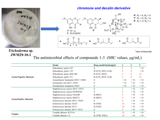
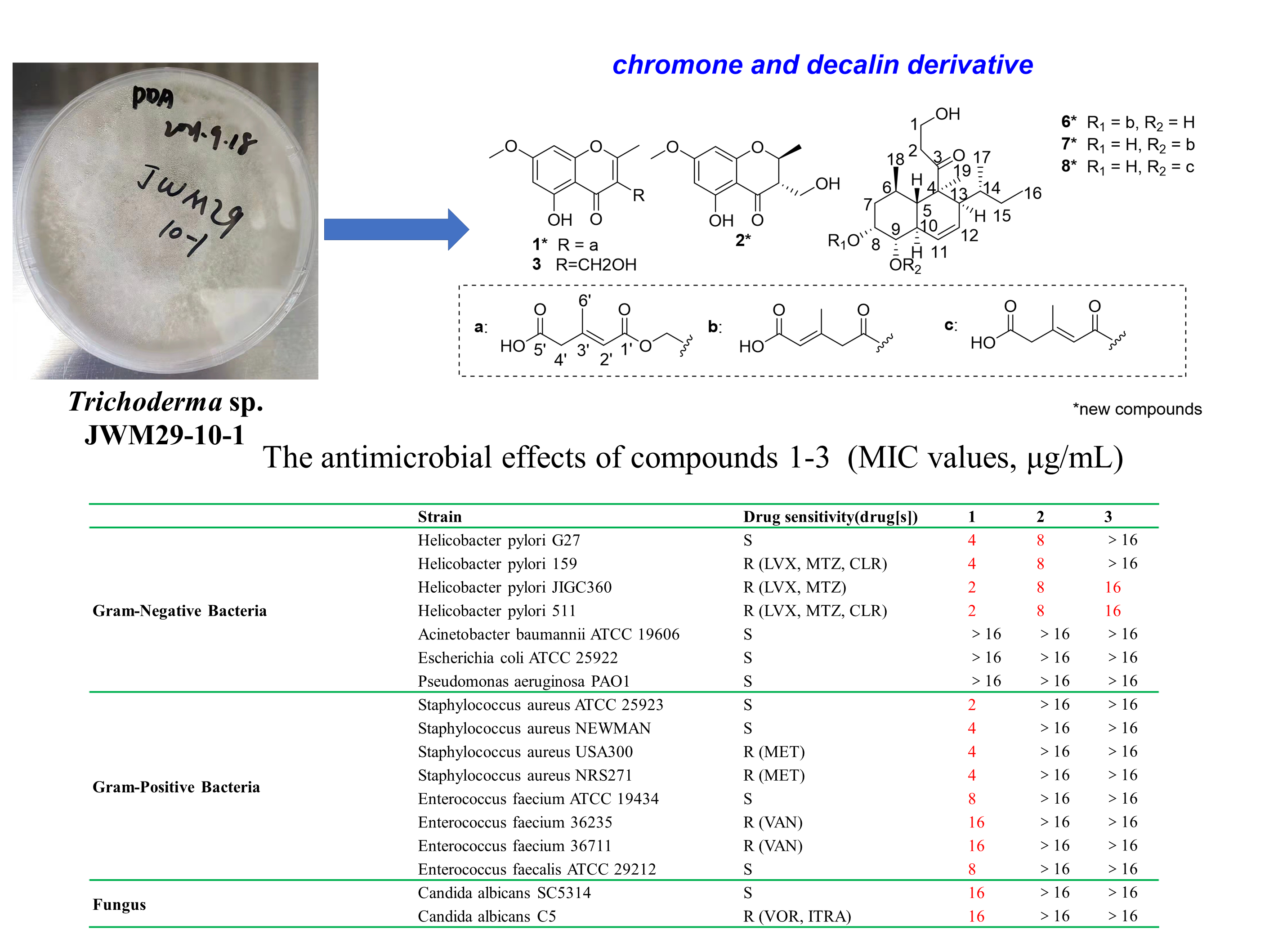
New Polyketides from a Hydrothermal Vent Sediment Fungus Trichoderma sp. JWM29-10-1 and Their Antimicrobial Effects

1
Key Laboratory of Pharmacodynamic Constituents of Traditional Chinese Medicine and New Drug Research, International Cooperative Laboratory of Traditional Chinese Medicine Modernization and Innovative Drug Development of Ministry of Education (MOE) of China, Institute of Traditional Chinese Medicine and Natural Products, College of Pharmacy, Jinan University, Guangzhou 510632, China
2
Helicobacter pylori Research Centre, Jiangsu Key Laboratory of Pathogen Biology, Department of Pathogen Biology, Nanjing Medical University, Nanjing 211166, China
3
Ocean College, Zhejiang University, Zhoushan Campus, Zhoushan 316021, China
*
Authors to whom correspondence should be addressed.
These authors contributed equally to this work.
Mar. Drugs 2022, 20(11), 720; https://doi.org/10.3390/md20110720
Submission received: 13 October 2022 / Revised: 10 November 2022 / Accepted: 14 November 2022 / Published: 16 November 2022

Abstract

:
Marine fungi-derived secondary metabolites are still an important source for the discovery of potential antimicrobial agents. Here, five new polyketides (1, 2, and 68) and seven known compounds (35 and 912) were obtained from the culture of the marine-derived fungus Trichoderma sp. JWM29-10-1. Their structures were identified by extensive spectrographic data analyses, including 1D and 2D NMR, UV, IR, and HR-ESI-MS. Further, the absolute configurations of new compounds were determined by circular dichroism (CD) spectrum and alkali-hydrolysis in combination with the in situ dimolybdenum CD method. Subsequently, the antimicrobial effects of these isolated compounds were assessed by examining the minimal inhibition concentration (MIC) with the broth microdilution assay. Compounds 1 and 2 exhibited potent antimicrobial activity against Helicobacter pylori, including multidrug-resistant strains, with MIC range values of 2–8 µg/mL. Moreover, compound 1 showed significant inhibitory effects on the growth of Gram-positive pathogens, including methicillin-resistant Staphylococcus aureus (MRSA), Enterococcus faecalis, and vancomycin-resistant Enterococcus faecium, which greatly threaten human health. This study demonstrates that chromone derivatives 12, especially for 1, could be potential lead compounds for the development of new antimicrobial agents and provides insight for future medicinal chemistry research.

Graphical Abstract

1. Introduction

Marine fungi have the ability to produce biologically active lead compounds due to their special living environment. Increasing numbers of marine natural products (MNP) from marine fungi have been newly discovered in recent years [1,2]. Trichoderma sp. fungus is a filamentous fungus that mainly exists in marine animals, plants, and sediments attached to the seafloor [3,4]. Numerous chemical and pharmacological investigations have proved that Trichoderma sp. strains can produce abundant secondary metabolites, which exert potential anti-phytopathogenic, insecticidal, cytotoxic, antibacterial, and antioxidant activities, etc. [4,5] Polyketides, an important type of secondary metabolites from Trichoderma sp., were reported with vital antibacterial and anti-phytopathogenic effects [6]. Khamthong et al. reported the isolation of two new polyketides from Trichoderma aureoviride PSU-F95 and discovered one compound exhibited a certain antibacterial activity against Methicillin-resistant Staphylococcus aureus (MRSA) [6]. As infectious diseases seriously threaten human health due to the frequent occurrence of antibiotic resistance [7], polyketides have attracted significant attention due to their diverse biological effects, especially for their remarkable antimicrobial effects [8].
As part of our ongoing search for antimicrobial bioactive compounds from marine fungi, twelve polyketides, including five new compounds (12 and 68) and seven known ones (35 and 912), were isolated from the ethyl acetate (EtOAc) extract of the culture of fungus Trichoderma sp. JWM29-10-1. Here we described the isolation and chemical characterization of these isolated compounds. Meantime, the antimicrobial effects of these compounds were evaluated, and their preliminary structure-activity relationship (SAR) was also discussed.

2. Results

Various chromatographic methods, including silica gel column chromatography (CC), ODS CC, and semi-preparative RP HPLC, were used to investigate the chemical components in Trichoderma sp. JWM29-10-1, which resulted in the isolation of five new polyketides (12 and 68) and seven known ones (35 and 912) (Figure 1). The known compounds were identified as 5-hydroxy-3-hydroxymethyl-2-methyl-7-methoxychromone (3) [9], 5-hydroxy-2,3-dimethyl-7-methoxychromone (4) [9], trichoharin A (5) [10], tandyukisin D (9) [11], tandyukisin C (10) [11], trichoharzin (11) [12], and deoxynortrichoharzin (12) [12] by comparing their MS, 1H and 13C NMR data, and specific rotation with those reported.

2.1. Structure Elucidation

Compound 1 was obtained as a colorless powder. It has the molecular formula of C18H18O8 with ten degrees of unsaturation by the HR-ESI-MS data at m/z 363.1084 [M + H]+ (calcd. m/z 363.1080, see Supplementary Materials). The UV absorptions at 208, 230, 250, 257, and 292 nm, and IR bands at 1677 cm−1 (conjugated carbonyl) and 1625, 1545 cm−1 (aromatic system) implicated the presence of a chromone core structure [9]. The 1H NMR spectrum showed resonances for phenolic hydroxyl at δH 12.63 (1H, s, 5-OH), two aromatic protons at δH 6.34 (2H, brs, H-6 and H-8), a methoxy at δH 3.85 (3H, s, 7-OMe), an oxymethylene at δH 5.11 (2H, s, H-10), a methylene at δH 3.15 (2H, s, H-4′), and two methyls at δH 2.50 (3H, s, H-9) and 2.25 (3H, s, H-6′) (Table 1). The 13C NMR spectrum aided by the DEPT135 spectrum revealed the presence of 18 carbon resonances for ten quaternary carbons, including three carbonyl carbons at δC 181.0 (C-4), 165.7 (C-1′), and 174.6 (C-5′), three oxygenated aromatic carbons at δC 167.6 (C-2), 162.3 (C-5), and 165.9 (C-7), and four aromatic or olefinic carbons; three methines at δC 98.2 (C-6), 92.6 (C-8), and 119.6 (C-2′); two methylenes at δC 56.0 (C-10) and 45.5 (C-4′); a methoxyl at δC 55.9 (7-OMe) and two methyls at δC 18.6 (C-9) and 19.2 (C-6′). A comparison of the NMR data with that reported revealed the core structure of 1 as 5-hydroxy-3-hydroxymethyl-2-methyl-7-methoxychromone [13]. Additionally, analyses of the 1H and 13C NMR data in combination with the HMBC correlations of H-6′ with C-2′–4′, of H-4′ with C-3′ and C-5′, and of H-2′ with C-1′ and C-4′ implicated the presence of a 3-methyl-2-pentenedioic acid side chain [14]. The HMBC correlation of H-10 with C-1′ attached the side chain at the hydroxymethyl of C-10. Thus, the planar structure of 1 was identified, which was confirmed by key HMBC correlations (Figure 2). Furthermore, the NOESY correlation of H-2′ with H-4′ indicated the E configuration of a double bond at C-2′ and C-3′. Thus, the structure of 1 was identified and named as (2E)-1-[(5-hydroxy-7-methoxy-2-methyl-4-oxo-4H-1-benzopyran-3-yl)methyl]3-methyl-2-pentenedioate.
Compound 2 was isolated as a solid white powder. It has the molecular formula of C12H14O5 characterized by the HR-ESI-MS data at m/z 239.0925 [M + H]+ (calcd. m/z 239.0919), implying six degrees of unsaturation. Comparison of the 1H and 13C NMR data with that of 5,7-dihydroxy-2,3-dimethyl-4-chromanone suggested that 2 was also a chromone derivative [9], and the main difference laid in the 13C resonance of C-10. The 13C resonance of C-10 at 58.1 ppm in 2 instead of 9.7 ppm in 5,7-dihydroxy-2,3-dimethyl-4-chromanone indicated that 2 was the oxidative derivative of C-10 in 5,7-dihydroxy-2,3-dimethyl-4-chromanone [15]. Thus, the planar structure of 2 was identified, which was confirmed by 1H-1H COSY and HMBC correlations (Figure 2). The relative configuration of 2 was established by a NOESY experiment (Figure 3). In the NOESY spectrum, the correlations of H-3 with H-9 and of H-2 with H-10 indicated the trans orientation of H-2 and H-3, leading to the relative configuration of 2S* and 3S*. Since 2 showed opposite Cotton effects at 218 (∆ε, −2.4), 286 (∆ε, 1.1), and 310 (∆ε, −0.4) nm with those of (2R, 3R)-5,7-dihydroxy-2,3-dimethyl-4-chromanone [15], the absolute configuration of 2 was determined to be 2S, 3S. The opposite specific optical rotation of 2 with that of (2R, 3R)-5,7-dihydroxy-2,3-dimethyl-4-chromanone confirmed the above deduction [15]. Thus, the structure of 2 was determined and named (2S,3S)-5-hydroxy-3-hydroxymethyl-7-methoxy-2-methyl-4-chromanone.
Compound 6 was isolated as a pale-yellow oil, and its molecular formula was determined to be C25H38O7 by the HR-ESI-MS data at m/z 451.2707 [M + H]+ (calcd. 451.2696), accounting for seven degrees of unsaturation. Its IR spectrum exhibited characteristic bands at 3449, 1711, and 1623 cm−1 for hydroxyl groups, esters, and ketone, respectively. Analyses of the 1H and 13C NMR data aided by HSQC revealed five quaternary carbons, including three carbonyls at δC 215.5 (C-3), 170.2 (C-1′), and 170.4 (C-5′); ten methines including three olefinic methines at δH 6.01 (1H, brd, J = 10.4 Hz, H-11)/δC 125.7 (C-11), δH 5.70 (1H, brd, J = 10.4 Hz, H-12)/δC 124.1 (C-12), and δH 5.82 (1H, s, H-4′)/δC 119.3 (C-4′); two oxymethines at δH 5.24 (1H, d, J = 3.2 Hz, H-8)/δC 74.0 (C-8) and δH 3.56 (1H, dd, J = 10.8, 3.2 Hz, H-9)/δC 74.3 (C-9); five methylenes including one oxymethylene at δH 3.82 (1H, m, H-1b)/ 3.90 (1H, m, H-1a)/δC 58.1 (C-1), and five methyls (Table 2). A comparison of the NMR data with that of Tandyukisin D (9) implicated that they shared the same eujavanicol A core structure, and the main difference lay in the side chain. The side chain was identified to be (2E)-3-methyl-2-pentenedioic acid by the 13C NMR data analysis and key HMBC correlations (Figure 2). Further, the relative configuration of eujavanicol A fragment in 6 was identified by NOESY experiments. The NOESY correlations of H-19 with H-6, H-10, and H-13, and of H-13 with H-17 suggested that they were on one face (Figure 4). The NOESY correlations of H-5 with H-9 and H-18 indicated that they were on the other face and the decalin was trans. Thus, the relative configuration of 6 was identified as 4S*, 5S*, 6R*, 8R*, 9S*, 10R*, 13S*, 14R*, in line with that in 9. Additionally, the 13C resonance of C-6′ at δC 19.4 ppm, less than 20 ppm, supported the E configuration of the double bond in 6 [16]. The absolute configuration of 6 was determined by chemical derivatization in combination with in situ dimolybdenum CD method. Treatment of 6 with NaOH aqueous in MeOH resulted in the acquisition of 6A, which was identified to be eujavanicol A [17] according to their 1H and 13C NMR data and optical rotation value (6A: [α ] D 25 +19.0, eujavanicol A: [α ] D 25 +21.1), confirming the relative configuration of 6. Subsequently, the CD spectrum of complexes formed by vic-diols in 6A with dimolybdenum tetraacetate [Mo2(OAc)4] was measured. The negative Cotton effects at 310 and 400 nm arising within the d-d absorption bands of the metal complexes inferred the 8R and 9S configurations in 6A (Figure 4) [18,19]. Thus, the absolute configuration of 6 was deduced as 4S, 5S, 6R, 8R, 9S, 10R, 13S, and 14R. Finally, the structure of 6 was identified and named Tandyukisin G.
Compound 7 was isolated as a pale-yellow oil. The HR-ESI-MS showed a quasimolecular ion at m/z 451.2699 [M + H]+ (calcd. 451.2696), indicating a molecular formula of C25H38O7 and accounting for seven degrees of unsaturation. Analyses of the 1H and 13C NMR data aided by HSQC, 1H-1H COSY, and HMBC revealed 7 was also a new decalin derivative containing a eujavanicol. A skeleton and a (2E)-3-methyl-2-pentenedioic acid side chain, the same as that of 6 (Figure 2). The HMBC correlation of H-9 with C-1′ attached the side chain at C-9 of the eujavanicol A fragment. In addition, the 13C resonance of C-6′ at δC 19.5 ppm deduced the E configuration of the double bond in the side chain [16]. The relative configuration of 7 was also determined by the NOESY experiment. Subsequently, alkali-hydrolysis of 7 produced 7A, which was identified to be eujavanicol A by comparing their NMR data and optical rotation values with those reported [17]. Meanwhile, the negative Cotton effects of the complex formed by 8,9-diol in 7A with Mo2(OAc)4 at 310 and 400 nm determined the 8R, 9S configurations of the eujavanicol A skeleton (Figure 4). Thus, the structure of 7 was unambiguously identified with the configurations of 4S, 5S, 6R, 8R, 9S, 10R, 13S, and 14R and named Tandyukisin H.
Compound 8 was obtained as a pale-yellow oil. Its molecular formula was elucidated as C25H38O7 based on the HR-ESI-MS (m/z 451.2697 [M + H]+, calcd. 451.2696) and 13C NMR data, implying seven degrees of unsaturation. A comparison of the 1H and 13C NMR data with that of 7 suggested that they had great similarity except for the side chain. The resonance signals of an olefinic methine at C-2′ [δH 5.89 (1H, s)/δC 119.4] and xa methylene at C-4′ [3.19 (2H, s)/δC 45.7] in 8 instead of methylene at δH 3.27 (2H, s)/δC 46.3 (C- 2′) and an olefinic methine at δH 5.85 (1H, s)/δC 119.5 (C-4′) in 7 suggested the presence of Δ2′ double bond in 8. Meanwhile, the double bond was determined as an E configuration by the NOESY correlation of H-2′ with H-4′ in combination with the 13C resonance of C-6′ at δC 19.5 ppm [16]. The HMBC and 1H-1H COSY correlations confirmed the planar structure of 8 (Figure 2). The absolute configuration of 8,9-diols in 8 was also determined by alkali-hydrolysis followed by in situ dimolybdenum CD method. The negative Cotton effects of the complex formed by 8A with Mo2(OAc)4 at 310 and 400 nm inferred the 8R and 9S configurations in 8. Thus, the configurations of 8 were determined as 4S, 5S, 6R, 8R, 9S, 10R, 13S, and 14R. Finally, the structure of 8 was identified and named Tandyukisin I.

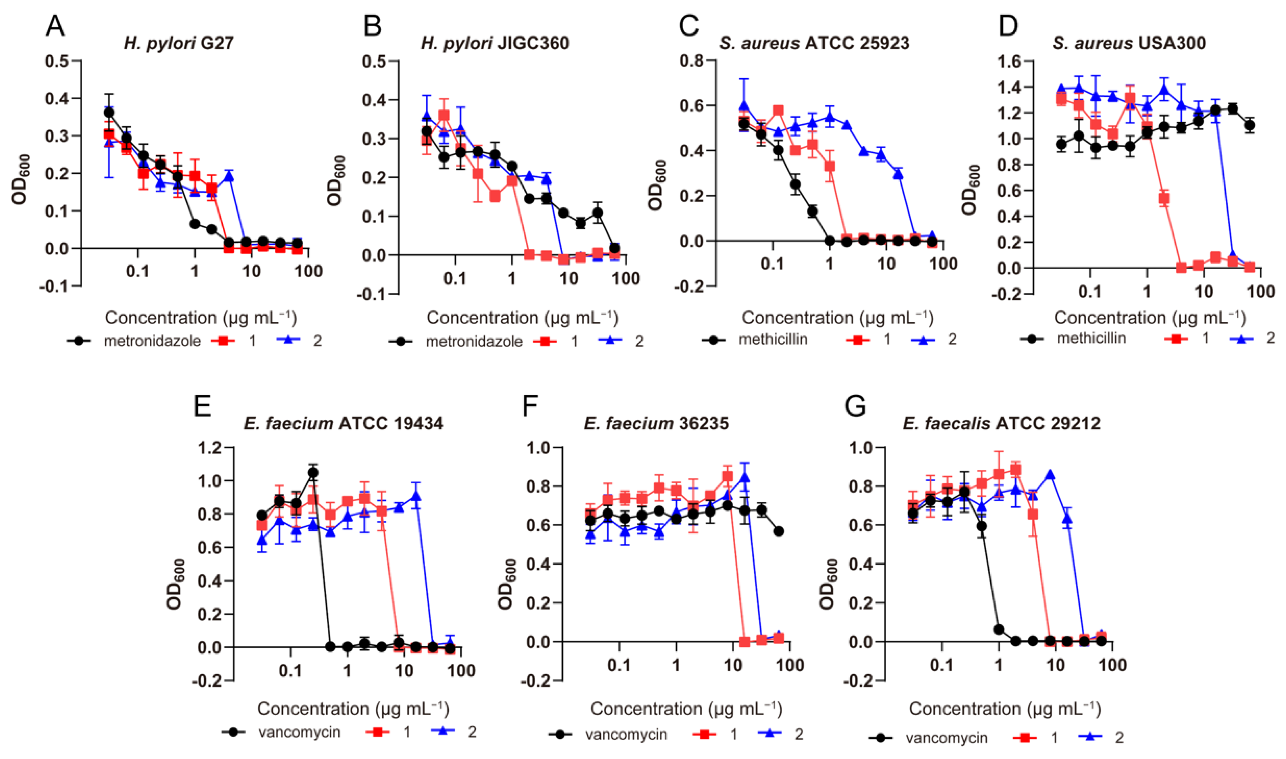
2.2. Antimicrobial Effects of Compounds 112

The antimicrobial effects of compounds 112 were evaluated by the broth microdilution assay. Results showed that compounds 1 and 2 have efficient antibacterial activities against Helicobacter pylori standard strains and clinical isolates, including three multidrug-resistant strains, with minimal inhibition concentration (MIC) values ranging from 2–8 µg/mL (Table 3). Interestingly, compound 1 also exhibited significant inhibitory effects on the growth of Gram-positive pathogens, including Staphylococcus aureus, methicillin-resistant S. aureus (MRSA), vancomycin-resistant Enterococcus faecium (VRE) and Enterococcus faecalis with MIC values of 2 to 16 µg/mL (Table 3 and Figure 5). In addition, compound 1 exerted moderate antimicrobial activity against the important fungal pathogen Candida albicans and Aspergillus fumigatus with MIC values of 16 and 64 µg/mL, indicating that compound 1 has broad-spectrum antimicrobial activity. Preliminary structure-activity relationship (SAR) analysis revealed that the double bond at C-2 and C-3 of the chromone core structure might be unfavorable for its anti-H. pylori effects based on our limited results since compound 2 showed a stronger inhibitory effect than 3. To our surprise, the introduction of a 3-methyl-2-pentenedioic acid side chain at C-13 could not only dramatically increase the anti-H. pylori activities of chromone derivatives, but also broad the antimicrobial spectrum from Gram-negative to Gram-positive bacteria and fungi.

3. Conclusions and Discussion

Marine fungal secondary metabolites have played a tremendous role in the discovery of anti-infectious drugs in the last 50 years [20,21,22]. In this study, five chromone derivatives (15) and seven decalin derivatives (612), including five new compounds (1, 2, and 68), were isolated from the culture of Trichoderma sp. JWM29-10-1. Chromone derivatives with 4-oxo-4H-1-benzopyran core scaffold represent a class of polyketides that were widely distributed in Trichoderma sp. and were reported with antifungal and cytotoxic effects. However, the systemic antimicrobial effect evaluation against bacteria and fungi and SAR were not performed up to now. Here compounds 12 exhibited potent antimicrobial effects with MIC values ranging from 2–16 µg/mL. Interestingly, compound 1 exhibited broad antimicrobial effects, especially for killing multidrug-resistant H. pylori. In addition, our study revealed that compound 1 showed significant inhibitory effects on the growth of MRSA and VRE, two of the major causes of community-acquired and hospital-acquired infections that threaten human health.
The rapid spread of multidrug-resistant (MDR) bacteria is a major concern for global public health. This threat is aggravated by an increasingly depleted antibiotic pipeline [23,24], with alarmingly few new classes of antibiotics introduced into clinical use over the past decades. In 2017, the World Health Organization (WHO) released a list of antibiotic-resistant “priority pathogens”—a catalogue of 12 bacterial species in urgent need of new antibiotics [25]. In this list, clarithromycin-resistant H. pylori, MRSA and VRE were ranked as ‘high” priority pathogens. In this study, compound 1 displayed a potent killing activity against these three important bacterial pathogens. To sum up, our studies shed light on the discovery of novel broad-spectrum antimicrobial agents and provide insight for future medicinal chemistry research.

4. Materials and Methods

4.1. General Experimental Procedure

Optical rotations were taken on a P-1020 digital polarimeter (JASCO International Co. Ltd., Tokyo, Japan). The IR spectra were measured on a JASCO FT/IR-480 plus spectrometer (JASCO, Tokyo, Japan), and UV/VIS spectra were recorded using a JASCO V-550 UV/VIS spectrometer (JASCO, Tokyo, Japan). Mass spectra were acquired with a Synapt G2 mass spectrometer (Waters, Wilmslow, UK). NMR data were taken by a Bruker AV 400 (Bruker Co. Ltd., Bremen, Germany) with signals of CD3OD (δH 3.31/δC 49.0) and CDCl3 (δH 7.26/δC 77.2) as internal references. A Chirascan plus (Applied Photo Physics Ltd., Leatherhead, UK) was used to acquire the CD spectra. The analytical and semi-preparative HPLC was carried out on a Shimadzu LC-20AB and LC-20AT Liquid Chromatography, respectively, with SPD-20A UV/VIS detector (Shimadzu, Tokyo, Japan). Columns for analytical and preparative HPLC were YMC-Triart C18 column (5 µm, ϕ 4.6 mm × 250 mm) and YMC Pack ODS-A column (5 µm, ϕ 10 mm × 250 mm), respectively. Silica gel for column chromatography (CC) was the product of Qingdao Marine Chemical Ltd. (Qingdao, China). ODS for CC were purchased from YMC Ltd. (YMC, Kyoto, Japan). MeOH and CH3CN with HPLC grade were purchased from Thermo Fisher (Waltham, MA, USA). Molybdenum acetate [Mo2(OAc)4] was purchased from Shanghai Yuanye Biological Co., Ltd (Shanghai, China).
The microorganism test strains were: Gram-negative bacteria (H. pylori strains G27, 159, JIGC360, and 511, A. baumannii ATCC 19,606, E. coli ATCC 25,922, and P. aeruginosa PAO1); Gram-positive bacteria (S. aureus ATCC 25,923, USA300, Newman, and NRS271, E. faecium ATCC 19,434, 36,235 and 36,711, E. faecalis ATCC 29,212); Fungus (C. albicans strains SC5314 and C5, A. fumigatus Af293). All were provided by the Department of Pathogen Biology & Jiangsu Key Laboratory of Pathogen Biology, Nanjing Medical University, Nanjing, China.

4.2. Fungal Material

The fungal strain JWM29-10-1 was collected and separated from hydrothermal vent sediments of Kueishantao, Taiwan, China and identified as Trichoderma sp. according to the morphological characteristics and the 18s rDNA sequence (OP501833), which is 99.9% similar to that of Trichoderma reesei (CBS999.97). The strain was preserved in Ocean College, Zhejiang University, Zhejiang, China.

4.3. Fermentation and Extraction

Strain Trichoderma sp. JWM29-10-1 was inoculated on a PDA agar plate, which consisted of 200 g potatoes, 20 g glucose, and 20 g agar in 1 L ddH2O. The spores from the agar plate were transferred into a triangular flask containing 100 mL PDA liquid medium and put in a constant temperature shaking incubator for 5 days (28 °C, 180 rpm/min) to obtain 1000 mL of seed culture solution (100 mL × 10). Then 20 mL of seed culture solution was inoculated to a solid rice medium composed of 100 g rice in 150 mL ddH2O. A total of 2.5 kg of large-scale fermentation was executed in a solid rice medium and cultured at room temperature for 45 days. The fermentation broth was extracted with EtOAc, and the filtrate was concentrated to dryness under reduced pressure to get crude extracts (30.0 g).

4.4. Compound Isolation

The crude extracts were chromatographed by silica gel CC (ϕ 8.0 × 50.0 cm, 200–300 mu, 400 g) eluted with gradient Petroleum ether-EtOAc (100:0, 98:2, 95:5, 9:1, 8:2, 7:3, 6:4, and 0:100) and EtOAc-MeOH (9:1) to obtain 6 fractions (Fr.1–6) based on TLC analyses.
The Fr.2 (1.25 g, Petroleum ether-EtOAc 80:20) was subjected to an ODS CC eluted with gradient MeOH–H2O (25–100%) to get 13 subfractions (Fr.2–1 to Fr.2–13). Fr.2–5 (39.2 mg, 45% MeOH–H2O) was purified by semi-preparative RP HPLC with an eluent of 60% MeOH–H2O (0.1% HCOOH) to get compound 2 (2.8 mg). Fr.2–6 (19.1 mg, 55% MeOH–H2O) was applied to semi-preparative RP HPLC eluted with 65% MeOH–H2O (0.1% HCOOH) to obtain compound 5 (6.5 mg). Fr.3 (2.06 g, Petroleum ether-EtOAc 70:30) was subjected to an ODS CC with stepwise gradient elution of 25%–100% MeOH–H2O to get 16 subfractions (Fr. 3–1–Fr. 3–16). Fr.3–7 (124.4 mg, 55% MeOH–H2O) was isolated by semi-preparative RP HPLC with an eluent of 65% MeOH–H2O (0.1%HCOOH) to yield compounds 3 (7.9 mg) and 4 (37.9 mg). Fr. 3–11 (74.1 mg, 65% MeOH-H2O) was applied to semi-preparative RP HPLC (70% MeOH-H2O with 0.1% HCOOH) to produce compounds 1 (3.4 mg), 11 (9.1 mg), and 12 (29.4 mg). Fr.4 (3.08 g, Petroleum ether-EtOAc 60:40) was applied to an ODS CC with gradient elution of 25–100% MeOH–H2O to get 13 subfractions (Fr. 4–1–Fr. 4–13). Fr.4–9 (224.4 mg, 65% MeOH–H2O) was purified by semi-preparative RP HPLC with an eluent of 35% CH3CN–H2O (0.1% HCOOH) to produce compounds 6 (28.4 mg), 7 (14.2 mg), 8 (22.7 mg), 9 (45.7 mg), and 10 (37.9 mg).

4.5. Spectroscopic Data of New Compounds

(2E)-1-[(5-hydroxy-7-methoxy-2-methyl-4-oxo-4H-1-benzopyran-3-yl)methyl]3-methyl-2-pentenedioate (1): White solid powder; UV (MeOH) λmax (log ε): 208 (4.1), 230 (4.1), 250 (4.0), 257 (3.8), and 292 (3.5) nm; IR (KBr) νmax: 3437, 3263, 2953, 2930, 1677, 1625, 1545, 1475, 1453, 1387, and 1212 cm−1; HR-ESI-MS: m/z 363.1084 [M + H]+ (calcd for C18H19O8, m/z 363.1080); 1H and 13C NMR spectral data (Table 1).
(2S,3S)-5-hydroxy-3-hydroxymethyl-7-methoxy-2-methyl-4-chromanone (2): White solid powder; [α ] D 25 −26.8 (c 0.5, in MeOH); UV (MeOH) λmax (log ε): 215 (3.9), 229 (3.7), 287 (3.8) nm; IR (KBr) νmax: 3439, 2984, 2950, 1727, 1668, 1572, 1433, 1374, 1173, 1068, 927, and 854 cm−1; CD (MeOH) λmax (∆ε): 218 (−2.4), 286 (1.1), 310 (−0.4); HR-ESI-MS: m/z 239.0925 [M + H]+ (calcd for C12H15O5, m/z 239.0919); 1H and 13C NMR spectral data (Table 1).
Tandyukisin G (6): Pale yellow oil; [α ] D 25 +19.6 (c 0.5, in MeOH); UV (MeOH) λmax (log ε): 215 (3.9) nm; IR (KBr) νmax: 3449, 2939, 1711, 1623, 1415, 1383, 1215, 1078, 988, and 926 cm−1; HR-ESI-MS: m/z 451.2707 [M + H]+ (calcd for m/z C25H39O7, 451.2696); 1H and 13C NMR spectral data (Table 2).
Tandyukisin H (7): Pale yellow oil; [α ] D 25 +16.8 (c 0.5, in MeOH); UV (MeOH) λmax (log ε): 214 (3.9) nm; IR (KBr) νmax: 3449, 2939, 1711, 1623, 1415, 1383, 1215, 1078, 988, and 926 cm−1; HR-ESI-MS: m/z 451.2699 [M + H]+ (calcd for C25H39O7, m/z 451.2696); 1H and 13C NMR spectral data (Table 2).
Tandyukisin I (8): Pale yellow oil; [α ] D 25 −11.0 (c 0.5, in MeOH); UV (MeOH) λmax (log ε): 220 (3.9) nm; IR (KBr) νmax: 3449, 2939, 1711, 1623, 1415, 1383, 1215, 1078, 988, and 926 cm−1; HR-ESI-MS: m/z 451.2697 [M + H]+ (calcd for C25H39O7, m/z 451.2696); 1H and 13C NMR spectral data (Table 2).

4.6. Absolute Configuration Determination of Compounds 68

To a solution of compound 6 (16 mg) in 2.5 mL, MeOH was added, 2 mL of aqueous NaOH (1.0 M). Subsequently, the mixture was reacted at room temperature for 24 h. Then, the reaction mixture was extracted with MeOH thrice, and the organic layer was dried under reduced pressure to afford 6A (11.5 mg). Following the same procedure, 7 (4.1 mg) and 8 (4.9 mg) were hydrolyzed with 0.3 M NaOH (aq.) to produce 7A (2.9 mg) and 8A (3.5 mg), respectively. Then, a mixture of 6A, 7A, or 8A with dimolybdenum tetraacetate [Mo2(OAc)4] (1:1.2) in DMSO solution was kept to react for 30 min to form the chiral complexes. Then, the CD spectra of the complexes were measured.

4.7. Antimicrobial Assays

Antimicrobial assays were assessed by the broth microdilution assay following the previous literature [26,27,28] according to CLSI guidelines. Firstly, H. pylori strains were cultured in Brain Heart Infusion (BHI) medium containing 10% fetal calf serum (FCS) under microaerophilic conditions (85% N2, 10% CO2, 5% O2, and 90% relative humidity) using a double-gas CO2 incubator (Binder, model CB160; Tuttlingen, Germany), while other bacterial pathogens were aerobically cultured in Mueller-Hinton (MH) broth. Candida albicans and Aspergillus fumigatus strains were cultivated in Roswell Park Memorial Institute (RPMI) 1640 medium (Sigma, Kawasaki, Japan) containing L-glutamine and buffered with 165 mM MOPS at pH 7.0 (denoted as RPMI medium). Subsequently, a single colony was picked and continuously incubated in BHI/MH/RPMI broth to reach a logarithmic growth phase. Then, the test compounds were dissolved in DMSO and serially diluted two-fold to different concentrations on a 96-well plate. An aliquot (10 µL) of microbial suspension was added to each well, and cell concentration was adjusted to approximately 5 × 10⁵ cells/mL for H. pylori, 5 × 10⁴ cells/mL for other bacterial pathogens and 1 × 10³ cells/mL for fungi. The concentration range tested for each of the compounds was 64–0.125 µg/mL, and each compound was tested in triplicate. The negative control group was treated with sterile water. Metronidazole, methicillin, vancomycin, and amphotericin B were used as the positive control for H. pylori, S. aureus, and other bacteria and fungi, respectively. After incubating H. pylori at 37 °C for 72 h and other bacteria or fungi for 24 h, the plates were examined, and the MIC was defined as the lowest concentration of the compounds with no visible growth. Growth of seven bacterial strains exposed to compounds 1 and 2 at various concentrations after 72 h for H. pylori strains and after 24 h for other bacterial strains was examined at 600 nm for optical density, and the OD600 was recorded. Experiments were performed with three biological replicates.

Supplementary Materials

The following supporting information can be downloaded at: https://www.mdpi.com/article/10.3390/md20110720/s1, the HR-ESI-Q-TOF-MS, UV, IR, and 1D-, 2D-NMR spectra of compounds 12 (Figures S1–S19), 68 (Figures S20–S49), and 6A (Figures S50–S52).

Author Contributions

C.L., J.C., D.T. and J.L., investigation, formal analysis, validation, data curation, writing—original draft preparation; D.L. and T.L., investigation, formal analysis; J.T., H.B. and B.W., conceptualization, resources, supervision, writing—original draft preparation, writing—review and editing, funding acquisition. All authors have read and agreed to the published version of the manuscript.

Funding

This research was funded by grants from the National Key Research and Development Program of China (Grant Numbers: 2018YFC0311002) and NSFC (No. 42176098 and 41876148).

Institutional Review Board Statement

Not applicable.

Informed Consent Statement

Not applicable.

Data Availability Statement

The authors confirm that the data supporting the reported results are available within the article and its supplementary materials.

Acknowledgments

We are grateful to P. Lin for the HR-ESI-MS measurement.

Conflicts of Interest

The authors declare no conflict of interest.

References

  1. Banerjee, P.; Mandhare, A.; Bagalkote, V. Marine natural products as source of new drugs: An updated patent review (July 2018–July 2021). Expert Opin. Ther. Pat. 2022, 32, 317–363. [Google Scholar] [CrossRef] [PubMed]
  2. Liu, Z.; Frank, M.; Yu, X.Q.; Yu, H.Q.; Tran-Cong, N.; Gao, Y.; Proksch, P. Secondary metabolites from marine-derived fungi from China. Prog. Chem. Org. Nat. Prod. 2020, 111, 81–153. [Google Scholar] [PubMed]
  3. Sood, M.; Kapoor, D.; Kumar, V.; Sheteiwy, M.; Ramakrishnan, M.; Landi, M.; Araniti, F.; Sharma, A. Trichoderma: The “secrets” of a multitalented biocontrol agent. Plants 2020, 9, 762. [Google Scholar] [CrossRef] [PubMed]
  4. Su, D.; Ding, L.J.; He, S. Marine-derived Trichoderma species as a promising source of bioactive secondary metabolites. Mini Rev. Med. Chem. 2018, 18, 1702–1713. [Google Scholar] [CrossRef] [PubMed]
  5. Zhang, J.; Tang, W.; Huang, Q.; Li, Y.; Wei, M.; Jiang, L.; Liu, C.; Yu, X.; Zhu, H. Trichoderma: A treasure house of structurally diverse secondary metabolites with medicinal importance. Front. Microbiol. 2021, 12, 2037. [Google Scholar] [CrossRef] [PubMed]
  6. Khamthong, N.; Rukachaisirikul, V.; Tadpetch, K.; Kaewpet, M.; Phongpaichit, S.; Preedanon, S.; Sakayaroj, J. Tetrahydroanthraquinone and xanthone derivatives from the marine-derived fungus Trichoderma aureoviride PSU-F95. Arch. Pharm. Res. 2012, 35, 461–468. [Google Scholar] [CrossRef]
  7. Gou, X.S.; Jia, J.; Xue, Y.X.; Ding, W.J.; Dong, Z.T.; Tian, D.M.; Chen, M.; Bi, H.K.; Hong, K.; Tang, J.S. New pyrones and their analogs from the marine mangrove-derived Aspergillus sp. DM94 with antibacterial activity against Helicobacter pylori. Appl. Microbiol. Biot. 2020, 104, 7971–7978. [Google Scholar] [CrossRef]
  8. De, J.; Kumla, D.; Dethoup, T.; Kijjoa, A. Bioactive compounds from terrestrial and marine-derived fungi of the genus Neosartorya. Molecules 2022, 27, 2351. [Google Scholar]
  9. Tanahashi, T.; Takenaka, Y.; Nagakura, N.; Hamada, N. 2,3-Dialkylchromones from Mycobiont Cultures of the Lichen Graphis scripta. Heterocycles 2000, 53, 1589–1593. [Google Scholar] [CrossRef]
  10. Wang, X.; Xu, T.; Sun, L.; Ceng, R.; Su, S.; Yang, X.; Yang, Y.; Ding, Z. The chemical diversity, the attractant, anti-acetylcholinesterase, and antifungal activities of metabolites from biocontrol Trichoderma harzianum uncovered by OSMAC strategy. Bioorg. Chem. 2021, 114, 105148. [Google Scholar] [CrossRef]
  11. Yamada, T.; Umebayashi, Y.; Kawashima, M.; Kikuchi, T.; Tanaka, R. Determination of the chemical structures of tandyukisins B–D, isolated from a Marine Sponge-Derived Fungus. Mar. Drugs. 2015, 13, 3231–3240. [Google Scholar] [CrossRef]
  12. Rahbæk, L.; Sperry, S.; Piper, J. Deoxynortrichoharzin, a new polyketide from the saltwater culture of a sponge-derived Paecilomyces fungus. J. Nat. Prod. 1998, 61, 1571–1573. [Google Scholar] [CrossRef]
  13. Rukachaisirikul, V.; Chinpha, S.; Phongpaichit, S.; Saikhwan, N.; Sakayaroj, J.; Preedanon, S. Sesquiterpene and monoterpene derivatives from the soil-derived fungus Trichoderma reesei PSU-SPSF013. Phytochem. Lett. 2019, 30, 124–129. [Google Scholar] [CrossRef]
  14. Ding, W.Y.; Cao, W.G.; Yao, Y. Synthesis of dimethyl-3-perfluoroalkyl-4-(3-oxo-2-triphenyl-phosphoranylidenbutanylidene)-pent-2-enedioate and its cyclization. Chin. J. Chem. 2010, 13, 468–474. [Google Scholar] [CrossRef]
  15. Mutanyatta-Comar, J.; Phale, O.J.K.; Abegaz, B.; Croft, K. Phloroglucinol derivatives and flavones from Helichrysum paronychioides. Bull. Chem. Soc. Ethiopia. 2006, 20, 61–68. [Google Scholar]
  16. Duh, C.Y.; Wang, S.K.; Chung, S.G.; Chou, G.C.; Dai, C.F. Cytotoxic Cembrenolides and Steroids from the Formosan Soft Coral Sarcophyton crassocaule. J. Nat. Prod. 2000, 63, 1634–1637. [Google Scholar] [CrossRef]
  17. Nakadate, S.; Nozawa, K.; Horie, H.; Fujii, Y.; Nagai, M.; Hosoe, T.; Kawai, K.I.; Yaguchi, T.; Fukushima, K. Eujavanicols A–C, decalin derivatives from Eupenicillium javanicum. J. Nat. Prod. 2007, 70, 1510–1512. [Google Scholar] [CrossRef]
  18. DiBari, L.; Pescitelli, G.; Pratelli, C.; Pini, D.; Salvadori, P. Determination of absolute configuration of acyclic 1,2-diols with Mo2(OAc)4. 1. Snatzke’s method revised. J. Org. Chem. 2001, 66, 4819–4825. [Google Scholar]
  19. Liu, J.; Du, D.; Si, Y.; Lu, H.; Wu, X.; Li, Y.; Liu, Y.; Yu, S. Application of dimolybdenum reagent Mo2(OAc)4 for determination of the absolute configurations of vic-diols. Chin. J. Org. Chem. 2010, 30, 1270–1278. [Google Scholar]
  20. Bovio, E. Marine fungi from sponges: Biodiversity, chemodiversity and biotechnological applications. Mar. Drugs 2019, 17, 220. [Google Scholar] [CrossRef] [Green Version]
  21. Keswani, C.; Mishra, S.; Sarma, B.; Singh, S.; Singh, H. Unraveling the efficient applications of secondary metabolites of various Trichoderma spp. Appl. Microbiol. Biot. 2014, 98, 533–544. [Google Scholar] [CrossRef] [PubMed]
  22. Shi, Z.; Liu, X.H.; Li, X.N.; Ji, N.Y. Antifungal and antimicroalgal trichothecene sesquiterpenes from the marine algicolous fungus Trichoderma brevicompactum A-DL-9-2. J. Agric. Food. Chem. 2020, 68, 15440–15448. [Google Scholar] [CrossRef] [PubMed]
  23. Lewis, K. The science of antibiotic discovery. Cell 2020, 18, 29–45. [Google Scholar] [CrossRef] [PubMed]
  24. Cooper, M.A.; Shlaes, D. Fix the antibiotics pipeline. Nature 2011, 472, 32. [Google Scholar] [CrossRef]
  25. Govindaraj Vaithinathan, A.; Vanitha, A. WHO global priority pathogens list on antibiotic resistance: An urgent need for action to integrate One Health data. Perspect. Public Health 2018, 138, 87–88. [Google Scholar] [CrossRef]
  26. Huang, Y.Q.; Hang, X.D.; Jiang, X.Q.; Zeng, L.P.; Jia, J.; Xie, Y.; Li, F.; Bi, H.K. In vivo activities of zinc linolenate, a delective antibacterial agent against Helicobacter pylori. Antimicrob. Agents Chemother. 2019, 63, e00004-19. [Google Scholar] [CrossRef] [Green Version]
  27. Qadi, M.; Jaradat, N. In vitro antitumor, antibacterial, and antifungal activities of phenylthio-ethyl benzoate derivatives. Arab. J. Sci. Eng. 2022, 46, 5339–5344. [Google Scholar]
  28. Lv, H.; Wang, K.; Xue, Y.; Chen, J.; Su, H.; Zhang, J.; Wu, Y.; Jia, J.; Bi, H.K.; Wang, H.; et al. Three new metabolites from the marine-derived fungus Aspergillus sp. WHUF03110. Nat. Prod. Commun. 2021, 16, 10936–10940. [Google Scholar] [CrossRef]
Figure 1. The chemical structures of compounds 112.
Marinedrugs 20 00720 g001
Figure 2. The key 1H-1H COSY and HMBC correlations of compounds 1, 2, and 68.
Marinedrugs 20 00720 g002
Figure 3. Key NOESY correlations (A) and CD spectrum (B) of 2.
Marinedrugs 20 00720 g003
Figure 4. Key NOESY correlations of 6 (A), the hydrolytic products of 6-8, 6A-8A (B), and CD spectra of complexes of 6A-8A with Mo2(OAc)4 (C).
Marinedrugs 20 00720 g004
Figure 5. Growth of seven bacterial strains exposed to compounds 1 and 2 at various concentrations after 72 h for H. pylori strains and after 24 h for other bacterial strains. OD600, optical density at 600 nm. (A), Growth of H. pylori G27. (B), Growth of H. pylori JIGC360. (C), Growth of S. aureus ATCC 25923. (D), Growth of S. aureus USA300. (E), Growth of E. faecium ATCC 19434. (F), Growth of E. faecium 36235. (G), Growth of E. faecalis ATCC 29212.
Marinedrugs 20 00720 g005
Table 1. 1H and 13C NMR data for compounds 1 and 2 (400 MHz for 1H and 100 MHz for 13C).
NO.1 a2 b
δCδH (J in Hz)δCδH (J in Hz)
2167.6 76.64.66 (1H, m)
3114.8 54.32.59 (1H, dt, 9.6, 4.0)
4181.0 198.2
4a104.9 104.1
5162.3 165.3
698.26.34 (1H, brs)95.56.02 (1H, d, 2.4)
7165.9 169.5
892.66.34 (1H, brs)94.76.00 (1H, d, 2.4)
8a157.6 164.0
918.62.50 (3H, s)19.31.54 (3H, d, 6.4)
1056.05.11 (2H, s)58.14.26 (1H, dd, 11.2, 4.0)
3.79 (1H, dd, 11.2, 4.0)
5-OH 12.63 (1H, s)
7-OMe55.93.85 (3H, s)56.23.82 (3H, s)
1′165.7
2′119.65.81 (1H, s)
3′151.5
4′45.53.15 (2H, s)
5′174.6
6′19.22.25 (3H, s)
a Measured in CDCl3; b Measured in CD3OD.
Table 2. 1H and 13C NMR data for compounds 68 (400 MHz for 1H and 100 MHz for 13C in CDCl3).
NO.678
δCδH (J in Hz)δCδH (J in Hz)δCδH (J in Hz)
158.13.90 (1H, m, Ha)58.13.90 (1H, m, Ha)58.13.93 (1H, m, Ha)
3.82 (1H, m, Hb)3.85 (1H, m, HB)3.85 (1H, m, Hb)
241.32.85 (1H, brd,18.8, Ha)41.32.88 (1H, brd, 18.8, Ha)41.22.89 (1H, ddd, 18.8, 6.0, 3.6, Ha)
2.66 (1H, brd, 18.8, Hb)2.69 (1H, brd, 18.4, Ha)2.70 (1H, ddd, 18.8, 7.2, 4.0, Ha)
3215.5 215.3 215.5
452.6 52.6 52.7
543.21.96 (1H, m)43.82.04 (1H, t, m)43.72.05 (1H, m)
631.61.57 (1H, m)30.41.80 (1H, m)30.41.83 (1H, m)
739.01.82 (1H, brd,12.0, Hα)41.01.84 (1H, m, Hα)40.81.86 (1H, m, Hα)
1.55 (1H, m, Hβ) 1.58 (1H, brd, 13.6, Hβ) 1.58 (1H, m, Hβ)
874.05.24 (1H, d, 3.2)67.64.15 (1H, brs)67.74.15 (1H, q, 3.2)
974.33.56 (1H, dd, 10.8, 3.2)79.14.75 (1H, d,11.2)77.54.77 (1H, dd, 11.6, 2.8)
1040.12.10 (1H, m)36.32.46 (1H, t, 11.2)36.32.50 (1H, m)
11125.76.01 (1H, brd,10.4)125.15.54 (1H, d, 10.8)125.25.61 (1H, m)
12124.15.70 (1H, brd,10.4)124.65.69 (1H, d,10.8)124.65.68 (1H, ddd, 10.4, 4.4,2.8)
1352.41.93 (1H, m)52.51.94 (1H, m)52.51.94 (1H, m)
1437.31.10 (1H, m)37.31.11 (1H, m)37.31.13 (1H, m)
1524.60.74 (1H, m, a)24.50.74 (1H, m)24.50.73 (1H, m)
1.45 (1H, m, b) 1.48 (1H, m) 1.49 (1H, m)
1612.70.76 (3H, m)12.60.77 (3H, t, 4.4)12.70.76 (3H, t, 6.4)
1719.40.92 (3H, d, 6.4)19.30.92 (3H, d, 6.4)19.30.91 (3H, d, 6.8)
1822.40.58 (3H, d, 5.6)22.40.60 (3H, d, 7.2)22.40.60 (3H, d, 6.8)
1919.51.23 (3H, s)19.51.25 (3H, s)19.41.25 (3H, s)
1′170.2 169.0 165.1
2′46.43.23 (2H, s)46.33.27 (2H, s)119.45.89 (1H, s)
3′153.7 153.4 152.5
4′119.35.82 (1H, s)119.55.85 (1H, s)45.73.19 (2H, s)
5′170.4 170.3 174.3
6′19.42.24(3H, s)19.52.27 (3H, s)19.52.28 (3H, s)
Table 3. The antimicrobial effects of compounds 112 from the Trichoderma sp. JWM29-10-1 (MIC values, µg/mL).
MIC (µg/mL) for:
StrainDrug Sensitivity(Drug [s]) a123456789101112
Gram-Negative BacteriaHelicobacter pylori G27S4832>6432>64>64>64>64>64>6464
Helicobacter pylori 159R (LVX, MTZ, CLR)4832>6432>64>64>64>64>64>6464
Helicobacter pylori JIGC360R (LVX, MTZ)2816>6432>64>64>64>64>64>6464
Helicobacter pylori 511R (LVX, MTZ, CLR)2816>6432>64>64>64>64>64>6464
Acinetobacter baumannii ATCC 19,606S>64>64>64>64>64>64>64>64>64>64>64>64
Escherichia coli ATCC 25,922S>64>64>64>64>64>64>64>64>64>64>64>64
Pseudomonas aeruginosa PAO1S>64>64>64>64>64>64>64>64>64>64>64>64
Gram-Positive BacteriaStaphylococcus aureus ATCC 25,923S232>64>64>64>64>64>64>64>64>64>64
Staphylococcus aureus NEWMANS432>64>64>64>64>64>64>64>64>64>64
Staphylococcus aureus USA300R (MET)432>64>64>64>64>64>64>64>64>64>64
Staphylococcus aureus NRS271R (MET)432>64>64>64>64>64>64>64>64>64>64
Enterococcus faecium ATCC 19,434S832>64>64>64>64>64>64>64>64>64>64
Enterococcus faecium 36,235R (VAN)1632>64>64>64>64>64>64>64>64>64>64
Enterococcus faecium 36,711R (VAN)1632>64>64>64>64>64>64>64>64>64>64
Enterococcus faecalis ATCC 29,212S832>64>64>64>64>64>64>64>64>64>64
FungusCandida albicans SC5314S16>64>64>64>64>64>64>64>64>64>64>64
Candida albicans C5R (VOR, ITRA)16>64>64>64>64>64>64>64>64>64>64>64
Aspergillus fumigatus Af293S64>64>64>64>64>64>64>64>64>64>64>64
a S, drug sensitive; R, drug resistant; LVX, levofloxacin; MTZ, metronidazole; CLR, clarithromycin; MET, methicillin; VAN, vancomycin; VOR, voriconazole; ITRA, itraconazole.
Publisher’s Note: MDPI stays neutral with regard to jurisdictional claims in published maps and institutional affiliations.

