Review

Marine Natural Products: Promising Candidates in the Modulation of Gut-Brain Axis towards Neuroprotection

by
Sajad Fakhri
1,†,
Akram Yarmohammadi
2,†,
Mostafa Yarmohammadi
2,
Mohammad Hosein Farzaei
3,* and
Javier Echeverria
4,*
1
Pharmaceutical Sciences Research Center, Health Institute, Kermanshah University of Medical Sciences, Kermanshah 6734667149, Iran
2
Student Research Committee, Faculty of Pharmacy, Kermanshah University of Medical Sciences, Kermanshah 6714415153, Iran
3
Medical Technology Research Center, Health Technology Institute, Kermanshah University of Medical Sciences, Kermanshah 6734667149, Iran
4
Departamento de Ciencias del Ambiente, Facultad de Química y Biología, Universidad de Santiago de Chile, Santiago 9170022, Chile
*
Authors to whom correspondence should be addressed.
These authors contributed equally to this work.
Mar. Drugs 2021, 19(3), 165; https://doi.org/10.3390/md19030165
Submission received: 28 February 2021 / Revised: 16 March 2021 / Accepted: 17 March 2021 / Published: 19 March 2021
(This article belongs to the Special Issue Marine Compounds and Human Intestinal Health)

Abstract

:
In recent decades, several neuroprotective agents have been provided in combating neuronal dysfunctions; however, no effective treatment has been found towards the complete eradication of neurodegenerative diseases. From the pathophysiological point of view, growing studies are indicating a bidirectional relationship between gut and brain termed gut-brain axis in the context of health/disease. Revealing the gut-brain axis has survived new hopes in the prevention, management, and treatment of neurodegenerative diseases. Accordingly, introducing novel alternative therapies in regulating the gut-brain axis seems to be an emerging concept to pave the road in fighting neurodegenerative diseases. Growing studies have developed marine-derived natural products as hopeful candidates in a simultaneous targeting of gut-brain dysregulated mediators towards neuroprotection. Of marine natural products, carotenoids (e.g., fucoxanthin, and astaxanthin), phytosterols (e.g., fucosterol), polysaccharides (e.g., fucoidan, chitosan, alginate, and laminarin), macrolactins (e.g., macrolactin A), diterpenes (e.g., lobocrasol, excavatolide B, and crassumol E) and sesquiterpenes (e.g., zonarol) have shown to be promising candidates in modulating gut-brain axis. The aforementioned marine natural products are potential regulators of inflammatory, apoptotic, and oxidative stress mediators towards a bidirectional regulation of the gut-brain axis. The present study aims at describing the gut-brain axis, the importance of gut microbiota in neurological diseases, as well as the modulatory role of marine natural products towards neuroprotection.

1. Introduction

The Modern lifestyle with the consumption of processed foods, meat, and wheat has changed the normal flora of the gastrointestinal tract (GIT) [1]. Recent studies are triggering the idea of revealing the relationship between gut flora and central nervous system (CNS) disorders like Parkinson’s disease (PD), Alzheimer’s disease (AD), multiple sclerosis (MS), amyotrophic lateral sclerosis (ALS), autism spectrum disorders (ASD), and mood disturbances such as anxiety and depression [2]. Growing evidence has proved the bidirectional communication of GIT and CNS named gut-brain axis. Regulation of the gut-brain human physiology may be affected by billions of bacteria that reside in the body. GIT is the main place that keeps the majority of this flora and these residents are called gut microbial (GM) [3]. Gut homeostasis can be compromised by several factors, including antibiotic exposure, diet, and infections and this alteration in the composition of GM takes part in the pathogenesis of gut-brain-associated diseases [4]. The Gut-brain axis is the main complex anatomical way in which the gut and brain hold their bidirectional relationship and could communicate with each other in health and diseases. Studies have shown the impact of GM on brain evolution, mood, and immune function [3]. GM communicates with gut epithelium to improve the body’s hemostasis and immunity. The strongest evidence for the role of GM on brain development was obtained from studies on germ-free (GF) mice [2]. In this regard, the dysregulated composition of gut bacterial plays a crucial role in the pathogenesis of gut-brain disorders [5]. This shows a bidirectional relationship through which the disturbance in the GM might influence the neurological signs and vice versa [2]. In another word, with the conception of a gut-brain axis being put forward, there is an increasing belief that this communication acts bidirectionally through which GM influence CNS, and the CNS affects the GM. Growing studies suggest that GM affects the development, functions, and disorders of the central nervous system through the regulation of associated receptors and signaling mediators [6]. The neuroimmune and neuroendocrine systems are two critical compositions of the gut-brain axis [7]. Revealing the detailed microbiota functions mediated by pivotal dysregulated pathways is essential to our finding of how the gut-brain axis may influence the neuronal outcomes [8]. Besides, dysregulation of intestinal permeability and gut integrity affects gut-bacteria-derived metabolites and related signaling pathways, towards the progression/development of different neurological diseases [9]. So, the GM helps to restore the normal function of the nervous system and gut-brain signaling. Several molecular mechanisms are behind the gut-brain bidirectional relationship. Regarding revealing the molecular insights into the influence of GM on CNS, it has been shown that gut-microbiota communicates CNS through producing multiple metabolites/neurotransmitters with neuromodulatory properties. Of those, inflammation, apoptosis, and oxidative stress, as well as associated signaling pathways/mediators play critical roles in facilitating the bidirectional relationship of gut and brain. Accordingly, gamma-aminobutyric acid (GABA), glutamine, 5-hydroxytryptamine (5-HT), histamine, glial cell function, synaptic pruning, blood-brain barrier function (BBB), and myelination are important players [6,10]. Considering the gut-brain axis, migration of toxic agents from gut to brain trigger astrocyte activation via the activation of phosphoinositide 3-kinase (PI3K)/protein kinase B (Akt)/mammalian target of rapamycin (mTOR) pathway [11]. Instead, during pathological conditions, the aforementioned pathways/mediators tend to be involved in many devastating neurological situations.
There are also several pathophysiological mechanisms behind neurodegeneration, including oxidative stress, neuroinflammation, apoptosis, imbalances of calcium ions, malfunction of mitochondria, impairment of signal transport through axons, DNA damage, and abnormalities in RNA processing [12,13]. Accordingly, modulation of these factors seems to pave the road in the prevention/treatment of neuronal-associated disorders. Without knowledge about the precise mechanism and etiology lying behind these disorders, they have some recurrent traits such as malfunction of mitochondria, protein misfolding, and inadequate clearance, which make them complicated to deal with. Complex pathological pathways of neurodegenerative diseases ensure the need of using natural molecules with diverse pharmacological properties [14]. By having 70% of plants covering the earth, and diverse organisms living, the marine environment is the most significant source for natural products. Rich biological and genetic diversity is owed to the harsh environmental conditions of the oceans. One of the preferences of natural medicine over synthetic is their better tolerance. It has been also shown that marine natural products have antioxidative, immunomodulatory, and anti-inflammatory properties [15].
Marine natural products such as carotenoids, polysaccharides, phytosterols, terpenoids, macrolactins, and alkaloids apply potential antioxidant and scavenging characteristics in modulating the properties of the gut-brain axis. Recent reports have shown the association of GM and neurodegenerative diseases [2,11,16] through those inflammatory/apoptotic/oxidative stress pathways. To the best of our knowledge, this is the first review highlighting the potential of marine-derived natural products in modulating the gut-brain axis towards neuroprotection. In this work, potential roles of marine-derived natural products have been reviewed on the gut-brain axis with respect to neurodegenerative diseases. Additionally, the association between gut microbial composition and CNS in physiological and pathological conditions has been described. Also, the applicable rationale for using marine-derived natural products in treating and managing neurodegenerative diseases has been reviewed. Marine natural products could be introduced as alternative candidates in the modulation of the gut-brain axis towards neuroprotection.

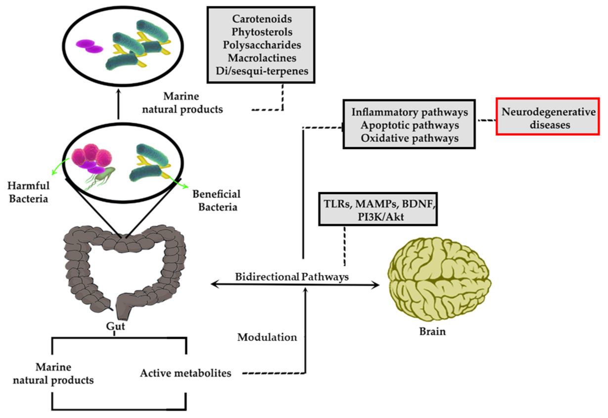
2. Gut Microbiome and Gut-Brain Axis in Diseases

Regulation of human physiology may be affected by billions of bacteria that reside in the body. It was estimated that there are 1011 bacteria per gram of the colon contents [17,18]. They are not the sole residents of this ecosystem, and viruses, protozoa, archaea, and fungi are also present [19]; however, the microbiota seems to be the king of GIT. The GIT is the main place that keeps the majority of this flora and these residents are called GM. Four major and two minor groups of GM are Bacteroidetes, Firmicutes, Proteobacteria, Actinobacteria, along with Verrucomicrobia, Fusobacteria, respectively [20].
Early investigations about interactions between GIT and the brain were focused on digestion and satiety [21]. Homeostasis of the gut is maintained by interactions of these bacteria with each other and with the epithelium layer of the gut and this homeostasis leads to improvement of host immunity [22,23]. The composition of GM is affected by so many factors and the hosted nature plays a key role on these inhabitants. Other factors include genetic, age, physical activity, environmental factors, infection, exposure to antibiotics [24], altering mucus secretion-induced stress [25], and nutrition [5,26]. In recent decades, nutrition and foods have attained special attention towards modulating GM.
The presence of GM has a double role in health and disease states, improving immune functionality and progression/suppression of the diseases, including neurodegeneration [22,23], such termed gut-brain axis. The gut-brain axis is the main complex anatomical way in gut and brain hold their bidirectional relationship and could communicate with each other in health and diseases [27]. The complexity of the relationship between GM and the CNS was exhibited in relevant studies. However, the exact mechanism of the bidirectional gut-brain relationship is not known. Studies showed that GF mice couldn’t develop a healthy intestinal tract in comparison to specific pathogen-free (SPF) and GF-conventional mice, confirming this hypothesis which GM has an essential function in the development of the enteric nervous system (ENS) [6,28], CNS, and hypothalamic-pituitary-adrenal (HPA) axis [29] in early stages of the postnatal life.
In this line, beneficial changing of GM via using antibiotics in SPF mice resulted in elevated pro-survival brain-derived neurotrophic factor (BDNF) in the hippocampus and depleted expression in the amygdala [30]. The Gut-brain axis consists of CNS (brain), ENS, and the digestive system [24]. Intrinsic innervation of the gut is achieved by the complex network of ENS neurons, including two networks, the myenteric and submucosal networks, that modulate gut functions like peristalsis, secretion, and absorption [31]. It is involved in peristalsis, hormone/acid secretion, bicarbonate, and mucus production [24]. The vagus nerve is the major pace to transmit visceral signals into CNS to cause reflexes and mind/moods change and the brain signals to gut for modulation of gut’s physiology and function [6,27].
The autonomic nervous system (ANS) in association with neuronal and neurohormonal signaling, controls many of the physiological functions such as breathing, heartbeat, digestion, peristalsis, bile secretion, permeability, carbohydrate level, mucosa state, homeostasis of mucosal fluids and osmolality, mucus production and mucosal immune functions [27,32]. ANS synapses are the places that sense the microbial metabolites as the tools of communication of microbiota with each other [33]. ANS directly signals gut via CNS leading to changes in its physiology. Gut epithelium’s role in the activation of immune responses can be modulated by ANS in both direct and indirect ways. In direct ways, it modulates the response of gut immune cells to microbes, and in indirect ways, it modulates microbes [34].
The microbiota-gut-brain (MGB) axis makes a bidirectional relationship between microbiota and brain [27], through which any disturbance in one of their functions, like the composition of the gut microbial, will affect the other one [2]. Any disruption in gut-brain crosstalk could result in the progression of cognitive and neural diseases [35,36]. The MGB axis is a component of a huge physiological network complex, including the endocrine (HPA axis), immune system (mediated by cytokines and chemokines), ANS, and the ENS. Gut microbiota exerts its effect on the HPA axis and vagus nerve by metabolites produced from the metabolism of tryptophan [24]. In this line, several microbial molecules take part in MGB-associated signaling and communication [37,38]. It has been revealed that Clostridium sporogenes decarboxylases could turn tryptophan to tryptamine, a neurotransmitter that causes serotonin and dopamine released by neurons [39]. Besides, one of the important inhibitory neurotransmitters like GABA is produced by Lactobacillus spp. and Bifidobacteria spp. from glutamate [40]. Cells that participate in these signaling pathways in association with bacterially derived compounds are enteroendocrine cells (EECs), enterochromaffin cells (ECCs), and mucosal immune cells. Stimulated EEC cells lead to the production of neuropeptides like, peptide YY, neuropeptide Y (NPY), and substance P that affect ENS [41]. To provide the precise molecular aspects of the effects of the GM on CNS, it has been shown that GM communicates CNS directionally through producing multiple neurotransmitters/metabolites with neuromodulatory properties. Amongst the mediators/pathways, glutamine, histamine, synaptic pruning, glial cell function, BBB function, and myelination are important players [6,10]. In addition to the above-mentioned methods of communication, the microbiota can directly synthesis the neuroactive mediators like GABA [42], 5-HT, norepinephrine, and dopamine [43]. Considering the gut-brain axis, the migration of toxic agents from gut to brain could trigger cell migration in astrocytes via the activation of the PI3K/Akt/mTOR pathway [11]. Consequently, the aforementioned mediators have critical roles in several body procedures, including apoptosis, inflammation, oxidative stress, as well as cell migration and proliferation towards homeostasis and even pathological situations of different organs.
The upstream factors such as growth receptors, G protein-coupled receptors (GPCRs), receptor tyrosine kinases (RTKs), and cytokines play a critical role in the attenuation of Janus kinase (JAK)/signal transducer and activator of transcription (STAT). Furthermore, Akt phosphorylates glycogen synthase kinase 3 (GSK-3β) with critical roles in several disorders especially neurodegenerative diseases. Akt also influences apoptotic pathways (e.g., Bax/Bcl-2, caspases), inflammatory mediators (Ils, COX, NF-κB), and oxidative factors (e.g., SOD, ROS, Nrf2, HO-1, CAT) in combating diseases/neurodegenerative disorders [44]. In this line, treatment with probiotics reduced depressive-like behaviors through increasing Bcl-2 and p-Akt, while decreasing malondialdehyde (MDA), cleaved caspase-3, and Bax in the serum [8].
As provided, the aforementioned GM plays a major role in the metabolization of natural compounds towards biological activities and health benefits, especially in neurodegenerative diseases. In this line, actively produced compounds can attenuate signaling mediators involved in neurodegeneration. Although, some of these metabolites cross the intestinal barrier and reach BBB via the bloodstream [38,45].

3. Gut-Brain Axis in Neurodegenerative Disease

Nowadays, neurodegenerative diseases have been considered global concerns and a lot of studies have focused on this area of research to lower burdens of associated disorders [46]. Neural diseases such as PD and AD consist of a group of disorders that 1% and 8% of the population suffer from CNS and peripheral nervous system (PNS) deteriorations, respectively [47].
Recent studies have shown that altered normal GM has a crucial role in neurodegenerative diseases like PD, AD, ALS, and depression; however, the exact mechanism underlying this phenomenon needs to be more scrutinized [48]. A recent study confirmed the relationship of microbiota dysbiosis (i.e., alteration of GM) and AD pathology [49], decreased count of gram-negative species and increased intestinal permeability also have been noted [48]. In AD, the reduction of GM biodiversity by taking antibiotics resulted in changes of neuroinflammatory and amyloidosis that confirmed the role of GM in the pathology of AD [50]. One of the key contributors to the pathogenesis of AD is microbiota diversity. For example, it was seen that in developed countries with a high hygienic condition, lower diversity of GM is correlated with AD incidence [16]. During AD, some changes have been seen in GM, including Bacteroides vulgatus, Bacteroides fragilis, Eggerthella lenta, Odoribacter splanchnicus, Butyrivibrio hungatei, Butyrivibrio proteoclasticus, Eubacterium eligens, Eubacterium hallii, Eubacterium rectale, Clostridium sp., Roseburia hominis, Bifidobacterium bifidum, and Faecalibacterium prausnitzii. This leads to increased accumulation of cerebral amyloid β (Aβ)/neuroinflammation, increasing bacterial lipopolysaccharides (LPS), as well as elevated interleukin (IL)-1 beta, NLR family pyrin domain containing 3 (NLRP3), and chemokine (C-X-C motif) ligand 2 (CXCL2). Dysregulated levels of toll-like receptors (TLRs), nuclear factor κB (NF-κB), IL-1β, IL-18, Aβ, and caspase-1 also results from the bidirectional dysregulation of the gut-brain axis in AD [51,52,53,54].
The role of human microbiota is more explored by advanced technology in the last two decades in both physiological and pathological states. Up to date methods of GF animal models, antibiotic aided microbiota manipulation and fecal microbial transplantation have been used in investigations [12]. For example, in an investigation to find the role of GM in the prevalence of AD in elder patients, they compared the fecal samples of AD patients with healthy elders. They found that a lower prevalence of butyrate-producing bacteria along with a higher abundance of bacterial taxa could be used as predictors of AD [51]. Some bacteria species such as Firmicutes, Bacteroidetes, and Proteobacteria are involved in the pathogenesis of chronic inflammatory diseases through produced amyloids including IL-17A and IL-22 cytokines [55]. The role of GM in the production of vitamin B12, which has a key role in cognitive abilities, is another example that emphasizes its importance [56]. In an investigation to find the role of GM in the prevalence of AD in elder patients, researchers compared the fecal samples of AD patients with healthy elders. They found that lower counts of butyrate-producing bacteria with a higher abundance of bacterial taxa could be used as predictors of AD [51].
One of the main barriers to restoring the GM is age and associated diseases. As the age of the patient goes up, the restoration will be more compromising. In patients with PD, the alteration of the GM and infection with H. pylori has been noticed [2]. In PD, dysregulated levels of the GM, Enterobacteriaceae, Prevotellaceae, Verrucomicrobiaceae, Lactobacillus, Porphyromonas, Parabacteroides, Mucispirillum, and Bacteroides fragilis results in an elevated rate of TLR4, IL-1β, IL-2, IL-4, IL-6, IL-13, IL-18, tumor necrosis factor-α (TNF-α), and interferon (IFN)-γ [3]. The PD metagenome includes more levels of genes taking part in LPS biosynthesis and type 3 bacterial secretion systems which show the higher potential of inflammation by microbial metabolites [57]. These studies reinforce the role of circulating inflammatory products in the periphery CNS inflammation presented in PD [50]. Toxic alpha-synuclein (α-Syn) aggregates are the hallmark of the Lewy bodies that are well known as the marker of PD substantia nigra pars compacta neurons [58]. In a study, it was shown that the first site of α-Syn deposition was the submucosal layer of the intestine [59]. In an analysis done on the fecal samples of PD patients, higher amounts of Enterobacteriaceae, and lower counts of Prevotellaceae were obvious in comparison to the control group with the same age. Raised levels of Enterobacteriaceae, as well as depleted amounts of Prevotellaceae, showed a correlation with postural and walking disabilities. The effect of Prevotellaceae is due to its ability in generating short-chain fatty acids (SCFAs), thiamine, and folate as by-products to create a healthy environment [60].
Probiotics are defined as living microorganisms, that have beneficial effects on consumer health when enough amounts of them have been digested. Their uses have increased in medical and clinical fields with altering impacts on other CNS disturbances, including anxiety and depression [61,62]. Their effect on GM proliferation has been proved [24]. In patients with anxiety/depression Bifidobacterium, Alistipes, Prevotella, Parabacteroides, Lachnospiraceae, Anaerostipes, Oscillibacter, Faecalibacterium, Ruminococcus, Clostridium, Megamonas, Streptococcus, Klebsiella, and Phascolarctobacterium are changing towards decreasing dopamine (DOPAC), homovanillic acid, hippocampus 5-HT, BDNF expression, and circulatory IL-10 while increasing plasma stress hormone [63,64]. Additionally, dysregulated levels of GABA, dopamine, 5-HT, and IL-10 have also been shown in anxiety/depression associated with the gut-brain axis [63,64,65]. Stress-related mental disorders like anxiety and irritable bowel syndrome (IBS) are highly correlated. This correlation triggered the idea of gut-brain axis studies. More than 50% of patients suffering from IBS have comorbidities of anxiety and depression [66]. In a study conducted by Sudo et al., it was shown that undisturbed GM composition at the early stages of life has a huge impact on adulthood stress management [29]. Later research showed that this matter will affect neurochemical compounds like, cortical and hippocampal brain-derived neurotrophic factor [29,67], hippocampal 5-HT receptor 1A magnitude [67], striatal monoamine turnover [68], and gene expression of synaptic plasticity [68] which emphasizes the potent influence of GM on CNS phenotype. Moreover, other effects of GM are in anxiety [67,68] and depression [69], pain response [70], feeding, taste, and metabolism areas [71].
In addition to AD, PD, anxiety/depression pain, and aging, there are other neurological deteriorations (e.g., ALS) that are usually associated with altered GM and lowered biodiversity of the gut flora suggesting the interrelationships of these factors. Neuroimmune activation may be achieved by increasing the levels of butyrate-producing species [48]. Also, in stool samples of ALS patients, higher levels of inflammatory Ruminococcaceae, Enterobacteria, and Escherichia coli have been detected in comparison to the control group [72]. Pre-clinical results showed increased gut permeability, damaged tight junction structure, and increased numbers of abnormal Paneth cells, a cell type responsible for antimicrobial defense in animal models of ALS. Besides, GM indicated a shifted relative abundance of microbial species including a decrease in butyrate-producing Butyrivibrio fibrisolvens [73]. Clinical evidence also confirmed a meaningful increase in Bacterioidetes and, consequently, a decreased Firmicutes to Bacteroidetes level, as well as a decrease in the beneficial Anaerostipes, Lachnospiraceae, and Oscillobacter in the GM of ALS patients. The aforementioned functional changes in ALS patients were concluded to be associated with the dysregulated levels of nitric oxide (NO), GABA, LPS, AMPA/N-methyl-D-aspartate (NMDA), and oxidative pathways [74,75,76].
Communication of gut and brain is mediated by some bacterial products towards neurological signs. In MS disease, a study by Farrokhi and co-workers showed lowered serum level of lipid 654 as the metabolite of Bactroidetes spp. In comparison to the control group [77]. In another study, it was demonstrated that Clostridium perfringens toxins B and D [78] could cause MS like-symptoms, including blurred vision and motor function disability [56]. Toxins-induced visual defects in MS patients are due to retina inflammation caused by formed defects in barrier veins and binding to vascular receptors [79]. Patients with MS experience the changes in levels of Acinetobacteria, Bacteroidetes, Desulfovibrionaceae, Firmicutes, Proteobacteria, Verrucomicrobia, and associated genus [77,80]. This is in agreement with dysregulated GABA, reduced levels of 5-HT and dopamine, while increased IFN-γ, monocyte chemoattractant protein (MCP-1), macrophage inflammatory protein (MIP)-1α, MIP-1β, and IL-6 in MS patients [77,80,81,82].
As another neurological disorder that has an undeniable interconnection with GM, ASD has several changes in gut bacteria, including Bifidobacteraceae, Veillonellaceae, Lactobacillaceae, Bifidobacterium, Megasphaera, Mitsuokella, Rumnicoccus, Lachnoclostridium, Clostridium, Sutterella, Desulfovibrio, Lactobacillus, Eubacterium, and Prevotella [83]. These changes are concomitant with pathophysiological changes in signaling mediators, including the upregulation of mTOR, TNF-α, IL-4, IL-5, IL-6, IL-8 while down-regulating IL-10, transforming growth factor beta (TGF-β), and 5-HT in ASD [11,81,84,85,86,87].
From the mechanistic point of view, behind the gut-brain association, oxidative stress and inflammation seem to play more important roles. Oxidative stress is one of the significant factors involved in mitochondrial dysfunction that has been observed in neurodegenerative diseases. It is the result of an imbalance between generated reactive oxygen species (ROS) and antioxidant defense arsenal. Biological targets for ROS molecules are lipids, proteins, and nucleic acids that lead to their destruction and degradation [88]. It has been seen that communication of microbiota with host cells can be done by merging them with mitochondrial activities. Potentially, interactions of microbiota-gut-brain axis with CNS oxidative stress may exist. In this line, increased amounts of ROS are associated with dysbiosis of microbiota leading to inflammation of the CNS. On the other hand, malfunction of CNS caused by brain lesions could lead to alterations in GM composition. Relationship between oxidative stress-mitochondria-microbiota and neurodegenerative diseases accents the importance of the gut-brain axis [18]. It has been shown that stress has an impact on postprandial gastrointestinal motility and induces a temporary reduction of gastric emptying in dogs [25]. Stress applies its effect through stress mediators, causing local immune activity via alteration of intestinal permeability [89] and can induce changes in germ composition [90].
Several studies have also shown the impact of GM on the CNS and immunity system. However leaky gut syndrome (LGS), which is the permeation of the normal flora into the outside of the intestinal lumen and consequently increased levels of neuroactive metabolites causes a neuroinflammatory response in the brain including cerebellum and hippocampus dysfunction [91,92]. It was evidenced that LGS is common in patients with multiple CNS disorders [93] and leaked metabolites into the blood compromises CNS [94]. Chronic mild inflammation leads to the release of cytokines into the blood and affecting the immune system. Inflammation-induced by microbiota is mediated by molecules such as LPS and peptidoglycans. Recognition of LPS is done by TLR4 which monocytes, macrophages, and brain microglia are rich in them [24]. Studies showed the presence of TLR4 mediated inflammatory responses in depressed IBS patients [95,96]. Blood levels of pro-inflammatory and anti-inflammatory chemokines can be modulated indirectly by microbiota and probiotics that have a direct effect on brain functions [24]. By introducing E. coli to the GF mice, macrophage activation and infiltration in adipose tissue led to high levels of pro-inflammatory cytokine and IFN expression [97].
From another mechanistic point of view, the GM has been shown to alter the evolution, activities, and abnormalities of the CNS and ENS by binding and stimulating pattern recognition receptors (PRRs) such as TLR2 and TLR4 [6,98]. Imbalance of the GM community, distortion of gut integrity and permeability leads to increasing levels of microbial products and microbes associated molecular patterns (MAMPs) in mesenteric lymphoid tissues that cause the occurrence of different neurological diseases [2,9]. Comparison of GF animals with conventional control mice confirmed that hormone signaling, BDNF expression, neurotransmission, and amino acid metabolism, was impaired in GF models [99]. The changes in microbiota composition due to taking antibiotics, influence the integrity and activities of the ENS, neurochemistry, and decrease the number of ganglia residing enteric glial cells in vivo [100].
The relatively fixed composition of the GM throughout life will be compromised in a series of situations such as, illnesses, exposure to antibiotics, and changing of diet or lifestyle [2]. Depending on the severity of the situation that the person encounters, the flora restores to the previous normal flora quickly or with a delay. But in some instances, it never goes back and turns to a chronic issue.
Altogether, GM and neurodegenerative diseases are in a bidirectional relationship, and modulating each of the aforementioned systems could affect the other. Table 1 shows changes in GM during some neurodegenerative diseases and related pathophysiological outcomes.

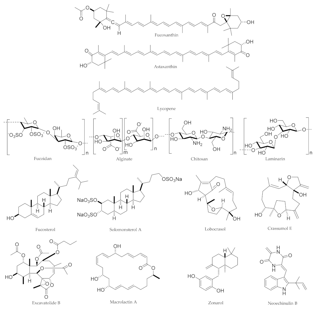
4. Marine-Derived Natural Products and Associated Sources

By covering 70% of planet earth and store a huge diversity of organisms, the ma-rine environment has been an outstanding source of natural products [88]. Associated genetic diversity and biological activities of marine active ingredients are owed to harsh environmental conditions of the oceans, living in conditions of high pressure, cold temperatures, dark fields, and adaptation to stressful conditions [101]. The molecular weight of marine-derived natural products ranges from 100 to 1000 Da and is specific to an individual taxonomic classification [102]. The survival of GIT microorganisms, which compete with other microorganisms, is greatly dependent on the produced or external administered marine-derived natural products. They take part in attraction, repletion, and even killing other competitors. Both eukaryotic and prokaryotic microorganisms can produce secondary metabolites. For example, Bacillus spp., Pseudomonas spp., eukaryotic fungi (e.g., Penicillium spp., Aspergillus spp.), filamentous actinomyces (e.g., Streptomyces spp.), and terrestrial plants have shown to produce secondary metabolites or associated metabolites.
Recently by discovering new secondary metabolites that are biologically active, one of the goals of the pharmaceutical and agrochemical industries is to produce them on large scale. Based on the diversity of the structures of secondary metabolites, they have huge potential to be used against diverse ranges of illnesses [103]. One of the features of marine natural sources is that they can act as a continuous source of bioactive molecules [88]. Two major sources of marine-derived natural products are marine organisms and fungi [104]. Investigations showed that the production of these metabolites is not a random phenomenon and is related to the ecological niche [105]. According to this fact, chemists who working on marine natural products, are trying to discover new species which produce these metabolites [104] through unique metabolic and genetic pathways [88]. So, the marine organisms and fungi absorbed a lot of attention [106]. Until now, more than 100 metabolites could be found that are produced by marine fungi [106].
Many marine-derived natural products have been found to elicit a broad range of bioactivities and, therefore, continue to be a prolific source for the production of new drugs or drug leads. We believe that the discovery of new and extreme habitats will advance the discovery of novel macro- and microorganisms and, thus, might lead to the detection and isolation of novel marine-associated natural products [107]. Of microorganism source of marine-derived natural products, Eubacteria are of major ones, including Actinobacteria, Cyanobacteria, and other bacteria. Besides, Archaebacteria, Euglenoids (Euglenozoa, Euglenoidea, Protozoa), Dinoflagellates, (Dinozoa, Dinoflagellatea, Protozoa), Ciliates (Protozoa, Ciliophora), Chrysophytes (Phaeophyta, Chrysophyceae, Chromista), Diatoms (Diatomae, Bacillariophyceae, Chromista), Eustigmatophytes (Phaeophyta, Eustigmatophyceae, Chromista), Raphidophytes (Chromista, Raphidophyta), Prymnesiophytes (Prymnesiophyta, Chromista), Cryptophytes (Chromista, Cryptophyceae, Cryptophyta), Prasinophytes or grass-green scaly algae (Plantae, Prasinophyta), Green microalgae (Chlorophyta, Plantae), Red microalgae (Rhodophyta, Plantae), and Fungi (Eumycota) [108]. Specifically, marine fungi are still an underestimated but rich source for new secondary metabolites, although their distribution and ecological role often remain scarce. Marine fungi are the sources of biologically active molecules with the known anticancer, neuroprotective, anti-angiogenesis, antibiotic, antiviral, antioxidative, and anti-inflammatory activities [109].

5. Marine-Derived Natural Products against Diseases: Approaches to the Gut-Brain Axis

As provided, marine-derived natural products could be extracted from several sources, especially bacteria, fungi, microalgae. Algae with a common name of seaweed are one of the major sources of marine-based compounds which are widely used in industries [110]. Some of those most important marine metabolites are carotenoids, polysaccharides, phytosterols, terpenoids, phenolic compounds, and alkaloids [111]. Based on their antioxidative, anti-inflammatory, and immune-regulatory characteristics, the aforementioned compounds showed satisfying results in the management of patients with diabetes, obesity, brain trauma, ischemic stroke, and other neurodegenerative diseases [112]. Neurodegenerative diseases are the result of physiological and pathological changes like ischemic strokes and brain injuries which end up in the loss of some neurons in specific regions of the brain [113]. Without knowledge about the precise mechanism and etiology lying behind these disorders, they all have features such as oxidative stress, neuroinflammation, malfunction of mitochondria, protein misfolding, and inadequate clearance, which make them complicated to deal with [114]. One of the preferences of natural medicine over synthetic is their better tolerance. It has been shown that marine natural products have antioxidative, immunomodulatory, and anti-inflammatory properties [115]. Complex pathological pathways of neurodegenerative diseases, ensure the need of using marine natural molecules with diverse pharmacological properties [116].

5.1. Carotenoids: Fucoxanthin, Astaxanthin, and Lycopene

As marine-derived compounds, carotenoids are fat-soluble pigments in plants, algae, fungi, and photosynthetic bacteria. These pigments generate bright yellow, red, and orange colors in plants, vegetables, and fruits. There are more than 600 different kinds of carotenoids with antioxidant effects [117]. Their relationship with photosynthesis is grouped into two classes, one group is directly involved in photosynthesis and another group protects the organism from photooxidation [118]. Major carotenoids produced by marine microorganisms include astaxanthin, fucoxanthin, lycopene, salinixanthin, saproxanthin, sioxanthin, siphonaxanthin, canthaxanthin, β-cryptoxanthin, diadinoxanthin, dinoxanthin, echinenone, lutein, zeaxanthin, and violaxanthin [23].
Compounds obtained from marine algae have potential antioxidative properties. Xanthones are marine natural products containing a tricyclic symmetric structure derived from dibenzo-γ-pirone [119]. Around 200 xanthone molecules have been recognized with the sources of plants, lichens, bacteria, and fungi [120]. Their biological activities include diverse ranges of antioxidative [121], antiproliferative [122], antimicrobial [123], antitumoral activities [124], and this diversity is due to their interactions with multiple molecular targets [125].
One of the most important xanthones is fucoxanthin with promising effects. Fucoxanthin is a carotenoid with several biological activities and health benefits through exerting anti-inflammatory effects in vitro and in vivo [126,127]. It has also been shown that fucoxanthin suppressed the cell cycle and induced apoptosis in combating cancer [128]. The hepatoprotective, cardioprotective, and anti-diabetic effects of fucoxanthin, as well as its effect on metabolic syndrome [129], are also indicated in concomitant studies [130]. Fucoxanthin is a product of Sargassum siliquastrum (a brown algae) and protects DNA from oxidation [16]. Brown algae, as an origin of fucoxanthin, showed antioxidant and anti-inflammatory effects in glial cells [131]. However, several other brown algae are marine sources for fucoxanthin, including Sargassum siliquastrum, Hijikia fusiformis, Undaria pinnatifida, Laminaria japonica, Alaria crassifolia, and Cladosiphon okamuranus [117]. Results of research recommend using algal metabolites, especially fucoxanthin in CNS diseases [132,133]. Fucoxanthin depleted the generation of Aβ1–42 fibril and Aβ1–42 oligomers, when co-incubated with Aβ1–42 monomers and showed the inhibitory effect of Aβ aggregation [134]. Besides, fucoxanthin prevents damages of DNA via H2O2 which is accompanied by increased levels of glutathione (GSH) and superoxide dis-mutase (SOD) [135]. It also protects LPS-activated BV-2 microglia via nuclear factor erythroid 2-related factor 2 (Nrf2)/heme oxygenase (HO)-1 pathway and promotes survival of the cell through the cAMP-dependent protein kinase (PKA)/cAMP response element-binding (CREB) pathway and increasing BDNF secretion [136]. Fucoxanthin also protects Aβ42-induced BV2 cells from inflammation via the reduction of pro-inflammatory mediators such as TNF-α, IL-6, IL-1β, and prostaglandin (PG)E2. Expression of inducible nitric oxide synthase (iNOS) and cyclooxygenase-2 (COX-2) and phosphorylation of mitogen-activated protein kinase (MAPK) pathway was reduced under influence of fucoxanthin [135]. Reduced expression of iNOS and COX-2 and secretion of inflammatory factors such as TNF-α, IL-6, PGE2, and NO take part in inhibition of Akt/NF-κB and MAPKs/stimulating protein-1 (AP-1) pathways, in LPS-activated BV-2 microglia was observed as the protectant activity of the fucoxanthin [135]. One of the major contributors to the pathologic processes of AD is the deposition of Aβ [137,138].
Oligomers of Aβ are notorious for their neurotoxicity and are one of the key compounds which take part in neurodegeneration of the AD. It is also demonstrated that fucoxanthin with its antioxidative and anti-apoptotic properties could play a protective role against oligomers of Aβ in SH-SY5Y cells. PI3K/Akt cascade as a protectant mechanism will be disturbed by oligomers of Aβ and the destructive series of reactions governed by the extracellular signal-regulated kinase (ERK) pathway will be activated. It has been also shown that inhibition of GSK-3β and mitogen-activated protein kinase (MEK) together could stop the destructive effects of Aβ. So, it could be concluded that PI3K/Akt and ERK pathways may contribute to Aβ oligomer-stimulated neurotoxicity. Effects of Aβ on PI3K/Akt and ERK pathways could be stopped by using fucoxanthin. Moreover, two PI3K inhibitor agents, LY294002 and wortmannin, when used, could stop the effects of fucoxanthin. This outcome suggested the mechanism by which fucoxanthin exerts its neuroprotective effects could be activation of PI3K/Akt cascade concurrently with stopping ERK pathway. Akt activation by fucoxanthin could also modulate NF-κB concerning reducing oxidative stress [139]. It also decreased apoptosis and oxidative stress in SH-SY5Y cells through activating a pro-survival PI3K/Akt pathway and suppressing the pro-apoptotic ERK pathway and preventing H2O2 stimulated apoptosis [140].
Scopolamine [141], and Aβ oligomer [141] can contribute to cognitive impairments in mice. Fucoxanthin, by inhibiting acetylcholinesterase (AChE) activity, regulation of choline acetyltransferase (ChAT) activity, and increasing BDNF expression has a protective role in these disorders. Nrf2/ARE and Nrf2-autophagy pathways-dependent neuroprotective mechanism is involved in fucoxanthin mediated traumatic brain injury amelioration [142]. Fucoxanthin has also shown promising results against human inflammation-related diseases through employing PI3K/Akt/CREB/peroxisome proliferator-activated receptor-gamma coactivator α, and Nrf2/ARE pathways [127,130]. In a recent study by Sun et al., fucoxanthin inhibited inflammation-related Lachnospiraceae and Erysipelotrichaceae while increased the Lactobacillus/Lactococcus, Bifidobacterium, and some butyrate-producing bacteria [143]. Guo et al. showed the critical role of fucoxanthin in modulating the ratio of Firmicutes/Bacteroidetes and the abundance of Akkermansia, thereby, it could be an auspicious microbiota-targeted functional food [144]. It has also shown promising interaction with intestinal Escherichia coli and lactobacilli towards the growth inhibition of pathogenic bacteria [145]. So, fucoxanthin develops the GM and modulates the neuronal inflammatory/oxidative/apoptotic pathways, thereby attenuates the gut-brain axis towards neuroprotective responses.
Most known carotenoids generated by marine fungi are commercially available astaxanthin and β-carotene [117], which the former is a xanthophyll carotenoid with the most potent antioxidant [146]. Extraction of astaxanthin is done due to its lipophilic nature by solvents, acids, microwave coupled, and enzyme aided methods [147]. In a red basidiomycetous yeast named Phaffia rhodozyma, astaxanthin has been extracted from the cytoplasmic membrane [88]. Cardinal microorganisms with the ability to synthesize astaxanthin are microalgae Chlorella zofingiensis, Chlorococcum spp., red yeast Phaffia rhodozyma, and the marine Agrobacterium aurantiacum [148]. Consequently, algae, yeast, and crustacean produce astaxanthin as a byproduct. Brain higher susceptibility to oxidative stress is due to its excessive metabolism, the existence of already oxidized molecules like catecholamine neurotransmitters, and polyunsaturated fatty acids which are in the structure of the cell membrane. Further exploration revealed other biological activities and health benefits could be obtained from this compound [149,150]. The anticancer [151], anti-obesity/triglyceride/cholesterol, cardioprotective, [152,153], hepatoprotective [154], and anti-diabetic [155] effects of astaxanthin are also reported [149].
The neuroprotective property of astaxanthin has been recently noticed passing through its anti-inflammatory, anti-apoptotic, and antioxidant effects besides maintenance of neural plasticity [156,157,158]. This collection of features, candidate astaxanthin as a therapeutic agent in neurodegenerative diseases. Although, because of its anti-inflammatory and antioxidative behavior, it has been investigated in cardiovascular health, metabolic syndrome, management of gastric ulcers, and malignancy, all of which share a common characteristic of inflammation and oxidative stress [159]. In building to cell membrane against oxidative stress, which is being exacerbated by aging, astaxanthin plays an important part by applying associated antioxidant agents like SOD, heme oxygenase-1 (HO-1), and GSH. Its unique structure of ketone-bearing ionone rings stabilizes radicals synergistically with the polyene backbone and improves antioxidative ability [160]. It was demonstrated that ROS could be depleted in vitro by using astaxanthin [161]. Suppression of microglial activation and as a result, lowering the production of cytotoxic compounds, is another function of astaxanthin [118]. Released NO from microglia during inflammation, reacts with superoxide leading to the formation of peroxynitrite that is a ROS, and destructing proteins/lipids/DNA is the final consequence [162]. It was demonstrated that the expression and release of IL-6, COX-2 was down-regulated by astaxanthin in an LPS-induced activated microglia, in vitro [163]. The host immune system can be boosted by the administration of astaxanthin. The result was the stimulation of T cells which results in the production of IFN-γ, B cells to end up to secretion of IgA, and natural killer cells, as well as generation of IFN-c and IL-6 [164].
In a recent study by Wu et al., astaxanthin made a shift in the GM towards the suppression of intestinal inflammation/oxidative stress pathways by inhibiting colon NLRP3 inflammasome activation [165]. Overall, on one hand, astaxanthin potentially attenuates neuronal dysregulated pathways. Besides, it modulated GM towards the modulation of intestinal inflammation/oxidative stress/apoptosis. Meanwhile, the expression of inflammatory factors including TNF-α and INF-γ decreased, while the expression of IL-10 increased. In another study, by regulating cecal microbiota, astaxanthin meaningfully decreased the expression of MyD88, TLR4, and p-p65, while increasing the p65 expression [166]. So, considering the simultaneous modulatory role of astaxanthin on the gut and brain, it introduces astaxanthin as a great candidate in regulating the gut-brain axis towards neuroprotection.
Lycopene is another carotenoid through which the gut and brain potentiate their beneficial relationship. It exists in vegetables and red fruits, such as red pepper, papaya, tomato, watermelon, and also in algae. Following 5 days of administration of diets on lycopene, the related plasma amount of lycopene is significantly increased towards distribution in blood and brain. Lycopene seems to be responsible for a gut-brain regulation then is particularly discharged undigested from the human body [167]. Studies indicated that the GM balance has a vital contribution to the occurrence/development of colitis, which affects brain function by the gut physiology regulation [3,168]. As a carotenoid, it has shown a critical role in colitis and the associated behavioral disorders. It is shown that 40 days of lycopene therapy (50 mg/kg body weight/day) in male mice prevented gut damages and inflammatory responses induced by dextran sulfate sodium (DSS). Lycopene attenuated DSS-induced dysfunctions of anxiety-like behavioral and depression by suppressing synaptic damages, inhibiting neuroinflammation, and increasing the expressions of neurotrophic factor and postsynaptic-density protein. Additionally, lycopene resulted in GM remodeling of colitis mice by reducing the relative abundance of Proteobacteria and elevating the relative abundance of Lactobacillus and Bifidobacterium. It has also stimulated the SCFAs production and suppressed the LPS permeability in colitis mice [169]. Recent results demonstrated that GM germinates SCFAs in Bacteroides and some others. Among those, Prevotella, Ruminococcus, Bacteroides, Clostridium, and Streptococcus produce acetic acid, Bacteroides, Coprococcus, and Ruminococcus, germinate propionic acid, and Lachnospiraceae Bifidobacterium, and Coprococcus harvest butyric acid [170]. The cell wall of Gram-negative bacteria (e.g., Proteobacteria and Bacteroides) is primarily composed of LPS, which is an activator antigen of TLR4 in triggering inflammatory responses [169]. The dietary supply of Bifidobacteria and Lactobacilli has also shown promising effects on behavioral disorders like depression [168]. Growing reports have indicated that the destruction of the gut barrier would also result in a gut permeability increment. Subsequently, the generation of some pathogenic compounds (e.g., LPS) is followed by passing through intestinal epithelium and the blood entrance, to trigger neuroinflammation [171]. Reports have revealed that microglia activation and pro-inflammatory cytokines (e.g., IL-1 and TNF-α) could dysregulate synaptic plasticity towards emotional disorders and depression-like behaviors [127]. Thus, lycopene attenuates DSS-induced behavioral disorders and colitis through making a balance in the microbes-gut-brain axis, thereby reducing brain microglia activation and gut/brain inflammatory cytokines [172]. In agreement with recent studies, a low level of BDNF may increase stress susceptibility to affect the structure of forebrain neurons. Therefore, it has been an auspicious marker of a positive antidepressant response of related drugs [3]. Moreover, lycopene has also protected the brain against behavioral disorders by modulating the brain expression of BDNF, as well as associated synaptic plasticity. Bidirectionally, it is controlling the DSS-induced change of GM metabolites including SCFAs and LPS. Thus, lycopene is reshaping the GM of DSS-induced colitis mice, indicating that gut-brain axis balance is an underlying mechanism [167]. Overall, the studies illustrated that lycopene could ameliorate DSS-induced colitis, as well as associated anxiety-like symptoms and depression. It could be clarified by the helpful effects of lycopene on gut barrier integrity as well as its modulatory roles on GM and associated production of metabolites [172].
As previously mentioned, lycopene has shown beneficial effects in CNS. Its activity in reducing oxidative stress and tert-butyl hydroperoxide-induced cell apoptosis promised using lycopene in AD. Lycopene has a variety of activities including raising GSH/GSSG enzyme levels, maintenance of mitochondrial membrane potential, depletion of ROS [173], reduction of inflammatory cytokine levels, and downregulation of TLR4 and NF-κB p65 mRNA and protein expressions [174]. Besides, the effects of lycopene on neurological recovery and anti-inflammation activities were also investigated on rat models for the management of spinal cord ischemia/reperfusion injury [175]. During a clinical trial, lycopene increased the relative abundance of GM profile, such as Bifidobacterium adolescentis and Bifidobacterium longum [176]. So, according to the critical role of lycopene in modulating GM and associated mediators of the gut-brain axis, it would be a promising agent towards neuroprotection.

5.2. Polysaccharides: Fucoidan, Chitosan, Alginate, and Laminarin

Other anti-inflammatory molecules that could be obtained from algal are polysaccharides, including chitosan, laminarin, fucoidan, and alginate [177]. Attenuation of NF-κB and ERK/MAPK/Akt pathways in BV2 microglia induced by LPS resulted in depressed anti-inflammatory responses by a sulfated polysaccharide named fucoidan [178]. In an in vivo study by Shang et al., fucoidan attenuated the GM in mice by increasing the abundance of Ruminococcaceae and Lactobacillus [179]. In another study, it alleviated colonic inflammation and GM dysbiosis by inhibiting IL-1β, IL-6, and TNF-α, while increasing IL-10 [180]. In a more recent study, feeding fucoidan from Okinawa mozuk altered the intestinal microbiota composition of adult zebrafish, which was characterized by the emergence and predominance of multiple bacterial affiliated with Comamonadaceae and Rhizobiaceae. They also found decreased expression of intestinal IL-1β [181]. Shi et al., also showed that fucoidan from Acaudina molpadioides increased the abundance of short SCFAs producer Coprococcus, Butyricicoccus, and Rikenella, through which mitigated intestinal mucosal injury [182]. In a recent double-blinded, placebo-controlled study, combination therapy of fucoidan increased fecal SCFAs, Pseudocatenulatum, Bifidobacterium, Bacteroides intestinalis, Eubacterium siraeum, while decreased Prevotella copri [183]. Additional studies have also focused on the potential of fucoidan in modulating GM by targeting antioxidative mediators (e.g., Nrf2, and GSH/GSSG) [184]. Sulfated oligosaccharides of gran algae Ulva lactuca and Enteromorpha prolifera decreased pro-inflammatory agents, reduced p53 and fork-head box protein O1 (FoxO1) genes, and caused overexpression of Sirt1 gene in SAMP8 mice [185]. Additionally, it upregulated the expression of BDNF through the ERK/CREB/tropomyosin-related kinase receptor B (TrkB) pathway and antioxidant enzymes such as HO-1, NAD(P)H quinine oxidoreductase-1 (NQO-1), and glutamate-cysteine ligase catalytic subunit (GCLC) via Akt [186].
The second most abundant polysaccharide is chitin after cellulose [187]. The deacetylated derivative, chitosan, has shown several therapeutic applications, including antimicrobial/antifungal [188,189], antioxidant [190], and antitumor [191] effects. Many studies showed the neuroprotective, antioxidative, and anti-inflammatory properties of chitosan. Deacetylation degree and chain size are two determinants of these properties [192]. In a study by He et al., chito-oligosaccharides were used as a protectant against oxidative stress caused by H2O2, and the lowest concentration with the highest efficacy was reported as 0.02 mg/mL. It was also demonstrated that carboxymethylated chitosan had a satisfactory effect in protecting Schwann cells against hydrogen peroxide-induced damage through a mitochondrial-dependent pathway [193]. In most recent reports, chitosan potentially modulated GM [194] by suppressing Helicobacter, promoting Akkermansia [195], and decreasing serum levels of IL-6 and IL-1β [196]. Yu et al., also showed that chitosan supplementation improved the Prevotella in the cecal of pigs [197]. Another polysaccharide that has been extracted from algae is seleno-polymannuronate was prepared from alginate-derived polymannuronate. Alginate is the major structural polysaccharide of brown macroalgae, which is widely used as food additives and functional food ingredients owing to the desired physicochemical properties and well-recognized beneficial effects on gut ecology [198]. Bacteroides ovatus is responsible for the fermentation of alginate in the gut. The fermentation outcome of alginate by gut microorganisms are SCFAs, which acts as an energy provider for intestinal and immune cells [199]. Treatment with alginate reduced IL-1β and CD11c inflammatory markers, and improved the growth of GM Lactobacillus gasseri, Lactobacillus reuteri, and Akkermansia muciniphila in mice [200]. Such beneficial roles of alginate on GM were also reported in other studies [201]. Additionally, alginate-derived oligosaccharide downregulated pro-inflammatory agent’s expression and enzymes in LPS/Aβ-induced BV2 microglia. This oligosaccharide also decreased the expression of TLR4 and NF-κB [202]. From the mechanistic point, alginate has targeted oxidative pathways (e.g., ROS) and neuroinflammation (e.g., TLR4) to protect neuronal damages. So, regulation of GM and neuronal dysregulated pathways by alginate pave the road in attaining a promising gut-brain axis [203].
Other polysaccharides which could be fermented by GM include Laminarin [204]. Brown seaweeds, such as Laminariaceae as the source and Laminaria, Saccharina, or Eisenia species as secondary [205] contain a linear polysaccharide named laminarin. Studies showed its neuroprotective properties in cerebral ischemic events through gliosis reduction and controlling pro-inflammatory microglia. In an in vivo study, laminarin meaningfully decreased GM Firmicutes while increased Bacteroidetes [206]. On the other hand, laminarin shows potential neuroprotective effects by attenuation of inflammation and oxidative stress. In this regard, laminarin reduced IL-1β, TNF-α, while increased SOD, IL-4, and IL-13 in aged gerbils [207]. So, by targeting GM and triggering anti-inflammatory and antioxidant mediators, laminarin could pave the road in the modulation of the gut-brain axis towards neuroprotection.
Besides, carrageenan derived from red algae is a high molecular weight biopolymer molecule, involves linear sulfated galactans [205]. κ-carrageenan extracted from Hypnea musciformis red algae showed the neuroprotective property in neurotoxicity caused via 6-hydroxydopamine on SH-SY5Y cells by regulating mitochondria transmembrane potential and decreasing activity of caspase-3 [208]. Porphyrin from Porphyra yezoensis suppressed NO generation in LPS-stimulated RAW264.7 cells by downregulation of iNOS expression [209]. Besides, sulfated polysaccharide fractions from Porphyra haitanesis had antioxidative properties and avoided lipid peroxidation in rat liver microsome [131].
In general, marine-derived polysaccharides either modulate GM and suppressed neuronal dysregulations towards the attenuation of the gut-brain axis and developing neuroprotection.

5.3. Macrolactins/Anthraquinones: Macrolactin A

Another example of marine natural products is macrolactins. Those are polyene cyclic backbone consisting of 24-membered ring lactones with alterations such as the binding of glucose β-pyranoside. Around 32 macrolactins are discovered so far including, 7-O-succinyl macrolactin A, macrolactins A–Z, 7-O-succinyl macrolactin F, 7-O-malonyl macrolactin A, and three ether-containing macrolactin A. Their sources are marine sediment, and soil isolates. Macrolactin A has exhibited antibacterial effects against S. aureus and B. subtilis at a concentration of 5 and 20 µg/disc, respectively, with the ability to inhibit B16-F10 murine melanoma cancer cells in vitro, mammalian Herpes simplex viruses, and protection of lymphoblast cells in opposition to HIV by inhibiting virus replication [210]. A Gram-positive bacterium which is a habitant of the deep-sea sediments is another source of macrolactin A. This compound has antibacterial, antiviral, and cytotoxic activities. It shows potent inhibition of mammalian herpes simplex virus (type I and II), protection of T cells against HIV replication, and suppression of B16-F10 murine melanoma cells in in vitro assays [211]. Marine microorganism-derived macrolactins inhibit inflammatory mediators through modulating HO-1/Nrf2 and suppressing TLR4 [212]. Yan et al. showed that treatment with macrolactins inhibited the mRNA expressions of iNOS, IL-1β, and IL-6, in vitro [213]. Those all could introduce macrolactins as promising modulators of the gut-brain axis.
In addition to macrolactin A, anthraquinones, like carotenoids, have antioxidative properties [88,214] and this property is owed to the quinoid structure that makes them capable to take part in reduction-oxidation reactions [215]. The antioxidant/pro-oxidant [216], antimicrobial/antiviral/antiparasitic [217], immunomodulatory [218], diuretic/laxative [109], vasorelaxant [219], lipid/glucose-lowering [220], and estrogenic activities [221] of anthraquinones extracted from marine sources are also reported [222]. These classes of marine drugs have shown potential neuroprotective responses by suppressing inflammatory/apoptotic/oxidative pathways [223]. Referring to the gut-brain axis, anthraquinones meaningfully promoted the dominant growth of Akkermansia muciniphila and suppressed the growth of Clostridium tyrobutyricum and Clostridium butyricum as well as butyrate-producing bacteria, thereby decreasing butyrate levels [224].
So, considering the neuroprotective role of macrolactins/anthraquinones as well as their modulatory effects on GM could make their acceptable future on the gut-brain axis towards neuroprotection.

5.4. Diterpenes/Sesquiterpenes: Lobocrasol, Excavatolide B, Crassumol E, and Zonarol

Soft corals are reservoirs of other marine-derived structurally named diterpenes as secondary metabolites with antioxidative properties. They participate in immune responses via regulation of the NF-κB signaling pathway at different levels [225,226]. A diterpene from cultured Taiwan gorgonian Briareum excavatum, called Excavatolide B, could suppress mRNA expression of COX-2 and iNOS in LPS-induced macrophage-activated mouse models through anti-inflammatory effects [227]. NF-κB activation is induced by TNF-α at sites of inflammation in various diseases, and thereby strong inhibition of NF-κB activation was observed by Lobocrasols A and B and other cembranoid diterpenes (crassumol E and (1R,4R,2E,7E,11E)-cembra-2,7,11-trien4-ol) that were obtained from methanolic extract of the Vietnamese soft coral Lobophytum crassum [228]. Novel diterpenoid compounds from L. crissum also showed anti-inflammatory effects by inhibiting COX-2 and iNOS [229]. Subsequently, as a sesquitepene, zonarol is derived from Dictyopteris undulata marine source with potential antioxidant effects (increasing NQO-1, HO-1, and PRDX4) to modulate the gut-brain axis [139,230]. These effects could introduce diterpenes/sesquiterpenes as promising therapeutic agents in modulating gut-brain axis and neuronal dysfunction.

5.5. Phytosterols: Fucosterol and Solomonsterol A

Steroids with strong anti-inflammatory activity and immune response suppression could be found in marine sponges. Receptors of farnesoid X and pregnane X were modulated by steroidal molecules [231]. Agonists of pregnane X receptors demonstrated promising effects in lowering intestinal inflammation and NF-κB activity [232]. Fucosterol, as a phytosterol, with a diverse range of activities including, antioxidant, anti-inflammatory, anticholinesterase, neuroprotection is extracted from brown algae. Besides, it can inhibit acetyl- and butyryl-cholinesterase (AChE and BChE) and β-secretase (an enzyme involved in Aβ production, which is a key player in AD) and diminish Aβ-induced neuronal death [233]. Recent studies have proved the potential of fucosterol to be used in neurodegenerative diseases. Although its precise mechanism is unknown. Due to its cholesterol-like structure, targets like intracellular proteins are reachable for this compound, and it can cross the cell membrane. Literature evidence showed that the target proteins of fucosterol are involved in inflammatory pathways like TNF, hypoxia-inducible factor 1-alpha (HIF-1), NF-κB, and vascular endothelial growth factor (VEGF) signaling. NF-κB as a transcription factor takes part in inflammatory and immune responses and so has a major role in neurodegeneration diseases. In research, it was showed that fucosterol has a weakening effect on inflammation induced by LPS in RAW 264.7 macrophages [234].
Cholesterol homeostasis in the brain is regulated by LXRs which are nuclear receptors with cholesterol-sensing ability. After activation of these receptors, they inhibit the production of pro-inflammatory cytokines such as TNF-α and IL-1β. Besides LXRs affect the expression of genes related to lipid metabolism, they also block inflammatory gene expression initiated by TLR activation [234]. PI3K/Akt pathway in parallel to MAPK signaling pathway modulates growth and survival of the cell. These pathways downstream compounds like CREB, Bcl-2, caspase-9, IKK, and NF-κB also participate in cell survival processes. It has been demonstrated that CREB which is a transcription regulator sensing the upstream signal from PI3K/Akt pathway was targeted by fucosterol. CREB has a pro-survival activity through upregulation of Bcl-2. Cholinergic, dopaminergic, and serotonergic synapses are highly abundant with CREB, ensuing that fucosterol might have a major effect on neuronal growth, survival, and activity. Maturation of evolving neurons, growth, and survival of mature neurons are dependent on the neurotrophin signaling pathway. Therefore, any abnormality in this pathway will cause neurodegeneration. Evidence showed that neurotrophin mimetic agents could be used in the therapy of AD. TrkB as a receptor of BDNF is one of the targets of the fucosterol which confirms its neurotrophin mimetic activity [234].
Aβ1-42-induced cytotoxicity through activating TrkB-mediated ERK1/2 signaling in primary hippocampal neurons could be inhibited by fucosterol, based on a work done by Oh and colleagues in an in vitro assay. Translation of in vitro results was done by showing the effects of fucosterol on the attenuation of Aβ1-42-stimulated mental impairment in aging rats [15]. Molecular docking and binding energy calculation in an in silico analysis confirmed strong binding affinity of fucosterol to TrkB which could be assumed as extra evidence of BDNF-mimetic activity of fucosterol.
Antioxidant activity of the fucosterol was proved by its effect of raising the levels of antioxidant enzymes such as glutathione peroxidase (GPX1), SOD, CAT, and HO-1 via Nrf2 activation and in silico data on TrkB binding [235]. Synaptic plasticity can be regulated by Ca2+ signaling at glutamatergic synapses. Synaptic proteins, such as GluN2A and AChE which have an affinity to fucosterol are associated with Ca2+ signaling, thus influences synaptic plasticity that affects memory and cognition. Based on the above information, fucosterol as a GluN2A NMDA receptor agonist could be used to improve AD patients [234].
The cholinergic deficit has a role in the pathology of AD. AChE inhibitor, by increasing availability of the acetylcholine, can compensate this deficiency, so fucosterol with AChE inhibitory property, (confirmed by molecular docking findings) can be used efficiently in the treatment of AD patients [236]. TLRs are key role-players of innate immunity by detecting pathogen-associated molecular patterns. Pathogenesis of chronic diseases including neurodegeneration is associated with the malfunction of TLR signaling. An in silico analysis with TLRs as protein target showed interaction of the fucosterol with both TLR2 and TLR4 which leads to the idea of using it in inflammation-induced neurodegeneration.
Levels of antioxidant activity of SOD, GPx, and CAT were raised in rat models induced by fucosterol [237]. Jung and colleagues showed that fucosterol avoided ROS generation in tert-butyl hydroperoxide (t-BHP)-induced RAW264.7 macrophages [238]. Also, in another research, it was concluded that HepG2 cells were protected against oxidation via fucosterol [239] and its mechanism by which the lung epithelial cells were protected was the elevation of SOD, CAT, and HO-1, and nuclear translocation of Nrf2 [240]. The glycoprotein of U. pinnatifida elevated activity of SOD (53.45%) and the activity of xanthine oxidase reduced by 82.05%. Diphlorethohydroxycarmalol and 6,6′-bieckol from Ishige okamurae showed antioxidant activity and decreased ROS level in RAW264.7 cells [131].
During the consequent studies, it was observed that inflammation induced by LPS was attenuated via using fucosterol in RAW 264.7 macrophage [112] and alveolar macrophage [241]. LPS- or Aβ-mediated neuroinflammation in stimulated microglial cells also was attenuated by fucosterol. Ecklonia spp. produces phlorotannins such as dieckol, phlorofucofuroeckol A [242], phlorofucofuroeckol B 6,6′-bieckol, and 8,8′-bieckol with anti-inflammatory properties via suppression of NF-κB and MAPK pathways [243].
As previously mentioned, some other classes of marine natural products also play critical roles in modulating the gut-brain axis. Of those, neoechinulin B, as an alkaloid extracted from Eurotium sp. SF-5989 marine fungi. is in this way through suppressing NF-κB and p38MAPK towards inhibiting neuroinflammation [244].
In general, carotenoids, polysaccharides, phytosterols, terpenoids, phenolic compounds, and alkaloids extracted from marine sources, possess several biological activities and health benefits. Figure 1 displays the chemical structure of the aforementioned candidate marine natural products capable of modulating the gut-brain axis towards neuroprotection.
They have the potential of modulating GM towards neuroprotection, thereby attenuate the gut-brain axis. Table 2 indicates marine-derived compounds, as well as associated sources towards neuroprotective responses.
The bidirectional relationship of GM and brain, as well as the critical role of marine natural products, are provided in Figure 2. As described, marine natural products are modifying the GM towards modulation of critical dysregulated inflammatory/apoptotic/oxidative stress pathways in neurodegenerative diseases, thereby reveal neuroprotection.

6. Conclusions

In addition to the role of GM in modulating dysbiosis, its critical role in preventing/treating neurodegenerative diseases (e.g., AD, ASD, ALS, MS, and PD), as well as associated complications is undeniable. Growing evidence implicates the gut-brain axis as a possible key target in the attenuation of neurological disorders.
Marine natural products are multi-target agents in a simultaneous modulation of intestinal and supra-intestinal disorders. Accordingly, carotenoids, polysaccharides, phytosterols, terpenoids, phenolic compounds, and alkaloids either by indirect modulation of GM or by direct suppression of neuroinflammation/apoptosis/oxidative stress, could be considered a potential/efficient strategies in combating neurodegenerative diseases. To do such, marine natural products potentially reduce the relative abundance of harmful GM, while increasing beneficial GM towards modulating the inflammatory mediators (e.g., NF-κB, TNF-α, ILs, COX-2, and TLRs), apoptosis (e.g., caspase, Bax/Bcl-2), and oxidative stress (e.g., ROS, Nrf2, HO-1, and AREs) in the gut. These compounds also regulate associated critical pathways in the gut, including PI3K/Akt/mTOR, MAMPs, BDNF, and ERK/CREB/MAPK. Considering the bidirectional relationship between gut and brain, modulation of these mediators/signaling mediators in the gut would result in their regulation in the brain towards neuroprotection.
A further area of research should focus on pre-clinical studies to reveal the precise molecular communication of the gut-brain axis, followed by well-controlled clinical trials. Such studies will help to investigate the more potential marine-derived natural products in the prevention, management, and treatment of the gut-brain axis towards neuroprotection.

Author Contributions

Conceptualization, S.F., M.H.F. and J.E.; software, S.F. and J.E., drafting the manuscript, S.F., A.Y. and M.Y.; review and editing the paper: S.F., M.H.F. and J.E. All authors have read, reviewed, and accepted to the published version of the manuscript. All authors have read and agreed to the published version of the manuscript.

Funding

J.E. gratefully acknowledges funding from CONICYT (PAI/ACADEMIA N° 79160109).

Conflicts of Interest

The authors declared that there is no conflict of interest.

References

  1. Gottlieb, M.G.V.; Closs, V.E.; Junges, V.M.; Schwanke, C.H.A. Impact of human aging and modern lifestyle on gut microbiota. Crit. Rev. Food Sci. Nutr. 2017, 58, 1557–1564. [Google Scholar] [CrossRef]
  2. Cryan, J.F.; O’Riordan, K.J.; Sandhu, K.; Peterson, V.; Dinan, T.G. The gut microbiome in neurological disorders. Lancet Neurol. 2020, 19, 179–194. [Google Scholar] [CrossRef]
  3. Lin, C.-H.; Chen, C.-C.; Chiang, H.-L.; Liou, J.-M.; Chang, C.-M.; Lu, T.-P.; Chuang, E.Y.; Tai, Y.-C.; Cheng, C.; Lin, H.-Y.; et al. Altered gut microbiota and inflammatory cytokine responses in patients with Parkinson’s disease. J. Neuroinflamm. 2019, 16, 129. [Google Scholar] [CrossRef] [PubMed]
  4. Vaiserman, A.M.; Koliada, A.K.; Marotta, F. Gut microbiota: A player in aging and a target for anti-aging intervention. Ageing Res. Rev. 2017, 35, 36–45. [Google Scholar] [CrossRef]
  5. Rogers, G.B.; Keating, D.J.; Young, R.L.; Wong, M.-L.; Licinio, J.; Wesselingh, S. From gut dysbiosis to altered brain function and mental illness: Mechanisms and pathways. Mol. Psychiatry 2016, 21, 738–748. [Google Scholar] [CrossRef] [PubMed] [Green Version]
  6. Heiss, C.N.; Olofsson, L.E. The role of the gut microbiota in development, function and disorders of the central nervous system and the enteric nervous system. J. Neuroendocr. 2019, 31, e12684. [Google Scholar] [CrossRef]
  7. Grenham, S.; Clarke, G.; Cryan, J.F.; Dinan, T.G. Brain–gut–microbe communication in health and disease. Front. Physiol. 2011, 2, 94. [Google Scholar] [CrossRef] [Green Version]
  8. Xu, N.; Fan, W.; Zhou, X.; Liu, Y.; Ma, P.; Qi, S.; Gu, B. Probiotics decrease depressive behaviors induced by constipation via activating the AKT signaling pathway. Metab. Brain Dis. 2018, 33, 1625–1633. [Google Scholar] [CrossRef] [PubMed]
  9. Patterson, T.T.; Grandhi, R. Gut Microbiota and Neurologic Diseases and Injuries. Taurine 9 2020, 73–91. [Google Scholar] [CrossRef]
  10. Mukhtar, K.; Nawaz, H.; Abid, S. Functional gastrointestinal disorders and gut-brain axis: What does the future hold? World J. Gastroenterol. 2019, 25, 552–566. [Google Scholar] [CrossRef]
  11. Suganya, K.; Koo, B.-S. Gut–Brain Axis: Role of Gut Microbiota on Neurological Disorders and How Probiotics/Prebiotics Beneficially Modulate Microbial and Immune Pathways to Improve Brain Functions. Int. J. Mol. Sci. 2020, 21, 7551. [Google Scholar] [CrossRef]
  12. Tilocca, B.; Pieroni, L.; Soggiu, A.; Britti, D.; Bonizzi, L.; Roncada, P.; Greco, V. Gut–Brain Axis and Neurodegeneration: State-of-the-Art of Meta-Omics Sciences for Microbiota Characterization. Int. J. Mol. Sci. 2020, 21, 4045. [Google Scholar] [CrossRef] [PubMed]
  13. Abbaszadeh, F.; Fakhri, S.; Khan, H. Targeting apoptosis and autophagy following spinal cord injury: Therapeutic approaches to polyphenols and candidate phytochemicals. Pharmacol. Res. 2020, 160, 105069. [Google Scholar] [CrossRef]
  14. Renaud, J.; Martinoli, M.-G. Considerations for the Use of Polyphenols as Therapies in Neurodegenerative Diseases. Int. J. Mol. Sci. 2019, 20, 1883. [Google Scholar] [CrossRef] [Green Version]
  15. Oh, J.H.; Choi, J.S.; Nam, T.-J. Fucosterol from an Edible Brown Alga Ecklonia stolonifera Prevents Soluble Amyloid Beta-Induced Cognitive Dysfunction in Aging Rats. Mar. Drugs 2018, 16, 368. [Google Scholar] [CrossRef] [Green Version]
  16. Grochowska, M.; Laskus, T.; Radkowski, M. Gut Microbiota in Neurological Disorders. Arch. Immunol. Ther. Exp. 2019, 67, 375–383. [Google Scholar] [CrossRef] [Green Version]
  17. Guarner, F.; Malagelada, J.-R. Gut flora in health and disease. Lancet 2003, 361, 512–519. [Google Scholar] [CrossRef]
  18. Wang, Y.; Xu, E.; Musich, P.R.; Lin, F. Mitochondrial dysfunction in neurodegenerative diseases and the potential countermeasure. CNS Neurosci. Ther. 2019, 25, 816–824. [Google Scholar] [CrossRef] [Green Version]
  19. Dinan, T.G.; Cryan, J.F. Regulation of the stress response by the gut microbiota: Implications for psychoneuroendocrinology. Psychoneuroendocrinology 2012, 37, 1369–1378. [Google Scholar] [CrossRef] [PubMed]
  20. Eckburg, P.B.; Bik, E.M.; Bernstein, C.N.; Purdom, E.; Dethlefsen, L.; Sargent, M.; Gill, S.R.; Nelson, K.E.; Relman, D.A. Diversity of the Human Intestinal Microbial Flora. Science 2005, 308, 1635–1638. [Google Scholar] [CrossRef] [Green Version]
  21. Konturek, S.J.; Konturek, J.W.; Pawlik, T.; Brzozowski, T. Brain-gut axis and its role in the control of food intake. J. Physiol. Pharmacol. 2004, 55, 137–154. [Google Scholar] [PubMed]
  22. Gubert, C.; Kong, G.; Renoir, T.; Hannan, A.J. Exercise, diet and stress as modulators of gut microbiota: Implications for neurodegenerative diseases. Neurobiol. Dis. 2020, 134, 104621. [Google Scholar] [CrossRef]
  23. Bălașa, A.F.; Chircov, C.; Grumezescu, A.M. Marine Biocompounds for Neuroprotection—A Review. Mar. Drugs 2020, 18, 290. [Google Scholar] [CrossRef] [PubMed]
  24. Kim, Y.K.; Shin, C. The Microbiota-Gut-Brain Axis in Neuropsychiatric Disorders: Pathophysiological Mechanisms and Novel Treatments. Curr. Neuropharmacol. 2018, 16, 559–573. [Google Scholar] [CrossRef] [PubMed]
  25. Rubio, C.A.; Huang, C.B. Quantification of the sulphomucin-producing cell population of the colonic mucosa during protracted stress in rats. In Vivo 1992, 6, 81–84. [Google Scholar] [PubMed]
  26. Nie, Y.; Luo, F.; Lin, Q. Dietary nutrition and gut microflora: A promising target for treating diseases. Trends Food Sci. Technol. 2018, 75, 72–80. [Google Scholar] [CrossRef]
  27. Mayer, E.A. Gut feelings: The emerging biology of gut–brain communication. Nat. Rev. Neurosci. 2011, 12, 453–466. [Google Scholar] [CrossRef]
  28. Neufeld, K.M.; Mao, Y.K.; Bienenstock, J.; Foster, J.A.; Kunze, W.A. The microbiome is essential for normal gut intrinsic primary afferent neuron excitability in the mouse. Neurogastroenterol. Motil. 2013, 25, 183-e88. [Google Scholar] [CrossRef]
  29. Sudo, N.; Chida, Y.; Aiba, Y.; Sonoda, J.; Oyama, N.; Yu, X.-N.; Kubo, C.; Koga, Y. Postnatal microbial colonization programs the hypothalamic-pituitary-adrenal system for stress response in mice. J. Physiol. 2004, 558, 263–275. [Google Scholar] [CrossRef] [PubMed]
  30. Bercik, P.; Denou, E.; Collins, J.; Jackson, W.; Lu, J.; Jury, J.; Deng, Y.; Blennerhassett, P.; Macri, J.; McCoy, K.D.; et al. The Intestinal Microbiota Affect Central Levels of Brain-Derived Neurotropic Factor and Behavior in Mice. Gastroenterology 2011, 141, 599–609.e3. [Google Scholar] [CrossRef] [PubMed] [Green Version]
  31. Furness, J.B. The enteric nervous system and neurogastroenterology. Nat. Rev. Gastroenterol. Hepatol. 2012, 9, 286–294. [Google Scholar] [CrossRef] [PubMed]
  32. Wehrwein, E.A.; Orer, H.S.; Barman, S.M. Overview of the Anatomy, Physiology, and Pharmacology of the Autonomic Nervous System. Compr. Physiol. 2016, 6, 1239–1278. [Google Scholar] [CrossRef]
  33. Rhee, S.H.; Pothoulakis, C.; Mayer, E.A. Principles and clinical implications of the brain–gut–enteric microbiota axis. Nat. Rev. Gastroenterol. Hepatol. 2009, 6, 306–314. [Google Scholar] [CrossRef] [PubMed] [Green Version]
  34. Alonso, C.; Guilarte, M.; Vicario, M.; Ramos, L.; Ramadan, Z.; Antolín, M.; Martínez, C.; Rezzi, S.; Saperas, E.; Kochhar, S.; et al. Maladaptive Intestinal Epithelial Responses to Life Stress May Predispose Healthy Women to Gut Mucosal Inflammation. Gastroenterology 2008, 135, 163–172. [Google Scholar] [CrossRef]
  35. Patrick, K.L.; Bell, S.L.; Weindel, C.G.; Watson, R.O. Exploring the “Multiple-Hit Hypothesis” of Neurodegenerative Disease: Bacterial Infection Comes Up to Bat. Front. Cell. Infect. Microbiol. 2019, 9, 138. [Google Scholar] [CrossRef] [Green Version]
  36. Pellegrini, C.; Antonioli, L.; Calderone, V.; Colucci, R.; Fornai, M.; Blandizzi, C. Microbiota-gut-brain axis in health and disease: Is NLRP3 inflammasome at the crossroads of microbiota-gut-brain communications? Prog. Neurobiol. 2020, 191, 101806. [Google Scholar] [CrossRef] [PubMed]
  37. Tolhurst, G.; Heffron, H.; Lam, Y.S.; Parker, H.E.; Habib, A.M.; Diakogiannaki, E.; Cameron, J.; Grosse, J.; Reimann, F.; Gribble, F.M. Short-Chain Fatty Acids Stimulate Glucagon-Like Peptide-1 Secretion via the G-Protein-Coupled Receptor FFAR2. Diabetes 2011, 61, 364–371. [Google Scholar] [CrossRef] [Green Version]
  38. Yano, J.M.; Yu, K.; Donaldson, G.P.; Shastri, G.G.; Ann, P.; Ma, L.; Nagler, C.R.; Ismagilov, R.F.; Mazmanian, S.K.; Hsiao, E.Y. Indigenous Bacteria from the Gut Microbiota Regulate Host Serotonin Biosynthesis. Cell 2015, 161, 264–276. [Google Scholar] [CrossRef] [Green Version]
  39. Williams, B.B.; Van Benschoten, A.H.; Cimermancic, P.; Donia, M.S.; Zimmermann, M.; Taketani, M.; Ishihara, A.; Kashyap, P.C.; Fraser, J.S.; Fischbach, M.A. Discovery and Characterization of Gut Microbiota Decarboxylases that Can Produce the Neurotransmitter Tryptamine. Cell Host Microbe 2014, 16, 495–503. [Google Scholar] [CrossRef] [Green Version]
  40. Jiang, C.; Li, G.; Huang, P.; Liu, Z.; Zhao, B. The Gut Microbiota and Alzheimer’s Disease. J. Alzheimer’s Dis. 2017, 58, 1–15. [Google Scholar] [CrossRef]
  41. Zhao, L.; Xiong, Q.; Stary, C.M.; Mahgoub, O.K.; Ye, Y.; Gu, L.; Xiong, X.; Zhu, S. Bidirectional gut-brain-microbiota axis as a potential link between inflammatory bowel disease and ischemic stroke. J. Neuroinflamm. 2018, 15, 139. [Google Scholar] [CrossRef]
  42. Barrett, E.; Ross, R.P.; O’Toole, P.W.; Fitzgerald, G.F.; Stanton, C. γ-Aminobutyric acid production by culturable bacteria from the human intestine. J. Appl. Microbiol. 2012, 113, 411–417. [Google Scholar] [CrossRef]
  43. Asano, Y.; Hiramoto, T.; Nishino, R.; Aiba, Y.; Kimura, T.; Yoshihara, K.; Koga, Y.; Sudo, N. Critical role of gut microbiota in the production of biologically active, free catecholamines in the gut lumen of mice. Am. J. Physiol. Liver Physiol. Gastrointest. Liver Physiol. 2012, 303, G1288–G1295. [Google Scholar] [CrossRef] [Green Version]
  44. Zarneshan, S.N.; Fakhri, S.; Farzaei, M.H.; Khan, H.; Saso, L. Astaxanthin targets PI3K/Akt signaling pathway toward potential therapeutic applications. Food Chem. Toxicol. 2020, 145, 111714. [Google Scholar] [CrossRef] [PubMed]
  45. Haghikia, A.; Jörg, S.; Duscha, A.; Berg, J.; Manzel, A.; Waschbisch, A.; Hammer, A.; Lee, D.-H.; May, C.; Wilck, N.; et al. Dietary Fatty Acids Directly Impact Central Nervous System Autoimmunity via the Small Intestine. Immunity 2015, 43, 817–829. [Google Scholar] [CrossRef] [PubMed] [Green Version]
  46. GBD 2015 Neurological Disorders Collaborator Group. Global, regional, and national burden of neurological disorders during 1990–2015: A systematic analysis for the Global Burden of Disease Study 2015. Lancet Neurol. 2017, 16, 877–897. [Google Scholar] [CrossRef] [Green Version]
  47. Endres, K.; Schäfer, K.-H. Influence of Commensal Microbiota on the Enteric Nervous System and Its Role in Neurodegenerative Diseases. J. Innate Immun. 2018, 10, 172–180. [Google Scholar] [CrossRef]
  48. Peterson, C.T. Dysfunction of the Microbiota-Gut-Brain Axis in Neurodegenerative Disease: The Promise of Therapeutic Modulation with Prebiotics, Medicinal Herbs, Probiotics, and Synbiotics. J. Evid. Based Integr. Med. 2020, 25, 2515690X20957225. [Google Scholar] [CrossRef] [PubMed]
  49. Zhuang, Z.-Q.; Shen, L.-L.; Li, W.-W.; Fu, X.; Zeng, F.; Gui, L.; Lü, Y.; Cai, M.; Zhu, C.; Tan, Y.-L.; et al. Gut Microbiota is Altered in Patients with Alzheimer’s Disease. J. Alzheimer’s Dis. 2018, 63, 1337–1346. [Google Scholar] [CrossRef] [PubMed] [Green Version]
  50. Minter, M.R.; Zhang, C.; Leone, V.; Ringus, D.L.; Zhang, X.; Oyler-Castrillo, P.; Musch, M.W.; Liao, F.; Ward, J.F.; Holtzman, D.M.; et al. Antibiotic-induced perturbations in gut microbial diversity influences neuro-inflammation and amyloidosis in a murine model of Alzheimer’s disease. Sci. Rep. 2016, 6, 30028. [Google Scholar] [CrossRef]
  51. Haran, J.P.; Bhattarai, S.K.; Foley, S.E.; Dutta, P.; Ward, D.V.; Bucci, V.; McCormick, B.A. Alzheimer’s Disease Microbiome Is Associated with Dysregulation of the Anti-Inflammatory P-Glycoprotein Pathway. mBio 2019, 10, e00632-19. [Google Scholar] [CrossRef] [Green Version]
  52. Tejera, D.; Mercan, D.; Sanchez-Caro, J.M.; Hanan, M.; Greenberg, D.; Soreq, H.; Latz, E.; Golenbock, D.; Heneka, M.T. Systemic inflammation impairs microglial Aβ clearance through NLRP 3 inflammasome. EMBO J. 2019, 38, e101064. [Google Scholar] [CrossRef] [PubMed]
  53. Domingues, C.; Silva, O.A.D.C.E.; Henriques, A.G. Impact of Cytokines and Chemokines on Alzheimer’s Disease Neuropathological Hallmarks. Curr. Alzheimer Res. 2017, 14, 870–882. [Google Scholar] [CrossRef] [PubMed] [Green Version]
  54. Lin, C.; Zhao, S.; Zhu, Y.; Fan, Z.; Wang, J.; Zhang, B.; Chen, Y. Microbiota-gut-brain axis and toll-like receptors in Alzheimer’s disease. Comput. Struct. Biotechnol. J. 2019, 17, 1309–1317. [Google Scholar] [CrossRef]
  55. Hill, J.M.; Lukiw, W.J. Microbial-generated amyloids and Alzheimer’s disease (AD). Front. Aging Neurosci. 2015, 7, 9. [Google Scholar] [CrossRef] [Green Version]
  56. Adamczyk-Sowa, M.; Medrek, A.; Madej, P.; Michlicka, W.; Dobrakowski, P. Does the Gut Microbiota Influence Immunity and Inflammation in Multiple Sclerosis Pathophysiology? J. Immunol. Res. 2017, 2017, 1–14. [Google Scholar] [CrossRef]
  57. Gerhardt, S.; Mohajeri, M.H. Changes of Colonic Bacterial Composition in Parkinson’s Disease and Other Neurodegenerative Diseases. Nutrients 2018, 10, 708. [Google Scholar] [CrossRef] [Green Version]
  58. Mantovani, S.; Smith, S.S.; Gordon, R.; O’Sullivan, J.D. An overview of sleep and circadian dysfunction in Parkinson’s disease. J. Sleep Res. 2018, 27, e12673. [Google Scholar] [CrossRef] [Green Version]
  59. Pouclet, H.; Lebouvier, T.; Coron, E.; Varannes, S.B.D.; Neunlist, M.; Derkinderen, P. A comparison between colonic submucosa and mucosa to detect Lewy pathology in Parkinson’s disease. Neurogastroenterol. Motil. 2012, 24, e202–e205. [Google Scholar] [CrossRef] [PubMed]
  60. Scheperjans, F.; Aho, V.; Pereira, P.A.; Koskinen, K.; Paulin, L.; Pekkonen, E.; Haapaniemi, E.; Kaakkola, S.; Eerola-Rautio, J.; Pohja, M.; et al. Gut microbiota are related to Parkinson’s disease and clinical phenotype. Mov. Disord. 2015, 30, 350–358. [Google Scholar] [CrossRef]
  61. Bravo, J.A.; Forsythe, P.; Chew, M.V.; Escaravage, E.; Savignac, H.M.; Dinan, T.G.; Bienenstock, J.; Cryan, J.F. Ingestion of Lactobacillus strain regulates emotional behavior and central GABA receptor expression in a mouse via the vagus nerve. Proc. Natl. Acad. Sci. USA 2011, 108, 16050–16055. [Google Scholar] [CrossRef] [PubMed] [Green Version]
  62. Abildgaard, A.; Elfving, B.; Hokland, M.; Wegener, G.; Lund, S. Probiotic treatment reduces depressive-like behaviour in rats independently of diet. Psychoneuroendocrinology 2017, 79, 40–48. [Google Scholar] [CrossRef]
  63. Roy, A.; Karoum, F.; Pollack, S. Marked Reduction in Indexes of Dopamine Metabolism Among Patients with Depression Who Attempt Suicide. Arch. Gen. Psychiatry 1992, 49, 447–450. [Google Scholar] [CrossRef]
  64. Cheung, S.G.; Goldenthal, A.R.; Uhlemann, A.-C.; Mann, J.J.; Miller, J.M.; Sublette, M.E. Systematic Review of Gut Microbiota and Major Depression. Front. Psychiatry 2019, 10, 34. [Google Scholar] [CrossRef] [Green Version]
  65. Du, Y.; Gao, X.-R.; Peng, L.; Ge, J.-F. Crosstalk between the microbiota-gut-brain axis and depression. Heliyon 2020, 6, e04097. [Google Scholar] [CrossRef]
  66. Whitehead, W.E.; Palsson, O.; Jones, K.R. Systematic review of the comorbidity of irritable bowel syndrome with other disorders: What are the causes and implications? Gastroenterology 2002, 122, 1140–1156. [Google Scholar] [CrossRef] [PubMed]
  67. Neufeld, K.M.; Kang, N.; Bienenstock, J.; Foster, J.A. Reduced anxiety-like behavior and central neurochemical change in germ-free mice. Neurogastroenterol. Motil. 2010, 23, 255-e119. [Google Scholar] [CrossRef] [PubMed]
  68. Heijtz, R.D.; Wang, S.; Anuar, F.; Qian, Y.; Björkholm, B.; Samuelsson, A.; Hibberd, M.L.; Forssberg, H.; Pettersson, S. Normal gut microbiota modulates brain development and behavior. Proc. Natl. Acad. Sci. USA 2011, 108, 3047–3052. [Google Scholar] [CrossRef] [PubMed] [Green Version]
  69. Zheng, P.; Zeng, B.; Zhou, C.; Liu, M.; Fang, Z.; Xu, X.; Zeng, L.; Chen, J.; Fan, S.; Du, X.; et al. Gut microbiome remodeling induces depressive-like behaviors through a pathway mediated by the host’s metabolism. Mol. Psychiatry 2016, 21, 786–796. [Google Scholar] [CrossRef]
  70. Kunze, W.A.; Mao, Y.K.; Wang, B.; Huizinga, J.D.; Ma, X.; Forsythe, P.; Bienenstock, J. Lactobacillus reuteri enhances excitability of colonic AH neurons by inhibiting calcium-dependent potassium channel opening. J. Cell. Mol. Med. 2009, 13, 2261–2270. [Google Scholar] [CrossRef]
  71. Duca, F.A.; Swartz, T.D.; Sakar, Y.; Covasa, M. Increased Oral Detection, but Decreased Intestinal Signaling for Fats in Mice Lacking Gut Microbiota. PLoS ONE 2012, 7, e39748. [Google Scholar] [CrossRef] [Green Version]
  72. Di Gioia, D.; Cionci, N.B.; Baffoni, L.; Amoruso, A.; Pane, M.; Mogna, L.; Gaggìa, F.; Lucenti, M.A.; Bersano, E.; Cantello, R.; et al. A prospective longitudinal study on the microbiota composition in amyotrophic lateral sclerosis. BMC Med. 2020, 18, 153. [Google Scholar] [CrossRef]
  73. Wu, S.; Yi, J.; Zhang, Y.-G.; Zhou, J.; Sun, J. Leaky intestine and impaired microbiome in an amyotrophic lateral sclerosis mouse model. Physiol. Rep. 2015, 3, e12356. [Google Scholar] [CrossRef] [Green Version]
  74. Fang, X.; Wang, X.; Yang, S.; Meng, F.; Wang, X.; Wei, H.; Chen, T. Evaluation of the Microbial Diversity in Amyotrophic Lateral Sclerosis Using High-Throughput Sequencing. Front. Microbiol. 2016, 7, 1479. [Google Scholar] [CrossRef] [PubMed] [Green Version]
  75. Erber, A.C.; Cetin, H.; Berry, D.; Schernhammer, E.S. The role of gut microbiota, butyrate and proton pump inhibitors in amyotrophic lateral sclerosis: A systematic review. Int. J. Neurosci. 2019, 130, 727–735. [Google Scholar] [CrossRef]
  76. Obrenovich, M.; Jaworski, H.; Tadimalla, T.; Mistry, A.; Sykes, L.; Perry, G.; Bonomo, R.A. The Role of the Microbiota–Gut–Brain Axis and Antibiotics in ALS and Neurodegenerative Diseases. Microorganisms 2020, 8, 784. [Google Scholar] [CrossRef] [PubMed]
  77. Farrokhi, V.; Nemati, R.; Nichols, F.C.; Yao, X.; Anstadt, E.; Fujiwara, M.; Grady, J.; Wakefield, D.; Castro, W.; Donaldson, J.; et al. Bacterial lipodipeptide, Lipid 654, is a microbiome-associated biomarker for multiple sclerosis. Clin. Transl. Immunol. 2013, 2, e8. [Google Scholar] [CrossRef] [PubMed]
  78. Uzal, F.A.; Kelly, W.R.; Morris, W.E.; Bermudez, J.; Baisón, M. The pathology of peracute experimental Clostridium perfringens type D enterotoxemia in sheep. J. Vet. Diagn. Investig. 2004, 16, 403–411. [Google Scholar] [CrossRef] [Green Version]
  79. Barnett, M.H.; Parratt, J.D.E.; Cho, E.-S.; Prineas, J.W. Immunoglobulins and complement in postmortem multiple sclerosis tissue. Ann. Neurol. 2009, 65, 32–46. [Google Scholar] [CrossRef]
  80. Park, A.M.; Omura, S.; Fujita, M.; Sato, F.; Tsunoda, I. Helicobacter pylori and gut microbiota in multiple sclerosis versus Alzheimer’s disease: 10 pitfalls of microbiome studies. Clin. Exp. Neuroimmunol. 2017, 8, 215–232. [Google Scholar] [CrossRef] [Green Version]
  81. Van Sadelhoff, J.H.J.; Pardo, P.P.; Wu, J.; Garssen, J.; Van Bergenhenegouwen, J.; Hogenkamp, A.; Hartog, A.; Kraneveld, A.D. The Gut-Immune-Brain Axis in Autism Spectrum Disorders; A Focus on Amino Acids. Front. Endocrinol. 2019, 10, 247. [Google Scholar] [CrossRef]
  82. Ochoa-Repáraz, J.; Mielcarz, D.W.; Ditrio, L.E.; Burroughs, A.R.; Foureau, D.M.; Haque-Begum, S.; Kasper, L.H. Role of Gut Commensal Microflora in the Development of Experimental Autoimmune Encephalomyelitis. J. Immunol. 2009, 183, 6041–6050. [Google Scholar] [CrossRef] [Green Version]
  83. Rao, A.V.; Bested, A.C.; Beaulne, T.M.; Katzman, A.M.; Iorio, C.; Berardi, J.M.; Logan, A.C. A randomized, double-blind, placebo-controlled pilot study of a probiotic in emotional symptoms of chronic fatigue syndrome. Gut Pathog. 2009, 1, 6. [Google Scholar] [CrossRef] [Green Version]
  84. Pulikkan, J.; Maji, A.; Dhakan, D.B.; Saxena, R.; Mohan, B.; Anto, M.M.; Agarwal, N.; Grace, T.; Sharma, V.K. Gut Microbial Dysbiosis in Indian Children with Autism Spectrum Disorders. Microb. Ecol. 2018, 76, 1102–1114. [Google Scholar] [CrossRef] [PubMed]
  85. Fattorusso, A.; Di Genova, L.; Dell’Isola, G.B.; Mencaroni, E.; Esposito, S. Autism Spectrum Disorders and the Gut Microbiota. Nutrients 2019, 11, 521. [Google Scholar] [CrossRef] [PubMed] [Green Version]
  86. Wang, T.; Fu, F.; Han, B.; Zhang, L.; Zhang, X. Danshensu ameliorates the cognitive decline in streptozotocin-induced diabetic mice by attenuating advanced glycation end product-mediated neuroinflammation. J. Neuroimmunol. 2012, 245, 79–86. [Google Scholar] [CrossRef]
  87. Ashwood, P.; Enstrom, A.; Krakowiak, P.; Hertz-Picciotto, I.; Hansen, R.L.; Croen, L.A.; Ozonoff, S.; Pessah, I.N.; DeWater, J.; Van De Water, J. Decreased transforming growth factor beta1 in autism: A potential link between immune dysregulation and impairment in clinical behavioral outcomes. J. Neuroimmunol. 2008, 204, 149–153. [Google Scholar] [CrossRef] [PubMed] [Green Version]
  88. Vitale, G.A.; Coppola, D.; Esposito, F.P.; Buonocore, C.; Ausuri, J.; Tortorella, E.; De Pascale, D. Antioxidant Molecules from Marine Fungi: Methodologies and Perspectives. Antioxidants 2020, 9, 1183. [Google Scholar] [CrossRef]
  89. Spitz, J.; Hecht, G.; Taveras, M.; Aoys, E.; Alverdy, J. The effect of dexamethasone administration on rat intestinal permeability: The role of bacterial adherence. Gastroenterology 1994, 106, 35–41. [Google Scholar] [CrossRef]
  90. Unsal, H.; Balkaya, M.; Unsal, C.; Biyik, H.; Başbülbül, G.; Poyrazoğlu, E. The short-term effects of different doses of dexamethasone on the numbers of some bacteria in the ileum. Dig. Dis. Sci. 2008, 53, 1842–1845. [Google Scholar] [CrossRef]
  91. Bengmark, S. Gut microbiota, immune development and function. Pharmacol. Res. 2013, 69, 87–113. [Google Scholar] [CrossRef] [PubMed]
  92. Daulatzai, M.A. Role of Stress, Depression, and Aging in Cognitive Decline and Alzheimer’s Disease. Curr. Top Behav. Neurobiol. 2014, 18, 265–296. [Google Scholar] [CrossRef]
  93. Maes, M.; Kubera, M.; Leunis, J.-C.; Berk, M. Increased IgA and IgM responses against gut commensals in chronic depression: Further evidence for increased bacterial translocation or leaky gut. J. Affect. Disord. 2012, 141, 55–62. [Google Scholar] [CrossRef] [PubMed]
  94. Julio-Pieper, M.; Bravo, J.A.; Aliaga, E.; Gotteland, M. Review article: Intestinal barrier dysfunction and central nervous system disorders—A controversial association. Aliment. Pharmacol. Ther. 2014, 40, 1187–1201. [Google Scholar] [CrossRef]
  95. Daulatzai, M.A. Chronic Functional Bowel Syndrome Enhances Gut-Brain Axis Dysfunction, Neuroinflammation, Cognitive Impairment, and Vulnerability to Dementia. Neurochem. Res. 2014, 39, 624–644. [Google Scholar] [CrossRef] [PubMed]
  96. Kelly, J.R.; Kennedy, P.J.; Cryan, J.F.; Dinan, T.G.; Clarke, G.; Hyland, N.P. Breaking down the barriers: The gut microbiome, intestinal permeability and stress-related psychiatric disorders. Front. Cell. Neurosci. 2015, 9, 392. [Google Scholar] [CrossRef] [PubMed] [Green Version]
  97. Caesar, R.; Reigstad, C.S.; Bäckhed, H.K.; Reinhardt, C.; Ketonen, M.; Lundén, G.Ö.; Cani, P.; Bäckhed, F. Gut-derived lipopolysaccharide augments adipose macrophage accumulation but is not essential for impaired glucose or insulin tolerance in mice. Gut 2012, 61, 1701–1707. [Google Scholar] [CrossRef] [Green Version]
  98. Hyland, N.P.; Cryan, J.F. Microbe-host interactions: Influence of the gut microbiota on the enteric nervous system. Dev. Biol. 2016, 417, 182–187. [Google Scholar] [CrossRef] [PubMed]
  99. Chen, K.; Luan, X.; Liu, Q.; Wang, J.; Chang, X.; Snijders, A.M.; Mao, J.-H.; Secombe, J.; Dan, Z.; Chen, J.-H.; et al. Drosophila Histone Demethylase KDM5 Regulates Social Behavior through Immune Control and Gut Microbiota Maintenance. Cell Host Microbe 2019, 25, 537–552. [Google Scholar] [CrossRef] [Green Version]
  100. Caputi, V.; Marsilio, I.; Filpa, V.; Cerantola, S.; Orso, G.; Bistoletti, M.; Paccagnella, N.; De Martin, S.; Montopoli, M.; Dall’Acqua, S.; et al. Antibiotic-induced dysbiosis of the microbiota impairs gut neuromuscular function in juvenile mice. Br. J. Pharmacol. 2017, 174, 3623–3639. [Google Scholar] [CrossRef] [Green Version]
  101. Nikapitiya, C. Bioactive Secondary Metabolites from Marine Microbes for Drug Discovery. Adv. Food Nutr. Res. 2012, 65, 363–387. [Google Scholar] [CrossRef]
  102. Petersen, L.-E.; Kellermann, M.Y.; Schupp, P.J. Secondary Metabolites of Marine Microbes: From Natural Products Chemistry to Chemical Ecology. YOUMARES 9 Ocean Res. Future 2019, 159–180. [Google Scholar] [CrossRef] [Green Version]
  103. Mayer, A.M.; Hamann, M.T. Marine pharmacology in 1999: Compounds with antibacterial, anticoagulant, antifungal, anthelmintic, anti-inflammatory, antiplatelet, antiprotozoal and antiviral activities affecting the cardiovascular, endocrine, immune and nervous systems, and other miscellaneous mechanisms of action. Comp. Biochem. Physiol. Part C Toxicol. Pharmacol. 2002, 132, 315–339. [Google Scholar] [CrossRef]
  104. Cheng, M.-M.; Tang, X.-L.; Sun, Y.-T.; Song, D.-Y.; Cheng, Y.-J.; Liu, H.; Li, P.-L.; Li, G.-Q. Biological and Chemical Diversity of Marine Sponge-Derived Microorganisms over the Last Two Decades from 1998 to 2017. Molecules 2020, 25, 853. [Google Scholar] [CrossRef] [PubMed] [Green Version]
  105. Höller, U.; Wright, A.D.; Matthee, G.F.; Konig, G.M.; Draeger, S.; Aust, H.-J.; Schulz, B. Fungi from marine sponges: Diversity, biological activity and secondary metabolites. Mycol. Res. 2000, 104, 1354–1365. [Google Scholar] [CrossRef]
  106. Hanif, N.; Murni, A.; Tanaka, C.; Tanaka, J. Marine Natural Products from Indonesian Waters. Mar. Drugs 2019, 17, 364. [Google Scholar] [CrossRef] [Green Version]
  107. Liang, X.; Luo, D.; Luesch, H. Advances in exploring the therapeutic potential of marine natural products. Pharmacol. Res. 2019, 147, 104373. [Google Scholar] [CrossRef]
  108. Pietra, F. Secondary metabolites from marine microorganisms: Bacteria, protozoa, algae and fungi. Achievements and prospects. Nat. Prod. Rep. 1997, 14, 453–464. [Google Scholar] [CrossRef] [PubMed]
  109. Blunt, J.W.; Carroll, A.R.; Copp, B.R.; Davis, R.A.; Keyzers, R.A.; Prinsep, M.R. Marine natural products. Nat. Prod. Rep. 2018, 35, 8–53. [Google Scholar] [CrossRef] [Green Version]
  110. Leandro, A.; Pereira, L.; Gonçalves, A.M.M. Diverse Applications of Marine Macroalgae. Mar. Drugs 2019, 18, 17. [Google Scholar] [CrossRef] [PubMed] [Green Version]
  111. Salehi, B.; Sharifi-Rad, J.; Seca, A.M.L.; Pinto, D.C.G.A.; Michalak, I.; Trincone, A.; Mishra, A.P.; Nigam, M.; Zam, W.; Martins, N. Current Trends on Seaweeds: Looking at Chemical Composition, Phytopharmacology, and Cosmetic Applications. Molecules 2019, 24, 4182. [Google Scholar] [CrossRef] [PubMed] [Green Version]
  112. Barbalace, M.C.; Malaguti, M.; Giusti, L.; Lucacchini, A.; Hrelia, S.; Angeloni, C. Anti-Inflammatory Activities of Marine Algae in Neurodegenerative Diseases. Int. J. Mol. Sci. 2019, 20, 3061. [Google Scholar] [CrossRef] [Green Version]
  113. Vasili, E.; Dominguez-Meijide, A.; Outeiro, T.F. Spreading of α-Synuclein and Tau: A Systematic Comparison of the Mechanisms Involved. Front. Mol. Neurosci. 2019, 12, 107. [Google Scholar] [CrossRef] [Green Version]
  114. Ganguly, G.; Chakrabarti, S.; Chatterjee, U.; Saso, L. Proteinopathy, oxidative stress and mitochondrial dysfunction: Cross talk in Alzheimer’s disease and Parkinson’s disease. Drug Des. Devel. Ther. 2017, 11, 797–810. [Google Scholar] [CrossRef] [PubMed] [Green Version]
  115. Islam, M.T. Oxidative stress and mitochondrial dysfunction-linked neurodegenerative disorders. Neurol. Res. 2017, 39, 73–82. [Google Scholar] [CrossRef] [PubMed]
  116. León, R.; Garcia, A.G.; Marco-Contelles, J. Recent advances in the multitarget-directed ligands approach for the treatment of Alzheimer’s disease. Med. Res. Rev. 2013, 33, 139–189. [Google Scholar] [CrossRef]
  117. Galasso, C.; Corinaldesi, C.; Sansone, C. Carotenoids from Marine Organisms: Biological Functions and Industrial Applications. Antioxidants 2017, 6, 96. [Google Scholar] [CrossRef] [PubMed] [Green Version]
  118. Grimmig, B.; Kim, S.-H.; Nash, K.; Bickford, P.C.; Shytle, R.D. Neuroprotective mechanisms of astaxanthin: A potential therapeutic role in preserving cognitive function in age and neurodegeneration. GeroScience 2017, 39, 19–32. [Google Scholar] [CrossRef]
  119. Masters, K.-S.; Bräse, S. Xanthones from Fungi, Lichens, and Bacteria: The Natural Products and Their Synthesis. Chem. Rev. 2012, 112, 3717–3776. [Google Scholar] [CrossRef]
  120. Anantachoke, N.; Tuchinda, P.; Kuhakarn, C.; Pohmakotr, M.; Reutrakul, V. Prenylated caged xanthones: Chemistry and biology. Pharm. Biol. 2011, 50, 78–91. [Google Scholar] [CrossRef] [Green Version]
  121. Chhouk, K.; Quitain, A.T.; Gaspillo, P.-A.D.; Maridable, J.B.; Sasaki, M.; Shimoyama, Y.; Goto, M. Supercritical carbon dioxide-mediated hydrothermal extraction of bioactive compounds from Garcinia Mangostana pericarp. J. Supercrit. Fluids 2016, 110, 167–175. [Google Scholar] [CrossRef]
  122. Yoo, J.-H.; Kang, K.; Jho, E.H.; Chin, Y.-W.; Kim, J.; Nho, C.W. α-and γ-Mangostin inhibit the proliferation of colon cancer cells via β-catenin gene regulation in Wnt/cGMP signalling. Food Chem. 2011, 129, 1559–1566. [Google Scholar] [CrossRef]
  123. Iinuma, M.; Tosa, H.; Tanaka, T.; Asai, F.; Kobayashl, Y.; Shimano, R.; Miyauchi, K.-I. Antibacterial Activity of Xanthones from Guttiferaeous Plants against Methicillin-resistant Staphylococcus aureus. J. Pharm. Pharmacol. 1996, 48, 861–865. [Google Scholar] [CrossRef]
  124. Shan, T.; Ma, Q.; Guo, K.; Liu, J.; Li, W.; Wang, F.; Wu, E. Xanthones from Mangosteen Extracts as Natural Chemopreventive Agents: Potential Anticancer Drugs. Curr. Mol. Med. 2011, 11, 666–677. [Google Scholar] [CrossRef] [PubMed]
  125. Abdel-Lateff, A.; Klemke, C.; König, G.M.; Wright, A.D. Two New Xanthone Derivatives from the Algicolous Marine Fungus Wardomyces anomalus. J. Nat. Prod. 2003, 66, 706–708. [Google Scholar] [CrossRef] [PubMed]
  126. Shiratori, K.; Ohgami, K.; Ilieva, I.; Jin, X.-H.; Koyama, Y.; Miyashita, K.; Yoshida, K.; Kase, S.; Ohno, S. Effects of fucoxanthin on lipopolysaccharide-induced inflammation In Vitro and In Vivo. Exp. Eye Res. 2005, 81, 422–428. [Google Scholar] [CrossRef]
  127. Liu, M.; Li, W.; Chen, Y.; Wan, X.; Wang, J. Fucoxanthin: A promising compound for human inflammation-related diseases. Life Sci. 2020, 255, 117850. [Google Scholar] [CrossRef]
  128. Rengarajan, T.; Rajendran, P.; Nandakumar, N.; Balasubramanian, M.P.; Nishigaki, I. Cancer Preventive Efficacy of Marine Carotenoid Fucoxanthin: Cell Cycle Arrest and Apoptosis. Nutrients 2013, 5, 4978–4989. [Google Scholar] [CrossRef] [PubMed] [Green Version]
  129. Miyashita, K.; Nishikawa, S.; Hosokawa, M. Therapeutic effect of fucoxanthin on metabolic syndrome and type 2 diabetes. In Nutritional and Therapeutic Interventions for Diabetes and Metabolic Syndrome; Bagchi, D., Sreejayan, N., Eds.; Academic Press: London, UK, 2012; Volume 1, pp. 367–379. [Google Scholar]
  130. Zhang, H.; Tang, Y.; Zhang, Y.; Zhang, S.; Qu, J.; Wang, X.; Kong, R.; Han, C.; Liu, Z. Fucoxanthin: A Promising Medicinal and Nutritional Ingredient. Evid. Based Complement. Altern. Med. 2015, 2015, 1–10. [Google Scholar] [CrossRef]
  131. Zou, Y.; Qian, Z.-J.; Li, Y.; Kim, M.-M.; Lee, S.-H.; Kim, S.-K. Antioxidant Effects of Phlorotannins Isolated from Ishige okamurae in Free Radical Mediated Oxidative Systems. J. Agric. Food Chem. 2008, 56, 7001–7009. [Google Scholar] [CrossRef]
  132. Schepers, M.; Martens, N.; Tiane, A.; Vanbrabant, K.; Liu, H.-B.; Lütjohann, D.; Mulder, M.; Vanmierlo, T. Edible seaweed-derived constituents: An undisclosed source of neuroprotective compounds. Neural Regen. Res. 2020, 15, 790–795. [Google Scholar] [CrossRef]
  133. Kim, S.-K.; Pangestuti, R. Biological Activities and Potential Health Benefits of Fucoxanthin Derived from Marine Brown Algae. Adv. Food Nutr. Res. 2011, 64, 111–128. [Google Scholar] [CrossRef] [PubMed]
  134. Xiang, S.; Liu, F.; Lin, J.; Chen, H.; Huang, C.; Chen, L.; Zhou, Y.; Ye, L.; Zhang, K.; Jin, J.; et al. Fucoxanthin Inhibits β-Amyloid Assembly and Attenuates β-Amyloid Oligomer-Induced Cognitive Impairments. J. Agric. Food Chem. 2017, 65, 4092–4102. [Google Scholar] [CrossRef]
  135. Pangestuti, R.; Vo, T.-S.; Ngo, D.-H.; Kim, S.-K. Fucoxanthin Ameliorates Inflammation and Oxidative Reponses in Microglia. J. Agric. Food Chem. 2013, 61, 3876–3883. [Google Scholar] [CrossRef] [PubMed]
  136. Zhao, D.; Kwon, S.H.; Chun, Y.S.; Gu, M.Y.; Yang, H.O. Anti-Neuroinflammatory Effects of Fucoxanthin via Inhibition of Akt/NF-κB and MAPKs/AP-1 Pathways and Activation of PKA/CREB Pathway in Lipopolysaccharide-Activated BV-2 Microglial Cells. Neurochem. Res. 2017, 42, 667–677. [Google Scholar] [CrossRef] [PubMed]
  137. Gammone, M.A.; Riccioni, G.; D’Orazio, N. Marine Carotenoids against Oxidative Stress: Effects on Human Health. Mar. Drugs 2015, 13, 6226–6246. [Google Scholar] [CrossRef] [PubMed]
  138. Gammone, M.A.; D’Orazio, N. Anti-Obesity Activity of the Marine Carotenoid Fucoxanthin. Mar. Drugs 2015, 13, 2196–2214. [Google Scholar] [CrossRef] [PubMed]
  139. Lin, J.; Yu, J.; Zhao, J.; Zhang, K.; Zheng, J.; Wang, J.; Huang, C.; Zhang, J.; Yan, X.; Gerwick, W.H.; et al. Fucoxanthin, a Marine Carotenoid, Attenuates β-Amyloid Oligomer-Induced Neurotoxicity Possibly via Regulating the PI3K/Akt and the ERK Pathways in SH-SY5Y Cells. Oxid. Med. Cell. Longev. 2017, 2017, 6792543. [Google Scholar] [CrossRef] [PubMed] [Green Version]
  140. Yu, J.; Lin, J.-J.; Yu, R.; He, S.; Wang, Q.-W.; Cui, W.; Zhang, J.-R. Fucoxanthin prevents H2O2-induced neuronal apoptosis via concurrently activating the PI3-K/Akt cascade and inhibiting the ERK pathway. Food Nutr. Res. 2017, 61, 1304678. [Google Scholar] [CrossRef] [Green Version]
  141. Lin, J.; Huang, L.; Yu, J.; Xiang, S.; Wang, J.; Zhang, J.; Yan, X.; Cui, W.; He, S.; Wang, Q. Fucoxanthin, A Marine Carotenoid, Reverses Scopolamine-Induced Cognitive Impairments in Mice and Inhibits Acetylcholinesterase In Vitro. Mar. Drugs 2016, 14, 67. [Google Scholar] [CrossRef] [Green Version]
  142. Zhang, L.; Wang, H.; Fan, Y.; Gao, Y.; Li, X.; Hu, Z.; Ding, K.; Wang, Y.; Wang, X. Fucoxanthin provides neuroprotection in models of traumatic brain injury via the Nrf2-ARE and Nrf2-autophagy pathways. Sci. Rep. 2017, 7, 46763. [Google Scholar] [CrossRef] [Green Version]
  143. Sun, X.; Zhao, H.; Liu, Z.; Sun, X.; Zhang, D.; Wang, S.; Xu, Y.; Zhang, G.; Wang, D. Modulation of Gut Microbiota by Fucoxanthin During Alleviation of Obesity in High-Fat Diet-Fed Mice. J. Agric. Food Chem. 2020, 68, 5118–5128. [Google Scholar] [CrossRef] [PubMed]
  144. Guo, B.; Yang, B.; Pang, X.; Chen, T.; Chen, F.; Cheng, K.-W. Fucoxanthin modulates cecal and fecal microbiota differently based on diet. Food Funct. 2019, 10, 5644–5655. [Google Scholar] [CrossRef]
  145. Liu, Z.; Sun, X.; Sun, X.; Wang, S.; Xu, Y. Fucoxanthin Isolated from Undaria pinnatifida Can Interact with Escherichia coli and lactobacilli in the Intestine and Inhibit the Growth of Pathogenic Bacteria. J. Ocean Univ. China 2019, 18, 926–932. [Google Scholar] [CrossRef]
  146. Corinaldesi, C.; Barone, G.; Marcellini, F.; Dell’Anno, A.; Danovaro, R. Marine Microbial-Derived Molecules and Their Potential Use in Cosmeceutical and Cosmetic Products. Mar. Drugs 2017, 15, 118. [Google Scholar] [CrossRef]
  147. Viera, I.; Pérez-Gálvez, A.; Roca, M. Bioaccessibility of Marine Carotenoids. Mar. Drugs 2018, 16, 397. [Google Scholar] [CrossRef] [Green Version]
  148. Yuan, J.-P.; Peng, J.; Yin, K.; Wang, J.-H. Potential health-promoting effects of astaxanthin: A high-value carotenoid mostly from microalgae. Mol. Nutr. Food Res. 2010, 55, 150–165. [Google Scholar] [CrossRef] [PubMed]
  149. Fakhri, S.; Abbaszadeh, F.; Dargahi, L.; Jorjani, M. Astaxanthin: A mechanistic review on its biological activities and health benefits. Pharmacol. Res. 2018, 136, 1–20. [Google Scholar] [CrossRef] [PubMed]
  150. Khoei, H.H.; Fakhri, S.; Parvardeh, S.; Mofarahe, Z.S.; Baninameh, Z.; Vardiani, M. Astaxanthin prevents the methotrexate-induced reproductive toxicity by targeting oxidative stress in male mice. Toxin Rev. 2019, 38, 248–254. [Google Scholar] [CrossRef]
  151. Faraone, I.; Sinisgalli, C.; Ostuni, A.; Armentano, M.F.; Carmosino, M.; Milella, L.; Russo, D.; Labanca, F.; Khan, H. Astaxanthin anticancer effects are mediated through multiple molecular mechanisms: A systematic review. Pharmacol. Res. 2020, 155, 104689. [Google Scholar] [CrossRef]
  152. Bhuvaneswari, S.; Arunkumar, E.; Viswanathan, P.; Anuradha, C.V. Astaxanthin restricts weight gain, promotes insulin sensitivity and curtails fatty liver disease in mice fed a obesity-promoting diet. Process. Biochem. 2010, 45, 1406–1414. [Google Scholar] [CrossRef]
  153. Inoue, M.; Tanabe, H.; Matsumoto, A.; Takagi, M.; Umegaki, K.; Amagaya, S.; Takahashi, J. Astaxanthin functions differently as a selective peroxisome proliferator-activated receptor γ modulator in adipocytes and macrophages. Biochem. Pharmacol. 2012, 84, 692–700. [Google Scholar] [CrossRef] [PubMed]
  154. Rao, A.R.; Sarada, R.; Shylaja, M.D.; Ravishankar, G.A. Evaluation of hepatoprotective and antioxidant activity of astaxanthin and astaxanthin esters from microalga-Haematococcus pluvialis. J. Food Sci. Technol. 2015, 52, 6703–6710. [Google Scholar] [CrossRef] [PubMed] [Green Version]
  155. Uchiyama, K.; Naito, Y.; Hasegawa, G.; Nakamura, N.; Takahashi, J.; Yoshikawa, T. Astaxanthin protects β-cells against glucose toxicity in diabetic db/db mice. Redox Rep. 2002, 7, 290–293. [Google Scholar] [CrossRef]
  156. Fakhri, S.; Dargahi, L.; Abbaszadeh, F.; Jorjani, M. Astaxanthin attenuates neuroinflammation contributed to the neuropathic pain and motor dysfunction following compression spinal cord injury. Brain Res. Bull. 2018, 143, 217–224. [Google Scholar] [CrossRef] [PubMed]
  157. Fakhri, S.; Dargahi, L.; Abbaszadeh, F.; Jorjani, M. Effects of astaxanthin on sensory-motor function in a compression model of spinal cord injury: Involvement of ERK and AKT signalling pathway. Eur. J. Pain 2019, 23, 750–764. [Google Scholar] [CrossRef]
  158. Fakhri, S.; Aneva, I.Y.; Farzaei, M.H.; Sobarzo-Sánchez, E. The Neuroprotective Effects of Astaxanthin: Therapeutic Targets and Clinical Perspective. Molecules 2019, 24, 2640. [Google Scholar] [CrossRef] [PubMed] [Green Version]
  159. Ambati, R.R.; Phang, S.-M.; Ravi, S.; Aswathanarayana, R.G. Astaxanthin: Sources, Extraction, Stability, Biological Activities and Its Commercial Applications—A Review. Mar. Drugs 2014, 12, 128–152. [Google Scholar] [CrossRef]
  160. Jackson, H.; Braun, C.L.; Ernst, H. The Chemistry of Novel Xanthophyll Carotenoids. Am. J. Cardiol. 2008, 101, S50–S57. [Google Scholar] [CrossRef] [PubMed]
  161. Liu, X.; Shibata, T.; Hisaka, S.; Osawa, T. Astaxanthin inhibits reactive oxygen species-mediated cellular toxicity in dopaminergic SH-SY5Y cells via mitochondria-targeted protective mechanism. Brain Res. 2009, 1254, 18–27. [Google Scholar] [CrossRef]
  162. Barros, M.P.; Poppe, S.C.; Bondan, E.F. Neuroprotective Properties of the Marine Carotenoid Astaxanthin and Omega-3 Fatty Acids, and Perspectives for the Natural Combination of Both in Krill Oil. Nutrients 2014, 6, 1293–1317. [Google Scholar] [CrossRef] [Green Version]
  163. Choi, H.D.; Kang, H.E.; Yang, S.H.; Lee, M.G.; Shin, W.G. Pharmacokinetics and first-pass metabolism of astaxanthin in rats. Br. J. Nutr. 2010, 105, 220–227. [Google Scholar] [CrossRef] [Green Version]
  164. Park, J.S.; Chyun, J.H.; Kim, Y.K.; Line, L.L.; Chew, B.P. Astaxanthin decreased oxidative stress and inflammation and enhanced immune response in humans. Nutr. Metab. 2010, 7, 18. [Google Scholar] [CrossRef] [PubMed] [Green Version]
  165. Wu, L.; Lyu, Y.; Srinivasagan, R.; Wu, J.; Ojo, B.; Tang, M.; El-Rassi, G.D.; Metzinger, K.; Smith, B.J.; Lucas, A.E.; et al. Astaxanthin-Shifted Gut Microbiota Is Associated with Inflammation and Metabolic Homeostasis in Mice. J. Nutr. 2020, 150, 2687–2698. [Google Scholar] [CrossRef] [PubMed]
  166. Chen, Y.; Zhao, S.; Jiao, D.; Yao, B.; Yang, S.; Li, P.; Long, M. Astaxanthin Alleviates Ochratoxin A-Induced Cecum Injury and Inflammation in Mice by Regulating the Diversity of Cecal Microbiota and TLR4/MyD88/NF-κB Signaling Pathway. Oxidative Med. Cell. Longev. 2021, 2021, 8894491. [Google Scholar] [CrossRef]
  167. Schweiggert, R.M.; Kopec, R.E.; Villalobos-Gutierrez, M.G.; Högel, J.; Quesada, S.; Esquivel, P.; Schwartz, S.J.; Carle, R. Carotenoids are more bioavailable from papaya than from tomato and carrot in humans: A randomised cross-over study. Br. J. Nutr. 2014, 111, 490–498. [Google Scholar] [CrossRef] [Green Version]
  168. Luna, R.A.; Foster, A.J. Gut brain axis: Diet microbiota interactions and implications for modulation of anxiety and depression. Curr. Opin. Biotechnol. 2015, 32, 35–41. [Google Scholar] [CrossRef] [PubMed]
  169. Akira, S.; Takeda, K. Toll-like receptor signalling. Nat. Rev. Immunol. 2004, 4, 499–511. [Google Scholar] [CrossRef]
  170. Yan, S.; Shi, R.; Li, L.; Ma, S.; Zhang, H.; Ye, J.; Wang, J.; Pan, J.; Wang, Q.; Jin, X.; et al. Mannan Oligosaccharide Suppresses Lipid Accumulation and Appetite in Western-Diet-Induced Obese Mice Via Reshaping Gut Microbiome and Enhancing Short-Chain Fatty Acids Production. Mol. Nutr. Food Res. 2019, 63, e1900521. [Google Scholar] [CrossRef] [PubMed]
  171. Burcelin, R.; Garidou, L.; Pomié, C. Immuno-microbiota cross and talk: The new paradigm of metabolic diseases. Semin. Immunol. 2012, 24, 67–74. [Google Scholar] [CrossRef]
  172. Zhao, B.; Wu, J.; Li, J.; Bai, Y.; Luo, Y.; Ji, B.; Xia, B.; Liu, Z.; Tan, X.; Lv, J.; et al. Lycopene Alleviates DSS-Induced Colitis and Behavioral Disorders via Mediating Microbes-Gut–Brain Axis Balance. J. Agric. Food Chem. 2020, 68, 3963–3975. [Google Scholar] [CrossRef] [PubMed]
  173. Huang, C.; Wen, C.; Yang, M.; Gan, D.; Fan, C.; Li, A.; Li, Q.; Zhao, J.; Zhu, L.; Lu, D. Lycopene protects against t-BHP-induced neuronal oxidative damage and apoptosis via activation of the PI3K/Akt pathway. Mol. Biol. Rep. 2019, 46, 3387–3397. [Google Scholar] [CrossRef] [PubMed]
  174. Liu, C.B.; Wang, R.; Yi, Y.F.; Gao, Z.; Chen, Y.Z. Lycopene mitigates β-amyloid induced inflammatory response and inhibits NF-κB signaling at the choroid plexus in early stages of Alzheimer’s disease rats. J. Nutr. Biochem. 2018, 53, 66–71. [Google Scholar] [CrossRef] [PubMed]
  175. Hua, Y.; Xu, N.; Ma, T.; Liu, Y.; Xu, H.; Lu, Y. Anti-Inflammatory Effect of Lycopene on Experimental Spinal Cord Ischemia Injury via Cyclooxygenase-2 Suppression. Neuroimmunomodulation 2019, 26, 84–92. [Google Scholar] [CrossRef] [PubMed]
  176. Wiese, M.; Bashmakov, Y.; Chalyk, N.; Nielsen, D.S.; Krych, Ł.; Kot, W.; Klochkov, V.; Pristensky, D.; Bandaletova, T.; Chernyshova, M. Prebiotic effect of lycopene and dark chocolate on gut microbiome with systemic changes in liver metabolism, skeletal muscles and skin in moderately obese persons. BioMed Res. Int. 2019, 2019, 4625279. [Google Scholar] [CrossRef]
  177. Florez, N.; Gonzalez-Munoz, M.J.; Ribeiro, D.; Fernandes, E.; Dominguez, H.; Freitas, M. Algae Polysaccharides’ Chemical Characterization and their Role in the Inflammatory Process. Curr. Med. Chem. 2017, 24, 149–175. [Google Scholar] [CrossRef]
  178. Park, H.Y.; Han, M.H.; Park, C.; Jin, C.-Y.; Kim, G.-Y.; Choi, I.-W.; Kim, N.D.; Nam, T.-J.; Kwon, T.K.; Choi, Y.H. Anti-inflammatory effects of fucoidan through inhibition of NF-κB, MAPK and Akt activation in lipopolysaccharide-induced BV2 microglia cells. Food Chem. Toxicol. 2011, 49, 1745–1752. [Google Scholar] [CrossRef]
  179. Shang, Q.; Shan, X.; Cai, C.; Hao, J.; Li, G.; Yu, G. Correction: Dietary fucoidan modulates the gut microbiota in mice by increasing the abundance of Lactobacillus and Ruminococcaceae. Food Funct. 2016, 7, 3224–3232. [Google Scholar] [CrossRef]
  180. Wang, L.; Ai, C.; Wen, C.; Qin, Y.; Liu, Z.; Wang, L.; Gong, Y.; Su, C.; Wang, Z.; Song, S. Fucoidan isolated from Ascophyllum nodosum alleviates gut microbiota dysbiosis and colonic inflammation in antibiotic-treated mice. Food Funct. 2020, 11, 5595–5606. [Google Scholar] [CrossRef]
  181. Ikeda-Ohtsubo, W.; Nadal, A.L.; Zaccaria, E.; Iha, M.; Kitazawa, H.; Kleerebezem, M.; Brugman, S. Intestinal Microbiota and Immune Modulation in Zebrafish by Fucoidan from Okinawa Mozuku (Cladosiphon okamuranus). Front. Nutr. 2020, 7, 67. [Google Scholar] [CrossRef] [PubMed]
  182. Shi, H.; Chang, Y.; Gao, Y.; Wang, X.; Chen, X.; Wang, Y.; Xue, C.; Tang, Q. Dietary fucoidan of Acaudina molpadioides alters gut microbiota and mitigates intestinal mucosal injury induced by cyclophosphamide. Food Funct. 2017, 8, 3383–3393. [Google Scholar] [CrossRef]
  183. Kan, J.; Cheng, J.; Xu, L.; Hood, M.; Zhong, D.; Cheng, M.; Liu, Y.; Chen, L.; Du, J. The combination of wheat peptides and fucoidan protects against chronic superficial gastritis and alters gut microbiota: A double-blinded, placebo-controlled study. Eur. J. Nutr. 2019, 59, 1655–1666. [Google Scholar] [CrossRef]
  184. Zhang, Y.; Zuo, J.; Yan, L.; Cheng, Y.; Li, Q.; Wu, S.; Chen, L.; Thring, R.; Yang, Y.; Gao, Y.; et al. Sargassum fusiforme fucoidan alleviates high-fat diet-induced obesity and insulin resistance associated with the improvement of hepatic oxidative stress and gut microbiota profile. J. Agric. Food Chem. 2020, 68, 10626–10638. [Google Scholar] [CrossRef]
  185. Liu, X.-Y.; Liu, D.; Lin, G.-P.; Wu, Y.-J.; Gao, L.-Y.; Ai, C.; Huang, Y.-F.; Wang, M.-F.; El-Seedi, H.R.; Chen, X.-H.; et al. Anti-ageing and antioxidant effects of sulfate oligosaccharides from green algae Ulva lactuca and Enteromorpha prolifera in SAMP8 mice. Int. J. Biol. Macromol. 2019, 139, 342–351. [Google Scholar] [CrossRef]
  186. Baek, S.Y.; Kim, M.R. Neuroprotective Effect of Carotenoid-Rich Enteromorpha prolifera Extract via TrkB/Akt Pathway against Oxidative Stress in Hippocampal Neuronal Cells. Mar. Drugs 2020, 18, 372. [Google Scholar] [CrossRef] [PubMed]
  187. Tang, Y.; Cui, Y.; De Agostini, A.; Zhang, L. Biological mechanisms of glycan- and glycosaminoglycan-based nutraceuticals. Prog. Mol. Biol. Transl. Sci. 2019, 163, 445–469. [Google Scholar] [CrossRef]
  188. Goy, R.C.; De Britto, D.; Assis, O.B.G. A review of the antimicrobial activity of chitosan. Polímeros 2009, 19, 241–247. [Google Scholar] [CrossRef]
  189. Younes, I.; Sellimi, S.; Rinaudo, M.; Jellouli, K.; Nasri, M. Influence of acetylation degree and molecular weight of homogeneous chitosans on antibacterial and antifungal activities. Int. J. Food Microbiol. 2014, 185, 57–63. [Google Scholar] [CrossRef] [PubMed]
  190. Park, P.-J.; Je, A.J.-Y.; Kim, S.-K. Free Radical Scavenging Activity of Chitooligosaccharides by Electron Spin Resonance Spectrometry. J. Agric. Food Chem. 2003, 51, 4624–4627. [Google Scholar] [CrossRef]
  191. Tokoro, A.; Takewaki, N.; Suzuki, K.; Mikami, T.; Suzuki, S.; Suzuki, M. Growth-inhibitory effect of hexa-N-acetylchitohexanse and chitohexaose against Meth-A solid tumor. Chem. Pharm. Bull. 1988, 36, 784–790. [Google Scholar] [CrossRef] [Green Version]
  192. Wiepjes, C.M.; Nota, N.M.; de Blok, C.J.; Klaver, M.; de Vries, A.L.; Wensing-Kruger, S.A.; de Jongh, R.T.; Bouman, M.-B.; Steensma, T.D.; Cohen-Kettenis, P.; et al. The Amsterdam Cohort of Gender Dysphoria Study (1972–2015): Trends in Prevalence, Treatment, and Regrets. J. Sex. Med. 2018, 15, 582–590. [Google Scholar] [CrossRef]
  193. He, B.; Wu, F.; Fan, L.; Li, X.-H.; Liu, Y.; Liu, Y.-J.; Ding, W.-J.; Deng, M.; Zhou, Y. Carboxymethylated chitosan protects Schwann cells against hydrogen peroxide-induced apoptosis by inhibiting oxidative stress and mitochondria dependent pathway. Eur. J. Pharmacol. 2018, 825, 48–56. [Google Scholar] [CrossRef]
  194. Zhang, X.; Yang, H.; Zheng, J.; Jiang, N.; Sun, G.; Bao, X.; Lin, A.; Liu, H. Chitosan oligosaccharides attenuate loperamide-induced constipation through regulation of gut microbiota in mice. Carbohydr. Polym. 2021, 253, 117218. [Google Scholar] [CrossRef] [PubMed]
  195. Zheng, J.; Yuan, X.; Cheng, G.; Jiao, S.; Feng, C.; Zhao, X.; Yin, H.; Du, Y.; Liu, H. Chitosan oligosaccharides improve the disturbance in glucose metabolism and reverse the dysbiosis of gut microbiota in diabetic mice. Carbohydr. Polym. 2018, 190, 77–86. [Google Scholar] [CrossRef]
  196. Xu, Y.; Mao, H.; Yang, C.; Du, H.; Wang, H.; Tu, J. Effects of chitosan nanoparticle supplementation on growth performance, humoral immunity, gut microbiota and immune responses after lipopolysaccharide challenge in weaned pigs. J. Anim. Physiol. Anim. Nutr. 2019, 104, 597–605. [Google Scholar] [CrossRef]
  197. Yu, T.; Wang, Y.; Chen, S.; Hu, M.; Wang, Z.; Wu, G.; Ma, X.; Chen, Z.; Zheng, C. Low-Molecular-Weight Chitosan Supplementation Increases the Population of Prevotella in the Cecal Contents of Weanling Pigs. Front. Microbiol. 2017, 8, 2182. [Google Scholar] [CrossRef] [PubMed]
  198. Holdt, S.L.; Kraan, S. Bioactive compounds in seaweed: Functional food applications and legislation. J. Appl. Phycol. 2011, 23, 543–597. [Google Scholar] [CrossRef]
  199. Michel, C.; Macfarlane, G. Digestive fates of soluble polysaccharides from marine macroalgae: Involvement of the colonic microflora and physiological consequences for the host. J. Appl. Bacteriol. 1996, 80, 349–369. [Google Scholar] [CrossRef]
  200. Wang, Y.; Li, L.; Ye, C.; Yuan, J.; Qin, S. Alginate oligosaccharide improves lipid metabolism and inflammation by modulating gut microbiota in high-fat diet fed mice. Appl. Microbiol. Biotechnol. 2020, 104, 3541–3554. [Google Scholar] [CrossRef]
  201. Zhang, P.; Liu, J.; Xiong, B.; Zhang, C.; Kang, B.; Gao, Y.; Li, Z.; Ge, W.; Cheng, S.; Hao, Y.; et al. Microbiota from alginate oligosaccharide-dosed mice successfully mitigated small intestinal mucositis. Microbiome 2020, 8, 112. [Google Scholar] [CrossRef] [PubMed]
  202. Zhou, R.; Shi, X.-Y.; Bi, D.-C.; Fang, W.-S.; Wei, G.-B.; Xu, X. Alginate-Derived Oligosaccharide Inhibits Neuroinflammation and Promotes Microglial Phagocytosis of β-Amyloid. Mar. Drugs 2015, 13, 5828–5846. [Google Scholar] [CrossRef] [PubMed] [Green Version]
  203. Xing, M.; Cao, Q.; Wang, Y.; Xiao, H.; Zhao, J.; Zhang, Q.; Ji, A.; Song, S. Advances in Research on the Bioactivity of Alginate Oligosaccharides. Mar. Drugs 2020, 18, 144. [Google Scholar] [CrossRef] [PubMed] [Green Version]
  204. Shang, Q.; Jiang, H.; Cai, C.; Hao, J.; Li, G.; Yu, G. Gut microbiota fermentation of marine polysaccharides and its effects on intestinal ecology: An overview. Carbohydr. Polym. 2018, 179, 173–185. [Google Scholar] [CrossRef]
  205. Zargarzadeh, M.; Amaral, A.J.; Custódio, C.A.; Mano, J.F. Biomedical applications of laminarin. Carbohydr. Polym. 2020, 232, 115774. [Google Scholar] [CrossRef]
  206. Nguyen, S.G.; Kim, J.; Guevarra, R.B.; Lee, J.-H.; Kim, E.; Kim, S.-I.; Unno, T. Laminarin favorably modulates gut microbiota in mice fed a high-fat diet. Food Funct. 2016, 7, 4193–4201. [Google Scholar] [CrossRef]
  207. Park, J.H.; Ahn, J.H.; Lee, T.-K.; Park, C.W.; Kim, B.; Lee, J.-C.; Kim, D.W.; Shin, M.C.; Cho, J.H.; Lee, C.-H.; et al. Laminarin Pretreatment Provides Neuroprotection against Forebrain Ischemia/Reperfusion Injury by Reducing Oxidative Stress and Neuroinflammation in Aged Gerbils. Mar. Drugs 2020, 18, 213. [Google Scholar] [CrossRef] [PubMed] [Green Version]
  208. Souza, R.B.; Frota, A.F.; Silva, J.; Alves, C.; Neugebauer, A.Z.; Pinteus, S.; Rodrigues, J.A.G.; Cordeiro, E.M.S.; de Almeida, R.R.; Pedrosa, R.; et al. In Vitro activities of kappa-carrageenan isolated from red marine alga Hypnea musciformis: Antimicrobial, anticancer and neuroprotective potential. Int. J. Biol. Macromol. 2018, 112, 1248–1256. [Google Scholar] [CrossRef] [PubMed]
  209. Jiang, Z.; Hama, Y.; Yamaguchi, K.; Oda, T. Inhibitory effect of sulphated polysaccharide porphyran on nitric oxide production in lipopolysaccharide-stimulated RAW264.7 macrophages. J. Biochem. 2011, 151, 65–74. [Google Scholar] [CrossRef] [Green Version]
  210. Mondol, M.A.M.; Shin, H.J.; Islam, M.T. Diversity of Secondary Metabolites from Marine Bacillus Species: Chemistry and Biological Activity. Mar. Drugs 2013, 11, 2846–2872. [Google Scholar] [CrossRef] [Green Version]
  211. Gustafson, K.; Roman, M.; Fenical, W. The macrolactins, a novel class of antiviral and cytotoxic macrolides from a deep-sea marine bacterium. J. Am. Chem. Soc. 1989, 111, 7519–7524. [Google Scholar] [CrossRef]
  212. Kim, E.-N.; Gao, M.; Choi, H.; Jeong, G.-S. Marine Microorganism-Derived Macrolactins Inhibit Inflammatory Mediator Effects in LPS-Induced Macrophage and Microglial Cells by Regulating BACH1 and HO-1/Nrf2 Signals through Inhibition of TLR4 Activation. Molecules 2020, 25, 656. [Google Scholar] [CrossRef] [Green Version]
  213. Yan, X.; Zhou, Y.-X.; Tang, X.-X.; Liu, X.-X.; Yi, Z.-W.; Fang, M.-J.; Wu, Z.; Jiang, F.-Q.; Qiu, Y.-K. Macrolactins from Marine-Derived Bacillus subtilis B5 Bacteria as Inhibitors of Inducible Nitric Oxide and Cytokines Expression. Mar. Drugs 2016, 14, 195. [Google Scholar] [CrossRef] [Green Version]
  214. Heo, S.-J.; Kim, J.-P.; Jung, W.-K.; Lee, N.-H.; Kang, H.-S.; Jun, E.-M.; Park, S.-H.; Kang, S.-M.; Lee, Y.-J.; Park, P.-J.; et al. Identification of chemical structure and free radical scavenging activity of diphlorethohydroxycarmalol isolated from a brown alga, Ishige okamurae. J. Microbiol. Biotechnol. 2008, 18, 676–681. [Google Scholar]
  215. Gessler, N.N.; Egorova, A.S.; Belozerskaia, T.A. Fungal anthraquinones (review). Prikl. Biokhim. Mikrobiol. 2013, 49, 109–123. [Google Scholar]
  216. Kosalec, I.; Kremer, D.; Locatelli, M.; Epifano, F.; Genovese, S.; Carlucci, G.; Randić, M.; Končić, M.Z. Anthraquinone profile, antioxidant and antimicrobial activity of bark extracts of Rhamnus alaternus, R. fallax, R. intermedia and R. pumila. Food Chem. 2013, 136, 335–341. [Google Scholar] [CrossRef] [PubMed]
  217. Zhang, J.-Y.; Tao, L.-Y.; Liang, Y.-J.; Chen, L.-M.; Mi, Y.-J.; Zheng, L.-S.; Wang, F.; She, Z.-G.; Lin, Y.-C.; To, K.K.W.; et al. Anthracenedione Derivatives as Anticancer Agents Isolated from Secondary Metabolites of the Mangrove Endophytic Fungi. Mar. Drugs 2010, 8, 1469–1481. [Google Scholar] [CrossRef]
  218. Tabolacci, C.; Cordella, M.; Turcano, L.; Rossi, S.; Lentini, A.; Mariotti, S.; Nisini, R.; Sette, G.; Eramo, A.; Piredda, L.; et al. Aloe-emodin exerts a potent anticancer and immunomodulatory activity on BRAF-mutated human melanoma cells. Eur. J. Pharmacol. 2015, 762, 283–292. [Google Scholar] [CrossRef] [PubMed]
  219. Huei-Chen, H.; Shu-Hsun, C.; Chao, P.-D.L. Vasorelaxants from Chinese herbs, emodin and scoparone, possess immunosuppressive properties. Eur. J. Pharmacol. 1991, 198, 211–213. [Google Scholar] [CrossRef]
  220. Zhao, X.-Y.; Qiao, G.-F.; Li, B.-X.; Chai, L.-M.; Li, Z.; Lu, Y.-J.; Yang, B.-F. Hypoglycaemic and hypolipidaemic effects of emodin and its effect on l-type calcium channels in dyslipidaemic-diabetic rats. Clin. Exp. Pharmacol. Physiol. 2009, 36, 29–34. [Google Scholar] [CrossRef] [PubMed]
  221. Matsuda, H.; Shimoda, H.; Morikawa, T.; Yoshikawa, M. Phytoestrogens from the roots of Polygonum cuspidatum (polygonaceae): Structure-Requirement of hydroxyanthraquinones for estrogenic activity. Bioorganic Med. Chem. Lett. 2001, 11, 1839–1842. [Google Scholar] [CrossRef]
  222. Fouillaud, M.; Venkatachalam, M.; Girard-Valenciennes, E.; Caro, Y.; Dufossé, L. Anthraquinones and Derivatives from Marine-Derived Fungi: Structural Diversity and Selected Biological Activities. Mar. Drugs 2016, 14, 64. [Google Scholar] [CrossRef] [PubMed] [Green Version]
  223. Li, X.; Chu, S.; Liu, Y.; Chen, N. Neuroprotective Effects of Anthraquinones from Rhubarb in Central Nervous System Diseases. Evid. Based Complement. Altern. Med. 2019, 2019, 3790728. [Google Scholar] [CrossRef] [Green Version]
  224. Wei, Z.; Wu, J.; Yang, Y.; Shen, P.; Cheng, P.; Tao, L.; Shan, Y.; Sun, Z.; Lu, Y. Anthraquinone laxative-altered gut microbiota induces colonic mucosal barrier dysfunction for colorectal cancer progression. Res. Sq. 2020, 1–35. [Google Scholar] [CrossRef]
  225. González, Y.; Doens, D.; Santamaría, R.; Ramos, M.; Restrepo, C.M.; De Arruda, L.B.; Lleonart, R.; Gutiérrez, M.; Fernández, P.L. A Pseudopterane Diterpene Isolated from the Octocoral Pseudopterogorgia acerosa Inhibits the Inflammatory Response Mediated by TLR-Ligands and TNF-Alpha in Macrophages. PLoS ONE 2013, 8, e84107. [Google Scholar] [CrossRef] [PubMed] [Green Version]
  226. de las Heras, B.; Hortelano, S. Molecular basis of the anti-inflammatory effects of terpenoids. Inflamm. Allergy Drug Targets 2009, 8, 28–39. [Google Scholar] [CrossRef] [PubMed]
  227. Lin, Y.-Y.; Lin, S.-C.; Feng, C.-W.; Chen, P.-C.; Su, Y.-D.; Li, C.-M.; Yang, S.-N.; Jean, Y.-H.; Sung, P.-J.; Duh, C.-Y.; et al. Anti-Inflammatory and Analgesic Effects of the Marine-Derived Compound Excavatolide B Isolated from the Culture-Type Formosan Gorgonian Briareum excavatum. Mar. Drugs 2015, 13, 2559–2579. [Google Scholar] [CrossRef] [PubMed] [Green Version]
  228. Tak, P.P.; Firestein, G.S. NF-kappaB: A key role in inflammatory diseases. J. Clin. Investig. 2001, 107, 7–11. [Google Scholar] [CrossRef]
  229. Thao, N.P.; Luyen, B.T.T.; Ngan, N.T.T.; Song, S.B.; Cuong, N.X.; Nam, N.H.; Van Kiem, P.; Kim, Y.H.; Van Minh, C. New anti-inflammatory cembranoid diterpenoids from the Vietnamese soft coral Lobophytum crassum. Bioorg. Med. Chem. Lett. 2014, 24, 228–232. [Google Scholar] [CrossRef]
  230. Shimizu, H.; Koyama, T.; Yamada, S.; Lipton, S.A.; Satoh, T. Zonarol, a sesquiterpene from the brown algae Dictyopteris undulata, provides neuroprotection by activating the Nrf2/ARE pathway. Biochem. Biophys. Res. Commun. 2015, 457, 718–722. [Google Scholar] [CrossRef] [Green Version]
  231. Fiorucci, S.; Distrutti, E.; Bifulco, G.; D’Auria, M.V.; Zampella, A. Marine sponge steroids as nuclear receptor ligands. Trends Pharmacol. Sci. 2012, 33, 591–601. [Google Scholar] [CrossRef]
  232. Sepe, V.; Ummarino, R.; D’Auria, M.V.; Mencarelli, A.; D’Amore, C.; Renga, B.; Zampella, A.; Fiorucci, S. Total Synthesis and Pharmacological Characterization of Solomonsterol A, a Potent Marine Pregnane-X-Receptor Agonist Endowed with Anti-Inflammatory Activity. J. Med. Chem. 2011, 54, 4590–4599. [Google Scholar] [CrossRef]
  233. Hannan, A.; Dash, R.; Sohag, A.A.M.; Moon, I.S. Deciphering Molecular Mechanism of the Neuropharmacological Action of Fucosterol through Integrated System Pharmacology and In Silico Analysis. Mar. Drugs 2019, 17, 639. [Google Scholar] [CrossRef] [PubMed] [Green Version]
  234. Yoo, M.S.; Shin, J.S.; Choi, H.E.; Cho, Y.W.; Bang, M.H.; Baek, N.I.; Lee, K.T. Fucosterol isolated from Undaria pinnatifida inhibits lipopolysaccharide-induced production of nitric oxide and pro-inflammatory cytokines via the inactivation of nuclear factor-κB and p38 mitogen-activated protein kinase in RAW264.7 macrophages. Food Chem. 2012, 135, 967–975. [Google Scholar] [CrossRef]
  235. Hannan, M.A.; Sohag, A.A.; Dash, R.; Haque, M.N.; Mohibbullah, M.; Oktaviani, D.F.; Hossain, M.T.; Choi, H.J.; Moon, I.S. Phytosterols of marine algae: Insights into the potential health benefits and molecular pharmacology. Phytomedicine 2020, 69, 153201. [Google Scholar] [CrossRef]
  236. Castro-Silva, E.S.; Bello, M.; Hernández-Rodríguez, M.; Correa-Basurto, J.; Murillo-Álvarez, J.I.; Rosales-Hernández, M.C.; Muñoz-Ochoa, M. In vitro and In Silico evaluation of fucosterol from Sargassum horridum as potential human acetylcholinesterase inhibitor. J. Biomol. Struct. Dyn. 2018, 37, 3259–3268. [Google Scholar] [CrossRef]
  237. Lee, S.; Lee, Y.S.; Jung, S.H.; Kang, S.S.; Shin, K.H. Anti-oxidant activities of fucosterol from the marine algae Pelvetia siliquosa. Arch. Pharmacal Res. 2003, 26, 719–722. [Google Scholar] [CrossRef]
  238. Jung, H.A.; Jin, S.E.; Ahn, B.R.; Lee, C.M.; Choi, J.S. Anti-inflammatory activity of edible brown alga Eisenia bicyclis and its constituents fucosterol and phlorotannins in LPS-stimulated RAW264.7 macrophages. Food Chem. Toxicol. 2013, 59, 199–206. [Google Scholar] [CrossRef]
  239. Choi, J.S.; Han, Y.R.; Byeon, J.S.; Choung, S.-Y.; Sohn, H.S.; Jung, H.A. Protective effect of fucosterol isolated from the edible brown algae, Ecklonia stolonifera and Eisenia bicyclis, on tert-butyl hydroperoxide- and tacrine-induced HepG2 cell injury. J. Pharm. Pharmacol. 2015, 67, 1170–1178. [Google Scholar] [CrossRef] [PubMed]
  240. Fernando, I.S.; Jayawardena, T.U.; Kim, H.-S.; Lee, W.W.; Vaas, A.; De Silva, H.; Abayaweera, G.; Nanayakkara, C.; Abeytunga, D.; Lee, D.-S.; et al. Beijing urban particulate matter-induced injury and inflammation in human lung epithelial cells and the protective effects of fucosterol from Sargassum binderi (Sonder ex J. Agardh). Environ. Res. 2019, 172, 150–158. [Google Scholar] [CrossRef]
  241. Brandhorst, S.; Choi, I.Y.; Wei, M.; Cheng, C.W.; Sedrakyan, S.; Navarrete, G.; Dubeau, L.; Yap, L.P.; Park, R.; Vinciguerra, M.; et al. A Periodic Diet that Mimics Fasting Promotes Multi-System Regeneration, Enhanced Cognitive Performance, and Healthspan. Cell Metab. 2015, 22, 86–99. [Google Scholar] [CrossRef] [Green Version]
  242. Kim, A.-R.; Lee, M.-S.; Choi, J.-W.; Utsuki, T.; Kim, J.-I.; Jang, B.-C.; Kim, H.-R. Phlorofucofuroeckol a Suppresses Expression of Inducible Nitric Oxide Synthase, Cyclooxygenase-2, and Pro-inflammatory Cytokines via Inhibition of Nuclear Factor-κB, c-Jun NH2-Terminal Kinases, and Akt in Microglial Cells. Inflammation 2012, 36, 259–271. [Google Scholar] [CrossRef]
  243. Kim, A.R.; Lee, B.; Joung, E.J.; Gwon, W.G.; Utsuki, T.; Kim, N.G.; Kim, H.R. 6,6’-Bieckol suppresses inflammatory responses by down-regulating nuclear factor-κB activation via Akt, JNK, and p38 MAPK in LPS-stimulated microglial cells. Immunopharmacol. Immunotoxicol. 2016, 38, 244–252. [Google Scholar] [CrossRef]
  244. Kim, K.-S.; Cui, X.; Lee, D.-S.; Sohn, J.H.; Yim, J.H.; Kim, Y.-C.; Oh, H. Anti-Inflammatory Effect of Neoechinulin A from the Marine Fungus Eurotium sp. SF-5989 through the Suppression of NF-κB and p38 MAPK Pathways in Lipopolysaccharide-Stimulated RAW264.7 Macrophages. Molecules 2013, 18, 13245–13259. [Google Scholar] [CrossRef] [Green Version]
  245. Hu, L.; Chen, W.; Tian, F.; Yuan, C.; Wang, H.; Yue, H. Neuroprotective role of fucoxanthin against cerebral ischemic/reperfusion injury through activation of Nrf2/HO-1 signaling. Biomed. Pharmacother. 2018, 106, 1484–1489. [Google Scholar] [CrossRef] [PubMed]
  246. Abdul, Q.A.; Choi, R.J.; Jung, H.A.; Choi, J.S. Health benefit of fucosterol from marine algae: A review. J. Sci. Food Agric. 2016, 96, 1856–1866. [Google Scholar] [CrossRef]
  247. Gan, S.Y.; Wong, L.Z.; Wong, J.W.; Tan, E.L. Fucosterol exerts protection against amyloid β-induced neurotoxicity, reduces intracellular levels of amyloid β and enhances the mRNA expression of neuroglobin in amyloid β-induced SH-SY5Y cells. Int. J. Biol. Macromol. 2019, 121, 207–213. [Google Scholar] [CrossRef]
  248. Mencarelli, A.; D’Amore, C.; Renga, B.; Cipriani, S.; Carino, A.; Sepe, V.; Perissutti, E.; D’Auria, M.V.; Zampella, A.; Distrutti, E.; et al. Solomonsterol A, a Marine Pregnane-X-Receptor Agonist, Attenuates Inflammation and Immune Dysfunction in a Mouse Model of Arthritis. Mar. Drugs 2013, 12, 36–53. [Google Scholar] [CrossRef]
  249. Shi, C.; Pan, T.; Cao, M.; Liu, Q.; Zhang, L.; Liu, G. Suppression of Th2 immune responses by the sulfated polysaccharide from Porphyra haitanensis in tropomyosin-sensitized mice. Int. Immunopharmacol. 2015, 24, 211–218. [Google Scholar] [CrossRef] [PubMed]
  250. Kim, H.; Ahn, J.H.; Song, M.; Kim, D.W.; Lee, T.-K.; Lee, J.-C.; Kim, Y.-M.; Kim, J.-D.; Cho, J.H.; Hwang, I.K.; et al. Pretreated fucoidan confers neuroprotection against transient global cerebral ischemic injury in the gerbil hippocampal CA1 area via reducing of glial cell activation and oxidative stress. Biomed. Pharmacother. 2019, 109, 1718–1727. [Google Scholar] [CrossRef]
  251. Vo, T.-S.; Kim, S.-K. Fucoidans as a natural bioactive ingredient for functional foods. J. Funct. Foods 2013, 5, 16–27. [Google Scholar] [CrossRef]
Figure 1. Chemical structure of candidate marine natural products.
Marinedrugs 19 00165 g001
Figure 2. Marine natural products and gut-brain axis. BDNF: brain-derived neurotrophic factor, MAMPs: microbes associated molecular patterns, PI3K: phosphoinositide 3-kinase, TLRs: tool-like receptors.
Marinedrugs 19 00165 g002
Table 1. Gut-brain axis in neurodegenerative diseases and associated outcomes.
Neurological
Disorder
Changes of MicrobiotaEffects/OutcomesReference
ADBacteroides vulgatus, Bacteroides fragilis, Eggerthella lenta,
Odoribacter splanchnicus, Butyrivibrio hungatei, Butyrivibrio proteoclasticus, Eubacterium eligens, Eubacterium hallii, Eubacterium rectale, Clostridium sp., Roseburia hominis, Bifidobacterium bifidum, Faecalibacterium prausnitzii
↑TLRs, ↑NF-κB, ↑IL-1β, ↑IL-18, ↑ Aβ, ↑caspase-1, CXCL2, ↑bacterial LPS, [51,52,53,54]
PDEnterobacteriaceae, Prevotellaceae, Verrucomicrobiaceae, Lactobacillus, Porphyromonas, Parabacteroides, Mucispirillum, Bacteroides fragilis↑TLR4, ↑IL-1β, ↑IL-2, ↑IL-4, ↑IL-6, ↑IL-13, ↑IL-18, ↑IFN-γ, ↑TNF-α [3]
ASDBifidobacteraceae, Veillonellaceae, Lactobacillaceae, Bifidobacterium, Megasphaera, Mitsuokella, Rumnicoccus, Lachnoclostridium, Clostridium, Sutterella, Desulfovibrio, Lactobacillus, Eubacterium, Prevotella↑mTOR, ↑TNF-α, ↑IL-4, ↑IL-5, ↑IL-6, ↑IL-8, ↑valeric acid, ↑intestinal serotonin, ↓IL-10, ↓TGF-β, ↓fecal acetic acid and butyrate,
↓cerebral 5-HT
[11,81,84,85,86,87]
DepressionBifidobacterium, Alistipes, Prevotella, Parabacteroides, Lachnospiraceae, Anaerostipes, Oscillibacter, Faecalibacterium, Ruminococcus, Clostridium, Megamonas, Streptococcus, Klebsiella, Phascolarctobacterium↓GABA, ↓dopamine, ↓5-HT, ↓BDNF, ↓IL-10 [63,64,65]
ALSRuminococcaceae, Bacteroidetes, Enterobacteria, Escherichia coli,
Butyrivibrio fibrisolvens,
Bacterioidetes, Oscillobacte,
Firmicutes, Anaerostipes, Lachnospiraceae
Dysregulated levels of NO, GABA, LPS, AMPA/NMDA, and oxidative pathways [74,75,76]
MSAcinetobacteria, Bacteroidetes, Desulfovibrionaceae, Firmicutes, Proteobacteria, Verrucomicrobia, and associated genus↓5-HT, ↓dopamine, dysregulated GABA, ↑IFN-γ, ↑MCP-1, ↑MIP-1α, ↑MIP-1β, ↑IL-6[77,80,81,82]
ASDBifidobacteraceae, Veillonellaceae, Lactobacillaceae, Bifidobacterium, Megasphaera, Mitsuokella, Rumnicoccus, Lachnoclostridium, Clostridium, Sutterella, Desulfovibrio, Lactobacillus, Eubacterium, Prevotella↑mTOR, ↑TNF-α, ↑IL-4, ↑IL-5, ↑IL-6, ↑IL-8, ↑valeric acid, ↑intestinal serotonin, ↓IL-10, ↓TGF-β, ↓fecal acetic acid and butyrate,
↓cerebral 5-HT
[11,81,84,85,86,87]
Aβ: amyloid-beta, AD: Alzheimer’s disease, ALS: amyotrophic lateral sclerosis, ASD: autism spectrum disorder, BDNF: brain-derived neurotrophic factor, CXCL2: chemokine (C-X-C motif) ligand 2, GABA: γ-aminobutyric acid, IFN-γ: interferon-gamma, IL: interleukin, LPS: lipopolysaccharide, MCP: monocyte chemoattractant protein, MIP: macrophage inflammatory protein, MS: multiple sclerosis, mTOR: mammalian target of rapamycin, NF-κB: nuclear factor-κB, NLRP3: NLR family pyrin domain containing 3, NMDA: N-methyl-D-aspartate, PD: Parkinson’s disease, TGF-β: transforming growth factor-beta, TLR: toll-like receptor, TNF-α: tumor necrosis factor-alpha, 5-HT: serotonin.
Table 2. Marine-derived compounds, sources, and associated neuroprotective responses.
Marine ClassCompoundMajor SourceEffect/OutcomeReference
CarotenoidFucoxanthinSargassum siliquastrum, Hijikia fusiformis,
Undaria pinnatifida, Laminaria japonica, Alaria crassifolia, Cladosiphon okamuranus
↑BDNF, ↑SOD, ↓ROS, ↓MDA, ↓cleaved caspase-3, ↑Bcl-2/Bax ratio[117,127]
↓ROS, ↑Beclin-1 (Atg6), ↑LC3 (Atg8) and ↓p62, ↓cleaved caspase-3, ↑HO-1, ↑NQO-1↑Nrf2[139,245]
AstaxanthinHematococcus pluvialis,
Chlorella zofingiensis, Chlorococcum sp., Phaffia rhodozyma, Agrobacterium aurantiacum
↓Bax/Bcl-2 ratio, ↓caspase-3, ↓Ca2+ influx, ↓ROS, ↓MDA, ↓LPO, ↓IL-1β, ↓TNF-α, ↓OS, ↓ NF-κB, ↓IL-1β, ↓ICAMs1[149]
LycopeneHaloarchaea belonging to the Haloferacaceae family↑GSH/GSSG, ↑BDNF, ↓TNF-α, ↓NF-κB, ↓ILs, ↓TLR4[167,174]
PhytosterolFucosterolAnthophycus longifolius, Chondria dasyphylla, Ecklonia
stolonifera, Undaria pinnatifida, Hizikia fusiformis
↑TrkB-mediated ERK1/2, ↓GRP78 ↑BDNF,
↑Ngb mRNA
↓APP, ↓Aβ levels
[15,246,247]
Solomonsterol ATheonella swinhoei↓Arthritic score in anti-type II collagen, antibody-induced arthritis mice model[248]
PolysaccharideSulfated polysaccharidePorphyra haitanensis, Ecklonia cava, Laminaria japonica,Cladosiphon okamuranus↓IgE level in tropomyosin-induced mouse
allergy model
[249]
Fucoidan↑p-PKC, ↓OS
↓caspases-9/3, ↓ROS, ↓LC3-II,
↑SOD, ↑GPx,
↓MDA,↑Bcl-2/Bax ratio,
↓cytochrome C,
↑livin and XIAP;
↑GSH, ↓Bax
[250,251]
ChitosanSpecies of crustaceous and cephalopodsModulating mitochondrial-dependent pathway[189,190,193]
LaminarinBrown seaweeds such as Laminariaceae, and Laminaria, Saccharina, or Eisenia↓Pro-inflammatory microglia[204,205]
AlginateMicroalgae↓TLR4, ↓NF-κB, ↓ROS [198,199,202]
DiterpeneExcavatolide BBriareum excavatum↓iNOS, ↓COX-2 [227]
Crassumol ESoft coral Lobophytum crassum↓NF-κB, ↓TNF-α [227]
LobocrasolSoft coral Lobophytum crassum↓NF-κB[228]
Hydroquinone
sesquiterpene
ZonarolDictyopteris
undulata
↑NQO-1, ↑HO-1, ↑PRDX4[139,230]
MacrolactinMacrolactin ABacillus subtilis, Marine sediment, and soil isolates↑Nrf2, ↑HO-1,
↓NF-κB, ↓TLR4, ↓IL-6, ↓iNOS
[212,213]
AlkaloidNeoechinulin BEurotium sp. SF-5989↓NF-κB, ↓p38 MAPK[244]
APP: amyloid-beta precursor protein, BDNF: brain-derived neurotrophic factor, ERK: extracellular-regulated kinase, GPx: glutathione peroxidase, GSH: glutathione, HO-1: heme oxygenase-1, ICAMs-1: soluble intercellular adhesion molecule 1, iNOS: inducible nitric oxide synthase, LC3: light chain 3, MAPK: mitogen-activated protein kinase, MDA: malondialdehyde, Ngb: neuroglobin, NO: nitric oxide, NF-κB: nuclear factor-κB, NQO-1: NAD(P)H Quinone Dehydrogenase 1, Nrf2: nuclear factor erythroid 2–related factor 2, OS: oxidative stress, PKC: protein kinase C, ROS: reactive oxygen species, SOD: superoxide dismutase, TLR: toll-like receptor, TNF-α: tumor necrosis factor-alpha, TrkB: tropomyosin-related kinase receptor B.
Publisher’s Note: MDPI stays neutral with regard to jurisdictional claims in published maps and institutional affiliations.

