Advanced Technologies for the Extraction of Marine Brown Algal Polysaccharides

 ,
,  , ,
, ,  ,
,  and
and
Abstract
:1. Introduction
2. The Chemical Structure and Bioactivity of Polysaccharides from Marine Brown Algae
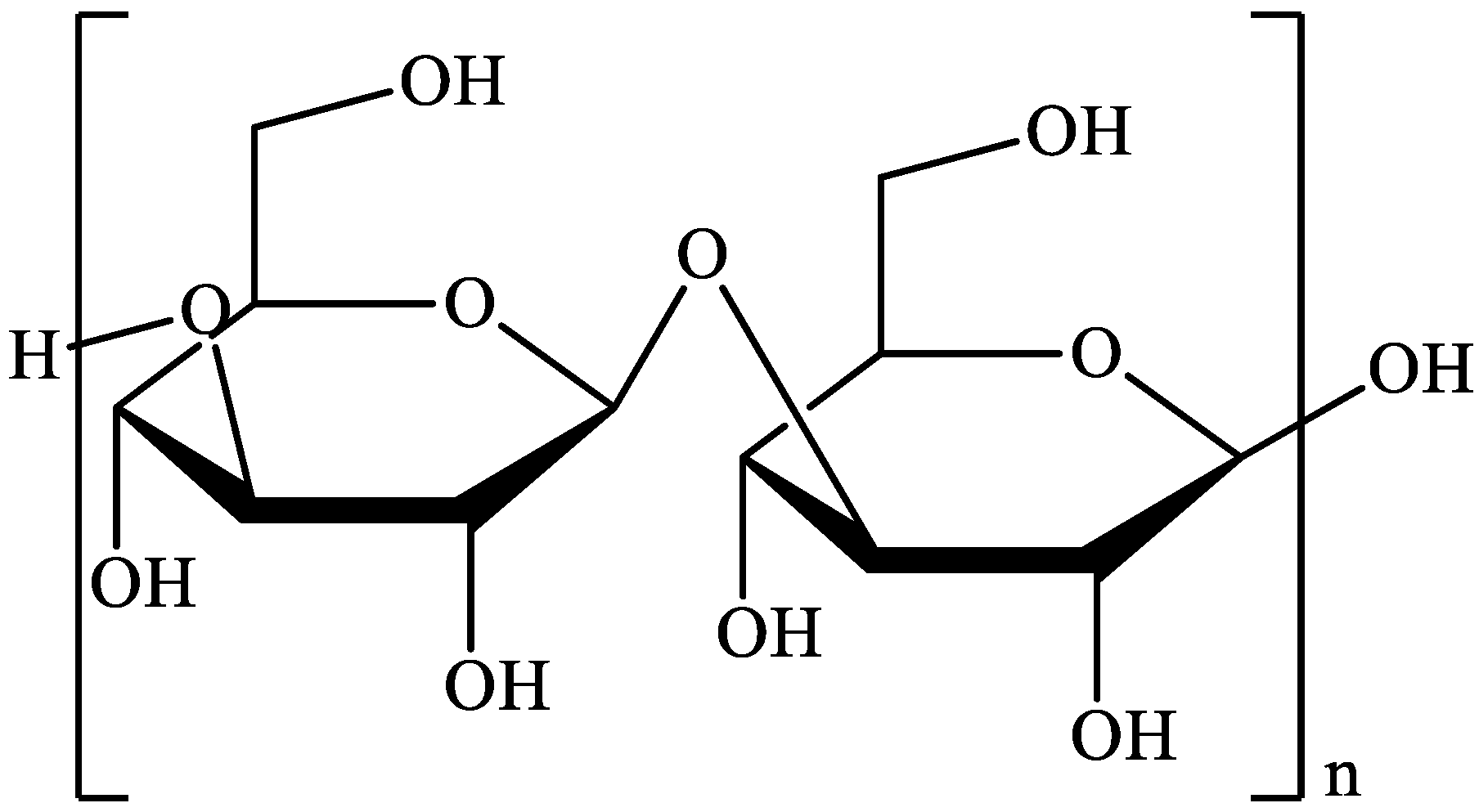
2.1. Laminarin
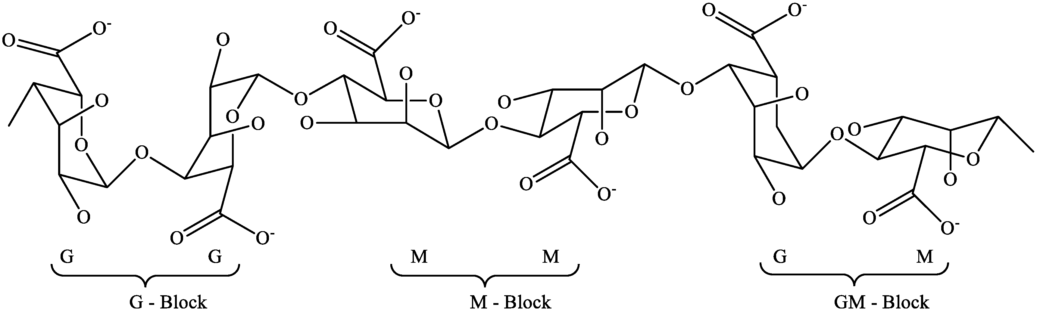
2.2. Alginates
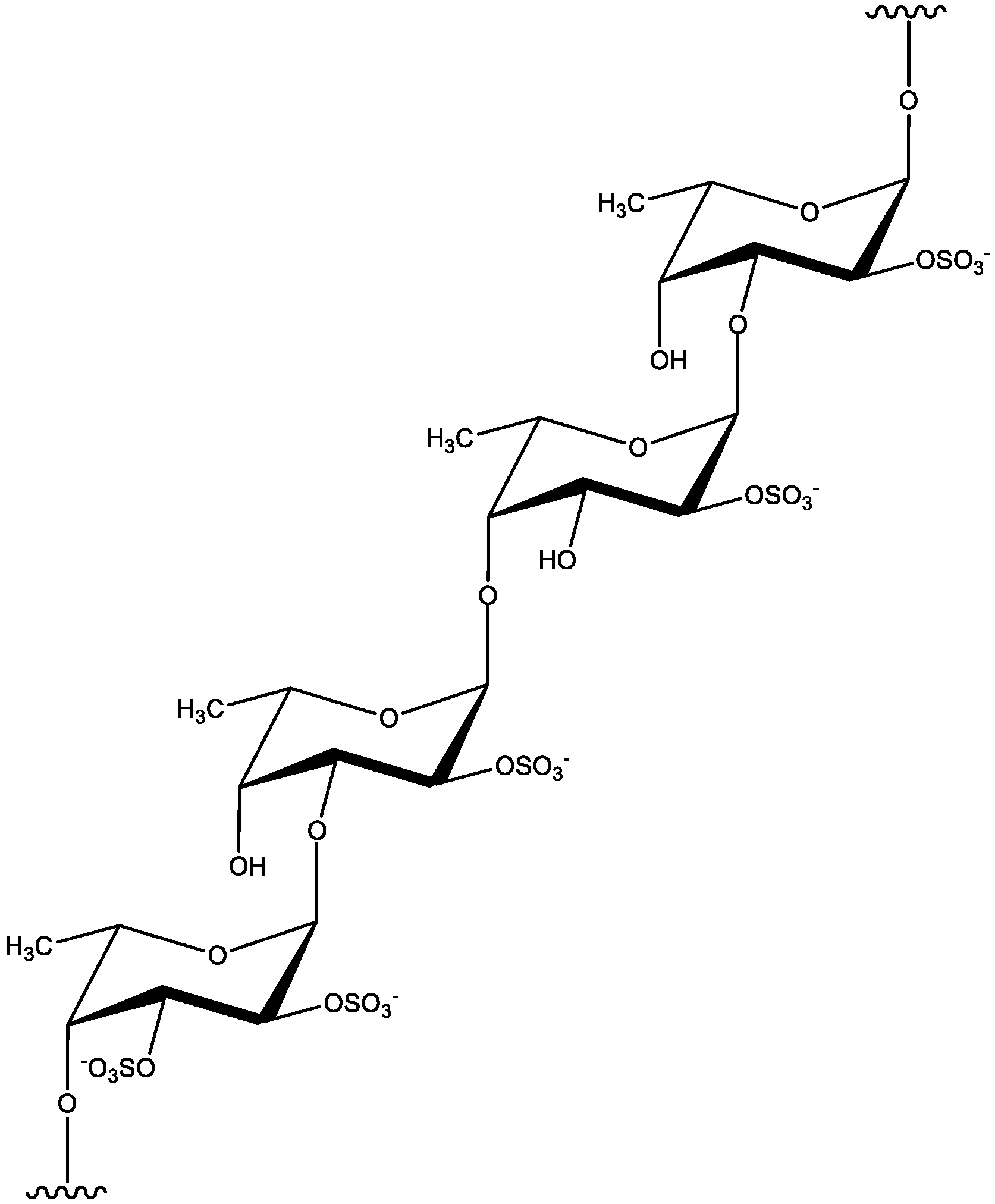
2.3. Fucoidan
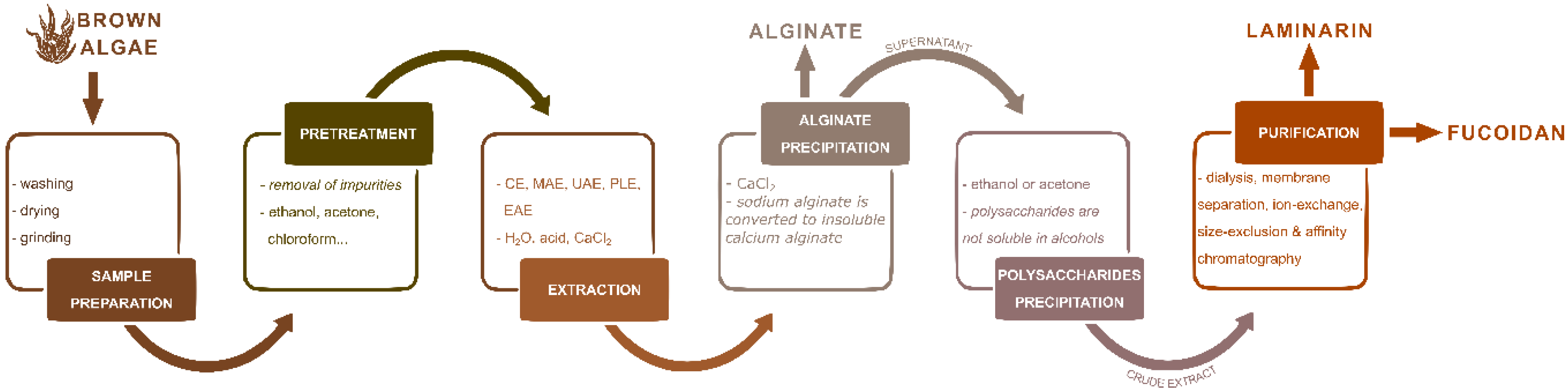
3. The Perspective of Advanced Technologies for Polysaccharide Extraction from Marine Brown Algae
3.1. Pre-treatment of Marine Brown Algae
3.2. Extraction Techniques
3.2.1. Conventional Extraction Technique (CE)
3.2.2. Advanced Extraction Techniques
Microwave Assisted Extraction (MAE)
Ultrasound Assisted Extraction (UAE)
Pressurized Liquid Extraction (PLE)
Enzymes Assisted Extraction (EAE)
3.3. Purification Procedure
4. Brown Algae Sulfated Polysaccharides as Drug Delivery Systems and Their Safety
5. Future Challenges and Potential Industry Application of Brown Algae
Author Contributions
Funding
Conflicts of Interest
References
- Cavalier-Smith, T. Evolution and relationships of algae: Major branches of the tree of life. In Unravelling the Algae the Past, Present, and Future of Algal Systematics; Juliet Brodie, J.L., Ed.; CRC Press: Boca Raton, FL, USA, 2007; pp. 21–55. ISBN 0-8493-7989-X. [Google Scholar]
- Rindi, F. Diversity and Classification of Marine Benthic Algae. Available online: http://marinespecies.org/introduced/wiki/Diversity_and_classification_of_marine_benthic_algae#cite_note-Cavalier-1 (accessed on 24 November 2019).
- Xu, S.Y.; Huang, X.; Cheong, K.L. Recent advances in marine algae polysaccharides: Isolation, structure, and activities. Mar. Drugs 2017, 15, 388. [Google Scholar] [CrossRef] [PubMed] [Green Version]
- Mišurcová, L.; Orsavová, J.; Ambrožová, J.V. Algal Polysaccharides and Health. In Polysaccharides; Springer: Berlin/Heidelberg, Germany, 2014; pp. 1–29. [Google Scholar]
- Zhao, C.; Yang, C.; Liu, B.; Lin, L.; Sarker, S.D.; Nahar, L.; Yu, H.; Cao, H.; Xiao, J. Bioactive compounds from marine macroalgae and their hypoglycemic benefits. Trends Food Sci. Technol. 2018, 72, 1–12. [Google Scholar] [CrossRef]
- Costa, L.S.; Fidelis, G.P.; Cordeiro, S.L.; Oliveira, R.M.; Sabry, D.A.; Câmara, R.B.G.; Nobre, L.T.D.B.; Costa, M.S.S.P.; Almeida-Lima, J.; Farias, E.H.C.; et al. Biological activities of sulfated polysaccharides from tropical seaweeds. Biomed. Pharmacother. 2010, 64, 21–28. [Google Scholar] [CrossRef] [PubMed]
- Pereira, L. Seaweeds as source of bioactive substances and skin care therapy-Cosmeceuticals, algotheraphy, and thalassotherapy. Cosmetics 2018, 5, 68. [Google Scholar] [CrossRef] [Green Version]
- Jiao, G.; Yu, G.; Zhang, J.; Ewart, H.S. Chemical structures and bioactivities of sulfated polysaccharides from marine algae. Mar. Drugs 2011, 9, 196–233. [Google Scholar] [CrossRef] [Green Version]
- Garcia-Vaquero, M.; Rajauria, G.; O’Doherty, J.V.; Sweeney, T. Polysaccharides from macroalgae: Recent advances, innovative technologies and challenges in extraction and purification. Food Res. Int. 2017, 99, 1011–1020. [Google Scholar] [CrossRef] [Green Version]
- Ale, M.T.; Mikkelsen, J.D.; Meyer, A.S. Important determinants for fucoidan bioactivity: A critical review of structure-function relations and extraction methods for fucose-containing sulfated polysaccharides from brown seaweeds. Mar. Drugs 2011, 9, 2106–2130. [Google Scholar] [CrossRef] [Green Version]
- Praveen, M.A.; Parvathy, K.R.K.; Balasubramanian, P.; Jayabalan, R. An overview of extraction and purification techniques of seaweed dietary fibers for immunomodulation on gut microbiota. Trends Food Sci. Technol. 2019, 92, 46–64. [Google Scholar] [CrossRef]
- De Jesus Raposo, M.F.; De Morais, A.M.B.; De Morais, R.M.S.C. Marine polysaccharides from algae with potential biomedical applications. Mar. Drugs 2015, 13, 2967–3028. [Google Scholar] [CrossRef]
- Hahn, T.; Lang, S.; Ulber, R.; Muffler, K. Novel procedures for the extraction of fucoidan from brown algae. Process Biochem. 2012, 47, 1691–1698. [Google Scholar] [CrossRef]
- Dore, C.M.P.G.; Faustino Alves, M.G.D.C.; Pofírio Will, L.S.E.; Costa, T.G.; Sabry, D.A.; De Souza Rêgo, L.A.R.; Accardo, C.M.; Rocha, H.A.O.; Filgueira, L.G.A.; Leite, E.L. A sulfated polysaccharide, fucans, isolated from brown algae Sargassum vulgare with anticoagulant, antithrombotic, antioxidant and anti-inflammatory effects. Carbohydr. Polym. 2013, 91, 467–475. [Google Scholar] [CrossRef] [PubMed]
- Hadj Ammar, H.; Hafsa, J.; Le Cerf, D.; Bouraoui, A.; Majdoub, H. Antioxidant and gastroprotective activities of polysaccharides from the Tunisian brown algae (Cystoseira sedoides). J. Tunis. Chem. Soc. 2016, 18, 80–88. [Google Scholar]
- Hentati, F.; Delattre, C.; Ursu, A.V.; Desbrières, J.; Le Cerf, D.; Gardarin, C.; Abdelkafi, S.; Michaud, P.; Pierre, G. Structural characterization and antioxidant activity of water-soluble polysaccharides from the Tunisian brown seaweed Cystoseira compressa. Carbohydr. Polym. 2018, 198, 589–600. [Google Scholar] [CrossRef] [PubMed]
- Mzibra, A.; Meftah Kadmiri, I.; El Arroussi, H. Enzymatic Technologies for Marine Polysaccharides; Trincone, A., Ed.; CRS PRESS: Boca Raton, FL, USA, 2019. [Google Scholar]
- Chaminda Lakmal, H.H.; Lee, J.-H.; Jeon, Y.-J. Enzyme-assisted extraction of a marine algal polysaccharide, fucoidan and bioactivities. In Polysaccharides: Bioactivity and Biotechnology; Springer: Berlin/Heidelberg, Germany, 2015; pp. 1–2241. ISBN 9783319162980. [Google Scholar]
- Lim, S.J.; Wan Aida, W.M. Extraction of sulfated polysaccharides (fucoidan) from brown seaweed. In Seaweed Polysaccharides; Elsevier: Amsterdam, The Netherlands, 2017; pp. 27–46. ISBN 9780128098172. [Google Scholar]
- Nisizawa, K.; Yamaguchi, T.; Handa, N.; Maeda, M.; Yamazaki, H. Chemical nature of a uronic acid-containing polysaccharide in the peritrophic membrane of the silkworm. J. Biochem. 1963, 54, 419–426. [Google Scholar] [CrossRef]
- Kadam, S.U.; Tiwari, B.K.; O’Donnell, C.P. Extraction, structure and biofunctional activities of laminarin from brown algae. Int. J. Food Sci. Technol. 2015, 50, 24–31. [Google Scholar] [CrossRef]
- Quillet, M. Glucide metabolism of brown algae. Presence of small quantities of Laminarin in numerous new species, distributed over the entire group of Phaeophyceae. Comptes Rendus l’Académie Sci. 1958, 246, 812–815. [Google Scholar]
- Chizhov, A.O.; Dell, A.; Morris, H.R.; Reason, A.J.; Haslam, S.M.; McDowell, R.A.; Chizhov, O.S.; Usov, A.I. Structural analysis of laminarans by MALDI and FAB mass spectrometry. Carbohydr. Res. 1998, 310, 203–210. [Google Scholar] [CrossRef]
- Menshova, R.V.; Ermakova, S.P.; Anastyuk, S.D.; Isakov, V.V.; Dubrovskaya, Y.V.; Kusaykin, M.I.; Um, B.H.; Zvyagintseva, T.N. Structure, enzymatic transformation and anticancer activity of branched high molecular weight laminaran from brown alga Eisenia bicyclis. Carbohydr. Polym. 2014, 99, 101–109. [Google Scholar] [CrossRef]
- Miao, H.Q.; Elkin, M.; Aingorn, E.; Ishai-Michaeli, R.; Stein, C.A.; Vlodavsky, I. Inhibition of heparanase activity and tumor metastasis by laminarin sulfate and synthetic phosphorothioate oligodeoxynucleotides. Int. J. Cancer 1999, 83, 424–431. [Google Scholar] [CrossRef]
- Bae, H.; Song, G.; Lee, J.; Hong, T.; Chang, M.; Lim, W. Laminarin-derived from brown algae suppresses the growth of ovarian cancer cells via mitochondrial dysfunction and ER stress. Mar. Drugs 2020, 18, 152. [Google Scholar] [CrossRef] [Green Version]
- Hernández-Carmona, G.; Freile-Pelegrín, Y.; Hernández-Garibay, E. Conventional and Alternative Technologies for the Extraction of Algal Polysaccharides; Woodhead Publishing: Shaston, UK, 2013; ISBN 9780857095121. [Google Scholar]
- Bixler, H.J.; Porse, H. A decade of change in the seaweed hydrocolloids industry. J. Appl. Phycol. 2011, 23, 321–335. [Google Scholar] [CrossRef]
- Rasmussen, R.S.; Morrissey, M.T. Marine biotechnology for production of food ingredients. Adv. Food Nutr. Res. 2007, 52, 237–292. [Google Scholar] [PubMed]
- Burtin, P. Nutritional value of seaweeds. Electron. J. Environ. Agric. Food Chem. 2003, 2, 498–503. [Google Scholar]
- Murata, M.; Nakazoe, J. Production and use of marine algae in Japan. Jpn. Agric. Res. Q. 2001, 35, 281–290. [Google Scholar] [CrossRef] [Green Version]
- Kimura, Y.; Watanabe, K.; Okuda, H. Effects of soluble sodium alginate on cholesterol excretion and glucose tolerance in rats. J. Ethnopharmacol. 1996, 54, 47–54. [Google Scholar] [CrossRef]
- Kim, I.H.; Lee, J.H. Antimicrobial activities against methicillin-resistant Staphylococcus aureus from macroalgae. J. Ind. Eng. Chem. 2008, 14, 568–572. [Google Scholar]
- Chapman, V.J.; Chapman, D.J. Seaweeds and Their Uses, 3rd ed.; Springer: Dordrecht, The Netherlands, 1980. [Google Scholar]
- Collado-González, M.; Cristina Ferreri, M.; Freitas, A.R.; Santos, A.C.; Ferreira, N.R.; Carissimi, G.; Sequeira, J.A.D.; Guillermo Díaz Baños, F.; Villora, G.; Veiga, F.; et al. Complex polysaccharide-based nanocomposites for oral insulin delivery. Mar. Drugs 2020, 18, 55. [Google Scholar] [CrossRef] [Green Version]
- Zvyagintseva, T.N.; Shevchenko, N.M.; Chizhov, A.O.; Krupnova, T.N.; Sundukova, E.V.; Isakov, V.V. Water-soluble polysaccharides of some far-eastern brown seaweeds. Distribution, structure, and their dependence on the developmental conditions. J. Exp. Mar. Biol. Ecol. 2003, 294, 1–13. [Google Scholar] [CrossRef]
- Zvyagintseva, T.N.; Shevchenko, N.M.; Popivnich, I.B.; Isakov, V.V.; Scobun, A.S.; Sundukova, E.V.; Elyakova, L.A. A new procedure for the separation of water-soluble polysaccharides from brown seaweeds. Carbohydr. Res. 1999, 322, 32–39. [Google Scholar] [CrossRef]
- Rioux, L.E.; Turgeon, S.L.; Beaulieu, M. Characterization of polysaccharides extracted from brown seaweeds. Carbohydr. Polym. 2007, 69, 530–537. [Google Scholar] [CrossRef]
- Bilan, M.I.; Grachev, A.A.; Ustuzhanina, N.E.; Shashkov, A.S.; Nifantiev, N.E.; Usov, A.I. A highly regular fraction of a fucoidan from the brown seaweed Fucus distichus L. Carbohydr. Res. 2004, 339, 511–517. [Google Scholar] [CrossRef]
- Li, B.; Lu, F.; Wei, X.; Zhao, R. Fucoidan: Structure and bioactivity. Molecules 2008, 13, 1671–1695. [Google Scholar] [CrossRef] [Green Version]
- Van Weelden, G.; Bobi, M.; Okła, K.; van Weelden, W.J.; Romano, A.; Pijnenborg, J.M.A. Fucoidan structure and activity in relation to anti-cancer mechanisms. Mar. Drugs 2019, 17, 32. [Google Scholar] [CrossRef] [Green Version]
- Sellimi, S.; Maalej, H.; Rekik, D.M.; Benslima, A.; Ksouda, G.; Hamdi, M.; Sahnoun, Z.; Li, S.; Nasri, M.; Hajji, M. Antioxidant, antibacterial and in vivo wound healing properties of laminaran purified from Cystoseira barbata seaweed. Int. J. Biol. Macromol. 2018, 119, 633–644. [Google Scholar] [CrossRef]
- January, G.G.; Naidoo, R.K.; Kirby-McCullough, B.; Bauer, R. Assessing methodologies for fucoidan extraction from South African brown algae. Algal Res. 2019, 40, 101517. [Google Scholar] [CrossRef]
- Sahera, M.F.; Thani, S.M.; Salha, S.Y. Characterization of sulphated polysaccharide with antiviral activity from marine brown alga Cystoseira myrica collected from Jazan coasts, KSA. Int. J. PharmTech Res. 2015, 8, 198–203. [Google Scholar]
- Deghrigue Abid, M.; Lajili, S.; Hadj Ammar, H.; Cherif, D.; Eltaief, N.; Majdoub, H.; Bouraoui, A. Chemical and biological properties of sodium alginates isolated from tow brown algae Dictyopteris Membranaceae and Padina Pavonica. Trends J. Sci. Res. 2019, 4, 62–67. [Google Scholar] [CrossRef]
- Sun, Q.L.; Li, Y.; Ni, L.Q.; Li, Y.X.; Cui, Y.S.; Jiang, S.L.; Xie, E.Y.; Du, J.; Deng, F.; Dong, C.X. Structural characterization and antiviral activity of two fucoidans from the brown algae Sargassum henslowianum. Carbohydr. Polym. 2020, 229, 115487. [Google Scholar] [CrossRef]
- Zhao, G.; Chen, X.; Wang, L.; Zhou, S.; Feng, H.; Chen, W.N.; Lau, R. Ultrasound assisted extraction of carbohydrates from microalgae as feedstock for yeast fermentation. Bioresour. Technol. 2013, 128, 337–344. [Google Scholar] [CrossRef]
- Harun, R.; Yip, J.W.S.; Thiruvenkadam, S.; Ghani, W.A.W.A.K.; Cherrington, T.; Danquah, M.K. Algal biomass conversion to bioethanol-a step-by-step assessment. Biotechnol. J. 2014, 9, 73–86. [Google Scholar] [CrossRef]
- Kadam, S.U.; Tiwari, B.K.; O’Connell, S.; O’Donnell, C.P. Effect of ultrasound pretreatment on the extraction kinetics of bioactives from brown seaweed (Ascophyllum nodosum). Sep. Sci. Technol. 2015, 50, 670–675. [Google Scholar] [CrossRef]
- Cui, Y.; Liu, X.; Li, S.; Hao, L.; Du, J.; Gao, D.H.; Kang, Q.; Lu, J. Extraction, characterization and biological activity of sulfated polysaccharides from seaweed Dictyopteris divaricata. Int. J. Biol. Macromol. 2018, 117, 256–263. [Google Scholar] [CrossRef]
- Fletcher, H.R.; Biller, P.; Ross, A.B.; Adams, J.M.M. The seasonal variation of fucoidan within three species of brown macroalgae. Algal Res. 2017, 22, 79–86. [Google Scholar] [CrossRef] [Green Version]
- Fawzy, M.A.; Gomaa, M.; Hifney, A.F.; Abdel-Gawad, K.M. Optimization of alginate alkaline extraction technology from Sargassum latifolium and its potential antioxidant and emulsifying properties. Carbohydr. Polym. 2017, 157, 1903–1912. [Google Scholar] [CrossRef]
- Ammar, H.H.; Lajili, S.; Said, R.B.; Le Cerf, D.; Bouraoui, A.; Majdoub, H. Physico-chemical characterization and pharmacological evaluation of sulfated polysaccharides from three species of Mediterranean brown algae of the genus Cystoseira. DARU J. Pharm. Sci. 2015, 23, 4–11. [Google Scholar]
- Liu, J.; Wu, S.-Y.; Chen, L.; Li, Q.-J.; Shen, Y.-Z.; Jin, L.; Zhang, X.; Chen, P.-C.; Wu, M.-J.; Choi, J.; et al. Different extraction methods bring about distinct physicochemical properties and antioxidant activities of Sargassum fusiforme fucoidans. Int. J. Biol. Macromol. 2019. [Google Scholar] [CrossRef]
- Yuan, Y.; Macquarrie, D. Microwave assisted extraction of sulfated polysaccharides (fucoidan) from Ascophyllum nodosum and its antioxidant activity. Carbohydr. Polym. 2015, 129, 101–107. [Google Scholar] [CrossRef]
- Rodriguez-Jasso, R.M.; Mussatto, S.I.; Pastrana, L.; Aguilar, C.N.; Teixeira, J.A. Microwave-assisted extraction of sulfated polysaccharides (fucoidan) from brown seaweed. Carbohydr. Polym. 2011, 86, 1137–1144. [Google Scholar] [CrossRef] [Green Version]
- Yuan, Y.; Zhang, J.; Fan, J.; Clark, J.; Shen, P.; Li, Y.; Zhang, C. Microwave assisted extraction of phenolic compounds from four economic brown macroalgae species and evaluation of their antioxidant activities and inhibitory effects on α-amylase, α-glucosidase, pancreatic lipase and tyrosinase. Food Res. Int. 2018, 113, 288–297. [Google Scholar] [CrossRef]
- Zhang, R.; Yuen, A.K.L.; Magnusson, M.; Wright, J.T.; de Nys, R.; Masters, A.F.; Maschmeyer, T. A comparative assessment of the activity and structure of phlorotannins from the brown seaweed Carpophyllum flexuosum. Algal Res. 2018, 29, 130–141. [Google Scholar] [CrossRef]
- Dang, T.T.; Bowyer, M.C.; Van Altena, I.A.; Scarlett, C.J. Optimum conditions of microwave-assisted extraction for phenolic compounds and antioxidant capacity of the brown alga Sargassum vestitum. Sep. Sci. Technol. 2018, 53, 1711–1723. [Google Scholar] [CrossRef]
- Magnusson, M.; Yuen, A.K.L.; Zhang, R.; Wright, J.T.; Taylor, R.B.; Maschmeyer, T.; de Nys, R. A comparative assessment of microwave assisted (MAE) and conventional solid-liquid (SLE) techniques for the extraction of phloroglucinol from brown seaweed. Algal Res. 2017, 23, 28–36. [Google Scholar] [CrossRef]
- Chen, C.; Zhang, B.; Huang, Q.; Fu, X.; Liu, R.H. Microwave-assisted extraction of polysaccharides from Moringa oleifera Lam. leaves: Characterization and hypoglycemic activity. Ind. Crops Prod. 2017, 100, 1–11. [Google Scholar] [CrossRef]
- Hu, W.; Zhao, Y.; Yang, Y.; Zhang, H.; Ding, C.; Hu, C.; Zhou, L.; Zhang, Z.; Yuan, S.; Chen, Y.; et al. Microwave-assisted extraction, physicochemical characterization and bioactivity of polysaccharides from Camptotheca acuminata fruits. Int. J. Biol. Macromol. 2019, 133, 127–136. [Google Scholar] [CrossRef]
- Senthil Kumar, C.; Sivakumar, M.; Ruckmani, K. Microwave-assisted extraction of polysaccharides from Cyphomandra betacea and its biological activities. Int. J. Biol. Macromol. 2016, 92, 682–693. [Google Scholar]
- Alboofetileh, M.; Rezaei, M.; Tabarsa, M.; Rittà, M.; Donalisio, M.; Mariatti, F.; You, S.G.; Lembo, D.; Cravotto, G. Effect of different non-conventional extraction methods on the antibacterial and antiviral activity of fucoidans extracted from Nizamuddinia zanardinii. Int. J. Biol. Macromol. 2018, 124, 131–137. [Google Scholar] [CrossRef]
- Okolie, C.L.; Mason, B.; Mohan, A.; Pitts, N.; Udenigwe, C.C. The comparative influence of novel extraction technologies on in vitro prebiotic-inducing chemical properties of fucoidan extracts from Ascophyllum nodosum. Food Hydrocoll. 2019, 90, 462–471. [Google Scholar] [CrossRef]
- Ren, B.; Chen, C.; Li, C.; Fu, X.; You, L.; Liu, R.H. Optimization of microwave-assisted extraction of Sargassum thunbergii polysaccharides and its antioxidant and hypoglycemic activities. Carbohydr. Polym. 2017, 173, 192–201. [Google Scholar] [CrossRef]
- Cao, C.; Huang, Q.; Zhang, B.; Li, C.; Fu, X. Physicochemical characterization and in vitro hypoglycemic activities of polysaccharides from Sargassum pallidum by microwave-assisted aqueous two-phase extraction. Int. J. Biol. Macromol. 2018, 109, 357–368. [Google Scholar] [CrossRef]
- Fayad, S.; Nehmé, R.; Tannoury, M.; Lesellier, E.; Pichon, C.; Morin, P. Macroalga Padina pavonica water extracts obtained by pressurized liquid extraction and microwave-assisted extraction inhibit hyaluronidase activity as shown by capillary electrophoresis. J. Chromatogr. 2017, 1497, 19–27. [Google Scholar] [CrossRef]
- Wang, J.; Zhang, J.; Zhao, B.; Wang, X.; Wu, Y.; Yao, J. A comparison study on microwave-assisted extraction of Potentilla anserina L. polysaccharides with conventional method: Molecule weight and antioxidant activities evaluation. Carbohydr. Polym. 2010, 80, 84–93. [Google Scholar] [CrossRef]
- Hanjabam, M.D.; Kumar, A.; Tejpal, C.S.; Krishnamoorthy, E.; Kishore, P.; Ashok Kumar, K. Isolation of crude fucoidan from Sargassum wightii using conventional and ultra-sonication extraction methods. Bioact. Carbohydr. Diet. Fibre 2019, 20, 100200. [Google Scholar] [CrossRef]
- Alboofetileh, M.; Rezaei, M.; Tabarsa, M.; You, S.G. Ultrasound-assisted extraction of sulfated polysaccharide from Nizamuddinia zanardinii: Process optimization, structural characterization, and biological properties. J. Food Process Eng. 2018, 42, 1–13. [Google Scholar] [CrossRef]
- Ying, Z.; Han, X.; Li, J. Ultrasound-assisted extraction of polysaccharides from mulberry leaves. Food Chem. 2011, 127, 1273–1279. [Google Scholar] [CrossRef]
- Yan, J.K.; Wang, Y.Y.; Ma, H.L.; Wang, Z. Bin Ultrasonic effects on the degradation kinetics, preliminary characterization and antioxidant activities of polysaccharides from Phellinus linteus mycelia. Ultrason. Sonochem. 2016, 29, 251–257. [Google Scholar] [CrossRef]
- Alboofetileh, M.; Rezaei, M.; Tabarsa, M.; You, S.G. Bioactivities of Nizamuddinia zanardinii sulfated polysaccharides extracted by enzyme, ultrasound and enzyme-ultrasound methods. J. Food Sci. Technol. 2019, 56, 1212–1220. [Google Scholar] [CrossRef]
- Kadam, S.U.; Donnell, C.P.O.; Rai, D.K.; Hossain, M.B.; Burgess, C.M.; Walsh, D.; Tiwari, B.K. Laminarin from Irish brown seaweeds Ascophyllum nodosum and Laminaria hyperborea. Mar. Drugs 2015, 13, 4270–4280. [Google Scholar] [CrossRef]
- Song, K.M.; Ha, S.J.; Lee, J.E.; Kim, S.H.; Kim, Y.H.; Kim, Y.; Hong, S.P.; Jung, S.K.; Lee, N.H. High yield ultrasonication extraction method for Undaria pinnatifida sporophyll and its anti-inflammatory properties associated with AP-1 pathway suppression. LWT Food Sci. Technol. 2015, 64, 1315–1322. [Google Scholar] [CrossRef]
- Hmelkov, A.B.; Zvyagintseva, T.N.; Shevchenko, N.M.; Rasin, A.B.; Ermakova, S.P. Ultrasound-assisted extraction of polysaccharides from brown alga Fucus evanescens. Structure and biological activity of the new fucoidan fractions. J. Appl. Phycol. 2018, 30, 2039–2046. [Google Scholar] [CrossRef]
- Youssouf, L.; Lallemand, L.; Giraud, P.; Soulé, F.; Bhaw-Luximon, A.; Meilhac, O.; D’Hellencourt, C.L.; Jhurry, D.; Couprie, J. Ultrasound-assisted extraction and structural characterization by NMR of alginates and carrageenans from seaweeds. Carbohydr. Polym. 2017, 166, 55–63. [Google Scholar] [CrossRef]
- Zhu, C.P.; Zhai, X.C.; Li, L.Q.; Wu, X.X.; Li, B. Response surface optimization of ultrasound-assisted polysaccharides extraction from pomegranate peel. Food Chem. 2015, 177, 139–146. [Google Scholar] [CrossRef]
- Wu, S.-C. Antioxidant activity of sulfated seaweeds polysaccharides by novel assisted extraction. In Solubility of Polysaccharides; Xu, Z., Ed.; IntechOpen: London, UK, 2017; pp. 89–108. [Google Scholar]
- Santoyo, S.; Plaza, M.; Jaime, L.; Ibañez, E.; Reglero, G.; Señorans, J. Pressurized liquids as an alternative green process to extract antiviral agents from the edible seaweed Himanthalia elongata. J. Appl. Phycol. 2011, 23, 909–917. [Google Scholar] [CrossRef] [Green Version]
- Saravana, P.S.; Cho, Y.J.; Park, Y.B.; Woo, H.C.; Chun, B.S. Structural, antioxidant, and emulsifying activities of fucoidan from Saccharina japonica using pressurized liquid extraction. Carbohydr. Polym. 2016, 153, 518–525. [Google Scholar] [CrossRef]
- Saravana, P.S.; Choi, J.H.; Park, Y.B.; Woo, H.C.; Chun, B.S. Evaluation of the chemical composition of brown seaweed (Saccharina japonica) hydrolysate by pressurized hot water extraction. Algal Res. 2016, 13, 246–254. [Google Scholar] [CrossRef]
- Saravana, P.S.; Cho, Y.N.; Patil, M.P.; Cho, Y.J.; Kim, G.D.; Park, Y.B.; Woo, H.C.; Chun, B.S. Hydrothermal degradation of seaweed polysaccharide: Characterization and biological activities. Food Chem. 2018, 268, 179–187. [Google Scholar] [CrossRef]
- Saravana, P.S.; Tilahun, A.; Gerenew, C.; Tri, V.D.; Kim, N.H.; Kim, G.D.; Woo, H.C.; Chun, B.S. Subcritical water extraction of fucoidan from Saccharina japonica: Optimization, characterization and biological studies. J. Appl. Phycol. 2018, 30, 579–590. [Google Scholar] [CrossRef]
- Vergara-Salinas, J.R.; Cuevas-Valenzuela, J.; Pérez-Correa, J.R. Pressurized hot water extraction of polyphenols from plant material. In Biotechnology of Bioactive Compounds; Gupta, V.K., Tuohy, M.G., Eds.; John Wiley & Sons: Hoboken, NJ, USA, 2015. [Google Scholar]
- Alboofetileh, M.; Rezaei, M.; Tabarsa, M.; You, S.G.; Mariatti, F.; Cravotto, G. Subcritical water extraction as an efficient technique to isolate biologically-active fucoidans from Nizamuddinia zanardinii. Int. J. Biol. Macromol. 2019, 128, 244–253. [Google Scholar] [CrossRef]
- Nadar, S.S.; Rao, P.; Rathod, V.K. Enzyme assisted extraction of biomolecules as an approach to novel extraction technology: A review. Food Res. Int. 2018, 108, 309–330. [Google Scholar] [CrossRef]
- Charoensiddhi, S.; Lorbeer, A.J.; Lahnstein, J.; Bulone, V.; Franco, C.M.M.; Zhang, W. Enzyme-assisted extraction of carbohydrates from the brown alga Ecklonia radiata: Effect of enzyme type, pH and buffer on sugar yield and molecular weight profiles. Process Biochem. 2016, 51, 1503–1510. [Google Scholar] [CrossRef]
- Alboofetileh, M.; Rezaei, M.; Tabarsa, M. Enzyme-assisted extraction of Nizamuddinia zanardinii for the recovery of sulfated polysaccharides with anticancer and immune-enhancing activities. J. Appl. Phycol. 2018, 31, 1391–1402. [Google Scholar] [CrossRef]
- Borazjani, N.J.; Tabarsa, M.; You, S.G.; Rezaei, M. Effects of extraction methods on molecular characteristics, antioxidant properties and immunomodulation of alginates from Sargassum angustifolium. Int. J. Biol. Macromol. 2017, 101, 703–711. [Google Scholar] [CrossRef] [Green Version]
- Hammed, A.M.; Jaswir, I.; Simsek, S.; Alam, Z.; Amid, A. Enzyme aided extraction of sulfated polysaccharides from Turbinaria turbinata brown seaweed. Int. Food Res. J. 2017, 24, 1660–1666. [Google Scholar]
- Rostami, Z.; Tabarsa, M.; You, S.G.; Rezaei, M. Relationship between molecular weights and biological properties of alginates extracted under different methods from Colpomenia peregrina. Process Biochem. 2017, 58, 289–297. [Google Scholar] [CrossRef]
- Sánchez-Camargo, A.D.P.; Montero, L.; Stiger-Pouvreau, V.; Tanniou, A.; Cifuentes, A.; Herrero, M.; Ibáñez, E. Considerations on the use of enzyme-assisted extraction in combination with pressurized liquids to recover bioactive compounds from algae. Food Chem. 2016, 192, 67–74. [Google Scholar] [CrossRef]
- Asanka Sanjeewa, K.K.; Shanura Fernando, I.P.; Kim, E.A.; Ahn, G.; Jee, Y.; Jeon, Y.J. Anti-inflammatory activity of a sulfated polysaccharide isolated from an enzymatic digest of brown seaweed Sargassum horneri in RAW 264.7 cells. Nutr. Res. Pract. 2017, 11, 3–10. [Google Scholar] [CrossRef] [Green Version]
- Kang, M.C.; Lee, H.G.; Choi, H.D.; Jeon, Y.J. Antioxidant properties of a sulfated polysaccharide isolated from an enzymatic digest of Sargassum thunbergii. Int. J. Biol. Macromol. 2019, 132, 142–149. [Google Scholar] [CrossRef]
- Scott, J.E. Fractionation by precipitation with quaternary ammonium salts. In Methods in Carbohydrate Chemistry; Whistler, R.L., BeMiller, J.N., Eds.; Academic Press: New York, NY, USA, 1965; pp. 38–44. [Google Scholar]
- Sosa-Hernández, J.E.; Escobedo-Avellaneda, Z.; Iqbal, H.M.N.; Welti-Chanes, J. State-of-the-art extraction methodologies for bioactive compounds from algal biome to meet bio-economy challenges and opportunities. Molecules 2018, 23, 2953. [Google Scholar] [CrossRef] [Green Version]
- Fernando, I.P.S.; Kim, D.; Nah, J.-W.; Jeon, Y.-J. Advances in functionalizing fucoidans and alginates (bio) polymers by structural modifications: A review. Chem. Eng. J. 2019, 355, 33–48. [Google Scholar] [CrossRef]
- De Jesus Raposo, M.F.; De Morais, R.M.S.C.; De Morais, A.M.M.B. Bioactivity and applications of sulphated polysaccharides from marine microalgae. Mar. Drugs 2013, 11, 233–252. [Google Scholar] [CrossRef] [Green Version]
- Lenstra, J.; van Hal, J.; Reith, H. Economic aspects of open ocean seaweed cultivation. In Proceedings of the Alg’n Chem Conference, Montpellier, France, 7–10 November 2011. [Google Scholar]
- Vishchuk, O.S.; Ermakova, S.P.; Zvyagintseva, T.N. The fucoidans from brown algae of Far-Eastern seas: Anti-tumor activity and structure–function relationship. Food Chem. 2013, 141, 1211–1217. [Google Scholar] [CrossRef]
- Pérez, M.; Falqué, E.; Domínguez, H. Antimicrobial action of compounds from marine seaweed. Mar. Drugs 2016, 14, 52. [Google Scholar] [CrossRef] [PubMed] [Green Version]
- Lee, J.Y.; Kim, Y.-J.; Kim, H.J.; Kim, Y.-S.; Park, W. Immunostimulatory effect of laminarin on RAW 264.7 mouse macrophages. Molecules 2012, 17, 5404–5411. [Google Scholar] [CrossRef] [PubMed] [Green Version]
- Wu, G.-J.; Shiu, S.-M.; Hsieh, M.-C.; Tsai, G.-J. Anti-inflammatory activity of a sulfated polysaccharide from the brown alga Sargassum cristaefolium. Food Hydrocoll. 2016, 53, 16–23. [Google Scholar] [CrossRef]
- Lopes, M.; Abrahim, B.; Veiga, F.; Seica, R.; Cabral, L.M.; Arnaud, P.; Andrade, J.C.; Ribeiro, A.J. Preparation methods and applications behind alginate-based particles. Expert Opin. Drug Deliv. 2017, 14, 769–782. [Google Scholar] [CrossRef] [PubMed]
- Lima, D.S.; Tenório-Neto, E.T.; Lima-Tenório, M.K.; Guilherme, M.R.; Scariot, D.B.; Nakamura, C.V.; Muniz, E.C.; Rubira, A.F. pH-responsive alginate-based hydrogels for protein delivery. J. Mol. Liq. 2018, 262, 29–36. [Google Scholar] [CrossRef]
- Sriamornsak, P.; Thirawong, N.; Korkerd, K. Swelling, erosion and release behavior of alginate-based matrix tablets. Eur. J. Pharm. Biopharm. 2007, 66, 435–450. [Google Scholar] [CrossRef]
- Agarwal, T.; Narayana, S.N.G.H.; Pal, K.; Pramanik, K.; Giri, S.; Banerjee, I. Calcium alginate-carboxymethyl cellulose beads for colon-targeted drug delivery. Int. J. Biol. Macromol. 2015, 75, 409–417. [Google Scholar] [CrossRef]
- Daemi, H.; Barikani, M. Synthesis and characterization of calcium alginate nanoparticles, sodium homopolymannuronate salt and its calcium nanoparticles. Sci. Iran. 2012, 19, 2023–2028. [Google Scholar] [CrossRef] [Green Version]
- Tran, T.T.-D.; Tran, P.H.-L.; Phan, M.L.-N.; Van, T.V. Colon specific delivery of fucoidan by incorporation of acidifier in enteric coating polymer. Polymer 2013, 9, 14. [Google Scholar]
- Ko, C.-L.; Wu, H.-Y.; Lin, Y.-S.; Yang, C.-H.; Chen, J.-C.; Chen, W.-C. Modulating the release of proteins from a loaded carrier of alginate/gelatin porous spheres immersed in different solutions. Biomed. Mater. Eng. 2017, 28, 515–529. [Google Scholar] [CrossRef]
- Guo, T.; Zhang, N.; Huang, J.; Pei, Y.; Wang, F.; Tang, K. A facile fabrication of core–shell sodium alginate/gelatin beads for drug delivery systems. Polym. Bull. 2019, 76, 87–102. [Google Scholar] [CrossRef]
- Chen, F.; Zhang, Z.; Deng, Z.; Zhang, R.; Fan, G.; Ma, D.; McClements, D.J. Controlled-release of antacids from biopolymer microgels under simulated gastric conditions: Impact of bead dimensions, pore size, and alginate/pectin ratio. Food Res. Int. 2018, 106, 745–751. [Google Scholar] [CrossRef] [PubMed]
- Jia, M.; Li, Z.-B.; Chu, H.-T.; Li, L.; Chen, K.-Y. Alginate-chitosan microspheres for controlled drug delivery of diltiazem hydrochloride in cardiac diseases. J. Biomater. Tissue Eng. 2015, 5, 246–251. [Google Scholar] [CrossRef]
- Kumar, S.; Chauhan, N.; Gopal, M.; Kumar, R.; Dilbaghi, N. Development and evaluation of alginate–chitosan nanocapsules for controlled release of acetamiprid. Int. J. Biol. Macromol. 2015, 81, 631–637. [Google Scholar] [CrossRef] [PubMed]
- Wang, Q.-S.; Wang, G.-F.; Zhou, J.; Gao, L.-N.; Cui, Y.-L. Colon targeted oral drug delivery system based on alginate-chitosan microspheres loaded with icariin in the treatment of ulcerative colitis. Int. J. Pharm. 2016, 515, 176–185. [Google Scholar] [CrossRef]
- Anal, A.K.; Stevens, W.F. Chitosan–alginate multilayer beads for controlled release of ampicillin. Int. J. Pharm. 2005, 290, 45–54. [Google Scholar] [CrossRef]
- Praveen, R.; Verma, P.R.P.; Singh, S.K.; George, J.K. Cross linked alginate gel beads as floating drug delivery system for cefdinir: Optimization using Box–Behnken design. J. Pharm. Investig. 2015, 45, 187–199. [Google Scholar] [CrossRef]
- Saha, T.; Masum, Z.U.; Ashrafi, S. Preparation and in-vitro evaluation of sodium alginate based gastroretentive floating tablet of domperidone. Galore Int. J. Health Sci. Res. 2018, 3, 1–4. [Google Scholar]
- Diós, P.; Nagy, S.; Pál, S.; Pernecker, T.; Kocsis, B.; Budán, F.; Horváth, I.; Szigeti, K.; Bölcskei, K.; Máthé, D. Preformulation studies and optimization of sodium alginate based floating drug delivery system for eradication of Helicobacter pylori. Eur. J. Pharm. Biopharm. 2015, 96, 196–206. [Google Scholar] [CrossRef]
- Zhu, X.; Su, M.; Tang, S.; Wang, L.; Liang, X.; Meng, F.; Hong, Y.; Xu, Z. Synthesis of thiolated chitosan and preparation nanoparticles with sodium alginate for ocular drug delivery. Mol. Vis. 2012, 18, 1973. [Google Scholar]
- Costa, J.R.; Silva, N.C.; Sarmento, B.; Pintado, M. Potential chitosan-coated alginate nanoparticles for ocular delivery of daptomycin. Eur. J. Clin. Microbiol. Infect. Dis. 2015, 34, 1255–1262. [Google Scholar] [CrossRef]
- Markeb, A.A.; El-Maali, N.A.; Sayed, D.M.; Osama, A.; Abdel-Malek, M.A.Y.; Zaki, A.H.; Elwanis, M.E.A.; Driscoll, J.J. Synthesis, structural characterization, and preclinical efficacy of a novel paclitaxel-loaded alginate nanoparticle for breast cancer treatment. Int. J. Breast Cancer 2016, 2016, 1–8. [Google Scholar] [CrossRef]
- Mukhopadhyay, P.; Chakraborty, S.; Bhattacharya, S.; Mishra, R.; Kundu, P.P. pH-sensitive chitosan/alginate core-shell nanoparticles for efficient and safe oral insulin delivery. Int. J. Biol. Macromol. 2015, 72, 640–648. [Google Scholar] [CrossRef] [PubMed]
- Lu, K.-Y.; Li, R.; Hsu, C.-H.; Lin, C.-W.; Chou, S.-C.; Tsai, M.-L.; Mi, F.-L. Development of a new type of multifunctional fucoidan-based nanoparticles for anticancer drug delivery. Carbohydr. Polym. 2017, 165, 410–420. [Google Scholar] [CrossRef]
- Joseph, J.J.; Sangeetha, D.; Shivashankar, M. In vitro release and cytotoxic studies of novel alginate nanocarrier for the antitumor drug: Sunitinib. Regen. Eng. Transl. Med. 2019, 5, 220–227. [Google Scholar] [CrossRef]
- Garg, T. Development and characterization of novel particulate carrier system for pulmonary delivery of antitubercular drugs. Ph.D. Thesis, I. K. Gujral Punjab Techncial University, Jalandhar, India, 2016. [Google Scholar]
- Bazban-Shotorbani, S.; Dashtimoghadam, E.; Karkhaneh, A.; Hasani-Sadrabadi, M.M.; Jacob, K.I. Microfluidic directed synthesis of alginate nanogels with tunable pore size for efficient protein delivery. Langmuir 2016, 32, 4996–5003. [Google Scholar] [CrossRef]
- Zhang, Z.; Zhang, R.; Zou, L.; McClements, D.J. Protein encapsulation in alginate hydrogel beads: Effect of pH on microgel stability, protein retention and protein release. Food Hydrocoll. 2016, 58, 308–315. [Google Scholar] [CrossRef] [Green Version]
- Arora, S.; Gupta, S.; Narang, R.K.; Budhiraja, R.D. Amoxicillin loaded chitosan–alginate polyelectrolyte complex nanoparticles as mucopenetrating delivery system for H. pylori. Sci. Pharm. 2011, 79, 673–694. [Google Scholar] [CrossRef] [Green Version]
- Boateng, J.S.; Matthews, K.H.; Stevens, H.N.E.; Eccleston, G.M. Wound healing dressings and drug delivery systems: A review. J. Pharm. Sci. 2008, 97, 2892–2923. [Google Scholar] [CrossRef]
- Lee, W.; Park, J.; Kim, K.; Kim, S.; Park, D.; Chae, M.; Suh, S.; Jeong, S.; Park, K. The biological effects of topical alginate treatment in an animal model of skin wound healing. Wound Repair Regen. 2009, 17, 505–510. [Google Scholar] [CrossRef] [PubMed]
- Park, J.-H.; Choi, S.-H.; Park, S.-J.; Lee, Y.; Park, J.; Song, P.; Cho, C.-M.; Ku, S.-K.; Song, C.-H. Promoting wound healing using low molecular weight fucoidan in a full-thickness dermal excision rat model. Mar. Drugs 2017, 15, 112. [Google Scholar] [CrossRef] [PubMed]
- Custódio, C.A.; Reis, R.L.; Mano, J.F. Photo-cross-linked laminarin-based hydrogels for biomedical applications. Biomacromolecules 2016, 17, 1602–1609. [Google Scholar] [CrossRef] [PubMed]
- Venkatesan, J.; Bhatnagar, I.; Manivasagan, P.; Kang, K.-H.; Kim, S.-K. Alginate composites for bone tissue engineering: A review. Int. J. Biol. Macromol. 2015, 72, 269–281. [Google Scholar] [CrossRef]
- Thomas, A.; Harding, K.G.; Moore, K. Alginates from wound dressings activate human macrophages to secrete tumour necrosis factor-α. Biomaterials 2000, 21, 1797–1802. [Google Scholar] [CrossRef]
- Doyle, J.W.; Roth, T.P.; Smith, R.M.; Li, Y.; Dunn, R.M. Effect of calcium alginate on cellular wound healing processes modeled in vitro. J. Biomed. Mater. Res. Off. J. Soc. Biomater. Jpn. Soc. Biomater. 1996, 32, 561–568. [Google Scholar] [CrossRef]
- Wang, W.; Lu, K.-J.; Yu, C.-H.; Huang, Q.-L.; Du, Y.-Z. Nano-drug delivery systems in wound treatment and skin regeneration. J. Nanobiotechnology 2019, 17, 82. [Google Scholar] [CrossRef]
- Kim, J.H.; Lee, J.-E.; Kim, K.H.; Kang, N.J. Beneficial effects of marine algae-derived carbohydrates for skin health. Mar. Drugs 2018, 16, 459. [Google Scholar] [CrossRef] [Green Version]
- Wang, J.; Jin, W.; Hou, Y.; Niu, X.; Zhang, H.; Zhang, Q. Chemical composition and moisture-absorption/retention ability of polysaccharides extracted from five algae. Int. J. Biol. Macromol. 2013, 57, 26–29. [Google Scholar] [CrossRef]
- Fernando, I.P.S.; Sanjeewa, K.K.A.; Samarakoon, K.W.; Kim, H.-S.; Gunasekara, U.; Park, Y.-J.; Abeytunga, D.T.U.; Lee, W.W.; Jeon, Y.-J. The potential of fucoidans from Chnoospora minima and Sargassum polycystum in cosmetics: Antioxidant, anti-inflammatory, skin-whitening, and antiwrinkle activities. J. Appl. Phycol. 2018, 30, 3223–3232. [Google Scholar] [CrossRef]
- Fitton, J.; Dell’Acqua, G.; Gardiner, V.-A.; Karpiniec, S.; Stringer, D.; Davis, E. Topical benefits of two fucoidan-rich extracts from marine macroalgae. Cosmetics 2015, 2, 66–81. [Google Scholar] [CrossRef] [Green Version]
- Hwang, P.A.; Yan, M.D.; Lin, H.T.V.; Li, K.L.; Lin, Y.C. Toxicological evaluation of low molecular weight fucoidan in vitro and in vivo. Mar. Drugs 2016, 14, 121. [Google Scholar] [CrossRef] [Green Version]
- Myers, S.P.; Mulder, A.M.; Baker, D.G.; Robinson, S.R.; Rolfe, M.I.; Brooks, L.; Fitton, J.H. Effects of fucoidan from Fucus vesiculosus in reducing symptoms of osteoarthritis: A randomized placebo-controlled trial. Biol. Targets Ther. 2016, 10, 81–88. [Google Scholar]
- Lim, S.J.; Mustapha, W.A.W.; Maskat, M.Y.; Latip, J.; Badri, K.H.; Hassan, O. Chemical properties and toxicology studies of fucoidan extracted from Malaysian Sargassum binderi. Food Sci. Biotechnol. 2016, 25, 23–29. [Google Scholar] [CrossRef]


| Algae | Polysaccharide | PRETREATMENT | EXTRACTION | Purification | Yield | References |
|---|---|---|---|---|---|---|
| Solvent; Time; Temperature | Solvent; Time; Temperature | |||||
| S. henslowianum | fucoidan | 95% EtOH; 2 × 12 h | H2O; 3 × 2 h; reflux | EtOH precipitation; dialysis (12000 Da) | 5.1% | [46] |
| S. fusiforme | fucoidan | 95% EtOH; 24 h; 30 °C | H2O; 3 h; 80 °C | EtOH precipitation; dialysis (3,5 kDa) | 3.94–11.24% | [54] |
| 1.0M HCl; 6 h; 25 °C | ||||||
| 2% CaCl2; 3 h; 50 °C | ||||||
| E. maxima L. pallida S. rugosum | fucoidan | / | H2O; 24 h; 70 °C | / | [43] | |
| 0.15M HCl; 2 h; 65 °C | EtOH precipitation | |||||
| methanol-chloroform-H2O (4:2:1); overnight; room temp. | 2% CaCl2; 5 h; 85 °C | 10% CTAB | ||||
| C. barbata | laminarin | acetone-methanol (7:3); 2 × 24 h; 30 °C chloroform; 2 × 24 h; 30 °C | 0.1M HCl; 2 × 2 h; 60 °C | EtOH precipitation; ultrafiltration (50, 10 & 1 kDa) | 7.27% | [42] |
| Cystoseira compressa | sodium alginate | acetone; 2 × 24 h; 25 °C methanol; 2 × 24 h; 25 °C | 0.1M HCl; 2 h; 60 °C 3% Na2CO3; 2 h; 60 °C | EtOH precipitation; dialysis (3,5 kDa) | fucoidan—5.2% | [16] |
| Dictyopteris divaricata | polysaccharides | / | H2O; 5–7 h; 80–100 °C; water to solid ratio 90–110 mL/g | EtOH precipitation | 3.05% | [50] |
| Sargassum latifolium | sodium alginate | / | 2% citric acid; 2 h; room temperature 3% Na2CO3; 1–3 h; 25–45 °C | EtOH precipitation | 18.89–40.43% | [52] |
| Fucus serratus F. vesiculosus A. nodosum | fucoidan | 85% EtOH; overnight; room temp. | 0.1M HCl; 4 h; 80 °C 1% CaCl2; overnight; 4 °C | EtOH precipitation | F. serratus—4.2–7.5% F. vesiculosus—8.1–12.2% A. nodosum—6.5–8.9% | [51] |
| D. Membranaceae P. Pavonica | sodium alginate | methanol-dichloromethane (1:1); 3x48h; room temp. petroleum ether; soxhlet acetone; soxhlet | 2% CaCl2; 3 × 3h 1M Na2CO3; 2 h | EtOH precipitation | D. Membranaceae - 18.93% P. Pavonica—66.72% | [45] |
| Cystoseira sedoides | fucoidan sodium alginate | acetone; 24 h; 25 °C 80% EtOH; 24 h; 25 °C 80% EtOH; 24 h; 78 °C | 2% CaCl2; 7 h; 70°C 2% Na2CO3; 70°C | dialysis (7 kDa) | fucoidan—4.2% alginate—11% | [15] |
| C. myrica | polysaccharides | petroleum ether acetone | H2O; 8 h; 80°C | EtOH precipitation; 10% CTAB; dialysis | 5.3% | [44] |
| Cystoseira crinite C. compressa C. sedoides | fucoidan | methanol-dichloromethane (1:1); 3 × 48 h; room temp. | 2% CaCl2; 3 × 3h | dialysis (30 kDa) | 2.8–3.7% | [55] |
| Algae | Polysaccharide | PRETREATMENT | EXTRACTION | Purification | Yield | References |
|---|---|---|---|---|---|---|
| Solvent; Time; Temperature | Solvent; Time; Temperature | |||||
| F. vesiculosus | fucoidan | / | H2O; 1–31 min; 30, 75, 120 psi 1% CaCl2; overnight; 4 °C | EtOH precipitation | 1.06–18.22% | [56] |
| S. thunbergii | polysaccharides | / | H2O; 15-25 min; 60–80 °C; 500–700 W; water to sample ratio 25:1, 30:1, 35:1 mL/g | EtOH precipitation | 2.41–2.75% | [66] |
| N. zanardinii | fucoidan | 85% EtOH; overnight; room temp. | H2O; 2 × 20 min; 90 °C; 700 W 1% CaCl2; 14 h; 4 °C | EtOH precipitation | 6.17% | [64] |
| A. nodosum | fucoidan | 80% EtOH; 20 h; room temp. 80% EtOH; 5 h; 70°C | 0.01M HCl; 15 min; 90 °C 2% CaCl2; overnight; 4 °C | EtOH precipitation | 5.71% | [65] |
| A. nodosum | fucoidan | 80% EtOH; 18 h; room temp. 80% EtOH; 4 h; 70°C | 0.01M HCl; 5–30 min; 90–150 °C 2% CaCl2; overnight; 4°C | EtOH precipitation | 6.48–16.08% | [55] |
| S. pallidum | polysaccharides | / | EtOH (19–27%) and ammonium sulfate (20–24%); 5–25 min; 70–110°C; 600–100 W | dialysis (3000Da); EtOH precipitation | 5.6–8.26% | [67] |
| Algae | Polysaccharide | PRETREATMENT | EXTRACTION | Purification | Yield | References |
|---|---|---|---|---|---|---|
| Solvent; Time; Temperature | Solvent; Time; Temperature | |||||
| L. hyperborean A. nodosum | laminarin | / | H2O and 0.03M HCl; 15 min; 60% amplitude; 20 Hz | EtOH precipitation | 5.29–6.24% | [75] |
| U. pinnatifida | polysaccharides | / | 0.01N HCl; 3-24 h; 80% amplitude | dialysis | 25% | [76] |
| F. evanescens | fucoidan | 70% EtOH; 10 days; 23 °C | H2O; 23 °C; 5–30 min | ion-exchange chromatography | 3.99–4.75% | [77] |
| S. witghtii | fucoidan | / | H2O; 30 min; 50% amplitude | EtOH precipitation | 14.61% | [70] |
| A. nodosum | fucoidan | 80% EtOH; 20 h; room temp. 80% EtOH; 5 h; 70 °C | 0.01M HCl; 35 min; 40% amplitude; 20 kHz 2% CaCl2; overnight; 4 °C | EtOH precipitation | 4.56% | [65] |
| N. zanardinii | fucoidan | 85% EtOH; overnight; room temp. | H2O; 2 × 20 min; 55 °C; 200 W; 20 kHz 1% CaCl2; 14 h; 4 °C | EtOH precipitation | 3.6% | [64] |
| N. zanardinii | fucoidan | 85% EtOH; 24h; room temp. | H2O; 59 min; 70 °C; 196 W; 20 kHz CaCl2; overnight; 4°C | EtOH precipitation | 3.6% | [74] |
| N. zanardinii | fucoidan | 85% EtOH; 24h; room temp. | H2O; 40–60 min; 70–90 °C; 100–200 W; 20 kHz 1% CaCl2; overnight; 4 °C | EtOH precipitation | 3.51% | [71] |
| S. binderi T. ornata | alginate | 80% EtOH; overnight; room temp. | H2O; 30 min; 30–90 °C; 75–150 W; 20 kHz | EtOH precipitation; 5% CaCl2 | 27% | [78] |
| Algae | Polysaccharide | PRETREATMENT | EXTRACTION | Purification | Yield | References |
|---|---|---|---|---|---|---|
| Solvent; Time; Temperature | Solvent; Time; Temperature | |||||
| N. zanardinii | fucoidan | 85% EtOH; overnight; room temp. | H2O; 2 × 10 min; 150 °C; 1500 W 1% CaCl2; 14 h; 4°C | EtOH precipitation | 13.5% | [64] |
| N. zanardinii | fucoidan | 85% EtOH; 24 h; room temp. | H2O; 10-30 min; 90–150 °C; 1500 W; 7.5 bar; 20–40 mL/g; 1% CaCl2; overnight; 4 °C | EtOH precipitation | 4.99–23.77% | [87] |
| S. japonica | fucoidan | / | H2O; 0.1% NaOH; 0.1% formic acid; 70% EtOH; 50% EtOH; 25% EtOH; 5 min; 80–200 °C; 5–100 bar; 200 rpm 1% CaCl2; overnight; 4°C | EtOH precipitation | 8.23% | [82] |
| S. japonica | fucoidan | supercritical CO2; 4 h; 50°C; 300 bar | 0.1% NaOH; 5–15 min; 100–180 °C; 20–80 bar; 100–300 rpm 0.04–0.09 mg/mL | EtOH precipitation | 0.1–12.89% | [85] |
| Himanthalia elongata | polysaccharides | / | H2O; 20 min; 100°C | EtOH precipitation; dialysis | 15.1% | [81] |
| Algae | Polysaccharide | EXTRACTION | Purification | Yield | Reference |
|---|---|---|---|---|---|
| Enzyme; Concentration; PH; Temperature; Time | |||||
| N. zanardinii | fucoidan | Alcalase (2.5 mL/g; pH7; 50°C; 24 h) | 1% CaCl2; overnight; 4°C EtOH precipitation | 5.58% | [74] |
| S. thunbergii | sulfated polysaccharide | 24h Viscozyme, Celluclast, AMG, Termamyl, Ultraflo, Protamex, Kojijyme, Neutrase, Flabourzyme, Alcalase | EtOH precipitation | 18.4–28.3% | [96] |
| N. zanardinii | fucoidan | Alcalase (5% v/v; pH8; 50 °C; 24 h) Celluclast (5% w/v; pH4.5; 50 °C, 24 h) Viscozyme (5% v/v; pH4.5; 50 °C; 24 h) Flavourzyme (5% v/v; pH7; 50 °C; 24 h) | CaCl2 - alginates removal EtOH precipitation | Alcalase—5.58% Celluclast—4.80% Viscozyme—4.28% Flavourzyme—4.36% | [90] |
| Colpomenia peregrina | alginates | Alcalase (ِ5% w/w; pH8; 50 °C; 24 h) Cellulase (5% w/w; pH4.5; 50 °C; 24 h) | 3% Na2CO3; pH11; 65°C; 3 h EtOH precipitation | Alcalase—3.8% Cellulase—6.6% | [93] |
| Sargassum angustifolium | alginates | Alcalase (ِ5% w/w; pH8; 50 °C; 24 h) Cellulase (5% w/w; pH4.5; 50 °C; 24 h) | 3% Na2CO3; pH11; 65°C; 3 h EtOH precipitation | Alcalase—3.5% Cellulase—3.47% | [91] |
| Turbinaria turbinata | polysaccharides | cellulase, amyloglucosidase and vicozyme | EtOH precipitation | 14–21% | [92] |
| S. horneri | sulfated polysaccharide | 24h AMG, Celluclast, Viscozyme, Alcalase | EtOH precipitation | AMG—71.63 Celluclast—88.7% Viscozyme—84.68% Alcalase—81.25% | [95] |
| Sargassum muticum | bioactive compounds | Viscozyme (pH4.5; 50 °C; 2 and 4 h) Alcalase (pH7; 50 °C; 2 and 4 h) | / | 13.6–23.5% | [94] |
| E. radiata | carbohydrates | 10% v/w; 50 °C; 24 h Viscozyme (pH 4.5); Celluclast (pH 4.5); Ultraflo (pH 7); Alcalase (pH 8); Neutrase (pH 6); Flavourzyme (pH 7) | EtOH precipitation | / | [89] |
© 2020 by the authors. Licensee MDPI, Basel, Switzerland. This article is an open access article distributed under the terms and conditions of the Creative Commons Attribution (CC BY) license (http://creativecommons.org/licenses/by/4.0/).
Share and Cite
Dobrinčić, A.; Balbino, S.; Zorić, Z.; Pedisić, S.; Bursać Kovačević, D.; Elez Garofulić, I.; Dragović-Uzelac, V. Advanced Technologies for the Extraction of Marine Brown Algal Polysaccharides. Mar. Drugs 2020, 18, 168. https://doi.org/10.3390/md18030168
Dobrinčić A, Balbino S, Zorić Z, Pedisić S, Bursać Kovačević D, Elez Garofulić I, Dragović-Uzelac V. Advanced Technologies for the Extraction of Marine Brown Algal Polysaccharides. Marine Drugs. 2020; 18(3):168. https://doi.org/10.3390/md18030168
Chicago/Turabian StyleDobrinčić, Ana, Sandra Balbino, Zoran Zorić, Sandra Pedisić, Danijela Bursać Kovačević, Ivona Elez Garofulić, and Verica Dragović-Uzelac. 2020. "Advanced Technologies for the Extraction of Marine Brown Algal Polysaccharides" Marine Drugs 18, no. 3: 168. https://doi.org/10.3390/md18030168