Article

New Monoterpenoids and Polyketides from the Deep-Sea Sediment-Derived Fungus Aspergillus sydowii MCCC 3A00324

1
Key Laboratory of Marine Genetic Resources, Third Institute of Oceanography, Ministry of Natural Resources, 184 Daxue Road, Xiamen 361005, China
2
Technology Innovation Center for Exploitation of Marine Biological Resources, Third Institute of Oceanography, Ministry of Natural Resources, Xiamen 361005, China
*
Authors to whom correspondence should be addressed.
The authors contributed equally to this work.
Mar. Drugs 2020, 18(11), 561; https://doi.org/10.3390/md18110561
Submission received: 16 October 2020 / Revised: 11 November 2020 / Accepted: 14 November 2020 / Published: 17 November 2020

Abstract

:
Chemical study of the secondary metabolites of a deep-sea-derived fungus Aspergillus sydowii MCCC 3A00324 led to the isolation of eleven compounds (111), including one novel (1) and one new (2) osmane-related monoterpenoids and two undescribed polyketides (3 and 4). The structures of the metabolites were determined by comprehensive analyses of the NMR and HRESIMS spectra, in association with quantum chemical calculations of the 13C NMR, ECD, and specific rotation data for the configurational assignment. Compound 1 possessed a novel monoterpenoid skeleton, biogenetically probably derived from the osmane-type monoperpenoid after the cyclopentane ring cleavage and oxidation reactions. Additionally, compound 3 was the first example of the α-pyrone derivatives bearing two phenyl units at C-3 and C-5, respectively. The anti-inflammatory activities of 111 were tested. As a result, compound 6 showed potent inhibitory nitric oxide production in lipopolysaccharide (LPS)-activated BV-2 microglia cells with an inhibition rate of 94.4% at the concentration of 10 µM. In addition, a plausible biosynthetic pathway for 1 and 2 was also proposed.

1. Introduction

Deep-sea-derived fungi inhabiting extreme sea environments have proven to be a new source for a wide array of structurally intriguing and biologically active secondary metabolites [1,2,3]. As of December 2019, about 700 new secondary metabolites were reported from deep-sea-derived fungi, such as terpenoids, polyketides, alkaloids, and steroids. Some of the metabolites featured a broad range of biological activities, for example, cytotoxicity [4,5] as well as antiviral [6,7], antibacterial [8,9], anti-inflammatory [10], and antiallergic effects [11]. In our continuing efforts to discover new or bioactive secondary metabolites from deep-sea-derived fungi [12,13,14,15,16], the fungus Aspergillus sydowii MCCC 3A00324, isolated from the South Atlantic Ocean deep-sea sediment (2246 m), attracted our attention due to the abundantly metabolic profile obtained by HPLC and TLC analysis (Figure S1, Supplementary Materials). A literature retrieval discovered that 52 out of 59 new compounds were isolated from the marine-derived Aspergillus sydowii fungus. The bisabolane-type sesquiterpenoids [17,18,19,20,21,22,23,24], xanthones [18,23,25], diphenyl ethers [20,26,27,28], and diketopiperazine alkaloids [29,30,31] were common secondary metabolites of the A. sydowii, some of which exhibited a wide range of pharmacological activities. To date, only three undescribed polyketides, namely asperentin B [32], 2,3,5-trimethyl-6-(3-oxobutan-2-yl)-4H-pyran-4-one, and (2R)-2,3-dihydro-7-hydroxy-6,8-dimethyl-2-[(E)-propl-enyl] chromen-4-one [33], have been reported from the deep-sea-derived A. sydowii. On the basis of the seldomly carried out chemical investigation and the abundantly metabolic profile of the A. sydowii MCCC 3A00324, a chemical study of the MCCC 3A00324 fungus was carried out. In our previous research, 17 undescribed and 10 known sesquiterpenoids were obtained from the fermented cultures of the fungus [34]. In our continuous research on this fungus, eleven compounds (111) (Figure 1), including one novel (1) and one new (2) monoterpenoids as well as two new polyketides (3 and 4) were discovered. Compounds 1 and 2 were the first representatives of the osmane-related monoterpenoids discovered from the fungi, while 3 was the first example of the α-pyrone derivatives with two phenyl units at C-3 and C-5, respectively. As terpenoids usually possess anti-inflammatory activities [35,36,37], all compounds were subsequently evaluated for their anti-inflammatory effects. As a result, compound 6 exhibited strong inhibitory nitric oxide production in lipopolysaccharide (LPS)-activated BV-2 microglia cells with a 94.4% inhibition rate at 10 µM. Herein, the isolation, structure elucidation, and anti-inflammatory activities of these metabolites as well as the biosynthetic pathway of 1 and 2 are reported.

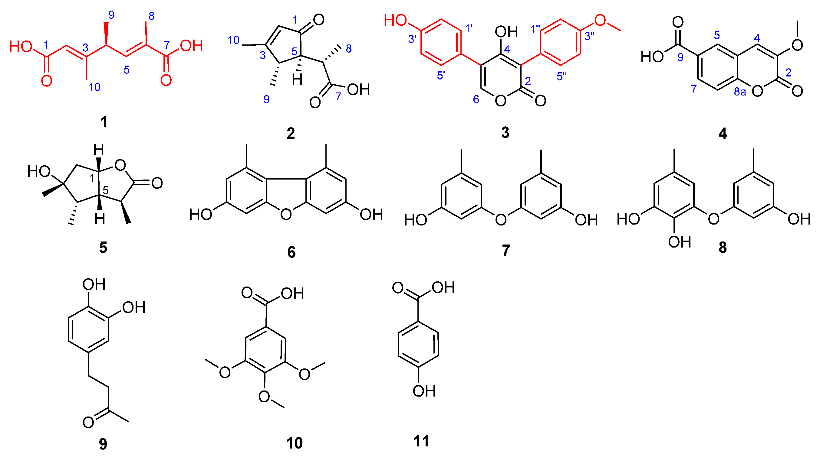
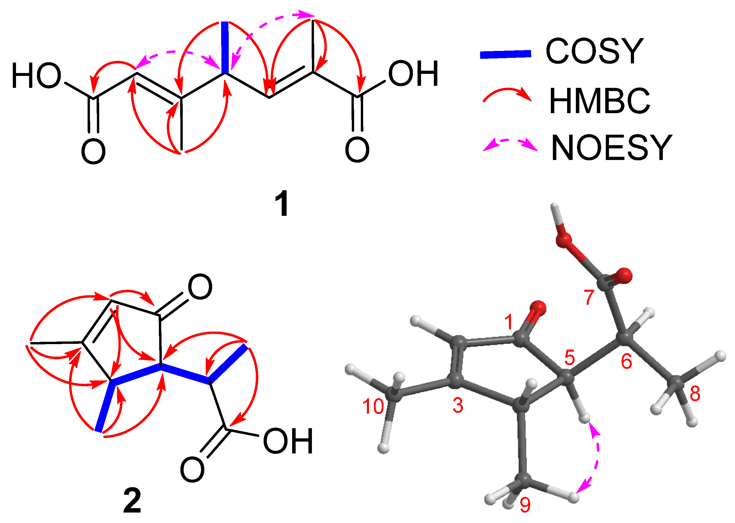
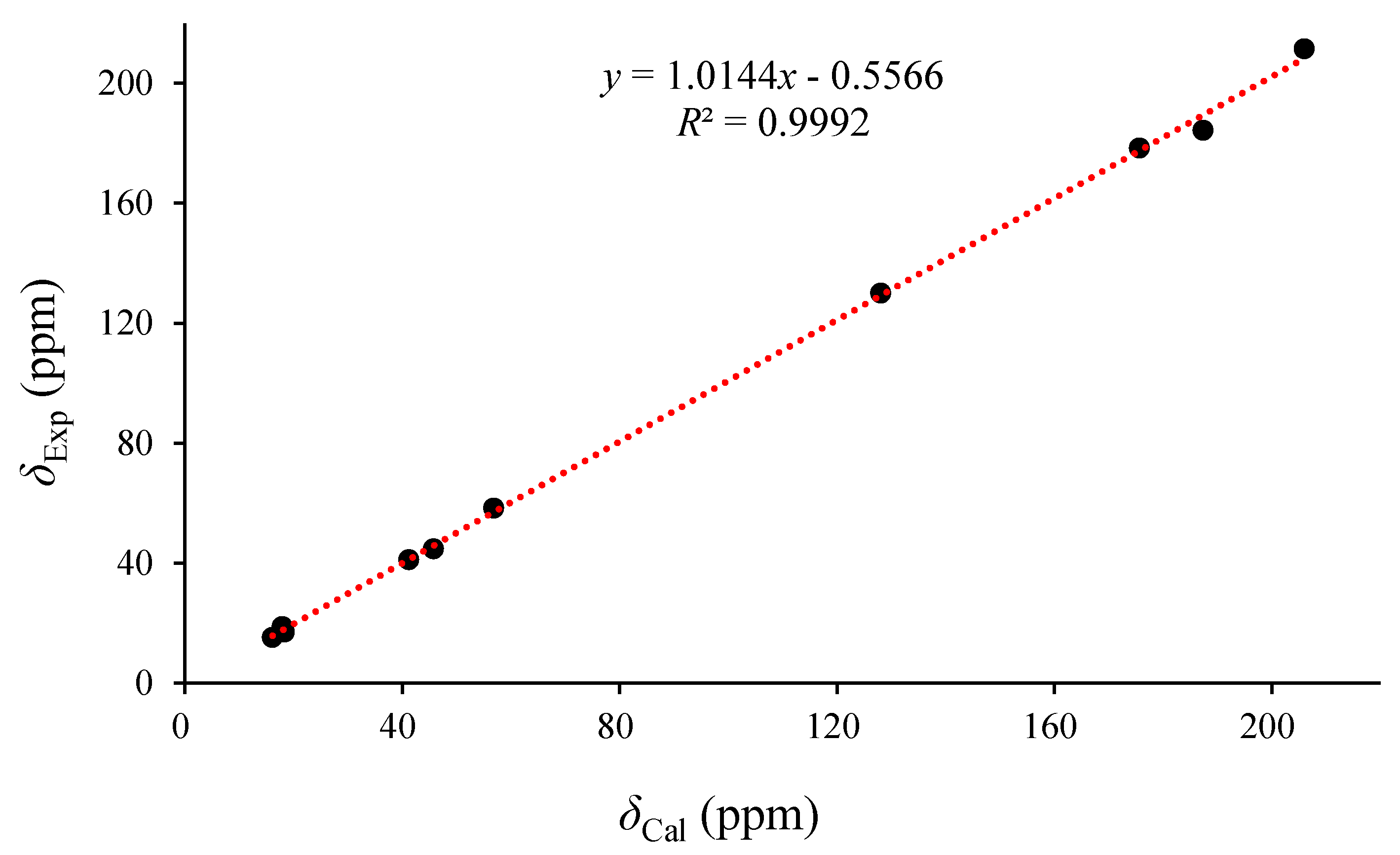
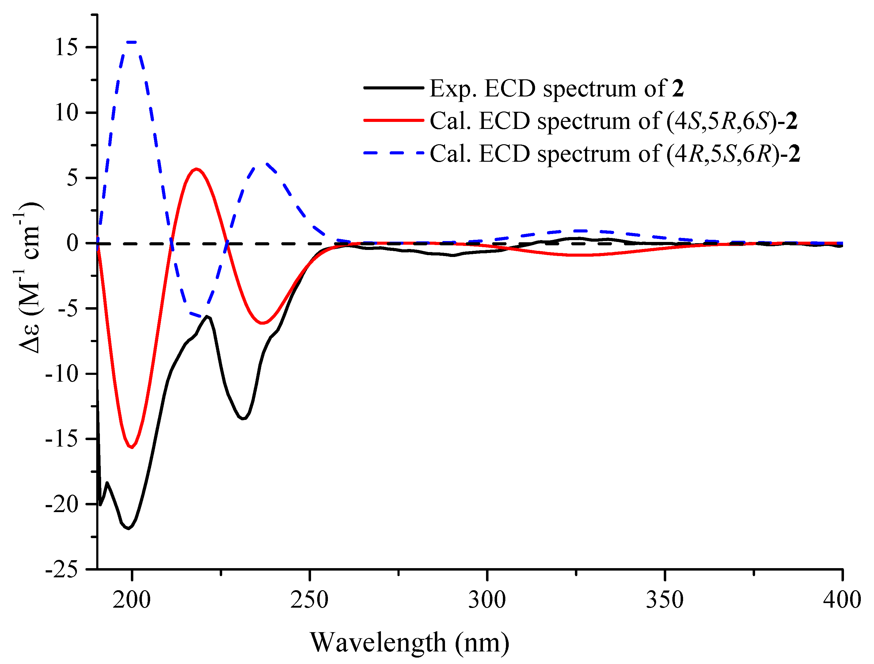
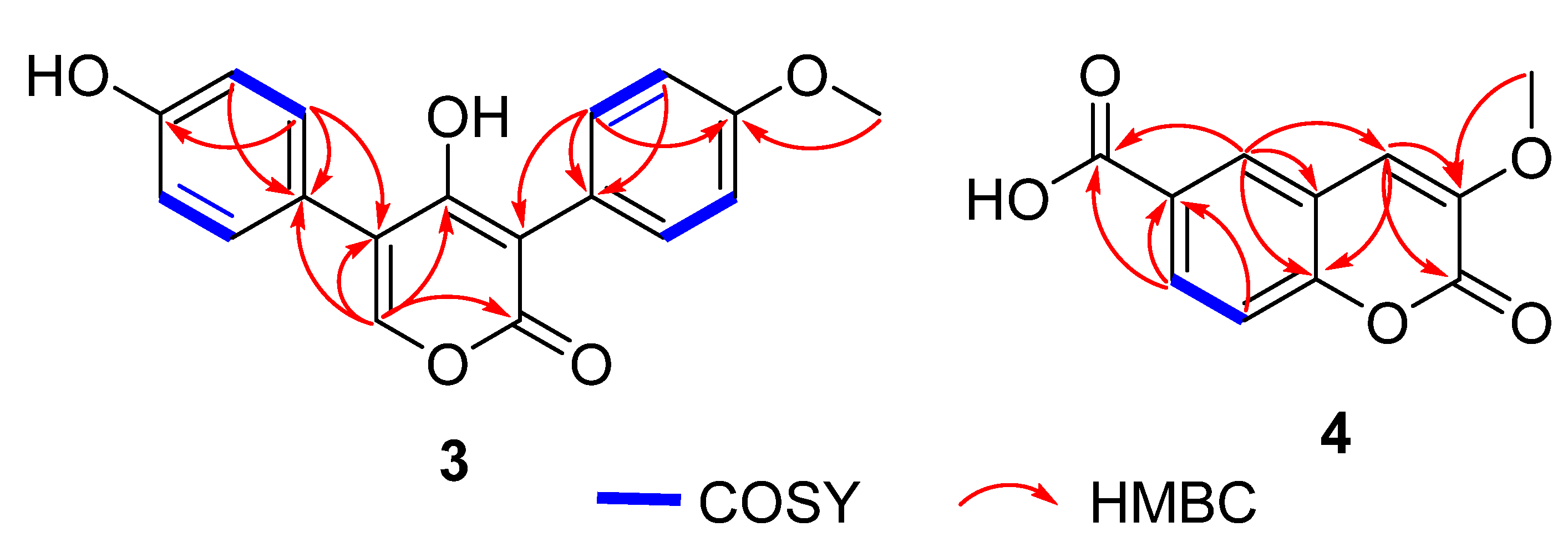
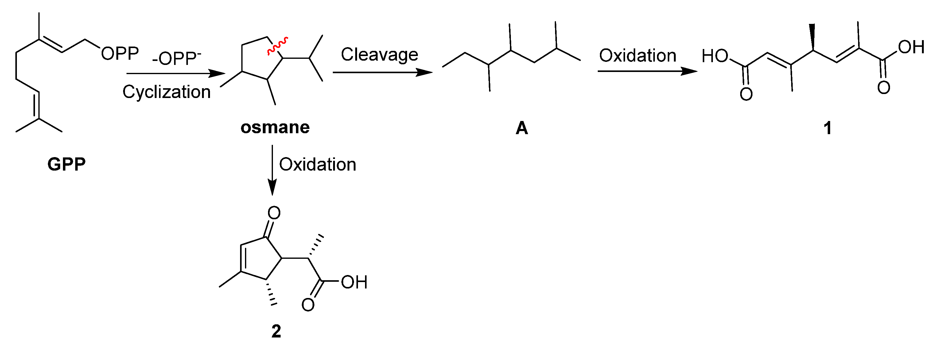
2. Results and Discussion

Aspermonoterpenoid A (1), colorless oil, has the molecular formula of C10H14O4 as determined by the 13C NMR data and the HRESIMS spectrum (m/z 221.0784, [M + Na]+), indicating four degrees of unsaturation. The 1H NMR spectrum showed two olefinic protons (δH 5.77, 6.66), a methine (δH 3.36), and three methyls (δH 1.24, 1.88, 2.14) (Table 1), while the 13C NMR and HSQC spectra exhibited four sp2 carbons (δC 117.0, 130.2, 144.5, 162.0) for two double bonds, two ester carbonyl carbons (δC 170.4, 171.5), one methine (δC 43.8), and three methyls (δC 12.8, 17.0, 19.0) (Table 1). As all degrees of unsaturation were accounted for by two carbonyl carbons and two double bonds, an acyclic structure was required in 1. The COSY data of H-4 (δH 3.36)/H3-9 (δH 1.24) and the HMBC cross-peaks from H3-8 (δH 1.88) to C-5 (δC 144.5)/C-6 (δC 130.2)/C-7 (δC 171.5), H3-9 to C-3 (δC 162.0)/C-4 (δC 43.8)/C-5, H3-10 (δH 2.14) to C-2 (δC 117.0)/C-3/C-4, and from H-2 (δH 5.77) to C-1 (δC 170.4)/C-4/C-10 (δC 17.0) established the planar structure of 1 as 2,4,5-trimethylhepta-2,5-dienedioic acid (Figure 2). The E geometries at Δ2 and Δ5 were resolved on the basis of the NOESY cross-peaks from H-2 to H-4 and from H3-8 to H-4 (Figure 2), which were further corroborated by the chemical shifts of Me-8 (δC 12.8) and Me-10 (δC 17.0). The sole chiral center of C-4 in 1 was resolved by the calculation of the specific rotation data using Gaussian 09 [38]. The specific rotation data of (4S*)-1 was calculated at the B3LYP/6-311+G(2d,p) level with the CPCM model in methanol, which has the same sign ([α ] D +138) as that of the experimental data ([α ] D 25 +54), revealing the 4S configuration. Therefore, the structure of 1 was determined to be (2E,5E,4S)-2,4,5-trimethylhepta-2,5-dienedioic acid, which was named aspermonoterpenoid A. Noteworthily, 1 possessed a novel chained monoperpenoid skeleton, biogenetically probably derived from the osmane-type monoperpenoid after the cyclopentane ring cleavage and oxidation reactions [39,40].
The molecular formula of aspermonoterpenoid B (2) was established to be C10H14O3 on the basis of the HRESIMS spectrum at the sodium adduct ion peak at m/z 205.0845 (calcd for C10H14O3Na, 205.0841), requiring four degrees of hydrogen deficiency. The 1H NMR spectrum showed one olefinic proton (δH 5.89), three methines (δH 2.21, 2.76, 2.94), and three methyls (δH 1.28, 1.29, 2.13). Analyses of the 13C NMR spectrum, with the aid of the HSQC spectrum, revealed ten carbon signals attributable to two olefinic carbons (δC 130.1, 184.4) for a double bond, two carbonyl carbons for a keto (δC 211.6) and a carboxylic (δC 178.5) functionalities, three methines (δC 41.2, 44.8, 58.4), and three methyls (δC 15.3, 17.1, 18.9), which accounted for three degrees of unsaturation. The remaining one double bond equivalent indicated that a monocyclic skeleton was required in 2. The COSY cross-peaks of H-4 (δH 2.76)/H-5 (δH 2.21)/H-6 (δH 2.94) and the HMBC correlations from H3-8 (δH 1.29) to C-5 (δC 58.4)/C-6 (δC 41.2)/C-7 (δC 178.5), H3-9 (δH 1.28) to C-3 (δC 184.4)/C-4 (δC 44.8)/C-5, H3-10 (δH 2.13) to C-2 (δC 130.1)/C-3/C-4, and from H-2 (δH 5.89) to C-1 (δC 211.6)/C-4/C-5 deduced the structure of 2 to be 3,4-dimethyl-5-isopropanic acidated cyclopentenone (Figure 2). The relative configuration of C-4 and C-5 in cyclopentenone ring moiety was determined as 4S* and 5R* on the basis of the NOESY correlation from H3-9 to H-5, in addition to the very weak interaction between H-4 and H-5 (Figure 2). Additionally, the relative configuration of C-6 was unreliable to be deduced by the NOESY data because of its location at the soft side chain. In order to assign the relative configuration, the two possible epimers (4S*,5R*,6S*)-2 (2a) and (4S*,5R*,6R*)-2 (2b) were subjected to the 13C NMR chemical shift theoretical calculation at the mPW1PW91/6-311+G(2d,p) level in methanol using Gaussian 09. As shown in Figure 3, the calculated 13C NMR data of 2a were nearly identical to those of the experimental NMR data, as corroborated by the correlation coefficient (R2) and root mean square error (RMSE) of 2a (R2 = 0.9992, RMSE = 2.4436) and 2b (R2 = 0.9988, RMSE = 2.7912), suggesting the 4S*, 5R*, and 6S* configurations. The absolute configuration of 2 was resolved on the basis of the ECD calculation, which was carried out at the B3LYP/6-311+G(2d,p) level in MeOH using the optimized geometries at the B3LYP/6-31+G(d,p) level in gas phase after conformational searches via the OPLS3 force field using Maestro 10.2. The ECD spectrum of 2a was calculated by the TDDFT method, which was in good agreement with the experimental ECD curve, indicating the 4S, 5R, and 6S configurations (Figure 4). Interestingly, 2 was the first representative of the osmane-type monoterpenoids discovered from the fungi.
Compound 3 was isolated as a colorless oil, and its molecular formula was assigned to be C18H14O5 on the basis of the HRESIMS spectrum at the sodium adduct ion peak at m/z 333.0741, implying 12 indices of hydrogen deficiency. The 1H NMR spectrum exhibited four aromatic protons with dual intensity at δH 6.85 (d, J = 8.5 Hz, 2H), 7.01 (d, J = 8.6 Hz, 2H), 7.29 (d, J = 8.5 Hz, 2H), and 7.33 (d, J = 8.6 Hz, 2H), indicating the presence of two para-substituted benzene rings. Moreover, a methoxy (δH 3.85) and one olefinic (δH 7.59) protons were also observed (Table 2). The 13C NMR spectrum showed a total of 18 carbon resonance signals, including twelve aromatic carbons (δC 115.1 × 2, 116.3 × 2, 123.5, 124.6, 131.9 × 2, 133.2 × 2, 158.9, and 160.9) for two benzene units, four olefinic carbons (δC 107.1, 120.4, 149.5, and 167.1) for two double bonds, a carbonyl carbon (δC 166.2), and one methoxy group (δC 55.7) (Table 2). The COSY data of H-1″ (δH 7.33) and H-2″ (δH 7.01) and the HMBC cross-peaks from H-1″/H-5″ to C-3″ (δC 160.9) and C-3 (δC 107.1), H-2″/H-4″ to C-3″ and C-6″ (δC 124.6), and from the methoxy protons (δH 3.85) to C-3″ deduced the p-methoxyphenyl moiety (ring C) to be tethered at C-3 (Figure 5). Additional COSY correlation between H-1′ (δH 7.29)/H-2′ (δH 6.85) and the HMBC correlations from H-1′/H-5′ to C-3′ (δC 158.9) and C-5 (δC 120.4) and from H-2′/H-4′ to C-3′ and C-6′ (δC 123.5) in association with the chemical shift of C-3′ established the p-hydroxyphenyl unit (ring A) to be resided at C-5. The remaining five olefinic carbons (δC 107.1, 120.4, 149.5, 166.2, and 167.1) constructed an α-pyrone moiety with a hydroxy at C-4 position, according to the HMBC cross-peaks from H-6 (δH 7.59) to C-2 (δC 166.2), C-4 (δC 167.2), C-5 (δC 120.4), and C-6′, as well as their chemical shifts and the molecular formula. Therefore, the structure of 3 was elucidated to be 4-hydroxy-5-(4-hydroxyphenyl)-3-(4-methoxyphenyl)-2H-pyran-2-one, which was named asperphenylpyrone. It is of note that 3 was the first example of the α-pyrone derivatives bearing two phenyl units at C-3 and C-5, respectively.
Compound 4 has the molecular formula of C11H8O5 as determined by the positive HRESIMS spectrum (m/z 243.0268, [M + Na]+) and the 13C NMR data, indicating eight degrees of unsaturation. The 1H NMR spectrum showed three aromatic protons (δH 7.45 (d, J = 8.6 Hz, H-8), 7.95 (dd, J = 8.6, 1.8 Hz, H-7), and 8.23 (d, J = 1.8 Hz, H-5)) for a 1,2,4-trisubstituted benzene ring, a singlet olefinic proton (δH 7.49), and one methoxy (δH 3.86), whereas the 13C NMR spectrum exhibited 11 carbon signals, including six aromatic carbons (δC 116.5, 120.4, 127.8, 128.9, 129.5, and 152.2) for a benzene ring, two olefinic carbons (δC 113.4, 144.8) for a double bond, two ester carbonyl carbons (156.6, 166.9), and a methoxy group (δC 56.8). The aforementioned NMR data were very similar to those of the known coumarin derivative [41], with the exception of the presence of an additional aromatic methine (δH 7.95, δC 129.5) in 4 instead of an oxygenated nonprotonated sp2 carbon. The additional olefinic proton was appointed at C-7 (δC 129.5) on the basis of the COSY cross-peaks from H-7 (δH 7.95) to H-8 (δH 7.45) and H-5 (δH 8.23) as well as the HMBC correlations from H-5 to C-4 (δC 113.4)/C-7/C-9 (δC 166.9)/C-8a (δC 152.2) and from H-7 to C-5 (δC 128.9), C-9, and C-8a (Figure 5). Thus, the structure of 4 was established as a 7-deoxylated derivative of the above known compound, which was given the trivial name of aspercoumarine acid.
In addition, seven known compounds were established to be pestalotiolactone A (5) [42,43], 3,7-dihydroxy-1,9-dimethyldibenzofuran (6) [44], diorcinol (7) [45], cordyol C (8) [46], 4-(3′,4′-dihydroxyphenyl)-2-butanone (9) [47], 3,4,5-trimethoxybenzoic acid (10) [48], and 4-hydroxybenzoic acid (11) [49] on the basis of comparison of their NMR and specific rotation data with those reported in the literature.
Compound 1 was a novel chained monoterpenoid. The plausible biosynthetic pathway for 1 and 2 was proposed (Scheme 1). Starting from the GPP, hydrosis, oxygenation, and cyclization reaction occurrence constructed the monocyclic ring monoterpenoid osmane. The osmane might undergo carbon–carbon bond cleavage to form a key intermediate A, which was further oxygenated to yield 1. Additionally, the osmane might also undergo oxygenation to form 2.
As terpenoids usually possess the anti-inflammatory activities, all isolated metabolites were evaluated for their inhibitory effects against NO secretion in LPS-activated BV-2 microglia cells. As a result, none of them showed obvious cytotoxic activities against BV-2 microglia cells at the concentration of 20 µM by the CCK-8 Kit, and all compounds exhibited dose-dependent inhibitory effects against NO production induced by the LPS at the concentrations of 20 and 10 µM, respectively (Table 3). Interestingly, compound 6 (10 µM) showed potent anti-inflammatory activities with the inhibition rate of 94.4%.

3. Materials and Methods

3.1. General Experimental Procedures

UV spectra were recorded using a UV8000 UV/Vis spectrophotometer (Shanghai Metash Instruments Inc., Shanghai, China). Optical rotation data were measured on the basis of the Rudolph Autopol IV automatic polarimeter (Rudolph Reaearch Analytical, Newburgh, NY, USA). ECD spectra were measured using a Chirascan spectrometer (Applied Photophysics inc., Leatherhead, Surrey, UK). HRESIMS data were measured using a Xevo G2 Q-TOF mass spectrometer (Waters, Milford, MA, USA). The 1H, 13C, HSQC, COSY, HMBC, and NOESY spectra were measured on the basis of the Bruker Avance 400 FT NMR spectrometer (Bruker Company, Fällanden, Switzerland) with tetramethylsilicane (TMS) as an internal standard. Semi-preparative HPLC was performed using an Alltech LS class pump with a model 201 variable wavelength UV/Vis detector, and a YMC packed ODS-A (250 × 10 mm, 5 μm) column (YMC Co., Ltd. Kyoto, Japan) was used for the purification. Column chromatography (CC) was carried out using a Sephadex LH-20 (Amersham Biosciences, San Francisco, CA, USA), ODS-A-HG (YMC Co., Ltd. Kyoto, Japan), and silica gel (Qingdao Marine Chemistry Co., Ltd., Qingdao, China). The TLC analyses were carried out with the precoated silica gel plates by heating after spraying with vanillin sulfuric acid chromogenic reagent (Xilong Scientific Co., Ltd., Shantou, China).

3.2. Fungal Material and Identifiation

The fungus was isolated from the deep-sea sediment at the depth of 2246 m sampling from the South Atlantic Ocean (W13.6639°, S14.2592°), and it was identified to be Aspergillus sydowii on the basis of the morphology and the internal transcribed spacer (ITS) region of the rDNA sequence. The ITS gene sequence was deposited in GenBank and assigned the accession no. MN918102. The fungus was preserved at the Marine Culture Collection of China (MCCC), and assigned the accession no. MCCC 3A00324. Therefore, the producing fungus was named Aspergillus sydowii MCCC 3A00324.

3.3. Fermentation, Extraction, and Isolation

The fungus was cultivated in a potato dextrose agar (PDA) plate under 25 °C for four days, and then the fresh mycelia and spores were inoculated into 500 mL Erlenmeyer flasks (×2), each containing 100 mL potato dextrose broth (PDB) medium and followed by cultivation in a rotary shaker under 25 °C at 200 rpm for four days. The seed cultures were subsequently inoculated to 30 Erlenmeyer flasks (1 L) (each containing 80 g rice and 120 mL sea water) after autoclaving at 121 °C for 22 min. The fermentation was performed under static conditions at 25 °C for 26 days.
The fermented material was fragmented using a stick and extracted successively with EtOAc three times, and then evaporated under reduced pressure to get an EtOAc extract (16.2 g). The extract was subjected to the vacuum liquid chromatography column on silica gel eluting with a gradient of CH2Cl2 and MeOH (1:0 to 0:1) to furnish fractions A and B. The fraction B (8.5 g) was subsequently separated by CC on ODS with MeOH/H2O elution (30–100%) to obtain fourteen fractions (Fr.1–Fr.14). Fraction Fr.3 (147 mg) was separated by CC on silica gel eluting with petroleum ether (PE)/EtOAc (10:1) to obtain two subfractions. The former was further purified by semi-preparative HPLC with the mobile phase of MeOH/H2O (3:7) to yield 2 (1.8 mg) and 5 (21.0 mg), whereas the latter was separated by semi-preparative HPLC (CH3CN/H2O, 1:9) to get 11 (0.5 mg). Fraction Fr.5 (333 mg) was chromatographed by CC on silica gel eluting with CH2Cl2/MeOH (30:1) to furnish two subfractions, whereas the former was further separately purified by semi-preparative HPLC using CH3CN/H2O (19:81) elution to obtain 10 (4.0 mg), and the latter using MeOH/H2O (7:18) elution to yield 4 (3.0 mg). Fraction Fr.7 (489 mg) was subjected to silica gel CC with CH2Cl2 and MeOH gradient elution (1:0→0:1) to yield two subfractions (Fr.7-1 and Fr.7-2). Subfraction Fr.7-1 was separated by semi-preparative HPLC purification (CH3CN/H2O, 23:77) to get 1 (1.4 mg) and 3 (3.8 mg). Subfraction Fr.7-2 was purified by semi-preparative HPLC (CH3CN/H2O, 37:63) to yield 9 (1.2 mg). Fraction Fr.11 (146 mg) was separated by silica gel CC eluting with CH2Cl2/acetone gradient from 1:0 to 0:1 to furnish two subfractions, whereas the former was further separated using CC on a Sephadex LH-20 (MeOH) and semi-preparative HPLC (CH3CN/H2O, 3:7) to obtain 7 (18.5 mg) and 8 (3.0 mg), respectively. Fraction Fr.13 (545 mg) was subjected to CC on silica gel eluting with PE/EtOAc (1:0→0:1) to get four subfractions (Fr.13-1–Fr.13-4). Subfraction Fr.13-1 was subjected to semi-preparative HPLC with the mobile phase of CH3CN/H2O (39:61) to yield 6 (1.0 mg).
Aspermonoterpenoid A (1): colorless oil; [ α ] D 25 +54 (c 0.06, MeOH); UV (MeOH) λmax (log ε) 223 (1.59) nm; 1H and 13C NMR data, see Table 1; HRESIMS m/z 221.0784 [M + Na]+ (calcd. for C10H14O4Na, 221.0790).
Aspermonoterpenoid B (2): colorless oil; [α ] D 25 −38 (c 0.07, MeOH); UV (MeOH) λmax (log ε) 229 (0.96) nm; ECD (MeOH) λmaxε) 199 (−21.88), 222 (−5.77), 231 (−13.46), 260 (−0.15) nm; 1H and 13C NMR data, see Table 1; HRESIMS m/z 205.0845 [M + Na]+ (calcd. for C10H14O3Na, 205.0841).
Asperphenylpyrone (3): colorless oil; UV (MeOH) λmax (log ε) 204 (1.67), 248 (0.85), 315 (0.28) nm; 1H and 13C NMR data, see Table 2; HRESIMS m/z 333.0741 [M + Na]+ (calcd. for C18H14O5Na, 333.0739).
Aspercoumarine acid (4): white powder; UV (MeOH) λmax (log ε) 204 (0.38), 296 (0.15) nm; 1H and 13C NMR data, see Table 2; HRESIMS m/z 243.0268 [M + Na]+ (calcd. for C11H8O5Na, 243.0269).

3.4. BV-2 Cell Culture and Treatment

The BV-2 microglia cells were cultured in DMEM medium containing 10% fetal bovine serum and antibiotics (100 units/mL of penicillin and 100 g/mL of streptomycin) and maintained in a humidified 5% CO2 incubator (Beijing Luxi Technology Co., Ltd., Beijing, China) at 37 °C. For the experiment, cells were seeded into 24-well plates (2 × 10⁴ cells/well) overnight. Next day, cells were incubated with fresh culture medium containing indicated concentration of the tested compounds for half an hour and following LPS treatment (1 μg/mL). Cells were treated vehicle (DMSO, 0.1%) as control.

3.5. Nitrite Quantification

The concentration of nitrite in culture medium was determined using a Griess Reagent Kit (Thermo Fisher, Shanghai, China). Briefly, 75 μL of cell culture supernatants were reacted with an equal volume of Griess Reagent Kit for 30 min at room temperature, and absorbance of diazonium was obtained at a wavelength of 560 nm. Nitrite production by vehicle stimulation was designated as 100% inhibition compared to LPS stimulation for the experiment.

3.6. Computational Details

3.6.1. 13C NMR Calculation of 2

Conformational searches were carried out using the Maestro 10.2 program (Schrödinger Inc., NY, USA) at the OPLS3 molecular mechanics force field within an energy window of 3.0 kcal/mol. The results exhibited 8 conformers for (4S*,5R*,6S*)-2 and (4S*,5R*,6R*)-2, respectively. The conformers were further optimized at the B3LYP/6-31+G(d,p) level in gas phase using Gaussian 09 (Gaussian, Inc., Wallingford, CT, USA) [38]. The conformers with a Boltzmann population over 1% were selected for the NMR calculations. The NMR data were calculated by the GIAO method at the mPW1PW91/6-311+G(2d,p) level with the IEFPCM model in methanol. Finally, the calculated NMR data were averaged according to the Boltzmann distribution for each conformer and then fitted to the experimental values by linear regression.

3.6.2. ECD Calculation of 2

The eight conformers of (4S*,5R*,6S*)-2 were optimized by density functional theory (DFT) calculations at the B3LYP/6-31+G(d,p) level in gas phase using Gaussian 09. The energies, oscillator strengths, and rotational strengths of the first 60 electronic excitations were calculated by the TDDFT method at the B3LYP/6-311+G(2d,p) level in methanol. The ECD spectrum was simulated using SpecDis (version1.70, Berlin, Germany) by applying the Gaussian band shapes with sigma = 0.3 eV. Finally, the calculated ECD data were weighted and then summed up each stable conformer on the basis of the Boltzmann population.

4. Conclusions

In summary, four new compounds, including one novel (1) and one new (2) monoterpenoids and two new polyketides (3 and 4), were obtained from the EtOAc extract of the deep-sea-derived fungus Aspergillus sydowii MCCC 3A00324, together with seven known compounds (511). The structures of metabolites were determined by comprehensive analyses of the NMR and HRESIMS spectra, in association with quantum chemical calculations of the ECD, 13C NMR, and specific rotation data for their configurational assignment. Compound 1 possessed a novel monoterpenoid skeleton, biogenetically probably derived from the osmane-type monoperpenoid after the cyclopentane ring cleavage and oxidation reactions. Additionally, 2 was the first osmane-type monoterpenoid representative discovered from the fungi, while 3 was the first example of the α-pyrone derivatives bearing two phenyl units at C-3 and C-5, respectively, indicating that the deep-sea-derived fungi are a unique source of the structurally novel compounds. Compound 6 exhibited significant inhibitory effects against NO secretion in LPS-activated BV-2 microglia cells (94.4% inhibition rate, 10 µM), suggesting the potential application for the anti-inflammatory agents.

Supplementary Materials

The following are available online at https://www.mdpi.com/1660-3397/18/11/561/s1, Figure S1: The HPLC and TLC metabolic profile of the study fungus, Figures S1-1–S4-7: HRESIMS, 1H, 13C, HSQC, COSY, and HMBC spectra of new compounds 14, and NOESY spectra of 1 and 2.

Author Contributions

S.N., Z.S., and S.P. isolated and identified the fungus. S.N. isolated and elucidated the structures. L.Y. and T.C. performed the anti-inflammatory assay. B.H. and G.Z. designed and coordinated the study. S.N., B.H., and G.Z. wrote and revised the manuscript. All authors have read and agreed to the published version of the manuscript.

Funding

This work was financially supported by the Scientific Research Foundation of the Third Institute of Oceanography (2018018), the National Natural Science Foundation of China (81901133), and the COMRA program (DY135-B2-08).

Conflicts of Interest

The authors declare no conflict of interest.

References

  1. Zain Ul Arifeen, M.; Ma, Y.N.; Xue, Y.R.; Liu, C.H. Deep-sea fungi could be the new arsenal for bioactive molecules. Mar. Drugs 2019, 18, 9. [Google Scholar] [CrossRef] [Green Version]
  2. Daletos, G.; Ebrahim, W.; Ancheeva, E.; El-Neketi, M.; Song, W.; Lin, W.; Proksch, P. Natural products from deep-sea-derived fungi−a new source of novel bioactive compounds? Curr. Med. Chem. 2018, 25, 186–207. [Google Scholar] [CrossRef]
  3. Carroll, A.R.; Copp, B.R.; Davis, R.A.; Keyzers, R.A.; Prinsep, M.R. Marine natural products. Nat. Prod. Rep. 2019, 36, 122–173. [Google Scholar] [CrossRef] [Green Version]
  4. Xu, J.; Liu, Z.; Chen, Y.; Tan, H.; Li, H.; Li, S.; Guo, H.; Huang, Z.; Gao, X.; Liu, H.; et al. Lithocarols A–F, six tenellone derivatives from the deep-sea derived fungus Phomopsis lithocarpus FS508. Bioorg. Chem. 2019, 87, 728–735. [Google Scholar] [CrossRef]
  5. Zhang, Z.; He, X.; Wu, G.; Liu, C.; Lu, C.; Gu, Q.; Che, Q.; Zhu, T.; Zhang, G.; Li, D. Aniline-tetramic acids from the deep-sea-derived fungus Cladosporium sphaerospermum L3P3 cultured with the HDAC inhibitor SAHA. J. Nat. Prod. 2018, 81, 1651–1657. [Google Scholar] [CrossRef]
  6. Liang, X.; Nong, X.; Huang, Z.; Qi, S. Antifungal and antiviral cyclic peptides from the deep-sea-derived fungus Simplicillium obclavatum EIODSF 020. J. Agric. Food Chem. 2017, 65, 5114–5121. [Google Scholar] [CrossRef]
  7. Huang, Z.; Nong, X.; Ren, Z.; Wang, J.; Zhang, X.; Qi, S. Anti-HSV-1, antioxidant and antifouling phenolic compounds from the deep-sea-derived fungus Aspergillus versicolor SCSIO 41502. Bioorg. Med. Chem. Lett. 2017, 27, 787–791. [Google Scholar] [CrossRef] [PubMed]
  8. Yu, G.; Sun, Z.; Peng, J.; Zhu, M.; Che, Q.; Zhang, G.; Zhu, T.; Gu, Q.; Li, D. Secondary metabolites produced by combined culture of Penicillium crustosum and a Xylaria sp. J. Nat. Prod. 2019, 82, 2013–2017. [Google Scholar] [CrossRef] [PubMed]
  9. Pang, X.; Lin, X.; Zhou, X.; Yang, B.; Tian, X.; Wang, J.; Xu, S.; Liu, Y. New quinoline alkaloid and bisabolane-type sesquiterpenoid derivatives from the deep-sea-derived fungus Aspergillus sp. SCSIO06786. Fitoterapia 2020, 140, 104406. [Google Scholar] [CrossRef] [PubMed]
  10. Wu, Y.; Zhang, Z.; Zhong, Y.; Huang, J.; Li, X.; Jiang, J.; Deng, Y.; Zhang, L.; He, F. Sumalactones A−D, four new curvularin-type macrolides from a marine deep sea fungus Penicillium sumatrense. RSC Adv. 2017, 7, 40015–40019. [Google Scholar] [CrossRef] [Green Version]
  11. Niu, S.; Xie, C.; Zhong, T.; Xu, W.; Luo, Z.; Shao, Z.; Yang, X. Sesquiterpenes from a deep-sea-derived fungus Graphostroma sp. MCCC 3A00421. Tetrahedron 2017, 73, 7267–7273. [Google Scholar] [CrossRef]
  12. Niu, S.; Xie, C.; Xia, J.; Liu, Q.; Peng, G.; Liu, G.; Yang, X. Botryotins A–H, tetracyclic diterpenoids representing three carbon skeletons from a deep-sea-derived Botryotinia fuckeliana. Org. Lett. 2020, 22, 580–583. [Google Scholar] [CrossRef] [PubMed]
  13. Niu, S.; Xia, M.; Chen, M.; Liu, X.; Li, Z.; Xie, Y.; Shao, Z.; Zhang, G. Cytotoxic polyketides isolated from the deep-sea-derived fungus Penicillium chrysogenum MCCC 3A00292. Mar. Drugs 2019, 17, 686. [Google Scholar] [CrossRef] [PubMed] [Green Version]
  14. Niu, S.; Liu, Q.; Xia, J.; Xie, C.; Luo, Z.; Shao, Z.; Liu, G.; Yang, X. Polyketides from the deep-sea-derived fungus Graphostroma sp. MCCC 3A00421 showed potent antifood allergic activities. J. Agric. Food Chem. 2018, 66, 1369–1376. [Google Scholar] [CrossRef]
  15. Niu, S.; Liu, D.; Shao, Z.; Proksch, P.; Lin, W. Eremophilane-type sesquiterpenoids in a deep-sea fungus Eutypella sp. activated by chemical epigenetic manipulation. Tetrahedron 2018, 74, 7310–7325. [Google Scholar] [CrossRef]
  16. Niu, S.; Liu, D.; Hu, X.; Proksch, P.; Shao, Z.; Lin, W. Spiromastixones A−O, antibacterial chlorodepsidones from a deep-sea-derived Spiromastix sp. fungus. J. Nat. Prod. 2014, 77, 1021–1030. [Google Scholar] [CrossRef]
  17. Liu, Y.; Zhang, J.; Li, C.; Mu, X.; Liu, X.; Wang, L.; Zhao, Y.; Zhang, P.; Li, X.; Zhang, X. Antimicrobial secondary metabolites from the seawater-derived fungus Aspergillus sydowii SW9. Molecules 2019, 24, 4596. [Google Scholar] [CrossRef] [Green Version]
  18. Liu, N.; Peng, S.; Yang, J.; Cong, Z.; Lin, X.; Liao, S.; Yang, B.; Zhou, X.; Zhou, X.; Liu, Y.; et al. Structurally diverse sesquiterpenoids and polyketides from a sponge-associated fungus Aspergillus sydowii SCSIO41301. Fitoterapia 2019, 135, 27–32. [Google Scholar] [CrossRef]
  19. Xu, X.; Zhao, S.; Yin, L.; Yu, Y.; Chen, Z.; Shen, H.; Zhou, L. A new sydonic acid derivative from a marine derived-fungus Aspergillus sydowii. Chem. Nat. Compd. 2017, 53, 1056–1058. [Google Scholar] [CrossRef]
  20. Liu, S.; Wang, H.; Su, M.; Hwang, G.J.; Hong, J.; Jung, J.H. New metabolites from the sponge-derived fungus Aspergillus sydowii J05B-7F-4. Nat. Prod. Res. 2017, 31, 1682–1686. [Google Scholar] [CrossRef]
  21. Wang, J.; Lin, X.; Qin, C.; Liao, S.; Wan, J.; Zhang, T.; Liu, J.; Fredimoses, M.; Chen, H.; Yang, B.; et al. Antimicrobial and antiviral sesquiterpenoids from sponge-associated fungus, Aspergillus sydowii ZSDS1-F6. J. Antibiot. 2014, 67, 581–583. [Google Scholar] [CrossRef] [PubMed]
  22. Chung, Y.; Wei, C.; Chuang, D.; El-Shazly, M.; Hsieh, C.; Asai, T.; Oshima, Y.; Hsieh, T.; Hwang, T.; Wu, Y.; et al. An epigenetic modifier enhances the production of anti-diabetic and anti-inflammatory sesquiterpenoids from Aspergillus sydowii. Bioorg. Med. Chem. 2013, 21, 3866–3872. [Google Scholar] [CrossRef] [PubMed]
  23. Trisuwan, K.; Rukachaisirikul, V.; Kaewpet, M.; Phongpaichit, S.; Hutadilok-Towatana, N.; Preedanon, S.; Sakayaroj, J. Sesquiterpene and xanthone derivatives from the sea fan-derived fungus Aspergillus sydowii PSU-F154. J. Nat. Prod. 2011, 74, 1663–1667. [Google Scholar] [CrossRef] [PubMed]
  24. Hamasaki, T.; Nagayama, K.; Hatsuda, Y. Two new metabolites, sydonic acid and hydroxysydonic acid, from Aspergillus sydowi. Agric. Biol. Chem. 1978, 42, 37–40. [Google Scholar] [CrossRef]
  25. Amin, M.; Liang, X.; Ma, X.; Dong, J.; Qi, S.; Amin, M.; Liang, X. New pyrone and cyclopentenone derivatives from marine-derived fungus Aspergillus sydowii SCSIO 00305. Nat. Prod. Res. 2019, 1–9. [Google Scholar] [CrossRef]
  26. Wang, Y.; Dong, Y.; Wu, Y.; Liu, B.; Bai, J.; Yan, D.; Zhang, L.; Hu, Y.; Mou, Y.; Dong, Y.; et al. Diphenyl ethers from a marine-derived Aspergillus sydowii. Mar. Drugs 2018, 16, 451. [Google Scholar] [CrossRef] [Green Version]
  27. Liu, X.; Song, F.; Ma, L.; Chen, C.; Xiao, X.; Ren, B.; Liu, X.; Dai, H.; Piggott, A.M.; Av-Gay, Y.; et al. Sydowiols A–C: Mycobacterium tuberculosis protein tyrosine phosphatase inhibitors from an East China Sea marine-derived fungus, Aspergillus sydowii. Tetrahedron Lett. 2013, 54, 6081–6083. [Google Scholar] [CrossRef]
  28. Taniguchi, M.; Kaneda, N.; Shibata, K.; Kamikawa, T. Isolation and biological activity of aspermutarubrol, a self-growth inhibitor from Aspergillus sydowii. Agric. Biol. Chem. 1978, 42, 1629–1630. [Google Scholar]
  29. Kaur, A.; Raja, H.A.; Darveaux, B.A.; Chen, W.-L.; Swanson, S.M.; Pearce, C.J.; Oberlies, N.H. New diketopiperazine dimer from a filamentous fungal isolate of Aspergillus sydowii. Magn. Reson. Chem. 2015, 53, 616–619. [Google Scholar] [CrossRef] [Green Version]
  30. He, F.; Sun, Y.; Liu, K.-S.; Zhang, X.; Qian, P.; Wang, Y.; Qi, S. Indole alkaloids from marine-derived fungus Aspergillus sydowii SCSIO 00305. J. Antibiot. 2012, 65, 109–111. [Google Scholar] [CrossRef] [Green Version]
  31. Zhang, M.; Wang, W.; Fang, Y.; Zhu, T.; Gu, Q.; Zhu, W. Cytotoxic alkaloids and antibiotic nordammarane triterpenoids from the marine-derived fungus Aspergillus sydowi. J. Nat. Prod. 2008, 71, 985–989. [Google Scholar] [CrossRef] [PubMed]
  32. Wiese, J.; Schmaljohann, R.; Imhoff, J.F.; Aldemir, H.; Gulder, T.A.M. Asperentin B, a new inhibitor of the protein tyrosine phosphatase 1B. Mar. Drugs 2017, 15, 191. [Google Scholar] [CrossRef] [PubMed]
  33. Li, D.; Cai, S.; Tian, L.; Lin, Z.; Zhu, T.; Fang, Y.; Liu, P.; Gu, Q.; Zhu, W. Two new metabolites with cytotoxicities from deep-sea fungus, Aspergillus sydowi YH11-2. Arch. Pharmacal Res. 2007, 30, 1051–1054. [Google Scholar]
  34. Niu, S.; Yang, L.; Zhang, G.; Chen, T.; Hong, B.; Pei, S.; Shao, Z. Phenolic bisabolane and cuparene sesquiterpenoids with anti-inflammatory activities from the deep-sea-derived Aspergillus sydowii MCCC 3A00324 fungus. Bioorg. Chem. 2020, 105, 104420. [Google Scholar] [CrossRef] [PubMed]
  35. Liu, M.; Sun, W.; Shen, L.; Hao, X.; Al Anbari, W.H.; Lin, S.; Li, H.; Gao, W.; Wang, J.; Hu, Z.; et al. Bipolaricins A–I, ophiobolin-type tetracyclic sesterterpenes from a phytopathogenic Bipolaris sp. Fungus. J. Nat. Prod. 2019, 82, 2897–2906. [Google Scholar] [CrossRef]
  36. Li, G.; Li, H.; Tang, W.; Guo, Y.; Li, X. Klyflaccilides A and B, diterpenoids with 6/5/8/3 fused tetracyclic carbon skeleton from the Hainan soft coral Klyxum flaccidum. Org. Lett. 2019, 21, 5660–5664. [Google Scholar] [CrossRef]
  37. Liu, M.; Wang, W.; Sun, H.; Pu, J. Diterpenoids from Isodon species: An update. Nat. Prod. Rep. 2017, 34, 1090–1140. [Google Scholar] [CrossRef]
  38. Frisch, M.J.; Trucks, G.W.; Schlegel, H.B.; Scuseria, G.E.; Robb, M.A.; Cheeseman, J.R.; Scalmani, G.; Barone, V.; Mennucci, B.; Petersson, G.A.; et al. Gaussian 09; Revision. D.01; Gaussian, Inc.: Wallingford, CT, USA, 2009. [Google Scholar]
  39. Leete, E.; Wemple, J.N. Biosynthesis of the cinchona alkaloids. The incorporation of geraniol-3-14C into quinine. J. Am. Chem. Soc. 1966, 88, 4743–4744. [Google Scholar] [CrossRef]
  40. Zhou, J.; Wu, Z.; Oyawaluja, B.O.; Coker, H.A.B.; Odukoya, O.A.; Yao, G.; Che, C.-T. Protein tyrosine phosphatase 1B inhibitory iridoids from Psydrax subcordata. J. Nat. Prod. 2019, 82, 2916–2924. [Google Scholar] [CrossRef]
  41. Yan, Z.; Wen, S.; Ding, M.; Guo, H.; Huang, C.; Zhu, X.; Huang, J.; She, Z.; Long, Y. The purification, characterization, and biological activity of new polyketides from mangrove-derived endophytic fungus Epicoccum nigrum SCNU-F0002. Mar. Drugs 2019, 17, 414. [Google Scholar] [CrossRef] [Green Version]
  42. Liu, S.; Dai, H.; Heering, C.; Janiak, C.; Lin, W.; Liu, Z.; Proksch, P. Inducing new secondary metabolites through co-cultivation of the fungus Pestalotiopsis sp. with the bacterium Bacillus subtilis. Tetrahedron Lett. 2017, 58, 257–261. [Google Scholar] [CrossRef]
  43. Li, X.; Li, X.; Yin, X.; Li, X.; Wang, B. Antimicrobial sesquiterpenoid derivatives and monoterpenoids from the deep-sea sediment-derived fungus Aspergillus versicolor SD-330. Mar. Drugs 2019, 17, 563. [Google Scholar] [CrossRef] [PubMed] [Green Version]
  44. Tanahashi, T.; Takenaka, Y.; Nagakura, N.; Hamada, N. Dibenzofurans from the cultured lichen mycobionts of Lecanora cinereocarnea. Phytochemistry 2001, 58, 1129–1134. [Google Scholar] [CrossRef]
  45. Fremlin, L.J.; Piggott, A.M.; Lacey, E.; Capon, R.J. Cottoquinazoline A and cotteslosins A and B, metabolites from an Australian marine-derived strain of Aspergillus versicolor. J. Nat. Prod. 2009, 72, 666–670. [Google Scholar] [CrossRef] [PubMed]
  46. Bunyapaiboonsri, T.; Yoiprommarat, S.; Intereya, K.; Kocharin, K. New diphenyl ethers from the insect pathogenic fungus Cordyceps sp. BCC 1861. Chem. Pharm. Bull. 2007, 55, 304–307. [Google Scholar] [CrossRef] [Green Version]
  47. Ayer, W.A.; Singer, P.P. Metabolites of bird’s nest fungi. Part 14. Phenolic metabolites of the bird’s nest fungus Nidula niveo-tomentosa. Phytochemistry 1980, 19, 2717–2721. [Google Scholar] [CrossRef]
  48. Chang, Y.C.; Chang, F.R.; Wu, Y.C. The constituents of Lindera glauca. J. Chin. Chem. Soc. (Taipei) 2000, 47, 373–380. [Google Scholar] [CrossRef]
  49. Sivakumar, S.; Reddy, M.L.P.; Cowley, A.H.; Vasudevan, K.V. Synthesis and crystal structures of lanthanide 4-benzyloxy benzoates: Influence of electron-withdrawing and electron-donating groups on luminescent properties. Dalton Trans. 2010, 39, 776–786. [Google Scholar] [CrossRef] [Green Version]
Figure 1. Chemical structures of 111 from Aspergillus sydowii MCCC 3A00324.
Marinedrugs 18 00561 g001
Figure 2. Key COSY, HMBC, and NOESY correlations of 1 and 2.
Marinedrugs 18 00561 g002
Figure 3. Linear regression analysis of the experimental 13C NMR data of 2 and calculated 13C NMR chemical shift data of 2a.
Marinedrugs 18 00561 g003
Figure 4. Experimental (Exp.) and calculated (Cal.) ECD spectra of 2 in methanol at the B3LYP/6-311+G(2d,p) level.
Marinedrugs 18 00561 g004
Figure 5. Selected COSY and HMBC correlations of compounds 3 and 4.
Marinedrugs 18 00561 g005
Scheme 1. Hypothetical biogenetic pathways for compounds 1 and 2.
Marinedrugs 18 00561 sch001
Table 1. 1H (400 MHz) and 13C (100 MHz) NMR spectroscopic data of 1 and 2 in CD3OD.
No.12
δCδHδCδH
1170.4, C 211.6, C
2117.0, CH5.77, s130.1, CH5.89, brs
3162.0, C 184.4, C
443.8, CH3.36, m44.8, CH2.76, m
5144.5, CH6.66, d (9.0)58.4, CH2.21, dd (4.9, 2.5)
6130.2, C 41.2, CH2.94, m
7171.5, C 178.5, C
812.8, CH31.88, d (0.6)15.3, CH31.29, d (7.2)
919.0, CH31.24, d (6.8)18.9, CH31.28, d (7.1)
1017.0, CH32.14, s17.1, CH32.13, s
Table 2. 1H (400 MHz) and 13C (100 MHz) NMR spectroscopic data of 3 and 4.
No.3 aNo.4 b
δCδHδCδH
2166.2 c, C 2156.6, C
3107.1, CH 3144.8, C
4167.1 c, C 4113.4, CH7.49, s
5120.4, C 4a120.4, C
6149.5, CH7.59, s5128.9, CH8.23, d (1.8)
1′131.9, CH7.29, d (8.5)6127.8, C
2′116.3, CH6.85, d (8.5)7129.5, CH7.95, dd (8.6, 1.8)
3′158.9, C 8116.5, CH7.45, d (8.6)
4′116.3, CH6.85, d (8.5)8a152.2, C
5′131.9, CH7.29, d (8.5)9166.9, C
6′123.5, C OMe56.8, CH33.86, s
1″133.2, CH7.33, d (8.6)
2″115.1, CH7.01, d (8.6)
3″160.9, C
4″115.1, CH7.01, d (8.6)
5″133.2, CH7.33, d (8.6)
6″124.6, C
OMe55.7, CH33.85, s
a Measured in CD3OD. b Measured in DMSO-d6. c Assignments in column could be interchanged.
Table 3. Inhibitory effects of 111 against NO production in LPS-activated BV-2 microglia cells and their cytotoxicities against BV-2 microglia cells.
CompoundsAnti-NO (%)Cell Viability Inhibition (%)
20 µM10 µM20 µM10 µM
133.5 ± 1.510.2 ± 2.00.7 ± 0.10.3 ± 1.2
234.0 ± 1.422.7 ± 1.44.6 ± 2.6−1.1 ± 0.4
322.7 ± 1.513.2 ± 1.34.1 ± 7.63.3 ± 3.1
428.3 ± 0.718.0 ± 2.03.5 ± 3.72.01 ± 1.4
539.1 ± 1.625.1 ± 0.810.0 ± 0.23.2 ± 1.7
6101.4 ± 2.494.4 ± 0.0−1.6 ± 5.1−0.8 ± 3.6
755.0 ± 1.435.4 ± 2.44.1 ± 3.8−1.5 ± 3.5
830.7 ± 0.818.0 ± 0.81.8 ± 4.7−0.1 ± 8.2
939.3 ± 0.730.4 ± 1.92.3 ± 0.1−1.1 ± 3.7
1044.8 ± 0.733.0 ± 0.70.4 ± 1.30.2 ± 1.8
1142.7 ± 1.330.8 ± 2.61.9 ± 2.4−0.5 ± 3.1
Publisher’s Note: MDPI stays neutral with regard to jurisdictional claims in published maps and institutional affiliations.

