Article

‘Messy’ Processing of χ-conotoxin MrIA Generates Homologues with Reduced hNET Potency

Institute for Molecular Bioscience, The University of Queensland, St. Lucia, Queensland 4072, Australia
*
Author to whom correspondence should be addressed.
Mar. Drugs 2019, 17(3), 165; https://doi.org/10.3390/md17030165
Submission received: 1 February 2019 / Revised: 26 February 2019 / Accepted: 12 March 2019 / Published: 14 March 2019

Abstract

:
Integrated venomics techniques have shown that variable processing of conotoxins from Conus marmoreus resulted in a dramatic expansion in the number of expressed conotoxins. One conotoxin from C. marmoreus, the χ-conotoxin MrIA, is a selective inhibitor of human norepinephrine transporters (hNET) and therefore a drug candidate for attenuating chronic neuropathic pain. It has been found that “messy” processing of the MrIA transcripts results in the expression of MrIA analogs with different truncations of the pro-peptide that contains portions of the MrIA molecule. The aim of this study was to investigate if variable processing of the expressed peptides results in modulation of the existing hNET pharmacology or creates new pharmacologies. To this end, a number of MrIA analogs found in C. marmoreus venom were synthesized and evaluated for their activity at hNET receptors. While several of the analogs exhibited norepinephrine transporter inhibitory activity comparable to that of MrIA, none significantly improved on the potency of conotoxin MrIA, and those analogs with disrupted pharmacophores produced greatly reduced NET inhibition, confirming previous structure-activity relationships seen on χ-class conopeptides. Additionally, analogs were screened for new activities on ion channels using calcium influx assays, although no major new pharmacology was revealed.

1. Introduction

Cone snails represent a large taxon of carnivorous gastropods that use specialized venom to hunt fish, worms, or fellow molluscs. The venom of these snails is prolific with small, disulfide-rich peptides that bind to various physiological targets. Because the disulfide bonding leads to highly stable and constrained 3-dimensional conformations [1], conotoxins, as they are known, have repeatedly shown high specificity for their targets, making them an excellent resource for the discovery of novel, medically significant compounds [1,2,3].
Due to recent transcriptomic sequencing of Conus venom glands, the discovery rate of novel conotoxins has increased dramatically [4,5,6]. Additionally, it has been revealed that, despite the large number of conotoxins found in a single snail’s venom, they originate from a relatively small number of genes [4]. In fact, it has been found that in a single species of cone snail, approximately only 100 genes are responsible for producing thousands of peptides [4]. This molecular diversity is possible via variable peptide processing (VPP), in which the use of alternative cleavage sites, post-translational modifications (PTMs), and variable N- and C-terminal truncations create a plethora of peptides from a single gene precursor, resulting in biological “messiness” at the proteomic level. Of particular interest was the gene coding for the χ-conotoxin MrIA (sequence NGVCCGYKLCHOC-NH2) because of its proven pharmacological relevance and its high expression in the venom. MrIA specifically inhibits human norepinephrine transporters (hNET) at an allosteric site, leading to an attenuation of neuropathic pain [7]. Because of this, an optimized version of MrIA, known as Xen2174 (sequence: ZGVCCGYKLCHOC-NH2), was progressed into phase II clinical trials to treat pain in post-surgical and cancer patients [8]. The high hNET selectivity of MrIA’s targeting is modulated by its pharmacophore, which is well understood [9]. The pharmacophore includes the stabilizing scaffold of two disulfide bonds joined in a 1-4, 2-3 ribbon connectivity. The scaffold stabilizes the pharmacophore residues, Tyr7, Lys8, and Leu9, and creates an inverse gamma turn that presents the pharmacophore residues and allows for selective binding on the hNET target [9] (Figure 1). It was found that modifications to any of the pharmacophore residues as well as slight structural changes could have large impacts on the hNET inhibition exhibited by the peptide [9].
In the study by Dutertre et al. [4] on C. marmoreus venom, 72 unique peptide masses related to MrIA were identified via proteomic methods that corresponded to various peptides originating from the MrIA parent peptide. A variety of different truncations contributed to this remarkable diversity, as well as PTMs, including C-terminal amidation and the inclusion of non-typical amino acids, such as pyroglutamic acid. MrIA and its deamidated form were much more dominant in the venom, with the next most intense mass precursor ion having an intensity of only approximately 4% the intensity of the deamidated form and 90% of the peptides with intensities of less than 1% of MrIA [4]. Currently, it is unknown how, if at all, peptides expressed at such low levels affect venom lethality.
A number of the MrIA analogs identified contained either the entire MrIA pharmacophore [4], or portions of it. However, the purpose of the analogs as venom components is unclear. Therefore, the aim of the present study was to investigate the activity of these MrIA analogs on hNET and ion channels that act as common conotoxin targets to gain a better understanding of their biological significance.

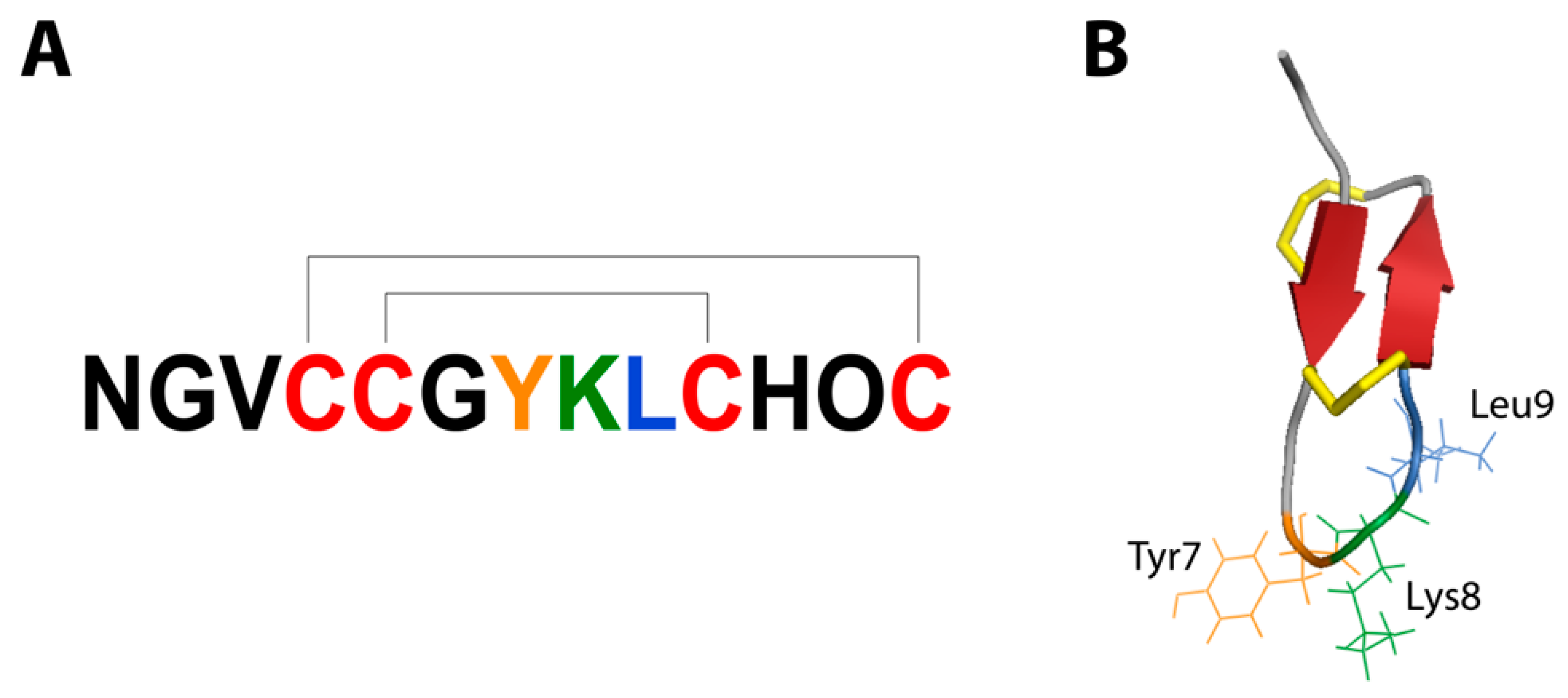
2. Results and Discussion

2.1. Peptide Synthesis

From the 72 MrIA analogs identified by Dutertre et al. [4], 18 sequences were chosen to be synthesized based on several factors, including size, the modified residues present, and the inclusion of fragments of the MrIA pharmacophore. Following synthesis and oxidation, a total of 26 peptide analogues were obtained, including O12P-MrIA in both its amidated 1 and acidic forms 2 (Table 1, 1 and 2, respectively). The total number of analogs obtained exceeded the number of sequences used, as several sequences produced disulfide isomers upon oxidation (Table 1). Additionally, those analogs containing three cysteine residues produced dimers when oxidized.

2.2. Norepinephrine Uptake Inhibition Assay

Based on previous work that showed the amidated form of MrIA (1) to be the slightly more potent version of MrIA [7], this analog was used as the standard comparison compound for the NE uptake inhibition assay. A majority of MrIA analogues tested were significantly (>10-fold) less potent than the MrIA standard 1 (Figure 2). As shown in Figure 2, analogs 2, 3, 11, 12-I, and 17 maintained activity comparable to that of the standard 1. All these peptides contained the VCCGYKLCHXC sequence, with X = proline or hydroxyproline. These peptides do contain the disulfide scaffolding as MrIA and are N-terminal truncated with post-translational modifications, such as N-terminal amidation or proline oxidation. These modifications have in previous work been shown that they are well tolerated in regards to hNET activity [9]. A slight loss of activity was observed for peptides 12II (non-native MrIA fold), and 13I and 13II (two isomers of the VCCGYKLCHOC sequence). None of the analogs significantly improved norepinephrine uptake inhibition.
Those analogs with comparable inhibitory activity to MrIA all maintained the three pharmacophore residues identified by Brust et al. [9], along with all four cysteine residues, while those with reduced activity lacked one or both of these pharmacophore characteristics. In peptides lacking all four cysteine residues, this was presumably due to conformational changes that did not allow the pharmacophore residues to be presented in a way that promoted optimal binding at the target. Interestingly, while the only peptide completely lacking all three pharmacophore residues (10) did have hNET inhibitory activity significantly less potent than the MrIA control, the activity was not significantly less than that of the other analogs that contained all three pharmacophore residues, but lacked one or more of the original cysteine residues. Therefore, while the pharmacophore residues are undoubtedly important in maintaining potent inhibitory hNET activity, they are not entirely necessary to elicit detectable hNET inhibition.
Of the peptide sequences that maintained both the pharmacophore residues and all four cysteine residues, two formed two different isomers upon oxidation (12I and 12II, 13I and 13II). While each of these analogs retained activity greater than those with disrupted pharmacophores, for each set of isomers, one with the native 1-4, 2-3 disulfide fold was significantly more active than the other (12I > 12II, 13II > 13I). Presumably, this was due to alternative disulfide connectivity that did not facilitate a hairpin gamma turn in the less active isomers.
Analog 18, which contained a sulfonated tyrosine residue, also demonstrated reduced activity. This is congruent with previous work by Brust et al. [9], where it was also found that, with the exception of methylation, modification of the tyrosine residue greatly attenuated MrIA activity. The loss of activity may be due to sulfonation adding a negative charge to the previously uncharged residue, or because it adds bulk to a side chain that has a narrow spatial window for optimal binding [9], but is most likely some combination of the two. These results confirm the importance of the MrIA pharmacophore determined by Brust et al. [9] for the NET inhibition displayed by MrIA.

2.3. Calcium Influx Assays

When tested against the CaV2.2 ion channel, the majority of analogs had no significant antagonistic effects (Figure 3). Only analog 3 significantly decreased activity at 10 μM with a 99.9% confidence interval, and the activity was not strong, as demonstrated by the comparison to the antagonistic activity of the ω-conotoxin CVID at 1 μM.
MrIA analogs exhibited no significant antagonistic activity against the voltage-gated sodium channels expressed by SH-SY5Y cells at 10 μM concentrations (Figure 4). These include the NaV subtypes 1.2, 1.3, 1.7 [10], and at lower levels, 1.4 and 1.5. Tetrodotoxin (TTX) at 1 μM concentration was used as an antagonist control for the NaV channels. Furthermore, these MrIA analogs exhibited no significant agonistic activity against receptors that induce intracellular calcium responses in SHSY5Y [11] (data not shown).

3. Materials and Methods

3.1. Materials and Apparatus

Protected Fmoc-amino acid residues were purchased from Novabiochem or Auspep (Melbourne, AU) with the exception of the neopentyl-protected sulfotyrosine, which was purchased from Bachem. The Fmoc-Rink resin and Fmoc-Wang resin were purchased by Auspep and Peptides International (Louisville, USA), while the 2-chlorotrityl resin was purchased from Novabiochem. Peptide-synthesis grade dimethylformamide (DMF), dichloromethane (DCM), diisopropylethylamine (DIEA), and trifluoroacetic acid (TFA) were supplied by Auspep, while 2-(1H-benzotriazol-1-yl)-1,1,3,3- tetramethyluronium hexafluorophosphate (HBTU), triisopropylsilane (TIPS), HPLC-grade acetonitrile, ammonium bicarbonate, and ammonium acetate were supplied by Sigma-Aldrich (Sydney, AU). Assay reagents at the highest grade were also supplied by Sigma-Aldrich unless otherwise stated.
The Symphony automated peptide synthesizer was supplied by Protein Technologies Inc. Peptides were analysed using a Shimadzu Prominence HPLC system coupled to a PE Sciex API 150EX turbo ionspray mass spectrometer and purified on a Shimadzu Preparative HPLC system. The high-throughput FLIPRTetra fluorescent plate reader was supplied by Molecular Devices (CA, USA).

3.2. Peptide Synthesis

Peptides were synthesised either manually or on a Symphony (Protein Technologies Inc, AZ, USA) automated peptide synthesiser using standard Fmoc-SPPS methods [12]. C-terminal amidated peptides were assembled on Fmoc-Rink resins, while those with C-terminal carboxylation were assembled on preloaded Fmoc-Wang resins. The exception to this was the peptide with the sequence, ZGVCCGYKLCHP (4I and 4II), which was assembled on a chlorotrityl resin to prevent diketopiperazine formation. Once assembled, peptides were cleaved with Trifluoroacetic acid (TFA)/water/Triisopropyl silane (TIPS) (90:5:5) for 3 hours, then were precipitated from chilled ether, filtered, and lyophilised from acetonitrile/water. Analytical RP-HPLC and electrospray mass spectrometry were used to confirm the purity and molecular masses of the final peptide products.
Reduced peptides were oxidised at pH 8.1 with a solution of 30% DMSO/70% NH4HCO3 for at least 2 hours at a concentration of 1 mg mL-1. The peptide with the sequence, LRNGVCCGYKLCHOC (12I and 12II), was insoluble in NH4HCO3 and was therefore oxidised in a solution of 20% DMSO/80% TFA. Once oxidised, peptides were purified by RP-HPLC using a C18 column. The peptides were eluted using a linear gradient from 0%–40% solvent B (0.043% TFA in 90% acetonitrile) with solvent A being 0.05% TFA.
The sulfotyrosine analog 18 was synthesized in the same manner as the other analogs using comercial neopentyl-protected sulfotyrosine (Bachem SA, Bubendorf, CH). Peptide 18 was cleaved from the resin with TFA/water/TIPS (90:5:5) for 1.5 h at 0 °C. The oxidation of Peptide 18 was performed in 2 M ammonium acetate at 37 °C overnight and subsequently purified using RP-HPLC with a linear gradient between 0.1 M ammonium acetate and acetonitrile as recommended by Simpson et al. [13]. This method was employed to protect the highly acid labile sulfate group.

3.3. NE Uptake Inhibition Assay

The norepinephrine (NE) uptake assay was performed as previously described [14,15]. Consistant with these earlier studies, native MrIA inhibited hNET with a pIC50 of 5.87 ± 0.03. COS-7 cells (ATCC, VA, USA) were cultured in Dulbecco’s modified Eagle’s medium (DMEM) containing 10% fetal bovine serum (FBS) in 96-well plates. Cells were transiently transfected at the confluency of 90% with NET cDNA using 10 μL FuGENE (Roche, AU) and 2 μg DNA per 106 cells. Twenty-four hours post-transfection, 10,000 cells from the overnight culture were evenly distributed per well in a 96-well plate format for an additional 24 hours incubation. An average cell count of approximately 50,000 cells per well was counted before uptake assays. NE uptake inhibition assay was performed using a fixed concentration of (3H)NE of 30 nM (40.5 Ci/mmol, Perkin Elmer, MA, USA) with increasing concentrations of peptide (1) and its peptide mimetics from 10−4 M to 10−15 M prepared in assay buffer (25 mM HEPES, pH 7.4, 125 mM NaCl, 1.2 mM MgSO4, 4.8 mM KCl, 1.2 mM KH2PO4, 1.3 mM CaCl2, 5.55 mM d-(+)-glucose, 1 mM ascorbic acid, 0.1% bovine serum albumin) using a 10-fold increment with a final volume of 50 μL per well in 96-well plates and incubated at 37 °C for 10 min, followed by two gentle washes using 100 μL assay buffer to remove excess (3H)NE. Cells were then lysed using 50 μL of 0.1 M NaOH with gentle shaking for 60 min. Lysed cells were transferred to the flexible 96-well plate (Perkin Elmer) with Optiphase Supermix scintillant (Perkin Elmer) for measurements. Each experiment was performed in triplicate and repeated three times.

3.4. Calcium Influx Assays

MrIA analogs (1 to 18, Table 1) were tested for antagonist activity at 10 μM concentrations against CaV2.2 [16] and NaV [10] ion channels using calcium influx assays and a high-throughput FLIPRTetra (Molecular Devices, CA, USA) fluorescent plate reader assay. SH-SY5Y cells were seeded in to black-walled, clear bottom 384-well imaging plates (Corning, NY, USA) at a concentration of approximately 40,000 cells per well two days prior to the assays. Cells were incubated in a Calcium 4 No Wash kit (Molecular Devices) in physiological salt solution (composition NaCl 140 mM, glucose 11.5 mM, KCl 5.9 mM, MgCl2 1.4 mM, NaH2PO4 1.2 mM, NaHCO3 5 mM, CaCl2 1.8 mM, HEPES 10 mM) at 37 °C for 30 min. An addition of 10 μM nifedipine was made to the dye used to test for activity at CaV2.2 in order to inhibit the L-type calcium channel activity. Using a cooled CCD camera at excitation 470–495 nM and emission 515–575 nM, 10 s baseline fluorescence readings were obtained, at which point the MrIA analogs were added to the cells and fluorescence readings were obtained every 2 s for 300 s. Standard agonists (90 mM KCl and 5 mM CaCl2 for CaV2.2 and 45 μΜ veratridine for NaVs) were then added and further fluorescent readings were obtained every second for 300 s. Each assay was performed at least three times with three replicates of each analog per assay. Resulting maximum fluorescence readings were normalized to F/Fmax using the average of the maximum fluorescence in positive control as Fmax. Analog activities were then compared to Fmax using ordinary one-way ANOVA.

4. Conclusions

While some of the MrIA analogs synthesized maintained hNET inhibitory activity comparable to that of MrIA, none were able to significantly improve upon it. Indeed, MrIA may have arisen from such analogues and become stabilized in the genome optimized through positive selection. Those analogs lacking pharmacophore elements previously identified had greatly attenuated activity, supporting the MrIA pharmacophore identified by Brust et al. [9]. Calcium influx assays revealed that one analog produced off-target effects with antagonistic activity at CaV2.2 channels. While this activity may not be biologically relevant, secondary conotoxin activities such as this have the potential to confer a competitive advantage. Based on this, we theorize that the “messiness” can provide the raw materials for evolutionary “experimentation” with the potential to enhance primary function and underpin the evolution of secondary function in conotoxins.

Author Contributions

R.Z. performed peptide synthesis, calcium influx assays, data analyses, and wrote the manuscript. A.B. contributed to peptide synthesis and provided guidance and ideas. P.J. performed hNET assays and partook in their analysis. F.C.C. assisted with calcium influx assays, the analysis of their results, and reviewing the manuscript. R.J.L. contributed by reviewing the manuscript. P.F.A. and R.J.L. conceived the project and provided guidance and ideas.

Funding

This work was supported by International Scholarship from the University of Queensland and Australian National Health & Medical Research Council (Program Grants APP1072113, 351446 and Principal Research Fellowships to R.J.L. and P.F.A).

Conflicts of Interest

The authors declare no conflict of interest.

References

  1. Akondi, K.B.; Muttenthaler, M.; Dutertre, S.; Kaas, Q.; Craik, D.J.; Lewis, R.J.; Alewood, P.F. Discovery, synthesis, and structure–activity relationships of conotoxins. Chem. Rev. 2014, 114, 5815–5847. [Google Scholar] [CrossRef] [PubMed]
  2. Lewis, R.J.; Garcia, M.L. Therapeutic potential of venom peptides. Nat. Rev. Drug Discov. 2003, 2, 790–802. [Google Scholar] [CrossRef] [PubMed]
  3. Lewis, R.J. Conotoxins as selective inhibitors of neuronal ion channels, receptors and transporters. IUBMB Life 2004, 56, 89–93. [Google Scholar] [CrossRef] [PubMed] [Green Version]
  4. Dutertre, S.; Jin, A.-H.; Kaas, Q.; Jones, A.; Alewood, P.F.; Lewis, R.J. Deep venomics reveals the mechanism for expanded peptide diversity in cone snail venom. Mol. Cell. Proteom. 2013, 12, 312–329. [Google Scholar] [CrossRef] [PubMed]
  5. Lavergne, V.; Dutertre, S.; Jin, A.-H.; Lewis, R.J.; Taft, R.J.; Alewood, P.F. Systematic interrogation of the Conus marmoreus venom duct transcriptome with ConoSorter reveals 158 novel conotoxins and 13 new gene superfamilies. BMC Genom. 2013, 14, 708. [Google Scholar] [CrossRef] [PubMed]
  6. Prashanth, J.R.; Brust, A.; Jin, A.-H.; Alewood, P.F.; Dutertre, S.; Lewis, R.J. Cone snail venomics: From novel biology to novel therapeutics. Future Med. Chem. 2014, 6, 1659–1675. [Google Scholar] [CrossRef] [PubMed]
  7. Sharpe, I.A.; Gehrmann, J.; Loughnan, M.L.; Thomas, L.; Adams, D.A.; Atkins, A.; Palant, E.; Craik, D.J.; Adams, D.J.; Alewood, P.F. Two new classes of conopeptides inhibit the α1-adrenoceptor and noradrenaline transporter. Nat. Neurosci. 2001, 4, 902–907. [Google Scholar] [CrossRef] [PubMed]
  8. Lewis, R.J. Discovery and development of the χ-conopeptide class of analgesic peptides. Toxicon 2012, 59, 524–528. [Google Scholar] [CrossRef] [PubMed]
  9. Brust, A.; Palant, E.; Croker, D.E.; Colless, B.; Drinkwater, R.; Patterson, B.; Schroeder, C.I.; Wilson, D.; Nielsen, C.K.; Smith, M.T. χ-Conopeptide pharmacophore development: Toward a novel class of norepinephrine transporter inhibitor (Xen2174) for pain. J. Med. Chem. 2009, 52, 6991–7002. [Google Scholar] [CrossRef] [PubMed]
  10. Vetter, I.; Mozar, C.A.; Durek, T.; Wingerd, J.S.; Alewood, P.F.; Christie, M.J.; Lewis, R.J. Characterisation of Nav types endogenously expressed in human SH-SY5Y neuroblastoma cells. Biochem. Pharmacol. 2012, 83, 1562–1571. [Google Scholar] [CrossRef] [PubMed] [Green Version]
  11. Yin, K.; Baillie, G.J.; Vetter, I. Neuronal cell lines as model dorsal root ganglion neurons: A transcriptomic comparison. Mol. Pain 2016, 12. [Google Scholar] [CrossRef] [PubMed]
  12. Fields, G.B.; Noble, R.L. Solid phase peptide synthesis utilizing 9-fluorenylmethoxycarbonyl amino acids. Int. J. Pept. Protein Res. 1990, 35, 161–214. [Google Scholar] [CrossRef]
  13. Simpson, L.S.; Zhu, J.Z.; Widlanski, T.S.; Stone, M.J. Regulation of chemokine recognition by site-specific tyrosine sulfation of receptor peptides. Chem. Biol. 2009, 16, 153–161. [Google Scholar] [CrossRef] [PubMed]
  14. Bryan-Lluka, L.J.; Bonisch, H.; Lewis, R.J. χ-Conopeptide MrIA partially overlaps desipramine and cocaine binding sites on the human norepinephrine transporter. J. Biol. Chem. 2003, 278, 40324–40329. [Google Scholar] [CrossRef] [PubMed]
  15. Sharpe, I.A.; Palant, E.; Schroeder, C.I.; Kaye, D.M.; Adams, D.J.; Alewood, P.F.; Lewis, R.J. Inhibition of the norepinephrine transporter by the venom peptide χ-MrIA. Site of action, Na+ dependence, and structure-activity relationship. J. Biol. Chem. 2003, 278, 40317–40323. [Google Scholar] [CrossRef] [PubMed]
  16. Sousa, S.R.; Vetter, I.; Ragnarsson, L.; Lewis, R.J. Expression and pharmacology of endogenous Cav channels in SH-SY5Y human neuroblastoma cells. PLoS ONE 2013, 8, e59293. [Google Scholar] [CrossRef] [PubMed]
Figure 1. (A) Primary structure of MrIA showing Cys1-Cys3, Cys2-Cys4 disulfide connectivity, Cys residues are coloured red, (B) 3D structure of MrIA. Yellow portions represent disulfide bonds and red arrows represent β-sheets. Pharmacophore residues are coloured orange (Tyr), green (Lys), and blue (Leu).
Marinedrugs 17 00165 g001
Figure 2. pIC50 values of MrIA analogs against human norepinephrine transporter (hNET) and their standard deviation bars. The MrIA standard (1) is coloured grey. Analogs whose hNET inhibitory activity do not significantly differ from the standard are marked by *.
Marinedrugs 17 00165 g002
Figure 3. Average F/Fmax values for MrIA analogs tested at 10 μM and CVID tested at 1 μM against CaV2.2 with associated standard deviation bars. Analog 3 was found to have statistically significant antagonistic activity at a 99.9% confidence interval and is marked *.
Marinedrugs 17 00165 g003
Figure 4. Average F/Fmax values for MrIA analogs tested at 10 μM and TTX tested at 1 μM against voltage-gated sodium channels (NaV) with associated standard deviation bars.
Marinedrugs 17 00165 g004
Table 1. List of peptide analogs used in this study, including their peptide sequence, relative abundance in Conus marmoreus venom as compared to the amidated form of MrIA (from Duterte et al. [4]), and expected and observed masses. Cysteine residues are coloured red. “Z” denotes pyroglutamic acid, “O” denotes hydroxyproline, “*” denotes an amidated C-terminus, Y(SO3) denotes sulfotyrosine.
AnalogSequenceRelative AbundanceExpected MW (Da)Observed MW (Da)
1NGVCCGYKLCHPC *100.001391.71392.0
2NGVCCGYKLCHPC49.771392.71392.8
3ZGVCCGYKLCHPC0.731389.71389.8
4IZGVCCGYKLCHP0.051288.52577.6
4II2577.6
5IZGVCCGYKLC0.341054.32106.0
5II2107.0
6INGVCCGYKLC0.121057.32115.8
6II2113.8
7ZGVCCGYKL0.03951.1951.6
8ZGVCCGYKL *0.01950.1950.4
9NGVCCGYK0.02841.0841.6
10ILRGILRNGVCC *0.021313.71314.0
11GILRNGVCCGYKLCHPC0.331831.21834.0
12-ILRNGVCCGYKLCHOC0.141678.01678.0
12-II1677.8
13IVCCGYKLCHOC0.881237.51237.4
13II1237.4
14ICGYKLCHOC0.211037.22073.6
14II2072.8
14III2073.0
14IV2071.8
15GYKLCHOC0.04934.1934.6
16YKLCHOC0.02877.1877.6
17GVCCGYKLCHPC0.011278.61278.6
18GVCCGY(SO3)KLCHOC0.111377.51376.6

