
Augmented Reality Integration in Skull Base Neurosurgery: A Systematic Review

 , , , ,

Abstract
:1. Introduction
2. Materials and Methods
2.1. Study Methodology and Registration
2.2. Search Strategy
2.3. Inclusion and Exclusion Criteria
2.4. Data Extraction
2.5. Statistical Analysis
3. Results
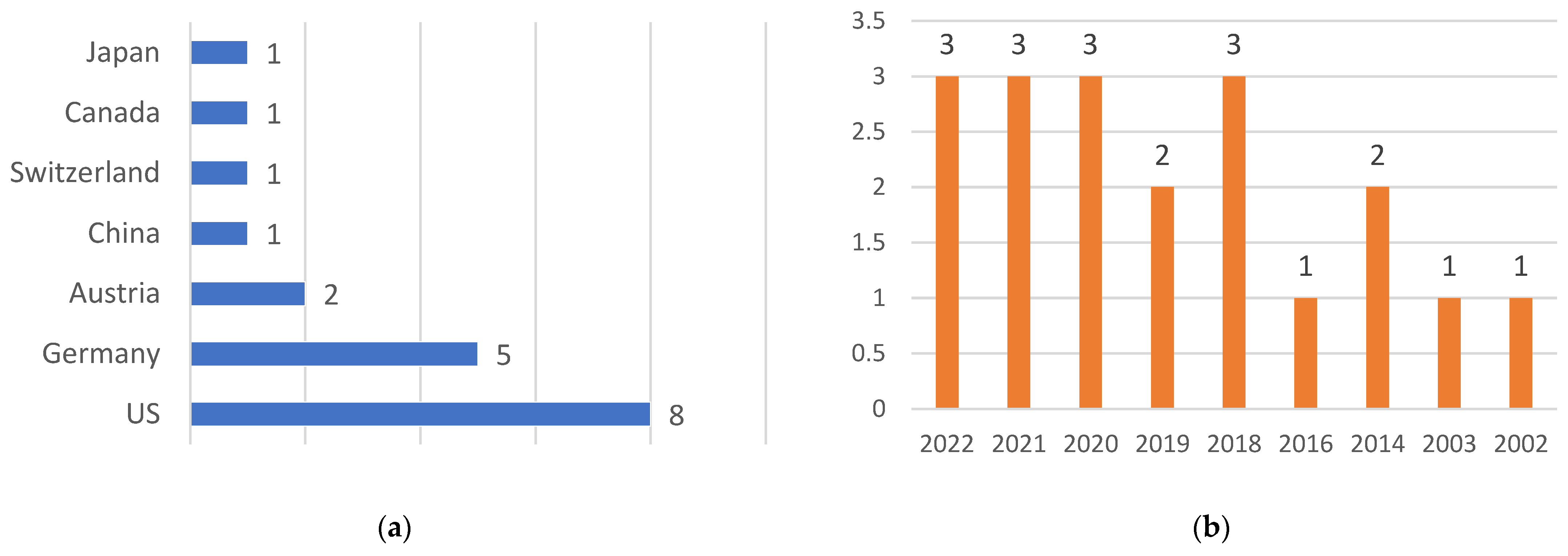
3.1. Characteristics of Included Studies
3.2. Study Sample Characteristics
3.3. Results from Laboratory Studies
3.4. Results from Clinical Studies
3.5. Results from Cadaveric Studies
4. Discussion
5. Conclusions
Author Contributions
Funding
Institutional Review Board Statement
Informed Consent Statement
Data Availability Statement
Conflicts of Interest
Appendix A
| Search | (Augmented reality OR AR) AND (skull-base) |
| Filter | none |
| Search details | (“augmented reality” [MeSH Terms] OR (“augmented” [All Fields] AND “reality” [All Fields]) OR “augmented reality” [All Fields]) AND (“skull base” [MeSH Terms] OR (“skull” [All Fields] AND “base” [All Fields]) OR “skull base” [All Fields]) |
Appendix B
| Section/Topic | # | Checklist Item | Reported on Page # |
| Title | |||
| Title | 1 | Identify the report as a systematic review, meta-analysis, or both. | 1 |
| Abstract | |||
| Structured summary | 2 | Provide a structured summary including, as applicable: background; objectives; data sources; study eligibility criteria, participants, and interventions; study appraisal and synthesis methods; results; limitations; conclusions and implications of key findings; systematic review registration number. | 1 |
| Introduction | |||
| Rationale | 3 | Describe the rationale for the review in the context of what is already known. | 2 |
| Objectives | 4 | Provide an explicit statement of questions being addressed with reference to participants, interventions, comparisons, outcomes, and study design (PICOS). | 2 |
| Methods | |||
| Protocol and registration | 5 | Indicate if a review protocol exists, if and where it can be accessed (e.g., Web address), and, if available, provide registration information including registration number. | 2 |
| Eligibility criteria | 6 | Specify study characteristics (e.g., PICOS, length of follow-up) and report characteristics (e.g., years considered, language, publication status) used as criteria for eligibility, giving rationale. | 3 |
| Information sources | 7 | Describe all information sources (e.g., databases with dates of coverage, contact with study authors to identify additional studies) in the search and date last searched. | 3 |
| Search | 8 | Present full electronic search strategy for at least one database, including any limits used, such that it could be repeated. | Appendix A |
| Study selection | 9 | State the process for selecting studies (i.e., screening, eligibility, included in systematic review, and, if applicable, included in the meta-analysis). | 3 |
| Data collection process | 10 | Describe method of data extraction from reports (e.g., piloted forms, independently, in duplicate) and any processes for obtaining and confirming data from investigators. | 3 |
| Data items | 11 | List and define all variables for which data were sought (e.g., PICOS, funding sources) and any assumptions and simplifications made. | 3 |
| Risk of bias in individual studies | 12 | Describe methods used for assessing risk of bias of individual studies (including specification of whether this was done at the study or outcome level), and how this information is to be used in any data synthesis. | N/A |
| Summary measures | 13 | State the principal summary measures (e.g., risk ratio, difference in means). | N/A |
| Synthesis of results | 14 | Describe the methods of handling data and combining results of studies, if done, including measures of consistency (e.g., I2) for each meta-analysis. | N/A |
| Risk of bias across studies | 15 | Specify any assessment of risk of bias that may affect the cumulative evidence (e.g., publication bias, selective reporting within studies). | N/A |
| Additional analyses | 16 | Describe methods of additional analyses (e.g., sensitivity or subgroup analyses, meta-regression), if done, indicating which were pre-specified. | N/A |
| Results | |||
| Study selection | 17 | Give numbers of studies screened, assessed for eligibility, and included in the review, with reasons for exclusions at each stage, ideally with a flow diagram. | Figure 1 |
| Study characteristics | 18 | For each study, present characteristics for which data were extracted (e.g., study size, PICOS, follow-up period) and provide the citations. | 3 |
| Risk of bias within studies | 19 | Present data on risk of bias of each study and, if available, any outcome level assessment (see item 12). | N/A |
| Results of individual studies | 20 | For all outcomes considered (benefits or harms), present, for each study: (a) simple summary data for each intervention group (b) effect estimates and confidence intervals, ideally with a forest plot. | Table 1, Table 2 and Table 3 |
| Synthesis of results | 21 | Present results of each meta-analysis done, including confidence intervals and measures of consistency. | N/A |
| Risk of bias across studies | 22 | Present results of any assessment of risk of bias across studies (see Item 15). | N/A |
| Additional analysis | 23 | Give results of additional analyses, if done (e.g., sensitivity or subgroup analyses, meta-regression [see Item 16]). | N/A |
| Discussion | |||
| Summary of evidence | 24 | Summarize the main findings including the strength of evidence for each main outcome; consider their relevance to key groups (e.g., healthcare providers, users, and policy makers). | 4–8 |
| Limitations | 25 | Discuss limitations at study and outcome level (e.g., risk of bias), and at review level (e.g., incomplete retrieval of identified research, reporting bias). | 10 |
| Conclusions | 26 | Provide a general interpretation of the results in the context of other evidence, and implications for future research. | 11 |
| Funding | |||
| Funding | 27 | Describe sources of funding for the systematic review and other support (e.g., supply of data); role of funders for the systematic review. | 11 |
References
- Begagić, E.; Bečulić, H.; Đuzić, N.; Džidić-Krivić, A.; Pugonja, R.; Muharemović, A.; Jaganjac, B.; Salković, N.; Sefo, H.; Pojskić, M. CRISPR/Cas9-Mediated Gene Therapy for Glioblastoma: A Scoping Review. Biomedicines 2024, 12, 238. [Google Scholar] [CrossRef] [PubMed]
- Sekhar, L.N.; Juric-Sekhar, G.; Qazi, Z.; Patel, A.; McGrath, L.B., Jr.; Pridgeon, J.; Kalavakonda, N.; Hannaford, B. The Future of Skull Base Surgery: A View Through Tinted Glasses. World Neurosurg. 2020, 142, 29–42. [Google Scholar] [CrossRef] [PubMed]
- Tzelnick, S.; Rampinelli, V.; Sahovaler, A.; Franz, L.; Chan, H.H.L.; Daly, M.J.; Irish, J.C. Skull-Base Surgery-A Narrative Review on Current Approaches and Future Developments in Surgical Navigation. J. Clin. Med. 2023, 12, 2706. [Google Scholar] [CrossRef] [PubMed]
- Pugonja, R.; Beculić, H.; Begagić, E.; Skomorac, R.; Selimović, E. Morphometric and Ki-67 proliferative index-related characteristics of meningiomas and their correlation with demographic, clinical, histopathological, and postoperative features. Med. Glas. 2024, 21, 132–139. [Google Scholar] [CrossRef]
- Wei, B.; Sun, G.; Hu, Q.; Tang, E. The Safety and Accuracy of Surgical Navigation Technology in the Treatment of Lesions Involving the Skull Base. J. Craniofac. Surg. 2017, 28, 1431–1434. [Google Scholar] [CrossRef] [PubMed]
- Hey, G.; Guyot, M.; Carter, A.; Lucke-Wold, B. Augmented Reality in Neurosurgery: A New Paradigm for Training. Medicina 2023, 59, 1721. [Google Scholar] [CrossRef] [PubMed]
- Panesar, S.S.; Magnetta, M.; Mukherjee, D.; Abhinav, K.; Branstetter, B.F.; Gardner, P.A.; Iv, M.; Fernandez-Miranda, J.C. Patient-specific 3-dimensionally printed models for neurosurgical planning and education. Neurosurg. Focus 2019, 47, E12. [Google Scholar] [CrossRef] [PubMed]
- Meola, A.; Cutolo, F.; Carbone, M.; Cagnazzo, F.; Ferrari, M.; Ferrari, V. Augmented reality in neurosurgery: A systematic review. Neurosurg. Rev. 2017, 40, 537–548. [Google Scholar] [CrossRef]
- Cho, J.; Rahimpour, S.; Cutler, A.; Goodwin, C.R.; Lad, S.P.; Codd, P. Enhancing Reality: A Systematic Review of Augmented Reality in Neuronavigation and Education. World Neurosurg. 2020, 139, 186–195. [Google Scholar] [CrossRef]
- Bečulić, H.; Begagić, E.; Skomorac, R.; Mašović, A.; Selimović, E.; Pojskić, M. ChatGPT’s contributions to the evolution of neurosurgical practice and education: A systematic review of benefits, concerns and limitations. Med. Glas. 2024, 21, 126–131. [Google Scholar] [CrossRef]
- Contreras López, W.O.; Navarro, P.A.; Crispin, S. Intraoperative clinical application of augmented reality in neurosurgery: A systematic review. Clin. Neurol. Neurosurg. 2019, 177, 6–11. [Google Scholar] [CrossRef] [PubMed]
- Bölek, K.A.; De Jong, G.; Henssen, D. The effectiveness of the use of augmented reality in anatomy education: A systematic review and meta-analysis. Sci. Rep. 2021, 11, 15292. [Google Scholar] [CrossRef] [PubMed]
- Cabrilo, I.; Sarrafzadeh, A.; Bijlenga, P.; Landis, B.N.; Schaller, K. Augmented reality-assisted skull base surgery. Neurochirurgie 2014, 60, 304–306. [Google Scholar] [CrossRef] [PubMed]
- Toader, C.; Eva, L.; Tataru, C.-I.; Covache-Busuioc, R.-A.; Bratu, B.-G.; Dumitrascu, D.-I.; Costin, H.P.; Glavan, L.-A.; Ciurea, A.V. Frontiers of Cranial Base Surgery: Integrating Technique, Technology, and Teamwork for the Future of Neurosurgery. Brain Sci. 2023, 13, 1495. [Google Scholar] [CrossRef] [PubMed]
- Bong, J.H.; Song, H.J.; Oh, Y.; Park, N.; Kim, H.; Park, S. Endoscopic navigation system with extended field of view using augmented reality technology. Int. J. Med. Robot. 2018, 14, e1886. [Google Scholar] [CrossRef]
- Li, L.; Yang, J.; Chu, Y.; Wu, W.; Xue, J.; Liang, P.; Chen, L. A Novel Augmented Reality Navigation System for Endoscopic Sinus and Skull Base Surgery: A Feasibility Study. PLoS ONE 2016, 11, e0146996. [Google Scholar] [CrossRef]
- Bopp, M.H.A.; Saß, B.; Pojskić, M.; Corr, F.; Grimm, D.; Kemmling, A.; Nimsky, C. Use of Neuronavigation and Augmented Reality in Transsphenoidal Pituitary Adenoma Surgery. J. Clin. Med. 2022, 11, 5590. [Google Scholar] [CrossRef]
- Zeiger, J.; Costa, A.; Bederson, J.; Shrivastava, R.K.; Iloreta, A.M.C. Use of Mixed Reality Visualization in Endoscopic Endonasal Skull Base Surgery. Oper. Neurosurg. 2020, 19, 43–52. [Google Scholar] [CrossRef]
- Carl, B.; Bopp, M.; Voellger, B.; Saß, B.; Nimsky, C. Augmented Reality in Transsphenoidal Surgery. World Neurosurg. 2019, 125, e873–e883. [Google Scholar] [CrossRef]
- Schwam, Z.G.; Kaul, V.F.; Bu, D.D.; Iloreta, A.C.; Bederson, J.B.; Perez, E.; Cosetti, M.K.; Wanna, G.B. The utility of augmented reality in lateral skull base surgery: A preliminary report. Am. J. Otolaryngol. 2021, 42, 102942. [Google Scholar] [CrossRef] [PubMed]
- Pojskić, M.; Bopp, M.H.A.; Saβ, B.; Carl, B.; Nimsky, C. Microscope-Based Augmented Reality with Intraoperative Computed Tomography-Based Navigation for Resection of Skull Base Meningiomas in Consecutive Series of 39 Patients. Cancers 2022, 14, 2302. [Google Scholar] [CrossRef] [PubMed]
- Steiert, C.; Behringer, S.P.; Kraus, L.M.; Bissolo, M.; Demerath, T.; Beck, J.; Grauvogel, J.; Reinacher, P.C. Augmented reality-assisted craniofacial reconstruction in skull base lesions—An innovative technique for single-step resection and cranioplasty in neurosurgery. Neurosurg. Rev. 2022, 45, 2745–2755. [Google Scholar] [CrossRef] [PubMed]
- Lai, M.; Skyrman, S.; Shan, C.; Babic, D.; Homan, R.; Edström, E.; Persson, O.; Burström, G.; Elmi-Terander, A.; Hendriks, B.H.W.; et al. Fusion of augmented reality imaging with the endoscopic view for endonasal skull base surgery; a novel application for surgical navigation based on intraoperative cone beam computed tomography and optical tracking. PLoS ONE 2020, 15, e0227312. [Google Scholar] [CrossRef]
- Birkfellner, W.; Figl, M.; Matula, C.; Hummel, J.; Hanel, R.; Imhof, H.; Wanschitz, F.; Wagner, A.; Watzinger, F.; Bergmann, H. Computer-enhanced stereoscopic vision in a head-mounted operating binocular. Phys. Med. Biol. 2003, 48, N49–N57. [Google Scholar] [CrossRef] [PubMed]
- Creighton, F.X.; Unberath, M.; Song, T.; Zhao, Z.; Armand, M.; Carey, J. Early Feasibility Studies of Augmented Reality Navigation for Lateral Skull Base Surgery. Otol. Neurotol. 2020, 41, 883–888. [Google Scholar] [CrossRef] [PubMed]
- Pennacchietti, V.; Stoelzel, K.; Tietze, A.; Lankes, E.; Schaumann, A.; Uecker, F.C.; Thomale, U.W. First experience with augmented reality neuronavigation in endoscopic assisted midline skull base pathologies in children. Childs Nerv. Syst. 2021, 37, 1525–1534. [Google Scholar] [CrossRef] [PubMed]
- Barber, S.R.; Wong, K.; Kanumuri, V.; Kiringoda, R.; Kempfle, J.; Remenschneider, A.K.; Kozin, E.D.; Lee, D.J. Augmented Reality, Surgical Navigation, and 3D Printing for Transcanal Endoscopic Approach to the Petrous Apex. OTO Open 2018, 2, 2473974X18804492. [Google Scholar] [CrossRef]
- Kawamata, T.; Iseki, H.; Shibasaki, T.; Hori, T. Endoscopic augmented reality navigation system for endonasal transsphenoidal surgery to treat pituitary tumors: Technical note. Neurosurgery 2002, 50, 1393–1397. [Google Scholar] [CrossRef]
- Leuze, C.; Neves, C.A.; Gomez, A.M.; Navab, N.; Blevins, N.; Vaisbuch, Y.; McNab, J.A. Augmented Reality for Retrosigmoid Craniotomy Planning. J. Neurol. Surgery. Part B Skull Base 2021, 83, e564–e573. [Google Scholar] [CrossRef]
- McJunkin, J.L.; Jiramongkolchai, P.; Chung, W.; Southworth, M.; Durakovic, N.; Buchman, C.A.; Silva, J.R. Development of a mixed reality platform for lateral skull base anatomy. Otol. Neurotol. Off. Publ. Am. Otol. Soc. Am. Neurotol. Soc. Eur. Acad. Otol. Neurotol. 2018, 39, e1137–e1142. [Google Scholar] [CrossRef]
- Dixon, B.J.; Daly, M.J.; Chan, H.; Vescan, A.; Witterick, I.J.; Irish, J.C. Augmented image guidance improves skull base navigation and reduces task workload in trainees: A preclinical trial. Laryngoscope 2011, 121, 2060–2064. [Google Scholar] [CrossRef] [PubMed]
- Cannizzaro, D.; Zaed, I.; Safa, A.; Jelmoni, A.J.M.; Composto, A.; Bisoglio, A.; Schmeizer, K.; Becker, A.C.; Pizzi, A.; Cardia, A.; et al. Augmented Reality in Neurosurgery, State of Art and Future Projections. A Systematic Review. Front. Surg. 2022, 9, 864792. [Google Scholar] [CrossRef] [PubMed]
- Sarica, C.; Egemen, E. Contribution of Countries to Main Neurosurgical Journals with Particular Emphasis on Turkey. Turk. Neurosurg. 2021, 31, 823–837. [Google Scholar] [CrossRef]
- Deng, W.; Li, F.; Wang, M.; Song, Z. Easy-to-use augmented reality neuronavigation using a wireless tablet PC. Ster. Funct. Neurosurg. 2014, 92, 17–24. [Google Scholar] [CrossRef] [PubMed]
- Tagaytayan, R.; Kelemen, A.; Sik-Lanyi, C. Augmented reality in neurosurgery. Arch. Med. Sci. 2018, 14, 572–578. [Google Scholar] [CrossRef]
- Kersten-Oertel, M.; Chen, S.S.; Drouin, S.; Sinclair, D.S.; Collins, D.L. Augmented reality visualization for guidance in neurovascular surgery. Stud. Health Technol. Inf. 2012, 173, 225–229. [Google Scholar]
- Scherschinski, L.; McNeill, I.T.; Schlachter, L.; Shuman, W.H.; Oemke, H.; Yaeger, K.A.; Bederson, J.B. Augmented reality-assisted microsurgical resection of brain arteriovenous malformations: Illustrative case. J. Neurosurg. Case Lessons 2022, 3, Case21135. [Google Scholar] [CrossRef]
- Abe, Y.; Sato, S.; Kato, K.; Hyakumachi, T.; Yanagibashi, Y.; Ito, M.; Abumi, K. A novel 3D guidance system using augmented reality for percutaneous vertebroplasty: Technical note. J. Neurosurg. Spine 2013, 19, 492–501. [Google Scholar] [CrossRef]
- Shmuylovich, L.; O’Brien, C.M.; Nwosu, K.; Achilefu, S. Low-cost augmented reality goggles enable precision fluorescence-guided cancer surgery. Res. Sq. 2023. [Google Scholar] [CrossRef]
- Yodrabum, N.; Rudeejaroonrung, K.; Chaikangwan, I.; Prompattanapakdee, J.; Noraset, T. Precision of Low-Cost Augmented Reality in Prefabricated Cutting Guide for Fibular Free Flap Surgery. J. Craniofac. Surg. 2022, 33, 916–919. [Google Scholar] [CrossRef] [PubMed]
- George, S.; Kesavadas, T. Low cost augmented reality for training of MRI-guided needle biopsy of the spine. Stud. Health Technol. Inf. 2008, 132, 138–140. [Google Scholar]
- Hong, W.; Huang, X.; Chen, Z.; Huang, S.; Wen, Y.; He, B.; Liu, Y.; Lin, Y. A Low-Cost Mobile-Based Augmented Reality Neuronavigation System for Retrosigmoid Craniotomy. Oper. Neurosurg. 2023. [Google Scholar] [CrossRef]
- Kubben, P.L.; Sinlae, R.S.N. Feasibility of using a low-cost head-mounted augmented reality device in the operating room. Surg. Neurol. Int. 2019, 10, 26. [Google Scholar] [CrossRef]
- Dennler, C.; Bauer, D.E.; Scheibler, A.G.; Spirig, J.; Götschi, T.; Fürnstahl, P.; Farshad, M. Augmented reality in the operating room: A clinical feasibility study. BMC Musculoskelet. Disord. 2021, 22, 451. [Google Scholar] [CrossRef]
- Sommer, F.; Hussain, I.; Kirnaz, S.; Goldberg, J.; McGrath, L.; Navarro-Ramirez, R.; Waterkeyn, F.; Schmidt, F.; Gadjradj, P.S.; Härtl, R. Safety and Feasibility of Augmented Reality Assistance in Minimally Invasive and Open Resection of Benign Intradural Extramedullary Tumors. Neurospine 2022, 19, 501–512. [Google Scholar] [CrossRef]
- Jean, W.C. Virtual and Augmented Reality in Neurosurgery: The Evolution of its Application and Study Designs. World Neurosurg. 2022, 161, 459–464. [Google Scholar] [CrossRef] [PubMed]
- Nguyen, N.Q.; Cardinell, J.; Ramjist, J.M.; Lai, P.; Dobashi, Y.; Guha, D.; Androutsos, D.; Yang, V.X.D. An augmented reality system characterization of placement accuracy in neurosurgery. J. Clin. Neurosci. 2020, 72, 392–396. [Google Scholar] [CrossRef] [PubMed]
- Satoh, M.; Nakajima, T.; Watanabe, E.; Kawai, K. Augmented Reality in Stereotactic Neurosurgery: Current Status and Issues. Neurol. Med. Chir. 2023, 63, 137–140. [Google Scholar] [CrossRef] [PubMed]
- Hasan, S.; Miller, A.; Higginbotham, D.; Saleh, E.S.; McCarty, S. Virtual and Augmented Reality in Spine Surgery: An Era of Immersive Healthcare. Cureus 2023, 15, e43964. [Google Scholar] [CrossRef] [PubMed]
- Pierzchajlo, N.; Stevenson, T.C.; Huynh, H.; Nguyen, J.; Boatright, S.; Arya, P.; Chakravarti, S.; Mehrki, Y.; Brown, N.J.; Gendreau, J.; et al. Augmented Reality in Minimally Invasive Spinal Surgery: A Narrative Review of Available Technology. World Neurosurg. 2023, 176, 35–42. [Google Scholar] [CrossRef] [PubMed]
- Olexa, J.; Cohen, J.; Alexander, T.; Brown, C.; Schwartzbauer, G.; Woodworth, G.F. Expanding Educational Frontiers in Neurosurgery: Current and Future Uses of Augmented Reality. Neurosurgery 2023, 92, 241–250. [Google Scholar] [CrossRef]
- Finger, T.; Schaumann, A.; Schulz, M.; Thomale, U.W. Augmented reality in intraventricular neuroendoscopy. Acta Neurochir. 2017, 159, 1033–1041. [Google Scholar] [CrossRef]
- Karmonik, C.; Boone, T.B.; Khavari, R. Workflow for Visualization of Neuroimaging Data with an Augmented Reality Device. J. Digit. Imaging 2018, 31, 26–31. [Google Scholar] [CrossRef]
- Sandrone, S.; Carlson, C.E. Future of Neurology & Technology: Virtual and Augmented Reality in Neurology and Neuroscience Education: Applications and Curricular Strategies. Neurology 2021, 97, 740–744. [Google Scholar] [CrossRef] [PubMed]
- Montemurro, N.; Condino, S.; Carbone, M.; Cattari, N.; D’Amato, R.; Cutolo, F.; Ferrari, V. Brain Tumor and Augmented Reality: New Technologies for the Future. Int. J. Environ. Res. Public Health 2022, 19, 6347. [Google Scholar] [CrossRef] [PubMed]
- Aguilar-Salinas, P.; Gutierrez-Aguirre, S.F.; Avila, M.J.; Nakaji, P. Current status of augmented reality in cerebrovascular surgery: A systematic review. Neurosurg. Rev. 2022, 45, 1951–1964. [Google Scholar] [CrossRef] [PubMed]
- Chiou, S.Y.; Zhang, Z.Y.; Liu, H.L.; Yan, J.L.; Wei, K.C.; Chen, P.Y. Augmented Reality Surgical Navigation System for External Ventricular Drain. Healthcare 2022, 10, 1815. [Google Scholar] [CrossRef] [PubMed]
- Zanier, E.R.; Zoerle, T.; Di Lernia, D.; Riva, G. Virtual Reality for Traumatic Brain Injury. Front. Neurol. 2018, 9, 345. [Google Scholar] [CrossRef] [PubMed]
- Carl, B.; Bopp, M.; Saß, B.; Nimsky, C. Microscope-Based Augmented Reality in Degenerative Spine Surgery: Initial Experience. World Neurosurg. 2019, 128, e541–e551. [Google Scholar] [CrossRef] [PubMed]
- Dixon, B.J.; Chan, H.; Daly, M.J.; Vescan, A.D.; Witterick, I.J.; Irish, J.C. The effect of augmented real-time image guidance on task workload during endoscopic sinus surgery. Int. Forum Allergy Rhinol. 2012, 2, 405–410. [Google Scholar] [CrossRef]
- Citardi, M.J.; Yao, W.; Luong, A. Next-Generation Surgical Navigation Systems in Sinus and Skull Base Surgery. Otolaryngol. Clin. N. Am. 2017, 50, 617–632. [Google Scholar] [CrossRef]
- Kia, K.; Hwang, J.; Kim, I.S.; Ishak, H.; Kim, J.H. The effects of target size and error rate on the cognitive demand and stress during augmented reality interactions. Appl. Erg. 2021, 97, 103502. [Google Scholar] [CrossRef]
- Parekh, P.; Patel, S.; Patel, N.; Shah, M. Systematic review and meta-analysis of augmented reality in medicine, retail, and games. Vis. Comput. Ind. Biomed. Art 2020, 3, 21. [Google Scholar] [CrossRef]
- Thompson, S.; Schneider, C.; Bosi, M.; Gurusamy, K.; Ourselin, S.; Davidson, B.; Hawkes, D.; Clarkson, M.J. In vivo estimation of target registration errors during augmented reality laparoscopic surgery. Int. J. Comput. Assist. Radiol. Surg. 2018, 13, 865–874. [Google Scholar] [CrossRef]
- Ciric, I.; Ragin, A.; Baumgartner, C.; Pierce, D. Complications of transsphenoidal surgery: Results of a national survey, review of the literature, and personal experience. Neurosurgery 1997, 40, 225–236; discussion 236–237. [Google Scholar] [CrossRef]
- Schiavina, R.; Bianchi, L.; Lodi, S.; Cercenelli, L.; Chessa, F.; Bortolani, B.; Gaudiano, C.; Casablanca, C.; Droghetti, M.; Porreca, A.; et al. Real-time Augmented Reality Three-dimensional Guided Robotic Radical Prostatectomy: Preliminary Experience and Evaluation of the Impact on Surgical Planning. Eur. Urol. Focus 2021, 7, 1260–1267. [Google Scholar] [CrossRef]
- Roethe, A.L.; Rösler, J.; Misch, M.; Vajkoczy, P.; Picht, T. Augmented reality visualization in brain lesions: A prospective randomized controlled evaluation of its potential and current limitations in navigated microneurosurgery. Acta Neurochir. 2022, 164, 3–14. [Google Scholar] [CrossRef] [PubMed]
- Ragnhildstveit, A.; Li, C.; Zimmerman, M.H.; Mamalakis, M.; Curry, V.N.; Holle, W.; Baig, N.; Uğuralp, A.K.; Alkhani, L.; Oğuz-Uğuralp, Z.; et al. Intra-operative applications of augmented reality in glioma surgery: A systematic review. Front. Surg. 2023, 10, 1245851. [Google Scholar] [CrossRef] [PubMed]
- Mishra, R.; Narayanan, M.D.K.; Umana, G.E.; Montemurro, N.; Chaurasia, B.; Deora, H. Virtual Reality in Neurosurgery: Beyond Neurosurgical Planning. Int. J. Environ. Res. Public Health 2022, 19, 1719. [Google Scholar] [CrossRef]
- Zhou, Z.; Yang, Z.; Jiang, S.; Zhuo, J.; Zhu, T.; Ma, S. Augmented reality surgical navigation system based on the spatial drift compensation method for glioma resection surgery. Med. Phys. 2022, 49, 3963–3979. [Google Scholar] [CrossRef] [PubMed]
- Begagić, E.; Pugonja, R.; Bečulić, H.; Čeliković, A.; Tandir Lihić, L.; Kadić Vukas, S.; Čejvan, L.; Skomorac, R.; Selimović, E.; Jaganjac, B.; et al. Molecular Targeted Therapies in Glioblastoma Multiforme: A Systematic Overview of Global Trends and Findings. Brain Sci. 2023, 13, 1602. [Google Scholar] [CrossRef]
- Begagić, E.; Pugonja, R.; Bečulić, H.; Selimović, E.; Skomorac, R.; Saß, B.; Pojskić, M. The New Era of Spinal Surgery: Exploring the Use of Exoscopes as a Viable Alternative to Operative Microscopes-A Systematic Review and Meta-Analysis. World Neurosurg. 2023. [Google Scholar] [CrossRef]
- Shafarenko, M.S.; Catapano, J.; Hofer, S.O.P.; Murphy, B.D. The Role of Augmented Reality in the Next Phase of Surgical Education. Plast. Reconstr. Surg. Glob. Open 2022, 10, e4656. [Google Scholar] [CrossRef]
- Suresh, D.; Aydin, A.; James, S.; Ahmed, K.; Dasgupta, P. The Role of Augmented Reality in Surgical Training: A Systematic Review. Surg. Innov. 2023, 30, 366–382. [Google Scholar] [CrossRef]
- Cai, S.; Zhou, Y.; Shen, J.; Guo, J.; Xiong, X.; Jiang, X. Augmented Reality Based Surgical Training and Education System for Neurosurgery. In Proceedings of the 2022 International Conference on Advanced Robotics and Mechatronics (ICARM), Guilin, China, 9–11 July 2022; pp. 678–681. [Google Scholar]
- Van Gestel, F.; Frantz, T.; Soomro, M.H.; Elprama, S.A.; Vannerom, C.; Jacobs, A.; Vandemeulebroucke, J.; Jansen, B.; Scheerlinck, T.; Duerinck, J. Augmented Reality-Assisted Neurosurgical Drain Placement (ARANED): Technical Note. Acta Neurochir. Suppl. 2021, 131, 267–273. [Google Scholar] [CrossRef]
- Sommer, F.; Hussain, I.; Kirnaz, S.; Goldberg, J.L.; Navarro-Ramirez, R.; McGrath, L.B., Jr.; Schmidt, F.A.; Medary, B.; Gadjradj, P.S.; Härtl, R. Augmented Reality to Improve Surgical Workflow in Minimally Invasive Transforaminal Lumbar Interbody Fusion—A Feasibility Study with Case Series. Neurospine 2022, 19, 574–585. [Google Scholar] [CrossRef]
- Bečulić, H.; Spahić, D.; Begagić, E.; Pugonja, R.; Skomorac, R.; Jusić, A.; Selimović, E.; Mašović, A.; Pojskić, M. Breaking Barriers in Cranioplasty: 3D Printing in Low and Middle-Income Settings-Insights from Zenica, Bosnia and Herzegovina. Medicina 2023, 59, 1732. [Google Scholar] [CrossRef]
- Haque, F.; Luscher, A.F.; Mitchell, K.-A.S.; Sutradhar, A. Optimization of Fixations for Additively Manufactured Cranial Implants: Insights from Finite Element Analysis. Biomimetics 2023, 8, 498. [Google Scholar] [CrossRef] [PubMed]
- Goto, Y.; Kawaguchi, A.; Inoue, Y.; Nakamura, Y.; Oyama, Y.; Tomioka, A.; Higuchi, F.; Uno, T.; Shojima, M.; Kin, T.; et al. Efficacy of a Novel Augmented Reality Navigation System Using 3D Computer Graphic Modeling in Endoscopic Transsphenoidal Surgery for Sellar and Parasellar Tumors. Cancers 2023, 15, 2148. [Google Scholar] [CrossRef] [PubMed]
- Khor, W.S.; Baker, B.; Amin, K.; Chan, A.; Patel, K.; Wong, J. Augmented and virtual reality in surgery-the digital surgical environment: Applications, limitations and legal pitfalls. Ann. Transl. Med. 2016, 4, 454. [Google Scholar] [CrossRef] [PubMed]
- Salehahmadi, F.; Hajialiasgari, F. Grand Adventure of Augmented Reality in Landscape of Surgery. World J. Plast. Surg. 2019, 8, 135–145. [Google Scholar] [CrossRef] [PubMed]
- Begagić, E.; Bečulić, H.; Skomorac, R.; Pojskić, M. Accessible Spinal Surgery: Transformation Through the Implementation of Exoscopes As Substitutes for Conventional Microsurgery in Low- and Middle-Income Settings. Cureus 2023, 15, e45350. [Google Scholar] [CrossRef]
- Thavarajasingam, S.G.; Vardanyan, R.; Arjomandi Rad, A.; Thavarajasingam, A.; Khachikyan, A.; Mendoza, N.; Nair, R.; Vajkoczy, P. The use of augmented reality in transsphenoidal surgery: A systematic review. Br. J. Neurosurg. 2022, 36, 457–471. [Google Scholar] [CrossRef]
- Su, L.-M.; Vagvolgyi, B.; Agarwal, R.; Reiley, C.; Taylor, R.; Hager, G. Augmented Reality During Robot-assisted Laparoscopic Partial Nephrectomy: Toward Real-Time 3D-CT to Stereoscopic Video Registration. Urology 2009, 73, 896–900. [Google Scholar] [CrossRef]
- Kalavakonda, N.; Sekhar, L.; Hannaford, B. Augmented Reality Application for Aiding Tumor Resection in Skull-Base Surgery. In Proceedings of the 2019 International Symposium on Medical Robotics (ISMR) 2019, Atlanta, GA, USA, 3–5 April 2019; pp. 1–6. [Google Scholar]
- Glas, H.H.; Kraeima, J.; van Ooijen, P.M.A.; Spijkervet, F.K.L.; Yu, L.; Witjes, M.J.H. Augmented Reality Visualization for Image-Guided Surgery: A Validation Study Using a Three-Dimensional Printed Phantom. J. Oral. Maxillofac. Surg. 2021, 79, 1943.e1–1943.e10. [Google Scholar] [CrossRef]
- Yoon, J.W.; Chen, R.E.; Kim, E.J.; Akinduro, O.O.; Kerezoudis, P.; Han, P.K.; Si, P.; Freeman, W.D.; Diaz, R.J.; Komotar, R.J.; et al. Augmented reality for the surgeon: Systematic review. Int. J. Med. Robot. 2018, 14, e1914. [Google Scholar] [CrossRef] [PubMed]
- Plewan, T.; Mättig, B.; Kretschmer, V.; Rinkenauer, G. Exploring the benefits and limitations of augmented reality for palletization. Appl. Erg. 2021, 90, 103250. [Google Scholar] [CrossRef] [PubMed]


| Reference | Year | Country | N | Target | Treated Pathology | Surgical Modality | Virtual Data Source | Tracking Modality | Registration Technique | Display Type | TRE or FRE (mm) |
|---|---|---|---|---|---|---|---|---|---|---|---|
| Lai et al. [23] | 2020 | USA | 1 | SP | Sphenoid Sinus and Pituitary gland | Endo | CBCT | OTS | OTS | Endoscope display | 0.55 ± 0.24 |
| Li et al. [16] | 2016 | China | 9 +15 | 9 SP + 15 C | Sphenoid Sinus and Pituitary gland | Endo | CT | OTS | Manual | LCD (workstation) | 1.28 ± 0.45 |
| Steiert et al. [22] | 2022 | Germany | 9 | SP | Skull defect implantations (Fronto-orbital with extension to anterior skull base) (PMMA) | Micro | CT | N/d | Manual | HDM | N/d |
| Bong et al. [15] | 2018 | USA | 7 | SP | Trans-phenoidal Anatomy | Endo | N/d | OTS | OTS | Monitor | ~1 |
| Creighton et al. [25] | 2020 | USA | 1 | SP | Skull Model | Endo | CT | Hololens | Hololens | HMD | 10.62 ± 5.90 |
| Birkfellner et al. [24] | 2003 | Austria | 1 | SP | Skull Model | Micro | CT | OTS | Fiducial markers | HDM | 0.9 |
| Reference | Year | Country | N | Target | Treated Pathology with AR | Surgical Modality | Virtual Data Source | Tracking Modality | Registration Technique | Display Type | TRE |
|---|---|---|---|---|---|---|---|---|---|---|---|
| Pennacchietti et al. [26] | 2021 | Germany | 17 | P | Craniopharyngioma, abscess, fibrous tumor, aneurysmatic bone cyst, germinoma, Rathke cleft cyst, osteochondromyxoma, Iatrogenous CSF leak, myxoma, GH-secreting adenoma, and papillary Craniopharyngioma | Endo | MRI | Optical reference frame for the endoscope | Hybrid anatomical landmark and surface mesh | Endoscope display | N/d |
| Pojskić et al. [21] | 2022 | Germany | 39 | P | 39 skull-based meningiomas | Micro | CT+MRI | Hybrid anatomical landmark | Surface matching registration | HUD | 0.82 ± 0.37 |
| Schwam et al. [20] | 2021 | USA | 40 (nearby, n/d) | P | SB tumors (N/d) | Micro | N/d | BrainLab Curve™ and Surgical Theater | N/d | HUD | N/d |
| Barber et al. [27] | 2018 | USA | 1 | P | Cystic mass in the left petrous apex | Endo | CT | Viva trackers | 3D printed model | N/a | N/d |
| Barber et al. [27] | 2018 | USA | 1 | P | Transnasal cyst mass drainage | Endo | CT | Viva trackers | 3D printed model | Otoendoscope | N/d |
| Kawamata et al. [28] | 2002 | Japan | 12 | P | 12 pituitary adenomas | Endo | CT+MRI | Optical system | Optical | Endoscope monitor | N/d |
| Zeiger et al. [18] | 2020 | USA | 134 | P | 68 pituitary tumors (68), meningiomas (16), Rathke’s cleft cyst (10), CSF leaks (3) | Endo | CT+MRI | OTS | Fiducial markers, automatic iCT | Endoscope monitor | N/d |
| Bopp et al. [17] | 2022 | Germany | 164 | P | Adenoma (81) | Endo | CT+MRI | Anatomical landmarks | Fiducial markers (iCT) | HUD | 0.76 ± 0.33 |
| Carl et al. [19] | 2019 | Germany | 47 | P | Adenoma (43), biopsy (4) | Micro | MRI, CT, RF | EMN | Fiducial markers | HUD | 2.33 ± 1.30 |
| Cabrilo et al. [13] | 2014 | Switzerland | 1 | P | Inferior Clivus Cordoma | Micro | N/d | Surface matching system | N/d | N/d |
| Reference | Year | Country | N | Target | Treated Pathology | Surgical Modality | Virtual Data Source | Tracking Modality | Registration Technique | Display Type | TRE |
|---|---|---|---|---|---|---|---|---|---|---|---|
| Leuze et al. [29] | 2021 | USA | 8 | C | Retrosigmoid approach | Micro | CBCT | Optical tracker | Fiducial markers, manual | HMD | N/d |
| McJunkin et al. [30] | 2019 | USA | N/d | C | N/a | N/a | CT+MRI | Optitrack | Surface matching registration | HoloLens MR headset | 5.76 ± 0.54 |
| Dixon et al. [31] | 2014 | Canada | 1 | C | Transsphenoidal SB aproach | Endo | CT | OTS | Fiducial markers | AR display | 2.6 |
Disclaimer/Publisher’s Note: The statements, opinions and data contained in all publications are solely those of the individual author(s) and contributor(s) and not of MDPI and/or the editor(s). MDPI and/or the editor(s) disclaim responsibility for any injury to people or property resulting from any ideas, methods, instructions or products referred to in the content. |
© 2024 by the authors. Licensee MDPI, Basel, Switzerland. This article is an open access article distributed under the terms and conditions of the Creative Commons Attribution (CC BY) license (https://creativecommons.org/licenses/by/4.0/).
Share and Cite
Begagić, E.; Bečulić, H.; Pugonja, R.; Memić, Z.; Balogun, S.; Džidić-Krivić, A.; Milanović, E.; Salković, N.; Nuhović, A.; Skomorac, R.; et al. Augmented Reality Integration in Skull Base Neurosurgery: A Systematic Review. Medicina 2024, 60, 335. https://doi.org/10.3390/medicina60020335
Begagić E, Bečulić H, Pugonja R, Memić Z, Balogun S, Džidić-Krivić A, Milanović E, Salković N, Nuhović A, Skomorac R, et al. Augmented Reality Integration in Skull Base Neurosurgery: A Systematic Review. Medicina. 2024; 60(2):335. https://doi.org/10.3390/medicina60020335
Chicago/Turabian StyleBegagić, Emir, Hakija Bečulić, Ragib Pugonja, Zlatan Memić, Simon Balogun, Amina Džidić-Krivić, Elma Milanović, Naida Salković, Adem Nuhović, Rasim Skomorac, and et al. 2024. "Augmented Reality Integration in Skull Base Neurosurgery: A Systematic Review" Medicina 60, no. 2: 335. https://doi.org/10.3390/medicina60020335