
Utilization of Noncontrast Magnetic Resonance Lymphangiography for Selection of Effective Surgical Method in Breast Cancer-Related Lymphedema
 ,
, 
Abstract
:1. Introduction
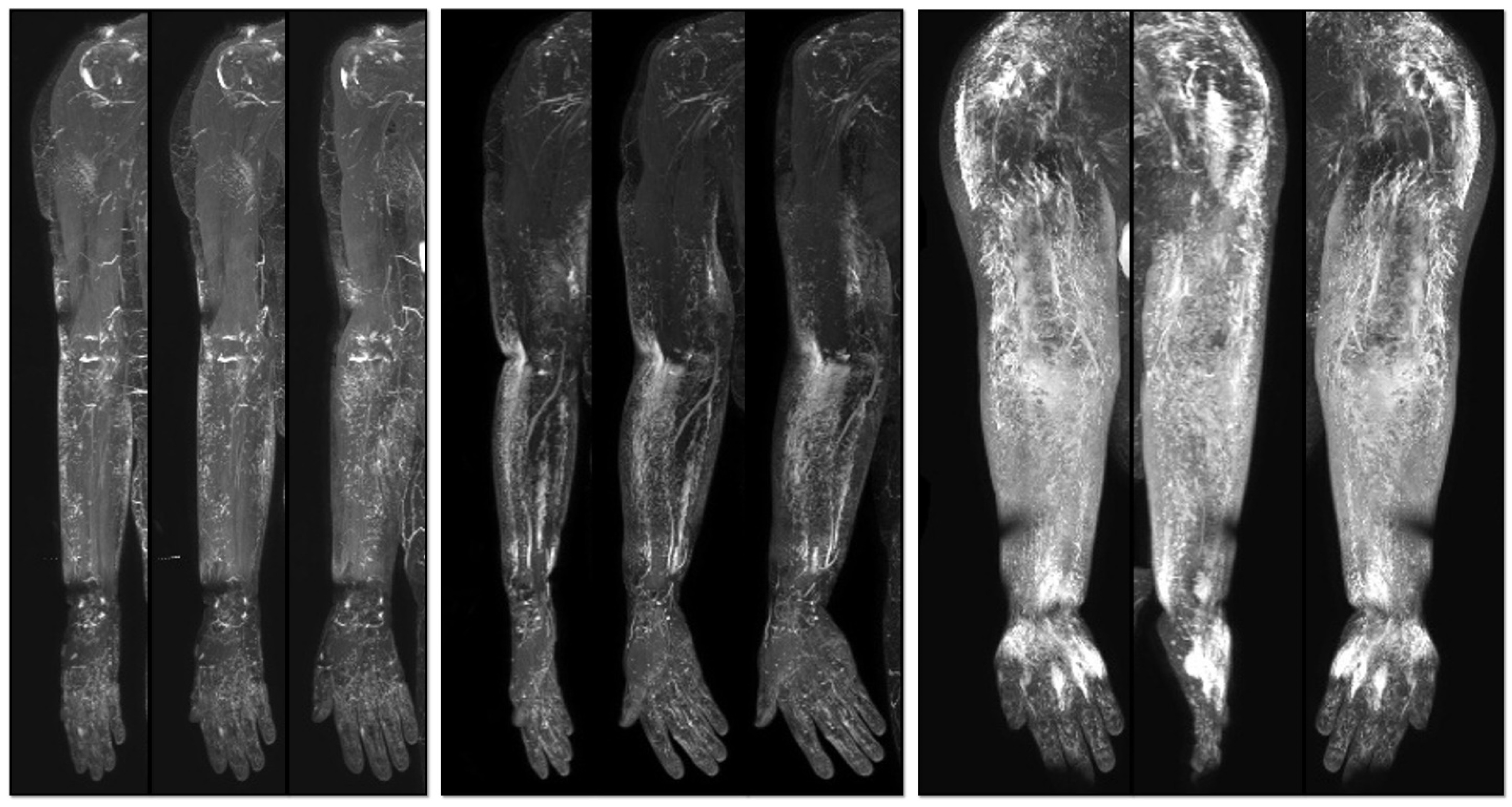
2. Methods
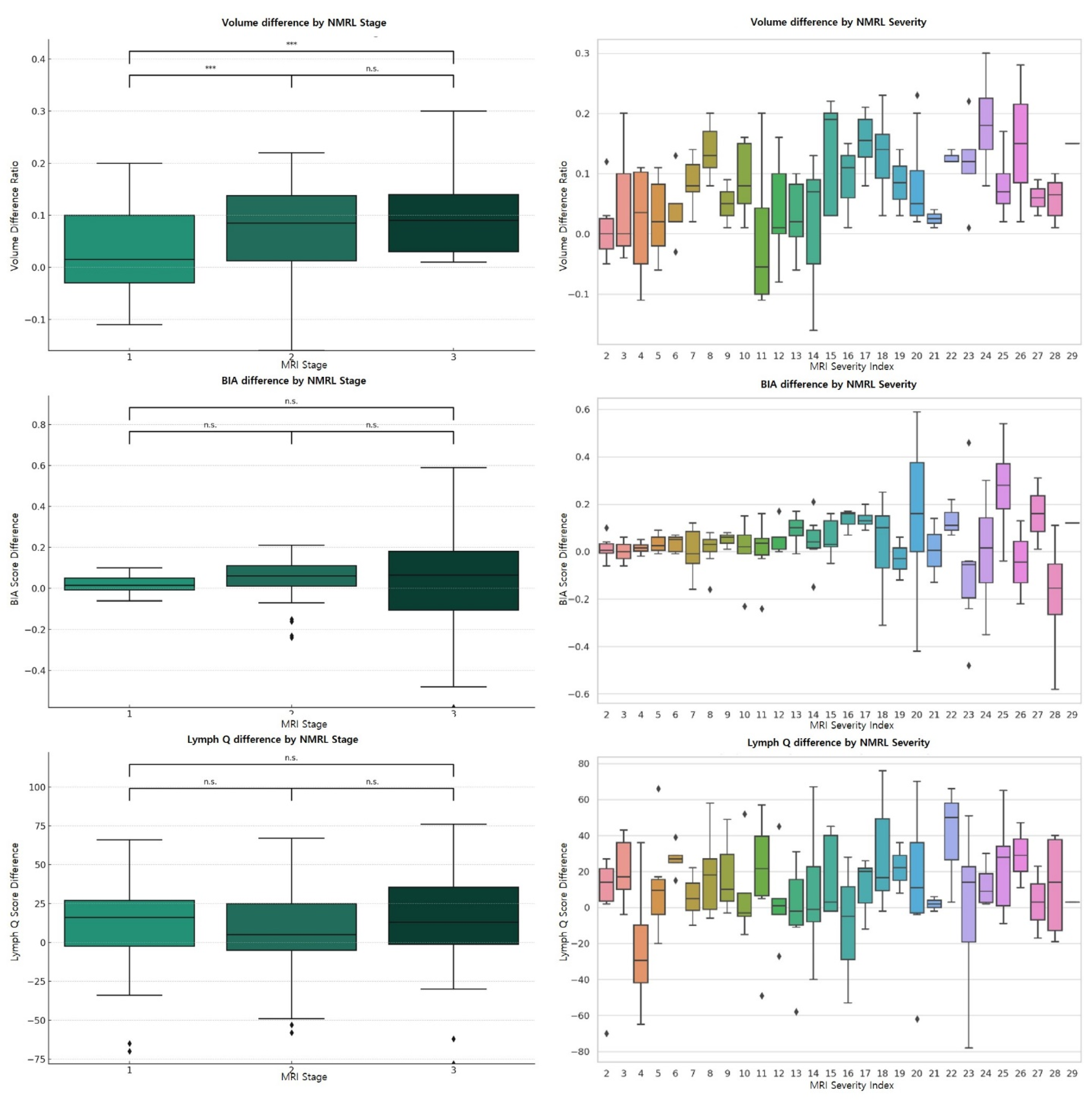
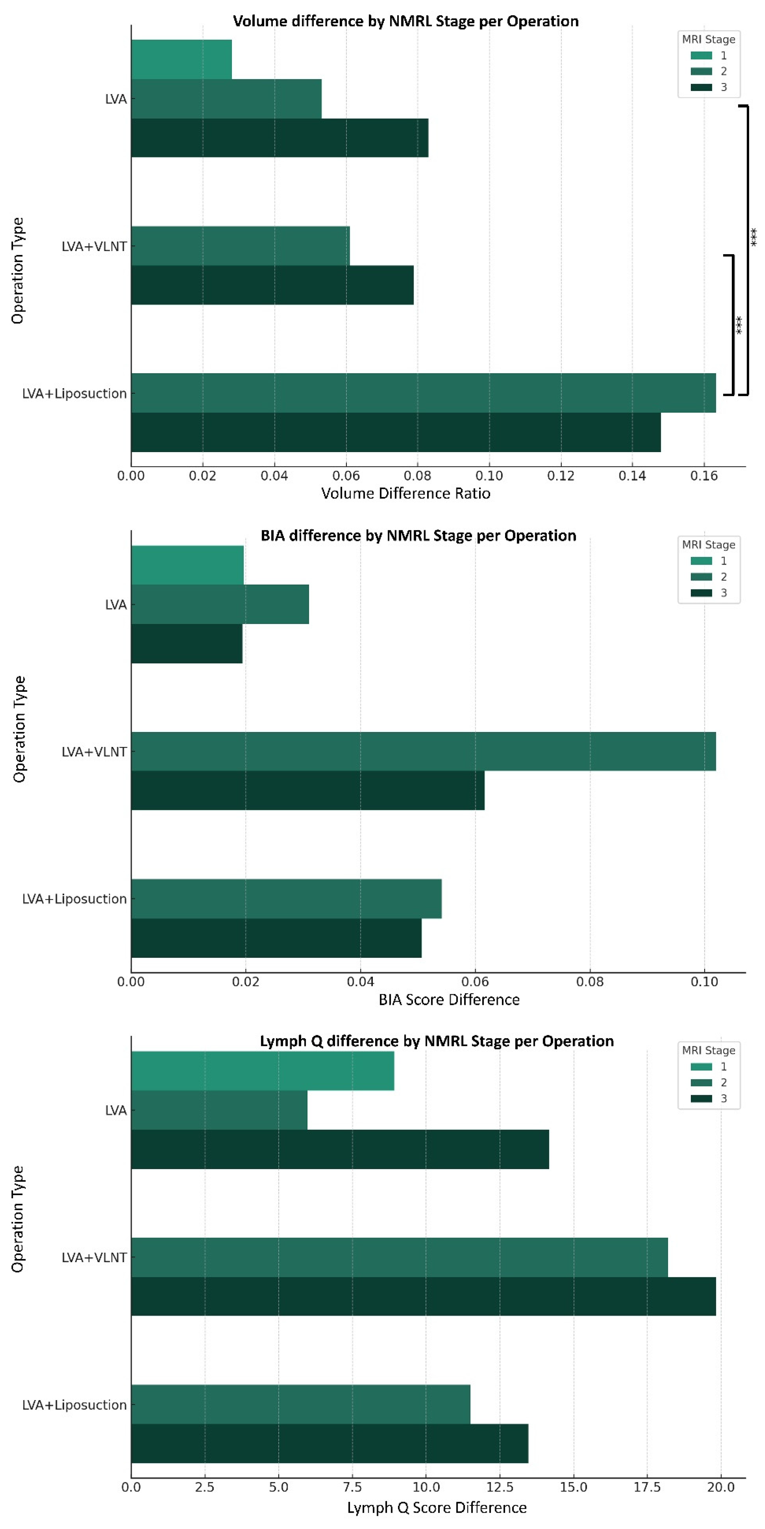
3. Results
4. Discussion
5. Conclusions
Supplementary Materials
Author Contributions
Funding
Institutional Review Board Statement
Informed Consent Statement
Data Availability Statement
Conflicts of Interest
References
- Mihara, M.; Hara, H.; Hayashi, Y.; Narushima, M.; Yamamoto, T.; Todokoro, T.; Iida, T.; Sawamoto, N.; Araki, J.; Kikuchi, K.; et al. Pathological steps of cancer-related lymphedema: Histological changes in the collecting lymphatic vessels after lymphadenectomy. PLoS ONE 2012, 7, e41126. [Google Scholar] [CrossRef] [PubMed]
- Card, A.; Crosby, M.A.; Liu, J.; Lindstrom, W.A.; Lucci, A.; Chang, D.W. Reduced incidence of breast cancer-related lymphedema following mastectomy and breast reconstruction versus mastectomy alone. Plast. Reconstr. Surg. 2012, 130, 1169–1178. [Google Scholar] [CrossRef] [PubMed]
- Taghian, N.R.; Miller, C.L.; Jammallo, L.S.; O’Toole, J.; Skolny, M.N. Lymphedema following breast cancer treatment and impact on quality of life: A review. Crit. Rev. Oncol. Hematol. 2014, 92, 227–234. [Google Scholar] [CrossRef] [PubMed]
- Koshima, I.; Inagawa, K.; Urushibara, K.; Moriguchi, T. Supermicrosurgical lymphaticovenular anastomosis for the treatment of lymphedema in the upper extremities. J. Reconstr. Microsurg. 2000, 16, 437–442. [Google Scholar] [CrossRef]
- Yamamoto, T.; Koshima, I. A prospective analysis of 100 consecutive lymphovenous bypass cases for treatment of extremity lymphedema. Plast. Reconstr. Surg. 2014, 133, 887e–888e. [Google Scholar] [CrossRef]
- Raju, A.; Chang, D.W. Vascularized lymph node transfer for treatment of lymphedema: A comprehensive literature review. Ann. Surg. 2015, 261, 1013–1023. [Google Scholar] [CrossRef]
- Park, J.K.-H.; Myung, Y. Two-team approach in lymphovenous anastomosis and omental lymph node flap harvest for upper limb lymphedema. Arch. Plast. Surg. 2021, 48, 131. [Google Scholar] [CrossRef]
- Bolletta, A.; di Taranto, G.; Losco, L.; Elia, R.; Sert, G.; Ribuffo, D.; Cigna, E.; Chen, H.C. Combined lymph node transfer and suction-assisted lipectomy in lymphedema treatment: A prospective study. Microsurgery 2022, 42, 433–440. [Google Scholar] [CrossRef]
- De Sire, A.; Losco, L.; Lippi, L.; Spadoni, D.; Kaciulyte, J.; Sert, G.; Ciamarra, P.; Marcasciano, M.; Cuomo, R.; Bolletta, A.; et al. Surgical Treatment and Rehabilitation Strategies for Upper and Lower Extremity Lymphedema: A Comprehensive Review. Medicina 2022, 58, 954. [Google Scholar] [CrossRef]
- Donahue, P.M.C.; MacKenzie, A.; Filipovic, A.; Koelmeyer, L. Advances in the prevention and treatment of breast cancer-related lymphedema. Breast Cancer Res. Treat. 2023, 200, 1–14. [Google Scholar] [CrossRef]
- Garza, R.M.; Beederman, M.; Chang, D.W. Physical and Functional Outcomes of Simultaneous Vascularized Lymph Node Transplant and Lymphovenous Bypass in the Treatment of Lymphedema. Plast. Reconstr. Surg. 2022, 150, 169–180. [Google Scholar] [CrossRef]
- Schaverien, M.V.; Asaad, M.; Selber, J.C.; Liu, J.; Chen, D.N.; Hall, M.S.; Butler, C.E. Outcomes of Vascularized Lymph Node Transplantation for Treatment of Lymphedema. J. Am. Coll. Surg. 2021, 232, 982–994. [Google Scholar] [CrossRef]
- Di Taranto, G.; Bolletta, A.; Chen, S.H.; Losco, L.; Elia, R.; Cigna, E.; Rubino, C.; Ribuffo, D.; Chen, H.C. A prospective study on combined lymphedema surgery: Gastroepiploic vascularized lymph nodes transfer and lymphaticovenous anastomosis followed by suction lipectomy. Microsurgery 2021, 41, 34–43. [Google Scholar] [CrossRef]
- Carl, H.M.; Walia, G.; Bello, R.; Clarke-Pearson, E.; Hassanein, A.H.; Cho, B.; Pedreira, R.; Sacks, J.M. Systematic Review of the Surgical Treatment of Extremity Lymphedema. J. Reconstr. Microsurg. 2017, 33, 412–425. [Google Scholar] [CrossRef]
- Chen, W.F.; Zhao, H.; Yamamoto, T.; Hara, H.; Ding, J. Indocyanine Green Lymphographic Evidence of Surgical Efficacy Following Microsurgical and Supermicrosurgical Lymphedema Reconstructions. J. Reconstr. Microsurg. 2016, 32, 688–698. [Google Scholar] [CrossRef]
- Spinelli, B.; Kallan, M.; Zhang, X.; Cheville, A.; Troxel, A.; Cohn, J.; Dean, L.; Sturgeon, K.; Evangelista, M.; Zhang, Z.; et al. Intra- and Interrater Reliability and Concurrent Validity of a New Tool for Assessment of Breast Cancer-Related Lymphedema of the Upper Extremity. Arch. Phys. Med. Rehabil. 2019, 100, 315–326. [Google Scholar] [CrossRef]
- Yamamoto, T.; Yamamoto, N.; Fuse, Y.; Narushima, M.; Koshima, I. Optimal Sites for Supermicrosurgical Lymphaticovenular Anastomosis: An Analysis of Lymphatic Vessel Detection Rates on 840 Surgical Fields in Lower Extremity Lymphedema Patients. Plast. Reconstr. Surg. 2018, 142, 924e–930e. [Google Scholar] [CrossRef] [PubMed]
- Hayashi, A.; Visconti, G.; Yamamoto, T.; Giacalone, G.; Hayashi, N.; Handa, M.; Yoshimatsu, H.; Salgarello, M. Intraoperative imaging of lymphatic vessel using ultra high-frequency ultrasound. J. Plast. Reconstr. Aesthet. Surg. 2018, 71, 778–780. [Google Scholar] [CrossRef] [PubMed]
- Myung, Y.; Park, S.; Kim, B.R.; Yang, E.J.; Park, J.K.-H.; Kang, Y. Validation of a Lymphedema Index Score Based on Noncontrast Magnetic Resonance Lymphangiography: Correlation with Clinical Staging and Indocyanine Green Lymphangiography. Lymphat. Res. Biol. 2023, 21, 70–77. [Google Scholar] [CrossRef] [PubMed]
- Devoogdt, N.; Van Kampen, M.; Geraerts, I.; Coremans, T.; Christiaens, M.R. Lymphoedema Functioning, Disability and Health questionnaire (Lymph-ICF): Reliability and validity. Phys. Ther. 2011, 91, 944–957. [Google Scholar] [CrossRef]
- Deptula, P.; Zhou, A.; Posternak, V.; He, H.; Nguyen, D. Multimodality Approach to Lymphedema Surgery Achieves and Maintains Normal Limb Volumes: A Treatment Algorithm to Optimize Outcomes. J. Clin. Med. 2022, 11, 598. [Google Scholar] [CrossRef] [PubMed]
- Chang, D.W.; Dayan, J.; Greene, A.K.; MacDonald, J.K.; Masia, J.; Mehrara, B.; Neligan, P.C.; Nguyen, D. Surgical Treatment of Lymphedema: A Systematic Review and Meta-Analysis of Controlled Trials. Results of a Consensus Conference. Plast. Reconstr. Surg. 2021, 147, 975–993. [Google Scholar] [CrossRef] [PubMed]
- Wolfs, J.A.; de Joode, L.G.; van der Hulst, R.R.; Qiu, S.S. Correlation between patency and clinical improvement after lymphaticovenous anastomosis (LVA) in breast cancer-related lymphedema: 12-month follow-up. Breast Cancer Res. Treat. 2020, 179, 131–138. [Google Scholar] [CrossRef] [PubMed]
- Rustad, K.C.; Chang, D.W. Surgical Approaches to the Prevention and Management of Breast Cancer–Related Lymphedema. Curr. Breast Cancer Rep. 2020, 12, 185–192. [Google Scholar] [CrossRef]
- Park, J.K.; Seo, J.; Yang, E.J.; Kang, Y.; Heo, C.Y.; Myung, Y. Association of lymphatic flow velocity with surgical outcomes in patients undergoing lymphovenous anastomosis for breast cancer-related lymphedema. Breast Cancer 2022, 29, 835–843. [Google Scholar] [CrossRef]
- Scaglioni, M.F.; Arvanitakis, M.; Chen, Y.C.; Giovanoli, P.; Chia-Shen Yang, J.; Chang, E.I. Comprehensive review of vascularized lymph node transfers for lymphedema: Outcomes and complications. Microsurgery 2018, 38, 222–229. [Google Scholar] [CrossRef]
- Imai, H.; Yoshida, S.; Mese, T.; Roh, S.; Fujita, A.; Sasaki, A.; Nagamatsu, S.; Koshima, I. Correlation between Lymphatic Surgery Outcome and Lymphatic Image-Staging or Clinical Severity in Patients with Lymphedema. J. Clin. Med. 2022, 11, 4979. [Google Scholar] [CrossRef]
- Schaverien, M.V.; Coroneos, C.J. Surgical Treatment of Lymphedema. Plast. Reconstr. Surg. 2019, 144, 738–758. [Google Scholar] [CrossRef]
- Cornelissen, A.J.M.; Beugels, J.; Ewalds, L.; Heuts, E.M.; Keuter, X.H.A.; Piatkowski, A.; van der Hulst, R.; Qiu Shao, S.S. Effect of Lymphaticovenous Anastomosis in Breast Cancer-Related Lymphedema: A Review of the Literature. Lymphat. Res. Biol. 2018, 16, 426–434. [Google Scholar] [CrossRef]
- Allen, R.J., Jr.; Cheng, M.H. Lymphedema surgery: Patient selection and an overview of surgical techniques. J. Surg. Oncol. 2016, 113, 923–931. [Google Scholar] [CrossRef]
- Rooke, T.W.; Felty, C.L. Lymphedema: Pathophysiology, classification, and clinical evaluation. In Handbook of Venous and Lymphatic Disorders; CRC Press: Boca Raton, FL, USA, 2017; pp. 707–712. [Google Scholar]
- Zampell, J.; Aschen, S.; Weitman, E.; Yan, A.; Elhadad, S.; Andrade, M.; Mehrara, B. Regulation of Adipogenesis by Lymphatic Fluid Stasis: Part I. Adipogenesis, Fibrosis, and Inflammation. Plast. Reconstr. Surg. 2012, 129, 825–834. [Google Scholar] [CrossRef] [PubMed]
- Imai, H.; Kawase, T.; Yoshida, S.; Mese, T.; Roh, S.; Fujita, A.; Uchiki, T.; Sasaki, A.; Nagamatsu, S.; Takazawa, A.; et al. Peripheral T cell profiling reveals downregulated exhaustion marker and increased diversity in lymphedema post-lymphatic venous anastomosis. iScience 2023, 26, 106822. [Google Scholar] [CrossRef] [PubMed]
- Park, J.H.; Myung, Y. Combination of lymphovenous anastomosis and lymph node transfer for breast cancer-related lymphedema. Plast. Aesthet. Res. 2023, 10, 40. [Google Scholar] [CrossRef]
- Brorson, H. Liposuction in Lymphedema Treatment. J. Reconstr. Microsurg. 2016, 32, 56–65. [Google Scholar] [CrossRef] [PubMed]


Disclaimer/Publisher’s Note: The statements, opinions and data contained in all publications are solely those of the individual author(s) and contributor(s) and not of MDPI and/or the editor(s). MDPI and/or the editor(s) disclaim responsibility for any injury to people or property resulting from any ideas, methods, instructions or products referred to in the content. |
© 2023 by the authors. Licensee MDPI, Basel, Switzerland. This article is an open access article distributed under the terms and conditions of the Creative Commons Attribution (CC BY) license (https://creativecommons.org/licenses/by/4.0/).
Share and Cite
Park, J.K.-h.; Choi, N.; Beom, J.; Lim, J.-Y.; Kang, Y.; Nam, S.-Y.; Myung, Y. Utilization of Noncontrast Magnetic Resonance Lymphangiography for Selection of Effective Surgical Method in Breast Cancer-Related Lymphedema. Medicina 2023, 59, 1656. https://doi.org/10.3390/medicina59091656
Park JK-h, Choi N, Beom J, Lim J-Y, Kang Y, Nam S-Y, Myung Y. Utilization of Noncontrast Magnetic Resonance Lymphangiography for Selection of Effective Surgical Method in Breast Cancer-Related Lymphedema. Medicina. 2023; 59(9):1656. https://doi.org/10.3390/medicina59091656
Chicago/Turabian StylePark, Joseph Kyu-hyung, Nakwon Choi, Jaewon Beom, Jae-Young Lim, Yusuhn Kang, Sun-Young Nam, and Yujin Myung. 2023. "Utilization of Noncontrast Magnetic Resonance Lymphangiography for Selection of Effective Surgical Method in Breast Cancer-Related Lymphedema" Medicina 59, no. 9: 1656. https://doi.org/10.3390/medicina59091656