
Simultaneous Free Fibula and Anterolateral Thigh Flap in Lower Extremity Reconstruction Following Osteomyelitis in a Trauma Patient: A Case Report
 , and
, and 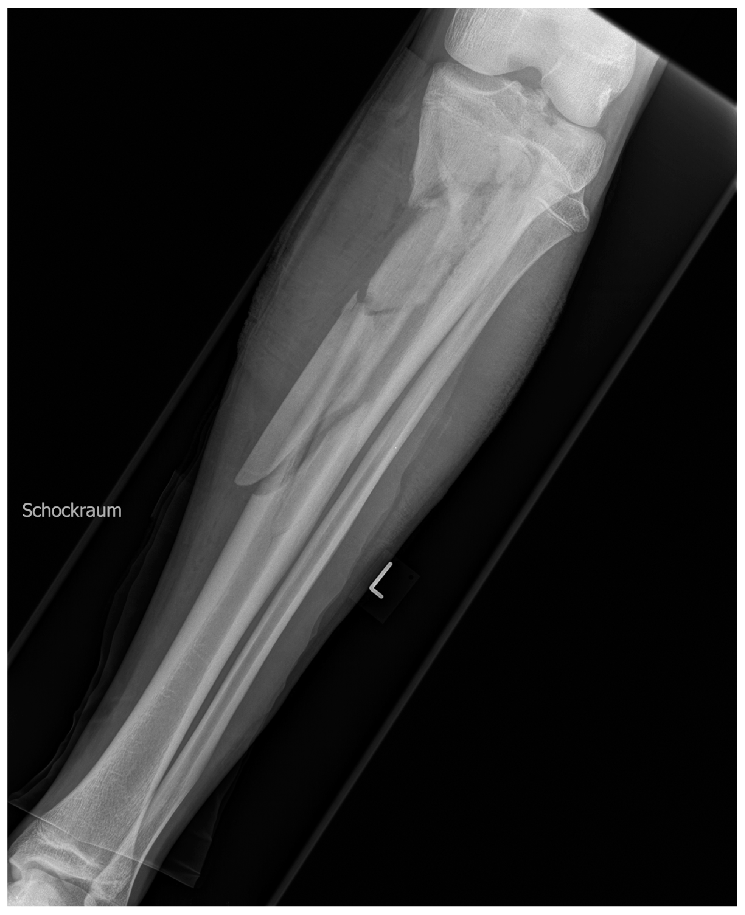
Abstract
:1. Introduction
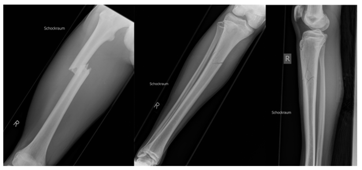
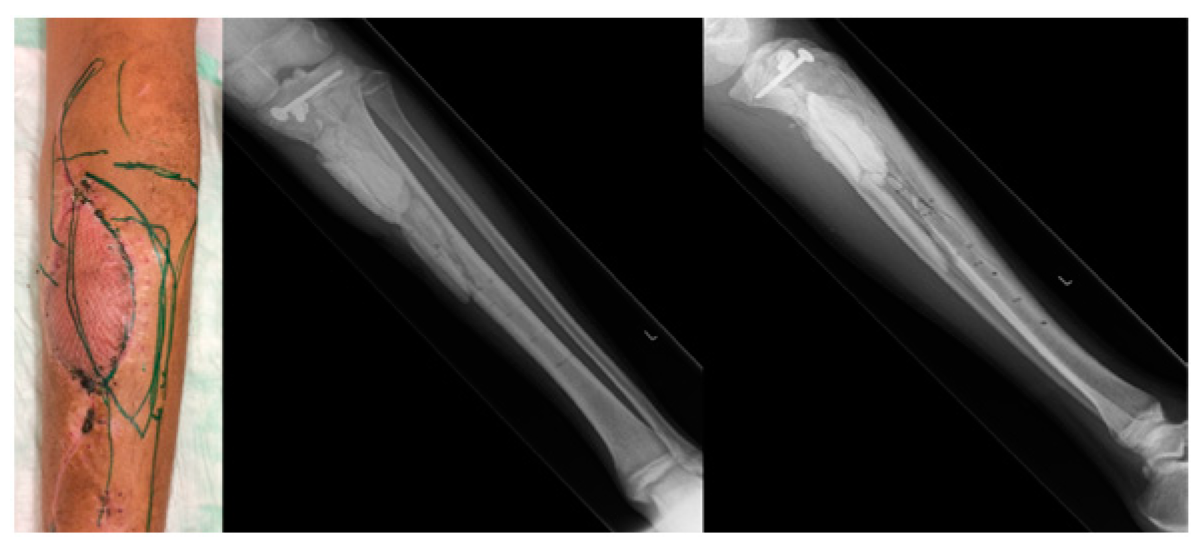
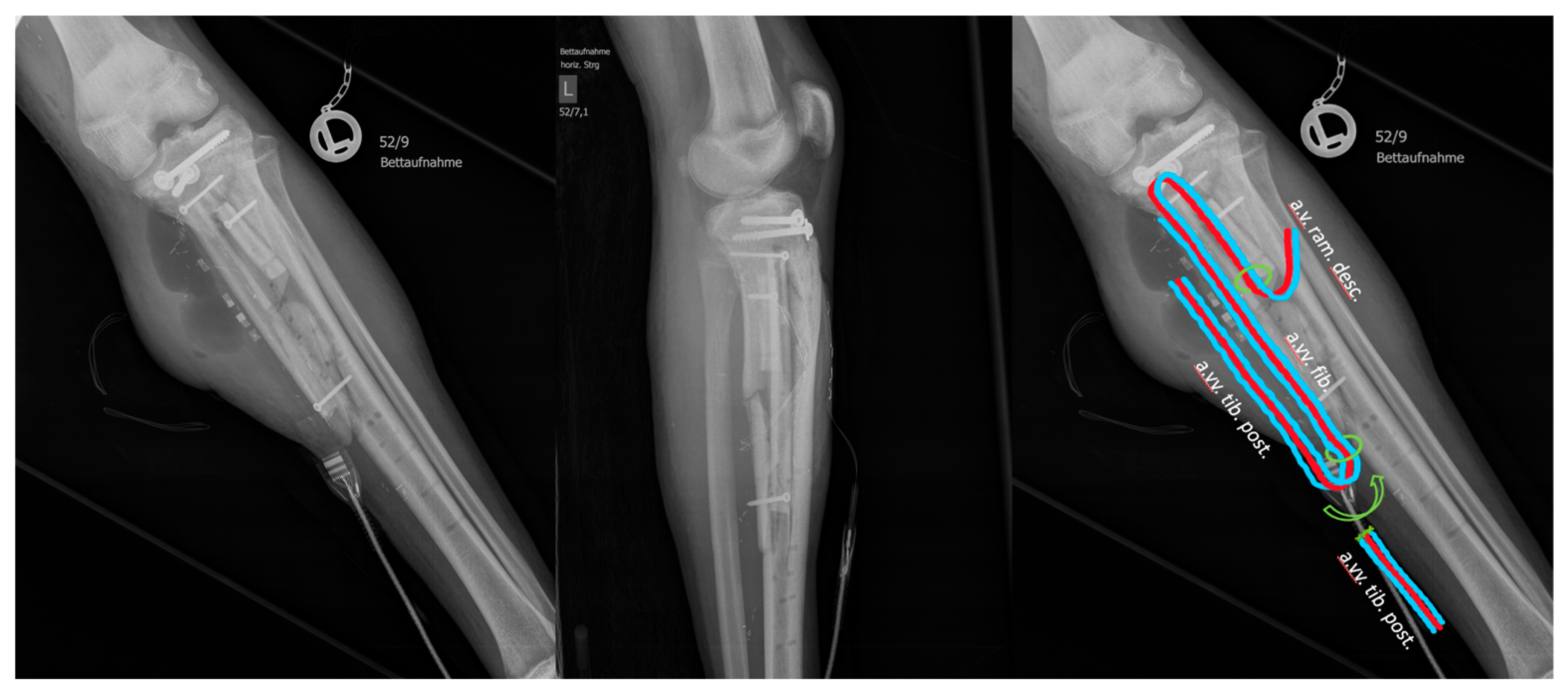
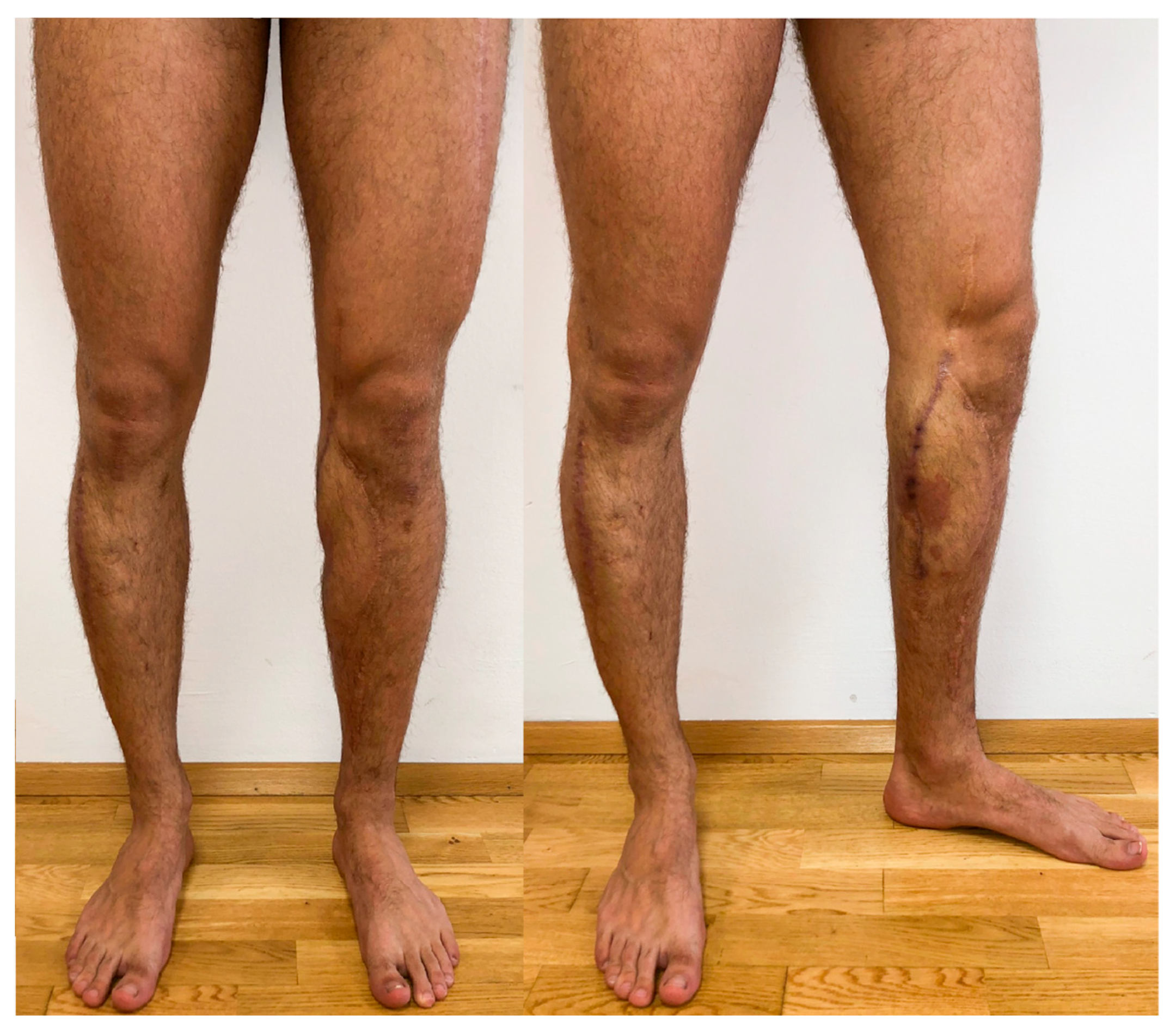
2. Case Description
3. Discussion
4. Conclusions
Author Contributions
Funding
Informed Consent Statement
Data Availability Statement
Conflicts of Interest
References
- Karami, R.A.; Ghieh, F.M.; Saghieh, S.S.; Ibrahim, A.E. The use of the fibula flap in post oncologic reconstruction of long bone in pediatric patients: A retrospective cohort study. J. Plast. Reconstr. Aesthet. Surg. 2021, 74, 2504–2511. [Google Scholar] [CrossRef]
- Yildirim, S.; Calikapan, G.T.; Akoz, T. Reconstructive microsurgery in pediatric population—A series of 25 patients. Microsurgery 2008, 28, 99–107. [Google Scholar] [CrossRef]
- Pototschnig, H.; Schaff, J.; Kovacs, L.; Biemer, E.; Papadopulos, N.A. The free osteofasciocutaneous fibula flap: Clinical applications and surgical considerations. Injury 2013, 44, 366–369. [Google Scholar] [CrossRef] [PubMed]
- Taylor, G.I.; Miller, G.D.; Ham, F.J. The free vascularized bone graft. A clinical extension of microvascular techniques. Plast. Reconstr. Surg. 1975, 55, 533–544. [Google Scholar] [CrossRef]
- Abed, Y.Y.; Beltrami, G.; Campanacci, D.A.; Innocenti, M.; Scoccianti, G.; Capanna, R. Biological reconstruction after resection of bone tumours around the knee: Long-term follow-up. J. Bone Joint Surg. Br. 2009, 91, 1366–1372. [Google Scholar] [CrossRef] [PubMed]
- Shi, L.L.; Garg, R.; Jawa, A.; Wang, Q.; Chai, Y.; Zeng, B.; Jupiter, J.B. Bony Hypertrophy in Vascularized Fibular Grafts. Hand 2022, 17, 106–113. [Google Scholar] [CrossRef]
- Feltri, P.; Solaro, L.; Di Martino, A.; Candrian, C.; Errani, C.; Filardo, G. Union, complication, reintervention and failure rates of surgical techniques for large diaphyseal defects: A systematic review and meta-analysis. Sci. Rep. 2022, 12, 9098. [Google Scholar] [CrossRef]
- Bodde, E.W.; de Visser, E.; Duysens, J.E.; Hartman, E.H. Donor-site morbidity after free vascularized autogenous fibular transfer: Subjective and quantitative analyses. Plast. Reconstr. Surg. 2003, 111, 2237–2242. [Google Scholar] [CrossRef] [PubMed] [Green Version]
- Hadouiri, N.; Feuvrier, D.; Pauchot, J.; Decavel, P.; Sagawa, Y. Donor site morbidity after vascularized fibula free flap: Gait analysis during prolonged walk conditions. Int. J. Oral Maxillofac. Surg. 2018, 47, 309–315. [Google Scholar] [CrossRef]
- Feuvrier, D.; Sagawa YJr Béliard, S.; Pauchot, J.; Decavel, P. Long-term donor-site morbidity after vascularized free fibula flap harvesting: Clinical and gait analysis. J. Plast. Reconstr. Aesthet. Surg. 2016, 69, 262–269. [Google Scholar] [CrossRef]
- Santamaría, E.; Galaso-Trujillo, J.R.; Palafox, D.; Mainardi, S.R.; García, R.A.; Romero, A.C.; Rangel, W.A.T. Donor-Site Morbidity Following Free Fibula Flap Harvest for Mandibular or Maxillary Reconstruction in Pediatric Patients. J. Craniofac. Surg. 2021, 32, e464–e468. [Google Scholar] [CrossRef]
- Kapukaya, R.; Ciloglu, O. Two-stage treatment with sliding fibular flap technique for chronic infected nonunion of the tibia. Chin. J. Traumatol. 2020, 23, 302–306. [Google Scholar] [CrossRef] [PubMed]
- Muramatsu, K.; Hashimoto, T.; Tominaga, Y.; Taguchi, T. Vascularized bone graft for oncological reconstruction of the extremities: Review of the biological advantages. Anticancer Res. 2014, 34, 2701–2707. [Google Scholar]
- Miyamoto, S.; Fujiki, M.; Nakatani, F.; Sakisaka, M.; Sakuraba, M. Free flow-through anterolateral thigh flap for complex knee defect including the popliteal artery. Microsurgery 2015, 35, 485–488. [Google Scholar] [CrossRef]
- Tsuge, I.; Yamanaka, H.; Katsube, M.; Sowa, Y.; Sakamoto, M.; Morimoto, N. Double-flap Mandibular Reconstruction around the Condylar Head Using Fibula and Anterolateral Thigh Flaps. Plast. Reconstr. Surg. Glob. Open 2022, 10, e4607. [Google Scholar] [CrossRef] [PubMed]
- Collins, J.; Ayeni, O.; Thoma, A. A systematic review of anterolateral thigh flap donor site morbidity. Can. J. Plast. Surg. 2012, 20, 17–23. [Google Scholar] [CrossRef] [Green Version]
- Yamamoto, T.; Yamamoto, N.; Kageyama, T.; Sakai, H.; Fuse, Y.; Tsuihiji, K.; Tsukuura, R. Definition of perforator flap: What does a “perforator” perforate? Glob. Health Med. 2019, 1, 114–116. [Google Scholar] [CrossRef] [PubMed]
- Hong, J.P.; Hur, J.; Kim, H.B.; Park, C.J.; Suh, H.P. The Use of Color Duplex Ultrasound for Local Perforator Flaps in the Extremity. J. Reconstr. Microsurg. 2022, 38, 233–237. [Google Scholar] [CrossRef] [PubMed]
- La Padula, S.; Pensato, R.; Pizza, C.; D’Andrea, F.; Roccaro, G.; Meningaud, J.P.; Hersant, B. The thoracodorsal artery perforator (TDAP) flap for the treatment of hidradenitis suppurativa of the axilla: A prospective comparative study. Plast. Reconstr. Surg. 2023. [Google Scholar] [CrossRef] [PubMed]
- Song, Y.G.; Chen, G.Z.; Song, Y.L. The free thigh flap: A new free flap concept based on the septocutaneous artery. Br. J. Plast. Surg. 1984, 37, 149–159. [Google Scholar] [CrossRef]
- La Padula, S.; Hersant, B.; Meningaud, J.P. Intraoperative use of indocyanine green angiography for selecting the more reliable perforator of the anterolateral thigh flap: A comparison study. Microsurgery 2018, 38, 738–744. [Google Scholar] [CrossRef] [PubMed]
- Trapero, A.; Pérez-García, A.; Andresen-Lorca, B.; Heredia-Alcalde, I. Anterolateral Thigh Free Flap Donor-Site Morbidity: A Retrospective Cohort Study. Plast. Aesthet. Nurs. 2022, 42, 152–155. [Google Scholar] [CrossRef] [PubMed]
- Jamari, J.; Ammarullah, M.I.; Santoso, G.; Sugiharto, S.; Supriyono, T.; Permana, M.S.; Winarni, T.I.; van der Heide, E. Adopted walking condition for computational simulation approach on bearing of hip joint prosthesis: Review over the past 30 years. Heliyon 2022, 8, e12050. [Google Scholar] [CrossRef] [PubMed]
- Giroux, P.A.; Hersant, B.; SidAhmed-Mezi, M.; Pizza, C.; La Padula, S.; Meningaud, J.P. The Outcomes Assessment of the Plasma Blade Technology in Upper Blepharoplasties: A Prospective Study on a Series of 25 Patients. Aesthetic. Plast. Surg. 2019, 43, 948–955. [Google Scholar] [CrossRef] [PubMed]
- Hersant, B.; Werkoff, G.; Sawan, D.; Sidahmed-Mezi, M.; Bosc, R.; La Padula, S.; Kalsoum, S.; Ouidir, N.; Meningaud, J.P.; Belkacemi, Y. Carbon dioxide laser treatment for vulvovaginal atrophy in women treated for breast cancer: Preliminary results of the feasibility EPIONE trial. Ann. Chir. Plast. Esthet. 2020, 65, e23–e31. [Google Scholar] [CrossRef] [PubMed]
- Palao, R.; Gallego-Escuredo, J.M.; Bohbot, S. Postabdominoplasty Scar Improvement after a Single Session with an Automated 1210-nm Laser. Plast. Reconstr. Surg. Glob. Open 2023, 11, e4866. [Google Scholar] [CrossRef]
- Monstrey, S.; Middelkoop, E.; Vranckx, J.J.; Bassetto, F.; Ziegler, U.E.; Meaume, S.; Téot, L. Updated scar management practical guidelines: Non-invasive and invasive measures. J. Plast. Reconstr. Aesthet. Surg. 2014, 67, 1017–1025. [Google Scholar] [CrossRef]


Disclaimer/Publisher’s Note: The statements, opinions and data contained in all publications are solely those of the individual author(s) and contributor(s) and not of MDPI and/or the editor(s). MDPI and/or the editor(s) disclaim responsibility for any injury to people or property resulting from any ideas, methods, instructions or products referred to in the content. |
© 2023 by the authors. Licensee MDPI, Basel, Switzerland. This article is an open access article distributed under the terms and conditions of the Creative Commons Attribution (CC BY) license (https://creativecommons.org/licenses/by/4.0/).
Share and Cite
Voljc, T.; Schintler, M.; Vasilyeva, A.; Kamolz, L.-P.; Buerger, H. Simultaneous Free Fibula and Anterolateral Thigh Flap in Lower Extremity Reconstruction Following Osteomyelitis in a Trauma Patient: A Case Report. Medicina 2023, 59, 1206. https://doi.org/10.3390/medicina59071206
Voljc T, Schintler M, Vasilyeva A, Kamolz L-P, Buerger H. Simultaneous Free Fibula and Anterolateral Thigh Flap in Lower Extremity Reconstruction Following Osteomyelitis in a Trauma Patient: A Case Report. Medicina. 2023; 59(7):1206. https://doi.org/10.3390/medicina59071206
Chicago/Turabian StyleVoljc, Tadej, Michael Schintler, Anna Vasilyeva, Lars-Peter Kamolz, and Heinz Buerger. 2023. "Simultaneous Free Fibula and Anterolateral Thigh Flap in Lower Extremity Reconstruction Following Osteomyelitis in a Trauma Patient: A Case Report" Medicina 59, no. 7: 1206. https://doi.org/10.3390/medicina59071206