

Reconstruction of High-Grade Trochlea Dysplasia in a Young Female with Recurrent Patella Dislocation: A Case Report

 and
and 
Abstract
:1. Introduction
2. Clinical Cases
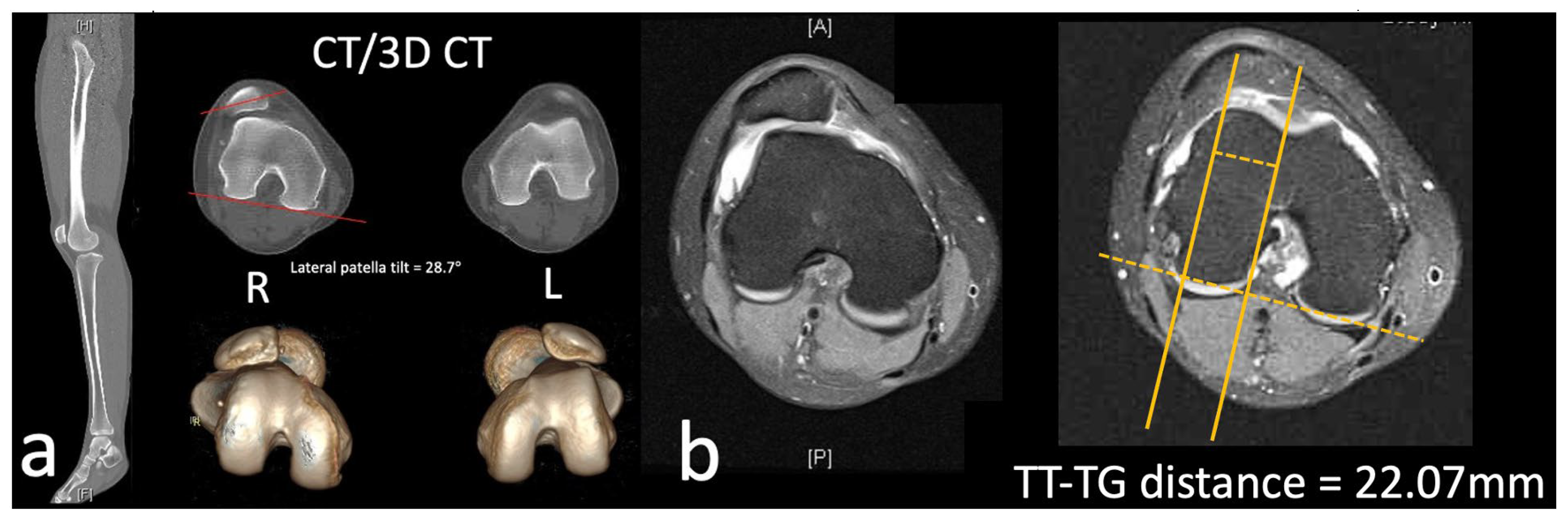
2.1. Case
2.2. Surgical Procedure
3. Discussion
3.1. Trochlea Dysplasia
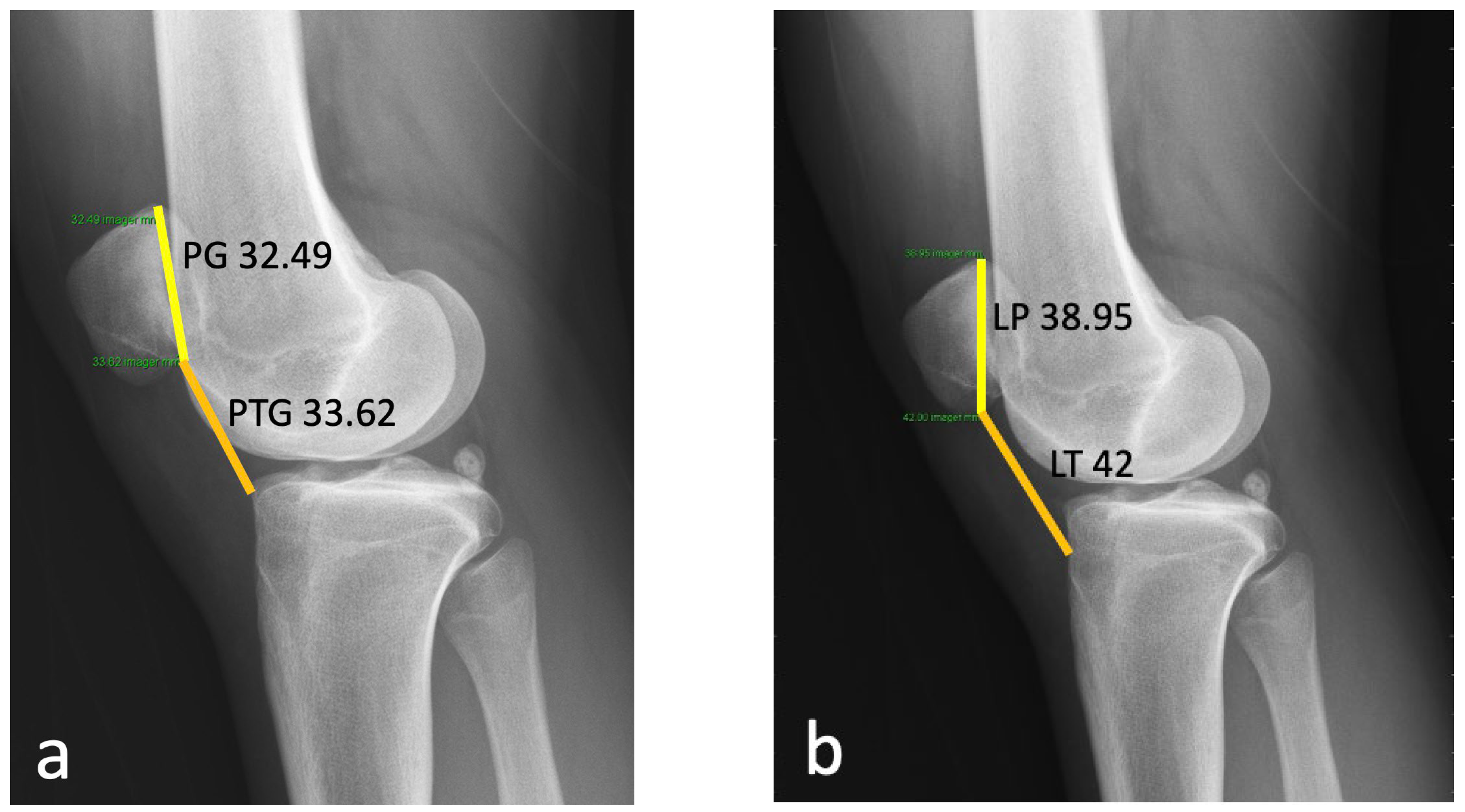
3.2. Patella Alta
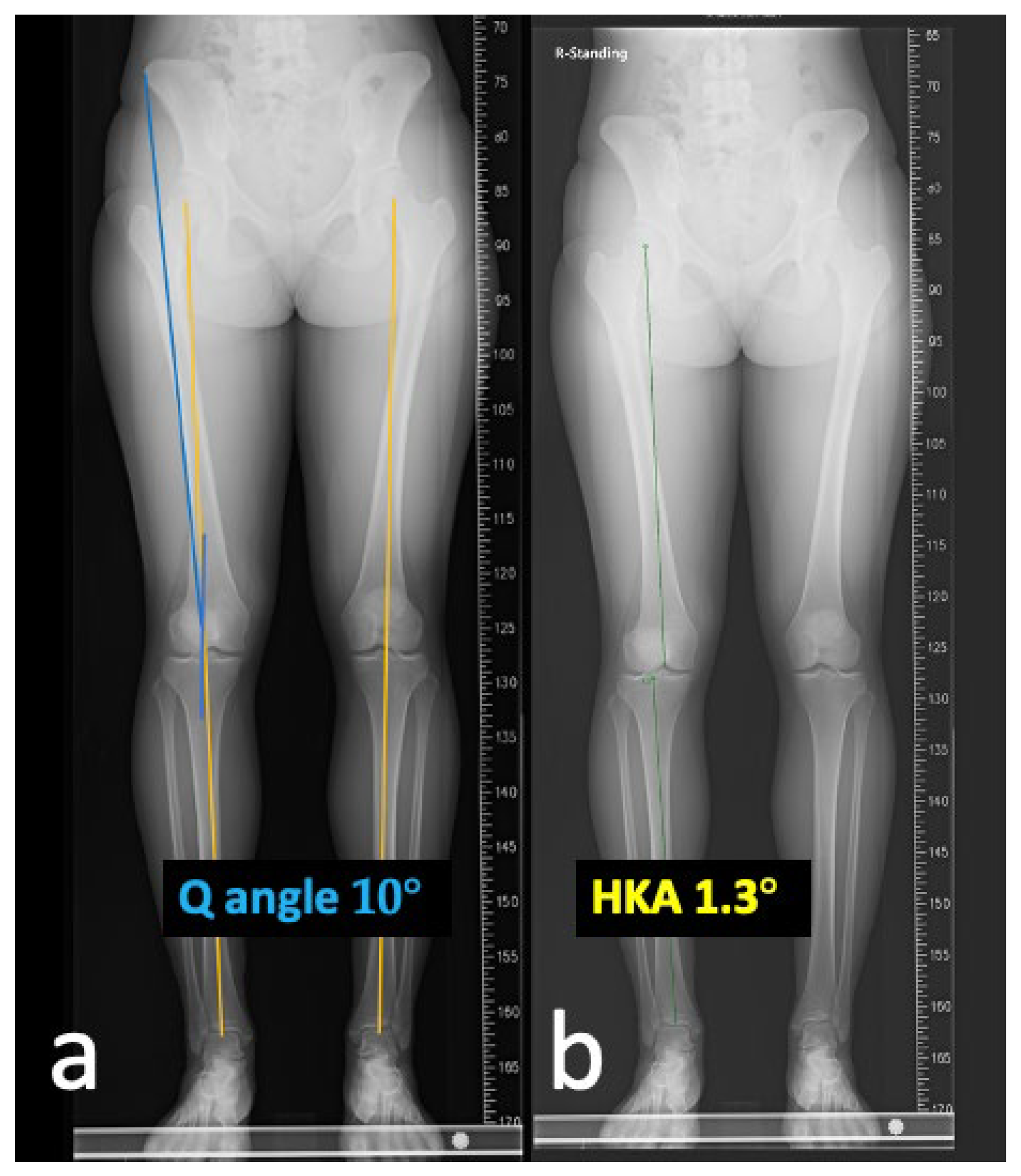
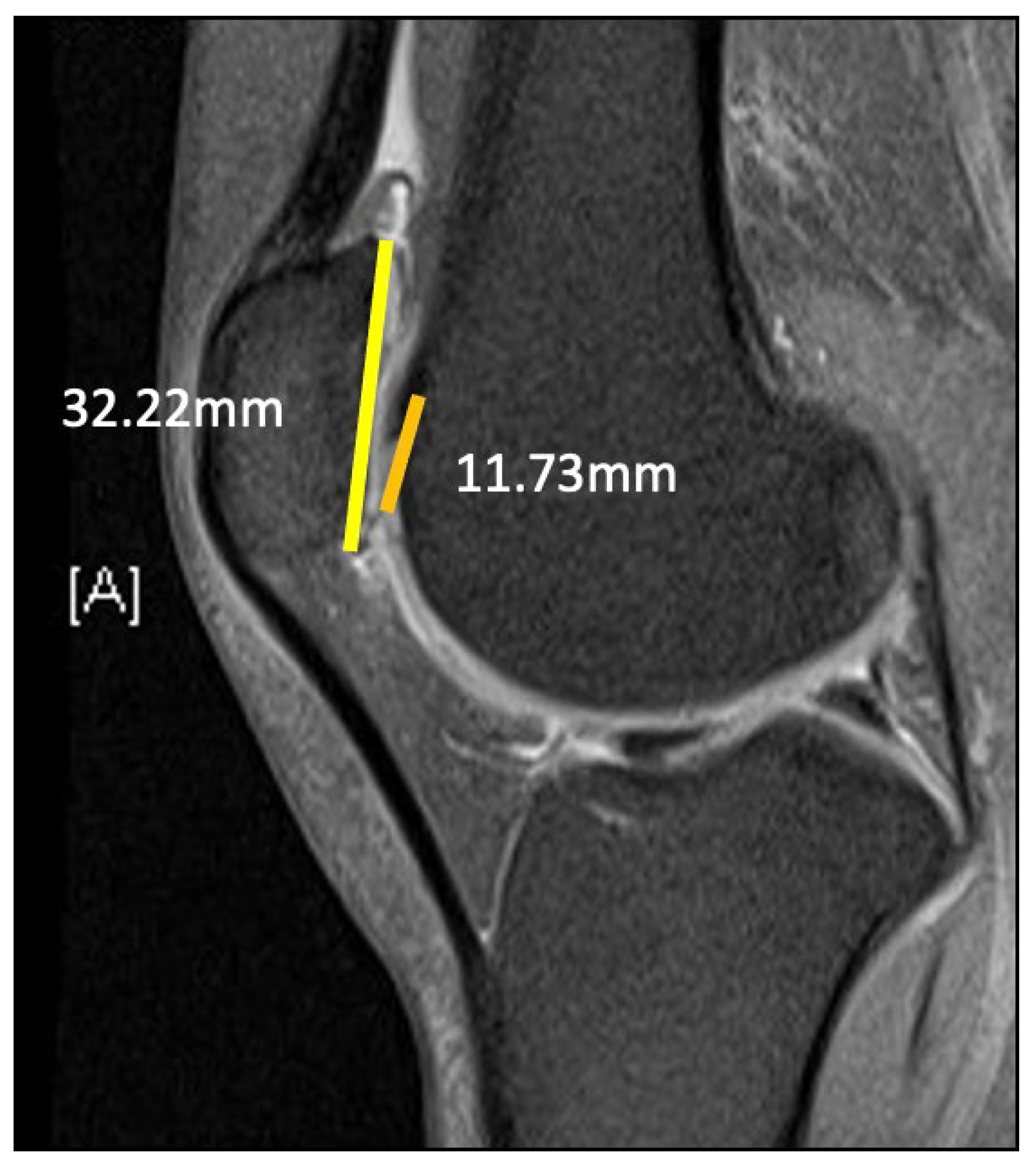
3.3. TT–TG Distance
3.4. Lateral Patella Tilt
3.5. MPFL and MQTFL Reconstruction
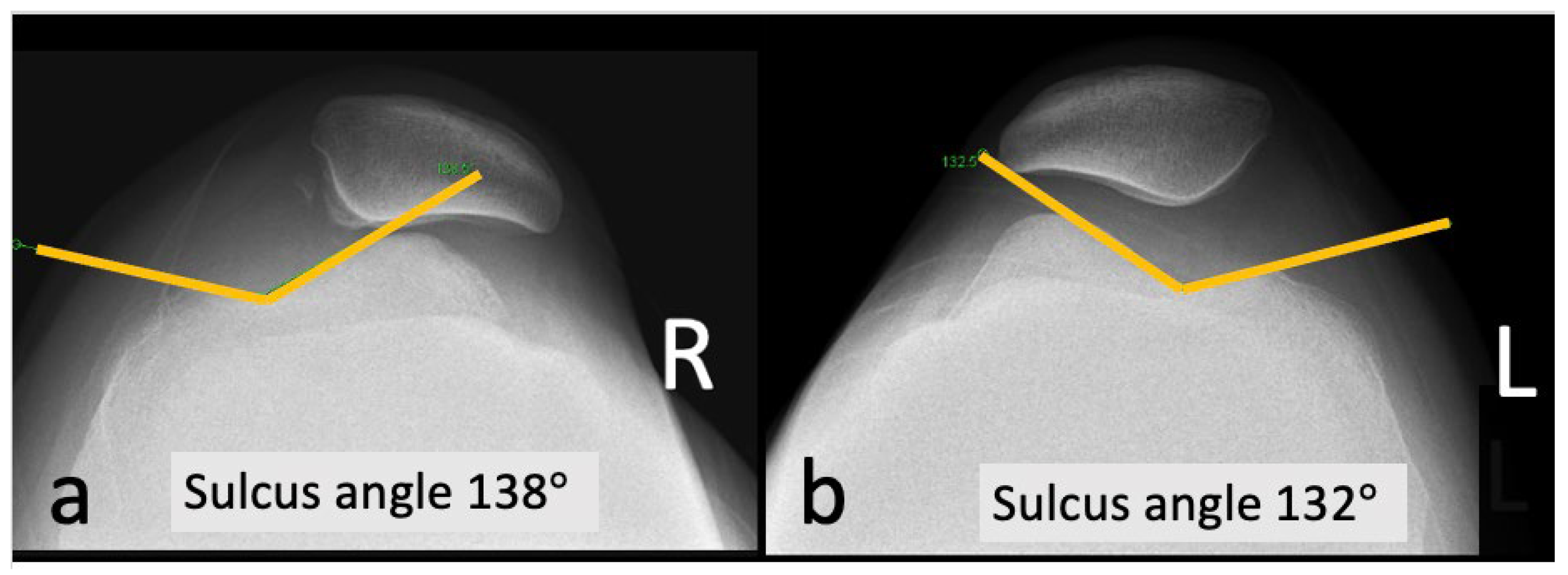
3.6. Sulcus Angle
3.7. Limitations
4. Conclusions
Author Contributions
Funding
Institutional Review Board Statement
Informed Consent Statement
Data Availability Statement
Acknowledgments
Conflicts of Interest
References
- Colvin, A.C.; West, R.V. Current Concepts Review: Patellar Instability. J. Bone Jt. Surg. 2008, 90, 2751–2762. [Google Scholar] [CrossRef] [PubMed]
- LaPrade, R.F.; Cram, T.R.; James, E.W.; Rasmussen, M.T. Trochlear Dysplasia and the Role of Trochleoplasty. Clin. J. Sport. Med. 2014, 33, 531–545. [Google Scholar] [CrossRef] [PubMed]
- Clifton, R.; Ng, C.Y.; Nutton, R.W. What is the role of lateral retinacular release? J. Bone Jt. Surg. 2010, 92-B, 1–6. [Google Scholar] [CrossRef] [PubMed]
- Dejour, H.; Walch, G.; Nove-Josserand, L.; Guier, C. Factors of patellar instability: An anatomic radiographic study. Knee Surg Sport. Traumatol Arthrosc 1994, 2, 19–26. [Google Scholar] [CrossRef] [PubMed]
- Dejour, D.H.; Mesnard, G.; de Sanctis, E.G. Updated treatment guidelines for patellar instability: “un menu à la carte”. J. Exp. Orthop. 2021, 8, 109. [Google Scholar] [CrossRef]
- McDaniel, G.; Mitchell, K.L.; Charles, C.; Kraus, V.B. A comparison of five approaches to measurement of anatomic knee alignment from radiographs. Osteoarthr. Cartil. 2010, 18, 273–277. [Google Scholar] [CrossRef]
- Brattstrom, H. Shape of the intercondylar groove normally and in recurrent dislocation of patella: A clinical and X-ray anatomical investigation. Acta Orthop Scand Suppl 1964, 35, 1–148. [Google Scholar] [CrossRef]
- Biedert, R.M.; Tscholl, P.M. Patella Alta: A Comprehensive Review of Current Knowledge. Am. J. Orthop. 2017, 46, 290–300. [Google Scholar]
- Mayer, C.; Magnussen, R.A.; Servien, E.; Demey, G.; Jacobi, M.; Neyret, P.; Lustig, S. Patellar tendon tenodesis in association with tibial tubercle distalisation with patella alta for the treatment of episodic patellar dislocation with patella alta. Am. J. Sport. Med. 2012, 40, 346–351. [Google Scholar] [CrossRef]
- Biedert, R.M.; Albrecht, S. The Patellotrochlear Index: A New Index for Assessing Patellar Height. Knee Surg Sport. Traumatol Arthrosc 2006, 14, 707–712. [Google Scholar] [CrossRef]
- Floyd, E.R.; Ebert, N.J.; Carlson, G.B.; Monson, J.K.; LaPrade, R.F. Medial Patellofemoral Reconstruction Using Quadriceps Tendon Autograft, Tibial Tubercle Osteotomy, and Sulcus-Deepening Trochleoplasty for Patellar Instability. Arthrosc. Tech. 2021, 10, 1249–1256. [Google Scholar] [CrossRef]
- Insall, J.; Goldberg, V.; Salvati, E. Recurrent dislocation and the high-riding patella. Clin. Orthop. Relat. Res. 1972, 88, 67–69. [Google Scholar] [CrossRef] [PubMed]
- Munch, J.L.; Sullivan, J.P.; Nguyen, J.T.; Mintz, D.; Green, D.W.; Stein, B.E.S.; Strickland, S. Patella articular overlap on MRI is a simple alternative to conventional measurements of patellar height. Orthop. J. Sport. Med. 2016, 4, 2325967116656328. [Google Scholar] [CrossRef] [PubMed]
- Caton, J.H.; Dejour, D. Tibial tubercle osteotomy in patellofemoral instability and in patellar height abnormality. Int. Orthop. 2010, 34, 305–309. [Google Scholar] [CrossRef] [PubMed]
- Barnett, A.J.; Prentice, M.; Mandalia, V.; Wakeley, C.J.; Eldridge, J.D.J. Patellar height measurement in trochlear dysplasia. Knee Surg Sport. Traumatol Arthrosc 2009, 17, 1412–1415. [Google Scholar] [CrossRef] [PubMed]
- Ali, S.A.; Helmer, R.; Terk, M.R. Patella Alta: Lack of Correlation between Patellotrochlear Cartilage Congruence and Commonly Used Patellar Height Ratios. Am. J. Roentgenol. 2009, 193, 1361–1366. [Google Scholar] [CrossRef] [PubMed]
- Tan, S.H.S.; Lim, B.Y.; Chng, K.S.J.; Doshi, C.; Wong, F.K.L.; Lim, A.K.S.; Hui, J.H. The difference between computed tomography and magnetic resonance imaging measurements of tibial tubercle-trochlear groove distance for patients with or without patellofemoral instability: A systematic review and meta-analysis. J. Knee Surg. 2020, 33, 768–776. [Google Scholar] [CrossRef]
- Vairo, G.L.; Moya-Angeler, J.; Siorta, M.A.; Anderson, A.H.; Sherbondy, P.S. Tibial tubercle-trochlear groove distance is a reliable and accurate indica- tor of patellofemoral instability. Clin. Orthop. Relat Res. 2019, 477, 1450–1458. [Google Scholar] [CrossRef]
- Camp, C.L.; Stuart, M.J.; Krych, A.J.; Levy, B.A.; Bond, J.R.; Collins, M.S.; Dahm, D.L. CT and MRI Measurements of Tibial Tubercle–Trochlear Groove Distances Are Not Equivalent in Patients with Patellar Instability. Am. Orthop. Soc. Sport. Med. 2013, 41, 1835–1840. [Google Scholar] [CrossRef]
- Ho, C.P.; James, E.W.; Surowiec, R.K.; Gatlin, C.C.; Ellman, M.B.; Cram, T.R.; Dornan, G.J.; LaPrade, R.F. Systematic Technique-Dependent Differences in CT Versus MRI Measurement of the Tibial Tubercle–Trochlear Groove Distance. Am. J. Sport. Med. 2015, 43, 675–682. [Google Scholar] [CrossRef]
- Schöttle, P.B.; Zanetti, M.; Seifert, B.; Pfirrmann, C.W.A. The tibial tuberosity-trochlear groove distance; a comparative study between CT and MRI scanning. Knee 2006, 13, 26–31. [Google Scholar] [CrossRef] [PubMed]
- Fulkerson, J.P. Diagnosis and treatment of patients with patellofemoral pain. Am. J. Sport. Med. 2002, 30, 447–456. [Google Scholar] [CrossRef] [PubMed]
- Migliorini, F.; Maffulli, N.; Eschweiler, J.; Quack, V.; Tingart, M.; Driessen, A. Lateral retinacular release combined with MPFL reconstruction for patellofemoral instability: A systematic review. Arch. Orthop. Trauma Surg. 2021, 141, 283–292. [Google Scholar] [CrossRef] [PubMed]
- Desio, S.M.; Burks, R.T.; Bachus, K.N. Soft tissue restraints to lateral patellar translation in the human knee. Am. J. Sports. Med. 1998, 26, 59–65. [Google Scholar] [CrossRef]
- Camp, C.L.; Krych, A.J.; Dahm, D.L.; Levy, B.A.; Stuart, M.J. Medial patellofemoral ligament repair for recurrent patellar dislocation. Am. J. Sport. Med. 2010, 38, 2248–2254. [Google Scholar] [CrossRef]
- Parikh, S.N.; Nathan, S.T.; Wall, E.J.; Eismann, E.A. Complications of medial patellofemoral ligament reconstruction in young patients. Am. J. Sport. Med. 2013, 41, 1030–1038. [Google Scholar] [CrossRef]
- Spang, R.; Egan, J.; Hanna, P.; Lechtig, A.; Haber, D.; DeAngelis, J.P.; Nazarian, A.; Ramappa, A.J. Comparison of Patellofemoral Kinematics and Stability after Medial Patellofemoral Ligament and Medial Quadriceps Tendon–Femoral Ligament Reconstruction. Am. J. Sport. Med. 2020, 48, 2252–2259. [Google Scholar] [CrossRef]
- Tecklenburg, K.; Dejour, D.; Hoser, C.; Fink, C. Bony and cartilaginous anatomy of the patellofemoral joint. Knee Surg Sport. Traumatol Arthrosc 2006, 14, 235–240. [Google Scholar] [CrossRef]


Disclaimer/Publisher’s Note: The statements, opinions and data contained in all publications are solely those of the individual author(s) and contributor(s) and not of MDPI and/or the editor(s). MDPI and/or the editor(s) disclaim responsibility for any injury to people or property resulting from any ideas, methods, instructions or products referred to in the content. |
© 2023 by the authors. Licensee MDPI, Basel, Switzerland. This article is an open access article distributed under the terms and conditions of the Creative Commons Attribution (CC BY) license (https://creativecommons.org/licenses/by/4.0/).
Share and Cite
Wu, C.-H.; Hsu, K.-Y.; Cheng, Y.-H.; Yang, C.-P.; Sheu, H.; Chang, S.-S.; Chen, C.-Y.; Chiu, C.-H. Reconstruction of High-Grade Trochlea Dysplasia in a Young Female with Recurrent Patella Dislocation: A Case Report. Medicina 2023, 59, 986. https://doi.org/10.3390/medicina59050986
Wu C-H, Hsu K-Y, Cheng Y-H, Yang C-P, Sheu H, Chang S-S, Chen C-Y, Chiu C-H. Reconstruction of High-Grade Trochlea Dysplasia in a Young Female with Recurrent Patella Dislocation: A Case Report. Medicina. 2023; 59(5):986. https://doi.org/10.3390/medicina59050986
Chicago/Turabian StyleWu, Chih-Hsuan, Kuo-Yao Hsu, You-Hung Cheng, Cheng-Pang Yang, Huan Sheu, Shih-Sheng Chang, Chao-Yu Chen, and Chih-Hao Chiu. 2023. "Reconstruction of High-Grade Trochlea Dysplasia in a Young Female with Recurrent Patella Dislocation: A Case Report" Medicina 59, no. 5: 986. https://doi.org/10.3390/medicina59050986