
Influence of Different Orthodontic Brackets on Cytokine and Cortisol Profile

Abstract
:1. Introduction
2. Materials and Methods
2.1. Collection of Saliva
2.2. Clinical Assessment of Oral Cavity
2.3. Cytokine Measurement
2.4. Statistical Analysis
3. Results
4. Discussion
5. Conclusions
Author Contributions
Funding
Institutional Review Board Statement
Informed Consent Statement
Data Availability Statement
Conflicts of Interest
References
- Chaushu, S.; Klein, Y.; Mandelboim, O.; Barenholz, Y.; Fleissig, O. Immune Changes Induced by Orthodontic Forces: A Critical Review. J. Dent. Res. 2022, 101, 11–20. [Google Scholar] [CrossRef] [PubMed]
- Luo, X.; Wan, Q.; Cheng, L.; Xu, R. Mechanisms of bone remodeling and therapeutic strategies in chronic apical periodontitis. Front. Cell. Infect. Microbiol. 2022, 12, 908859. [Google Scholar] [CrossRef] [PubMed]
- Tsukasaki, M.; Takayanagi, H. Osteoclast biology in the single-cell era. Inflamm. Regen. 2022, 42, 27. [Google Scholar] [CrossRef] [PubMed]
- Xie, Y.; Tang, Q.; Yu, S.; Zheng, W.; Chen, G.; Huang, X.; Chen, L. Orthodontic Force-Induced BMAL1 in PDLCs Is a Vital Osteoclastic Activator. J. Dent. Res. 2022, 101, 177–186. [Google Scholar] [CrossRef]
- Zhang, R.; Peng, S.; Zhu, G. The role of secreted osteoclastogenic factor of activated T cells in bone remodeling. Jpn. Dent. Sci. Rev. 2022, 58, 227–232. [Google Scholar] [CrossRef]
- Lee, S.H.; Kim, T.S.; Choi, Y.; Lorenzo, J. Osteoimmunology: Cytokines and the skeletal system. BMB Rep. 2008, 41, 495–510. [Google Scholar] [CrossRef] [Green Version]
- Padisar, P.; Hashemi, R.; Naseh, M.; Nikfarjam, B.A.; Mohammadi, M. Assessment of tumor necrosis factor alpha (TNFα) and interleukin 6 level in gingival crevicular fluid during orthodontic tooth movement: A randomized split-mouth clinical trial. Electron. Physician 2018, 10, 7146–7154. [Google Scholar] [CrossRef] [Green Version]
- Jeon, H.H.; Yang, C.Y.; Shin, M.K.; Wang, J.; Patel, J.H.; Chung, C.H.; Graves, D.T. Osteoblast lineage cells and periodontal ligament fibroblasts regulate orthodontic tooth movement that is dependent on Nuclear Factor-kappa B (NF-kB) activation. Angle Orthod. 2021, 91, 664–671. [Google Scholar] [CrossRef]
- Behm, C.; Nemec, M.; Weissinger, F.; Rausch, M.A.; Andrukhov, O.; Jonke, E. MMPs and TIMPs Expression Levels in the Periodontal Ligament during Orthodontic Tooth Movement: A Systematic Review of In Vitro and In Vivo Studies. Int. J. Mol. Sci. 2021, 22, 6967. [Google Scholar] [CrossRef]
- Florencio-Silva, R.; Sasso, G.R.; Sasso-Cerri, E.; Simões, M.J.; Cerri, P.S. Biology of Bone Tissue: Structure. Function. and Factors That Influence Bone Cells. Biomed. Res. Int. 2015, 2015, 421746. [Google Scholar] [CrossRef] [Green Version]
- Di Domenico, M.; D’apuzzo, F.; Feola, A.; Cito, L.; Monsurrò, A.; Pierantoni, G.M.; Berrino, L.; De Rosa, A.; Polimeni, A.; Perillo, L. Cytokines and VEGF induction in orthodontic movement in animal models. J. Biomed. Biotechnol. 2012, 2012, 201689. [Google Scholar] [CrossRef] [PubMed]
- Huang, H.; Williams, R.C.; Kyrkanides, S. Accelerated orthodontic tooth movement: Molecular mechanisms. Am. J. Orthod. Dentofac. Orthop. 2014, 146, 620e32. [Google Scholar] [CrossRef] [PubMed]
- Lee, B. Force and tooth movement. Aust. Orthod. J. 2007, 23, 155. [Google Scholar] [PubMed]
- Baloul, S.S. Osteoclastogenesis and osteogenesis during tooth movement. Front. Oral Biol. 2016, 18, 75e9. [Google Scholar]
- Teixeira, C.C.; Khoo, E.; Tran, J.; Chartres, I.; Liu, Y.; Thant, L.M.; Khabensky, I.; Gart, L.P.; Cisneros, G.; Alikhani, M. Cytokine expression and accelerated tooth movement. J. Dent. Res. 2010, 89, 1135–1141. [Google Scholar] [CrossRef] [Green Version]
- Garlet, T.P.; Coelho, U.; Silva, J.S.; Garlet, G.P. Cytokine expression pattern in compression and tension sides of the periodontal ligament during orthodontic tooth movement in humans. Eur. J. Oral Sci. 2007, 115, 355e62. [Google Scholar] [CrossRef]
- Cawley, K.M.; Bustamante-Gomez, N.C.; Guha, A.G.; MacLeod, R.S.; Xiong, J.; Gubrij, I.; Liu, Y.; Mulkey, R.; Palmieri, M.; Thostenson, J.D.; et al. Local Production of Osteoprotegerin by Osteoblasts Suppresses Bone Resorption. Cell Rep. 2020, 32, 108052. [Google Scholar] [CrossRef]
- Kanzaki, H.; Chiba, M.; Arai, K.; Takahashi, I.; Haruyama, N.; Nishimura, M.; Mitani, H. Local RANKL gene transfer to the periodontal tissue accelerates orthodontic tooth movement. Gene Ther. 2006, 13, 678–685. [Google Scholar] [CrossRef] [Green Version]
- Amarasekara, D.S.; Kim, S.; Rho, J. Regulation of Osteoblast Differentiation by Cytokine Networks. Int. J. Mol. Sci. 2021, 22, 2851. [Google Scholar] [CrossRef]
- Klein, Y.; Fleissig, O.; Polak, D.; Barenholz, Y.; Mandelboim, O.; Chaushu, S. Immunorthodontics: In vivo gene expression of orthodontic tooth movement. Sci. Rep. 2020, 10, 8172. [Google Scholar] [CrossRef]
- Topkara, A.; Karaman, A.I.; Kau, C.H. Apical root resorption caused by orthodontic forces: A brief review and a long-term observation. Eur. J. Dent. 2012, 6, 445–453. [Google Scholar] [CrossRef] [PubMed]
- Roscoe, M.G.; Meira, J.B.; Cattaneo, P.M. Association of orthodontic force system and root resorption: A systematic review. Am. J. Orthod. Dentofac. Orthop. 2015, 147, 610–626. [Google Scholar] [CrossRef]
- Silness, J.; Loe, H. Periodontal Disease in Pregnancy. II. Correlation between Oral Hygiene and Periodontal Condition. Acta Odontol. Scand. 1964, 22, 121–135. [Google Scholar] [CrossRef] [PubMed]
- Gujar, A.N.; Baeshen, H.A.; Alhazmi, A.; Bhandi, S.; Raj, A.T.; Patil, S.; Birkhed, D. Cytokine levels in gingival crevicular fluid during orthodontic treatment with aligners compared to conventional labial fixed appliances: A 3-week clinical study. Acta Odontol. Scand. 2019, 77, 474–481. [Google Scholar] [CrossRef]
- Pamukçu, H.; Polat-Özsoy, Ö.; Gülşahi, A.; Özemre, M.Ö. External apical root resorption after nonextraction orthodontic treatment with labial vs. lingual fixed appliances. J. Orofac. Orthop. 2020, 81, 41–51. [Google Scholar] [CrossRef]
- Ata-Ali, F.; Plasencia, E.; Lanuza-Garcia, A.; Ferrer-Molina, M.; Melo, M.; Ata-Ali, J. Effectiveness of lingual versus labial fixed appliances in adults according to the Peer Assessment Rating index. Am. J. Orthod. Dentofac. Orthop. 2019, 155, 819–825. [Google Scholar] [CrossRef] [PubMed]
- Bergamo, A.Z.N.; Nelson-Filho, P.; do Nascimento, C.; Casarin, R.C.V.; Casati, M.Z.; Andrucioli, M.C.D.; Kuchler, É.C.; Longo, D.L.; da Silva, L.A.B.; Matsumoto, M.A.N. Cytokine profile changes in gingival crevicular fluid after placement different brackets types. Arch. Oral Biol. 2018, 85, 79–83. [Google Scholar] [CrossRef]
- Danz, J.C.; Kantarci, A.; Bornstein, M.M.; Katsaros, C.; Stavropoulos, A. Impact of Orthodontic Forces on Plasma Levels of Markers of Bone Turnover and Inflammation in a Rat Model of Buccal Expansion. Front. Physiol. 2021, 12, 637606. [Google Scholar] [CrossRef]
- Widayati, R.; Adiwirya, M.; Soedarsono, N. Osteoprotegerin Level Differences in Orthodontic Treatment with Self-ligating and Conventional Preadjusted Brackets at Early Aligning and Leveling Phase. World J. Dent. 2018, 1, 2–7. [Google Scholar] [CrossRef]
- Mukherjee, U.; Nayak, K.; Nayak, A.; Adarsh, N.; Kuttappa, M.; Shett, A. Variations of Salivary Levels of Osteoprotegerin during Orthodontic Tooth Movement. J. Indian Orthod. Soc. 2019, 53, 10–13. [Google Scholar] [CrossRef] [Green Version]
- Kusumi, A.; Sakaki, H.; Kusumi, T.; Oda, M.; Narita, K.; Nakagawa, H.; Kubota, K.; Satoh, H.; Kimura, H. Regulation of synthesis of osteoprotegerin and soluble receptor activator of nuclear factor-kappaB ligand in normal human osteoblasts via the p38 mitogen-activated protein kinase pathway by the application of cyclic tensile strain. J. Bone Min. Metab. 2005, 23, 373–381. [Google Scholar] [CrossRef]
- Flórez-Moreno, G.A.; Isaza-Guzmán, D.M.; Tobón-Arroyave, S.I. Time-related changes in salivary levels of the osteotropic factors sRANKL and OPG through orthodontic tooth movement. Am. J. Orthod. Dentofac. Orthop. 2013, 143, 92–100. [Google Scholar] [CrossRef] [PubMed]
- Monteiro, M.R.; Silva, L.E.; Elias, C.N.; Vilella Ode, V. Frictional resistance of self-ligating versus conventional brackets in different bracket-archwire-angle combinations. J. Appl. Oral Sci. 2014, 22, 228–234. [Google Scholar] [CrossRef] [PubMed]
- Yu, Z.; Jiaqiang, L.; Weiting, C.; Wang, Y.; Zhen, M.; Ni, Z. Stability of treatment with self-ligating brackets and conventional brackets in adolescents: A long-term follow-up retrospective study. Head Face Med. 2014, 20, 41. [Google Scholar] [CrossRef] [Green Version]
- Costa, A.A.; Serra-Negra, J.M.; Bendo, C.B.; Pordeus, I.A.; Paiva, S.M. Impact of wearing fixed orthodontic appliances on quality of life among adolescents: Case-control study. Angle Orthod. 2016, 86, 121–126. [Google Scholar] [CrossRef] [PubMed] [Green Version]
- Doepel, M.; Soderling, E.; Ekberg, E.L.; Nılner, M.; Le Bell, Y. Salivary cortisol and IgA levels in patients with myofascial pain treated with occlusal appliances in the short term. J. Oral Rehabil. 2009, 36, 210–216. [Google Scholar] [CrossRef] [PubMed]
- Aksoy, A.; Cesur, M.G.; Dağdeviren, B.H.; Özkaynak, Y.A.; Karacin, G.; Gültekin, F. Assessment of Pain. Anxiety. and Cortisol Levels During the Initial Aligning Phase of Fixed Orthodontic Treatment. Turk. J. Orthod. 2019, 32, 34–40. [Google Scholar] [CrossRef] [PubMed]
- Silva Andrade, A.; Marcon Szymanski, M.; Hashizume, L.N.; Santos Mundstock, K.; Ferraz Goularte, J.; Hauber Gameiro, G. Evaluation of stress biomarkers and electrolytes in saliva of patients undergoing fixed orthodontic treatment. Minerva. Stomatol. 2018, 67, 172–178. [Google Scholar] [CrossRef]
- Chetan. Evaluation of cortisol level in saliva of patient undergoing orthodontic treatment. Int. J. Appl. Dent. Sci. 2022, 8, 91–98. [Google Scholar] [CrossRef]
- Canigur, B.N.; Bozkaya, E.; Isler, S.C.; Elbeg, S.; Uraz, A.; Yuksel, S. Assessment of salivary stress and pain biomarkers and their relation to self-reported pain intensity during orthodontic tooth movement: A longitudinal and prospective study. J. Orofac. Orthop. 2022, 83, 339–352. [Google Scholar] [CrossRef]
- Andrucioli, M.C.D.; Matsumoto, M.A.N.; Fukada, S.Y.; Saraiva, M.C.P.; Bergamo, A.Z.N.; Romano, F.L.; Silva, R.A.B.D.; Silva, L.A.B.D.; Nelson-Filho, P. Quantification of pro-inflammatory cytokines and osteoclastogenesis markers in successful and failed orthodontic mini-implants. J. Appl. Oral Sci. 2019, 27, e20180476. [Google Scholar] [CrossRef] [PubMed]
- Duarte, P.M.; de Mendonça, A.C.; Máximo, M.B.B.; Santos, V.R.; Bastos, M.F.; Nociti, F.H., Jr. Effect of Anti-Infective Mechanical Therapy on Clinical Parameters and Cytokine Levels in Human Peri-Implant Diseases. J. Periodontol. 2009, 80, 234–243. [Google Scholar] [CrossRef] [PubMed]


| I CB | Mean ± Std.Dev. | |||
| Total | Men | Women | p | |
| Age | 16.78 ± 3.56 | 17.00 ± 4.43 | 16.53 ± 3.22 | 0.78 |
| Cortisol | 23,19 ± 19,23 | 23.75 ± 25.55 | 23.01 ± 17.65 | 0.94 |
| OPG | 7.03 ± 6.57 | 6.42 ± 5.36 | 7.23 ± 7.02 | 0.80 |
| TNF-alpha | 13.49 ± 8.14 | 16.29 ± 9.53 | 12.50 ± 7.67 | 0.34 |
| IL-6 | 13.18 ± 25.17 | 16.23 ± 18.42 | 12.11 ± 27.58 | 0.74 |
| IFN-gamma | 7.97 ± 5.41 | 6.98 ± 1.58 | 8.28 ± 6.16 | 0.62 |
| II SB | Mean ± Std.Dev. | |||
| Total | Men | Women | p | |
| Age | 15.93 ± 3.42 | 17.4 ± 4.16 | 16.2 ± 3.70 | 0.58 |
| Cortisol | 25.720 ± 9.11 | 22.59 ± 6.56 | 27.29 ± 10.08 | 0.37 |
| OPG | 8.55 ± 5.33 | 9.13 ± 6.16 | 8.26 ± 5.20 | 0.78 |
| TNF-alpha | 13.36 ± 2.42 | 12.18 ± 2.29 | 13.94 ± 2.37 | 0.20 |
| IL-6 | 13.06 ± 20.16 | 20.27 ± 33.55 | 9.46 ± 9.40 | 0.35 |
| IFN-gamma | 8.84 ± 5.83 | 9.44 ± 6.91 | 8.54 ± 5.60 | 0.80 |
| Mean ± Std.Dev (Min–Max); Median (25–75%) Confidence Intervals 95% | p (T0–T2) | ||
| T0 | T2 | ||
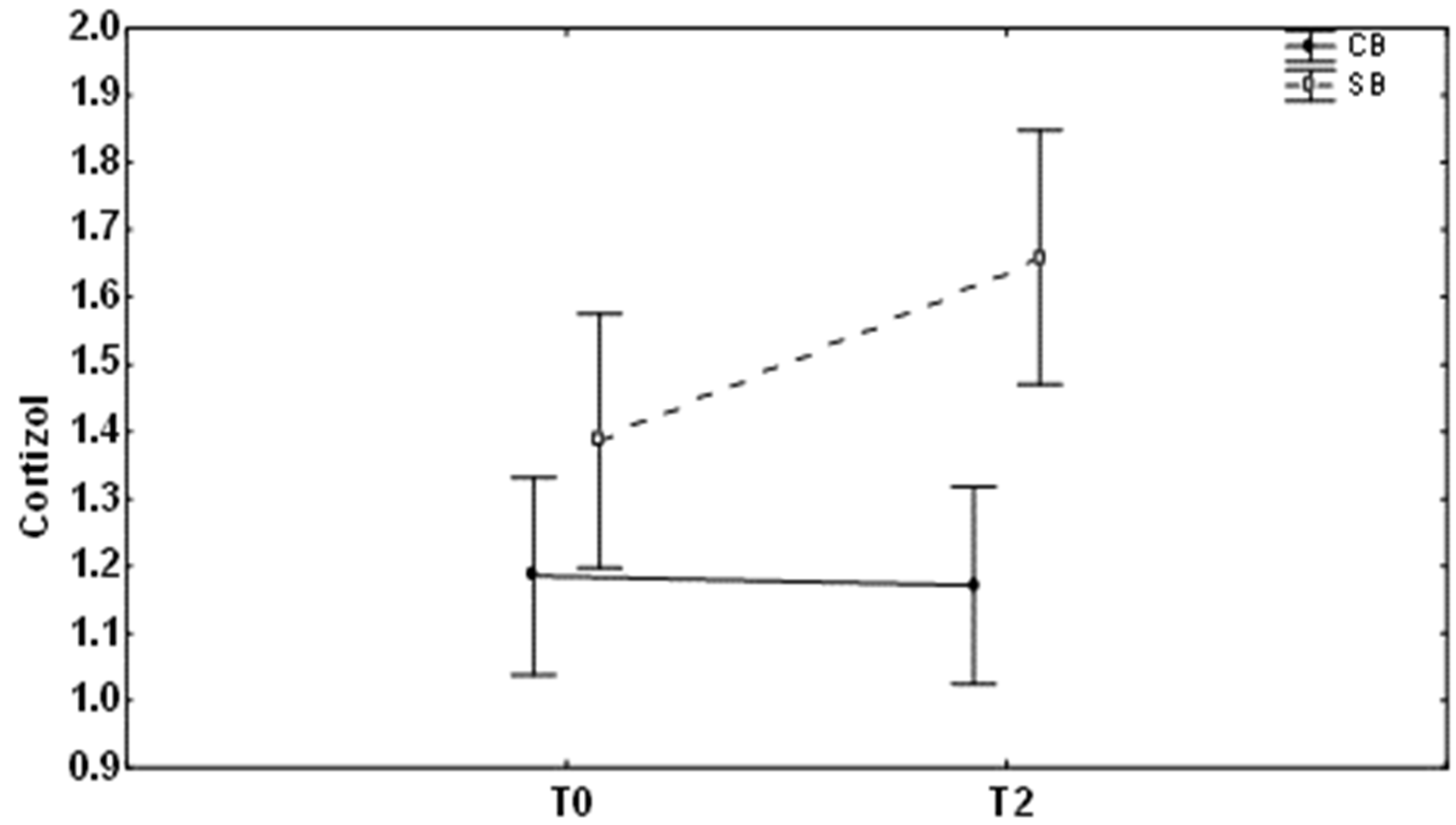
| Cortisol | 23.19 ± 19.23 (1.06–69.68) 15.76 (8.81–36.99) 15.25–31.13 | 22.44 ± 18.82 (1.64–63.83) 14. 60 (11.86–34.76) 14.67–30.21 | 0.90 |
| OPG | 7.03 ± 6.57 (2.28–11.70) 4.66 (2.04–25.00) 2.04–25.00 | 9.93 ± 10.59 (1.48–41.30) 5.51 (2.50–11.14) 5.56–14.30 | 0.42 |
| TNF-alpha | 13.49 ± 8.14 (6.93–37.50) 10.38 (7.90–16.09) 9.96–17.01 | 11.89 ± 11.40 (4.92–54.63) 9.06 (6.40–11.59) 7.18–16.59 | 0.10 |
| IL-6 | 13.18 ± 25.17 (2.42–116.27) 3.87 (2.552–9.05) 2.30–24.07 | 10.94 ± 13.60 (2.45–52.06) 3.76 (2.84–17.73) 5.33–16.56 | 0.67 |
| IFN-gamma | 7.97 ± 5.41 (4.94–27.40) 6.01 (5.78–7.33) 5.74–10.20 | 9.56 ± 14.00 (4.78–65.00) 5.67 (2.84–17.73) 3.78–15.33 | 0.25 |
| Mean ± Std.Dev (Min–Max); Median (25–75%) Confidence Intervals 95% | p (T0–T2) | ||
| T0 | T2 | ||
| Cortisol | 25.72 ± 9.11 (15.22–42.50) 25.61 (18.56–32.510 20.678–30.762 | 48.45 ± 18.11 (31.14–78.45) 38.92 (32.80–69.45) (38.43–58.48) | 0.001 |
| OPG | 8.55 ± 5.33 (3.33–18.07) 5.88 (5.13–15.26) 5.60–11.50 | 2.72 ± 0.73 (2.06–5.24) 2.58 (2.45–2.78) 2.32–3.13 | 0.003 |
| TNF-alpha | 13.36 ± 2.42 (10.08–18.51) 13.45 (11.01–15.47) 12.02–14.70 | 8.32 ± 1.50 (5.73–11.26) 8.60 (7.37–9.47) 7.49–9.15 | 0.002 |
| IL-6 | 13.06 ± 20.16 (3.55–80.25) 5.4 (4.47–7.04) 1.90–24.22 | 4.99 + 5.00 (2.42–18.27) 2.96 (2.75–4.12) 2.22–7.75 | 0.11 |
| IFN-gamma | 8.84 ± 5.83 (4.70–24.02) 6.41 (6.16–8.33) (5.61–12.07) | 5.95 ± 1.02 (4.83–9.37) 5.80 (5.39–5.98) (5.382–6.516) | 0.15 |
Disclaimer/Publisher’s Note: The statements, opinions and data contained in all publications are solely those of the individual author(s) and contributor(s) and not of MDPI and/or the editor(s). MDPI and/or the editor(s) disclaim responsibility for any injury to people or property resulting from any ideas, methods, instructions or products referred to in the content. |
© 2023 by the authors. Licensee MDPI, Basel, Switzerland. This article is an open access article distributed under the terms and conditions of the Creative Commons Attribution (CC BY) license (https://creativecommons.org/licenses/by/4.0/).
Share and Cite
Pantsulaia, I.; Orjonikidze, N.; Kvachadze, I.; Mikadze, T.; Chikovani, T. Influence of Different Orthodontic Brackets on Cytokine and Cortisol Profile. Medicina 2023, 59, 566. https://doi.org/10.3390/medicina59030566
Pantsulaia I, Orjonikidze N, Kvachadze I, Mikadze T, Chikovani T. Influence of Different Orthodontic Brackets on Cytokine and Cortisol Profile. Medicina. 2023; 59(3):566. https://doi.org/10.3390/medicina59030566
Chicago/Turabian StylePantsulaia, I., N. Orjonikidze, I. Kvachadze, T. Mikadze, and T. Chikovani. 2023. "Influence of Different Orthodontic Brackets on Cytokine and Cortisol Profile" Medicina 59, no. 3: 566. https://doi.org/10.3390/medicina59030566