
Application of Cortical Bone Plate Allografts Combined with Less Invasive Stabilization System (LISS) Plates in Fixation of Comminuted Distal Femur Fractures
Abstract
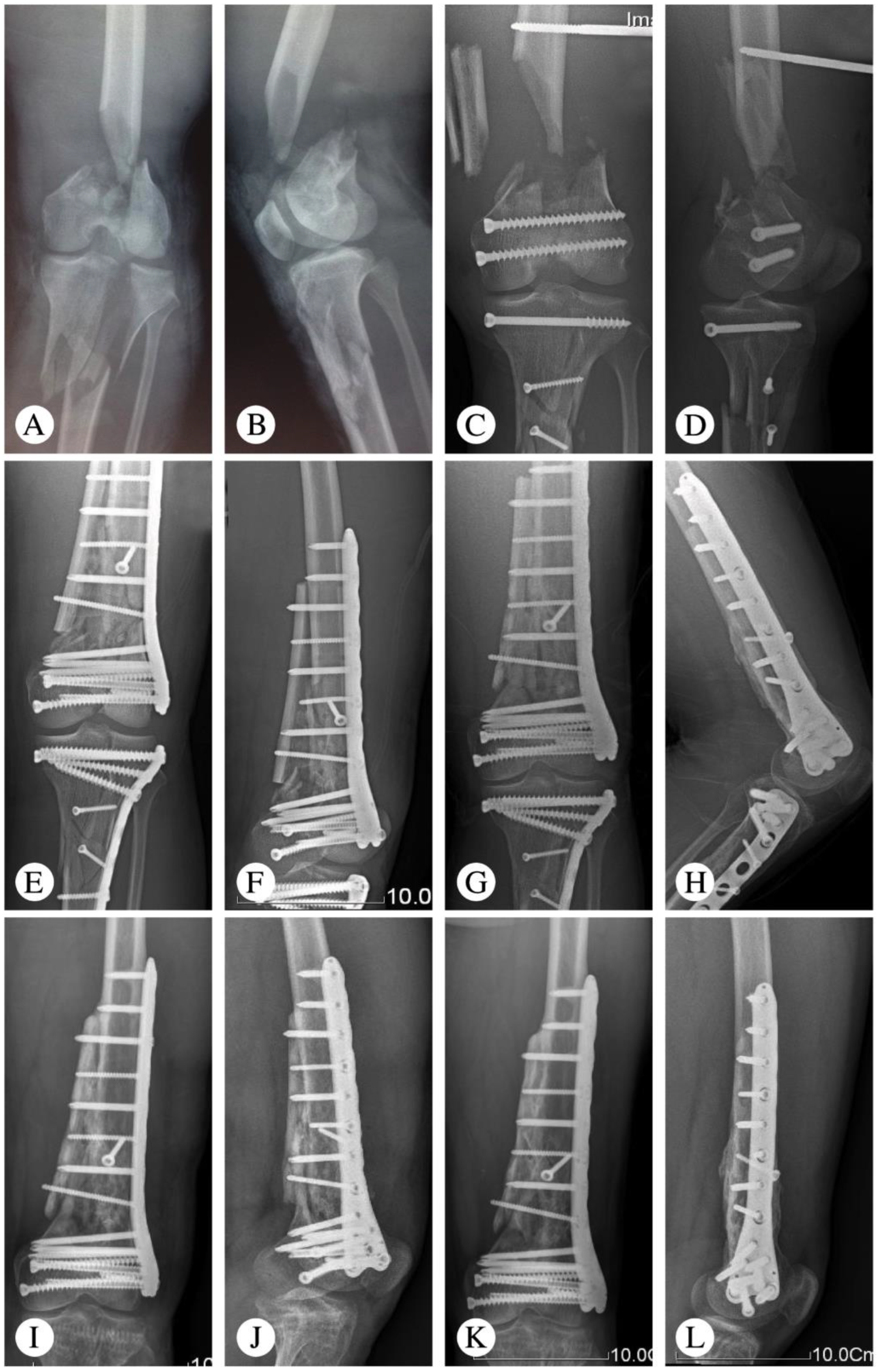
:1. Introduction
2. Patient and Method
2.1. Clinical Data
2.2. Preoperative Preparation
2.3. Surgical Procedure
2.4. Postoperative Management
2.5. Outcome Assessment
3. Results
4. Discussion
5. Conclusions
Author Contributions
Funding
Institutional Review Board Statement
Informed Consent Statement
Data Availability Statement
Conflicts of Interest
References
- Court-Brown, C.M.; Caesar, B. Epidemiology of adult fractures: A review. Injury 2006, 37, 691–697. [Google Scholar] [CrossRef] [PubMed]
- Martinet, O.; Cordey, J.; Harder, Y.; Maier, A.; Bühler, M.; Barraud, G.E. The epidemiology of fractures of the distal femur. Injury 2000, 31, 62–63. [Google Scholar] [CrossRef]
- Müller, M.E.; Nazarian, S.; Koch, P.; Schatzker, J. The Comprehensive Classification of Fractures of Long Bones; Springer: Berlin/Heidelberg, Germany; New York, NY, USA, 1990. [Google Scholar]
- Marsh, J.L.; Slongo, T.F.; Agel, J.; Broderick, J.S.; Creevey, W.; DeCoster, T.A.; Prokuski, L.; Sirkin, M.S.; Ziran, B.; Henley, B.; et al. Fracture and dislocation classification compendium—2007: Orthopaedic Trauma Association classification, database and outcomes committee. J. Orthop. Trauma. 2007, 21, S1–S133. [Google Scholar] [CrossRef]
- Johnson, K.D. Internal fixation of distal femoral fractures. Instr. Course. Lect. 1987, 36, 437–448. [Google Scholar] [PubMed]
- Davison, B.L. Varus collapse of comminuted distal femur fractures after: Open reduction and internal fixation with a lateral condylar buttress plate. Am. J. Orthop. 2003, 32, 27–30. [Google Scholar] [PubMed]
- Narsaria, N.; Singh, A.K.; Rastogi, A.; Singh, V. Biomechanical analysis of distal femoral fracture fixation: Dynamic condylar screw versus locked compression plate. J. Orthop. Sci. 2014, 19, 770–775. [Google Scholar] [CrossRef]
- Kulkarni, S.G.; Varshneya, A.; Kulkarni, G.S.; Kulkarni, M.G.; Kulkarni, V.S.; Kulkarni, R.M. Antegrade interlocking nailing for distal femoral fractures. J. Orthop. Surg. 2012, 20, 48–54. [Google Scholar] [CrossRef] [Green Version]
- Smith, T.O.; Hedges, C.; MacNair, R.; Wimhurst, J.A. The clinical and radiological outcomes of the LISS plate for distal femoral fractures: A systematic review. Injury 2009, 40, 1049–1063. [Google Scholar] [CrossRef]
- Ali, F.; Saleh, M. Treatment of isolated complex distal femoral fractures by external fixation. Injury 2000, 31, 139–146. [Google Scholar] [CrossRef]
- Wang, M.T.; An, V.V.G.; Sivakumar, B.S. Non-union in lateral locked plating for distal femoral fractures: A systematic review. Injury 2019, 50, 1790–1794. [Google Scholar] [CrossRef]
- Rollo, G.; Pichierri, P.; Grubor, P.; Marsilio, A.; Bisaccia, M.; Grubor, M.; Pace, V.; Lanzetti, R.M.; Giaracuni, M.; Filipponi, M.; et al. The challenge of nonunion and malunion in distal femur surgical revision. Med. Glas. 2019, 16. ahead of print. [Google Scholar] [CrossRef]
- Von Keudell, A.; Shoji, K.; Nasr, M.; Lucas, R.; Dolan, R.; Weaver, M.J. Treatment Options for Distal Femur Fractures. J. Orthop. Trauma. 2016, 30, S25–S27. [Google Scholar] [CrossRef] [PubMed]
- Henderson, C.E.; Kuhl, L.L.; Fitzpatrick, D.C.; Marsh, J.L. Locking plates for distal femur fractures: Is there a problem with fracture healing? J. Orthop. Trauma. 2011, 25, S8–S14. [Google Scholar] [CrossRef]
- Buckley, R.; Mohanty, K.; Malish, D. Lower limb malrotation following MIPO technique of distal femoral and proximal tibial fractures. Injury 2011, 42, 194–199. [Google Scholar] [CrossRef]
- Dumitru, M.; Vrinceanu, D.; Banica, B.; Cergan, R.; Taciuc, I.A.; Manole, F.; Popa-Cherecheanu, M. Management of Aesthetic and Functional Deficits in Frontal Bone Trauma. Medicina 2022, 58, 1756. [Google Scholar] [CrossRef] [PubMed]
- Hartin, N.L.; Harris, I.; Hazratwala, K. Retrograde nailing versus fixed-angle blade plating for supracondylar femoral fractures: A randomized controlled trial. ANZ J. Surg. 2006, 76, 290–294. [Google Scholar] [CrossRef]
- Canadian Orthopaedic Trauma Society. Are Locking Constructs in Distal Femoral Fractures Always Best? A Prospective Multicenter Randomized Controlled Trial Comparing the Less Invasive Stabilization System with the Minimally Invasive Dynamic Condylar Screw System. J. Orthop. Trauma. 2016, 30, 1–6. [Google Scholar] [CrossRef] [PubMed]
- Frigg, R.; Appenzeller, A.; Christense, R.; Frenk, A.; Gilbert, S.; Schavan, R. The development of the distal femur less invasive stabilization system (LISS). Injury 2001, 32, SC24-31. [Google Scholar] [CrossRef]
- Khalil, A.-S.; Ayoub, M.A. Highly unstable complex C3-type distal femur fracture: Can double plating via a modified Olerud extensile approach be a standby solution? J. Orthopaed. Traumatol. 2012, 13, 179–188. [Google Scholar] [CrossRef] [Green Version]
- Steinberg, E.L.; Elis, J.; Steinberg, Y.; Salai, M.; Ben-Tov, T. A double-plating approach to distal femur fracture: A clinical study. Injury 2017, 48, 2260–2265. [Google Scholar] [CrossRef]
- Kumar, P.; Patel, S.; Kumar, V.; Rajnish, R.K.; Hooda, A. A double-plating approach to distal femur fracture: A clinical study; how apt is the technique? How strong is the evidence? Injury 2018, 49, 737–738. [Google Scholar] [CrossRef]
- Shih, H.N.; Shih, L.Y.; Cheng, C.Y.; Hsu, K.Y.; Chang, C.H. Reconstructing humerus defects after tumor resection using an intramedullary cortical allograft strut. Chang. Gung. Med. J. 2002, 25, 656–662. [Google Scholar] [PubMed]
- Van Houwelingen, A.P.; McKee, M.D. Treatment of osteopenic humeral shaft nonunion with compression plating, humeral cortical allograft struts, and bone grafting. J. Orthop. Trauma. 2005, 19, 36–42. [Google Scholar] [CrossRef] [PubMed]
- Wang, J.W.; Wang, C.J. Supracondylar fractures of the femur above total knee arthroplasties with cortical allograft struts. J. Arthroplast. 2002, 17, 365–372. [Google Scholar] [CrossRef] [PubMed]
- Wang, J.W.; Weng, L.H. Treatment of distal femoral nonunion with internal fixation, cortical allograft struts, and autogenous bone-grafting. J. Bone. Joint. Surg. Am. 2003, 85, 436–440. [Google Scholar] [CrossRef]
- Keenan, O.J.F.; Ross, L.A.; Magill, M.; Moran, M.; Scott, C.E.H. Immediate weight-bearing is safe following lateral locked plate fixation of periprosthetic distal femoral fractures. Knee. Surg. Relat. Res. 2021, 33, 19. [Google Scholar] [CrossRef] [PubMed]


| Patients No | Gender | Age (Years) | Causes of Injury | Injury Type | Fracture Type | Other Injury |
|---|---|---|---|---|---|---|
| 1 | Male | 40 | Heavy object crushes | Closed fracture | C2 | - |
| 2 | Male | 61 | Implant failure | Closed fracture | A3 | - |
| 3 | Male | 18 | Fall from height | Closed fracture | A3 | - |
| 4 | Male | 35 | Traffic accident | Closed fracture | C2 | - |
| 5 | Male | 29 | Heavy object crushes | Closed fracture | C3 | - |
| 6 | Female | 69 | Fall from height | Closed fracture | C2 | - |
| 7 | Female | 40 | Traffic accident | Closed fracture | C3 | - |
| 8 | Male | 41 | Fall from height | Closed fracture | C2 | Traumatic brain injury |
| 9 | Female | 22 | Traffic accident | Closed fracture | C2 | - |
| 10 | Male | 21 | Traffic accident | Open fracture | C3, Gustilo III b | Ipsilateral tibial fracture |
| 11 | Male | 30 | Traffic accident | Closed fracture | C2 | Contralateral tibial and fibula fracture |
| 12 | Female | 34 | Traffic accident | Open fracture | A3, Gustilo I | - |
| 13 | Male | 23 | Heavy object crushes | Closed fracture | A3 | - |
| 14 | Female | 69 | Implant failure | Closed fracture | C3 | - |
| 15 | Female | 31 | Traffic accident | Closed fracture | C2 | - |
| 16 | Male | 40 | Traffic accident | Closed fracture | C2 | Ipsilateral tibial fracture |
| 17 | Male | 23 | Traffic accident | Closed fracture | A3 | - |
| 18 | Female | 59 | Traffic accident | Closed fracture | C3 | Hemopneumothorax |
| 19 | Male | 55 | Traffic accident | Closed fracture | C3 | - |
| 20 | Female | 71 | Fall from height | Closed fracture | A3 | - |
| 21 | Female | 47 | Heavy object crushes | Closed fracture | C2 | Ipsilateral patella fracture |
| 22 | Male | 33 | Traffic accident | Open fracture | A3, Gustilo III a | - |
| 23 | Male | 42 | Traffic accident | Closed fracture | C2 | Traumatic brain injury |
| 24 | Male | 51 | Traffic accident | Closed fracture | C3 | - |
| 25 | Female | 56 | Traffic accident | Closed fracture | C2 | Contralateral tibial and fibula fracture |
| 26 | Male | 59 | Traffic accident | Closed fracture | A3 | - |
| 27 | Female | 67 | Fall from height | Closed fracture | C3 | - |
| 28 | Female | 42 | Heavy object crushes | Closed fracture | C2 | Hemopneumothorax |
| 29 | Male | 53 | Traffic accident | Open fracture | A3, Gustilo II | - |
| 30 | Male | 46 | Traffic accident | Closed fracture | C3 | Contralateral tibial and fibula fracture |
| 31 | Male | 33 | Traffic accident | Closed fracture | C2 | - |
| 32 | Male | 78 | Traffic accident | Closed fracture | A3 | - |
| 33 | Female | 51 | Fall from height | Closed fracture | C3 | - |
| Patients No | Follow-Up (Months) | Bone Union (Months) | Knee Range of Motion | Outcomes * | Complications |
|---|---|---|---|---|---|
| 1 | 14 | 4 | 110° | Good | - |
| 2 | 73 | 5 | 100° | Fair | Quadricep strength grade 3 |
| 3 | 14 | 8 | 80° | Poor | Deep infection, secondary surgery |
| 4 | 26 | 5 | 100° | Excellent | - |
| 5 | 33 | 5 | 90° | Fair | Quadricep strength grade 4 |
| 6 | 29 | 6 | 110° | Excellent | - |
| 7 | 28 | 7 | 100° | Good | - |
| 8 | 35 | 6 | 110° | Excellent | - |
| 9 | 22 | 3 | 110° | Good | - |
| 10 | 69 | 9 | 80° | Poor | Quadricep strength grade 4 |
| 11 | 19 | 3 | 130° | Excellent | - |
| 12 | 28 | 5 | 110° | Good | - |
| 13 | 15 | 4 | 100° | Good | - |
| 14 | 17 | 6 | 90° | Fair | - |
| 15 | 26 | 7 | 110° | Fair | Superficial infection |
| 16 | 31 | 5 | 120° | Excellent | - |
| 17 | 43 | 4 | 130° | Excellent | - |
| 18 | 19 | 5 | 100° | Fair | - |
| 19 | 54 | 12 | 90° | Fair | Nonunion, secondary surgery |
| 20 | 27 | 3 | 110° | Excellent | - |
| 21 | Lost to follow-up | - | - | - | - |
| 22 | 50 | 8 | 110° | Good | - |
| 23 | 25 | 5 | 120° | Excellent | - |
| 24 | 60 | 4 | 110° | Good | Post-traumatic arthritis |
| 25 | 24 | 5 | 110° | Good | - |
| 26 | 17 | 4 | 110° | Excellent | - |
| 27 | 42 | 6 | 110° | Good | - |
| 28 | Lost to follow-up | - | - | - | - |
| 29 | 45 | 4 | 90° | Good | Superficial infection |
| 30 | 26 | 4 | 100° | Fair | - |
| 31 | 13 | 4 | 100° | Fair | - |
| 32 | 33 | 5 | 120° | Excellent | - |
| 33 | Lost to follow-up | - | - | - | - |
Disclaimer/Publisher’s Note: The statements, opinions and data contained in all publications are solely those of the individual author(s) and contributor(s) and not of MDPI and/or the editor(s). MDPI and/or the editor(s) disclaim responsibility for any injury to people or property resulting from any ideas, methods, instructions or products referred to in the content. |
© 2023 by the authors. Licensee MDPI, Basel, Switzerland. This article is an open access article distributed under the terms and conditions of the Creative Commons Attribution (CC BY) license (https://creativecommons.org/licenses/by/4.0/).
Share and Cite
Guo, Z.; Liu, H.; Luo, D.; Cai, T.; Zhang, J.; Wu, J. Application of Cortical Bone Plate Allografts Combined with Less Invasive Stabilization System (LISS) Plates in Fixation of Comminuted Distal Femur Fractures. Medicina 2023, 59, 207. https://doi.org/10.3390/medicina59020207
Guo Z, Liu H, Luo D, Cai T, Zhang J, Wu J. Application of Cortical Bone Plate Allografts Combined with Less Invasive Stabilization System (LISS) Plates in Fixation of Comminuted Distal Femur Fractures. Medicina. 2023; 59(2):207. https://doi.org/10.3390/medicina59020207
Chicago/Turabian StyleGuo, Zhimin, Hui Liu, Deqing Luo, Taoyi Cai, Jinhui Zhang, and Jin Wu. 2023. "Application of Cortical Bone Plate Allografts Combined with Less Invasive Stabilization System (LISS) Plates in Fixation of Comminuted Distal Femur Fractures" Medicina 59, no. 2: 207. https://doi.org/10.3390/medicina59020207