Review

The Interplay between Tumour Microenvironment Components in Malignant Melanoma

by
Cornelia Amalinei
*,†,
Adriana Grigoraș
*,
Ludmila Lozneanu
,
Irina-Draga Căruntu
,
Simona-Eliza Giușcă
and
Raluca Anca Balan
Department of Morphofunctional Sciences I, “Grigore T. Popa” University of Medicine and Pharmacy, 700115 Iasi, Romania
*
Authors to whom correspondence should be addressed.
These authors contributed equally to this work.
Medicina 2022, 58(3), 365; https://doi.org/10.3390/medicina58030365
Submission received: 22 December 2021 / Revised: 12 February 2022 / Accepted: 22 February 2022 / Published: 2 March 2022
(This article belongs to the Special Issue New Perspectives in the Treatment of Skin Disease)

Abstract

:
Malignant melanoma has shown an increasing incidence during the last two decades, exhibiting a large spectrum of locations and clinicopathological characteristics. Although current histopathological, biochemical, immunohistochemical, and molecular methods provide a deep insight into its biological behaviour and outcome, melanoma is still an unpredictable disease, with poor outcome. This review of the literature is aimed at updating the knowledge regarding melanoma’s clinicopathological and molecular hallmarks, including its heterogeneity and plasticity, involving cancer stem cells population. A special focus is given on the interplay between different cellular components and their secretion products in melanoma, considering its contribution to tumour progression, invasion, metastasis, recurrences, and resistance to classical therapy. Furthermore, the influences of the specific tumour microenvironment or “inflammasome”, its association with adipose tissue products, including the release of “extracellular vesicles”, and distinct microbiota are currently studied, considering their influences on diagnosis and prognosis. An insight into melanoma’s particular features may reveal new molecular pathways which may be exploited in order to develop innovative therapeutic approaches or tailored therapy.

1. Introduction

Normally located in the basal layer of the epidermis and dermis of the skin, by their ability of melanin synthesis, melanocytes cooperate with neighbouring cells, especially keratinocytes, to protect DNA from ultraviolet light (UV)-induced damage. Although malignant melanoma accounts for about 1% of all skin cancers, its incidence has been constantly increasing in the last two decades, mainly affecting light-skinned persons [1,2]. An estimated 420,000 new melanoma cases per year are registered worldwide [3], representing 5% of all newly diagnosed cancers [4] and exhibiting a large spectrum of locations and clinicopathological characteristics.
Although current histopathological, biochemical, immunohistochemical, and molecular methods provide a deep insight into its biological behaviour and outcome, melanoma is still an unpredictable disease, with poor outcome [5,6,7,8]. Recent progresses in immunomodulatory therapy have been added to the current arsenal in the fight against melanoma, but there are considerable efforts to identify suitable biomarkers for early diagnosis, staging, differential diagnosis, prognosis, and tailored therapy [5,9,10,11].
Microscopic analysis of biopsies or of the surgical specimens is important for the establishment of the histopathological diagnosis and prognosis parameters (tumour thickness or Breslow index, mitotic rate, and ulceration), added to specific immunohistochemical markers, playing together a very important role in melanoma management [5,12].
Additionally, melanoma biomarkers may be classified into different categories, such as diagnostic (showing a higher expression in melanoma cells than in normal tissue), prognostic or predictive markers (showing an increased expression in advanced stages of disease and providing valuable information regarding the treatment response or, on the contrary, correlated to disease recurrence), and progenitor and/or stem cell markers (specific for cell subpopulations that exhibit high carcinogenicity, metastatic potential, and treatment resistance) [5,6,11,13,14,15,16].
Circulating melanoma cells or melanoma-associated extracellular molecules provide noninvasive analytical access, considering the release of proteins and other molecules into the extracellular fluid, and may be considered potential serum biomarkers [5]. Different qualitative and semi-quantitative molecular assays have been used for melanocyte-associated monoclonal antibody (MelanA/MART1), Melanoma-associated antigen recognized by T cells 1 (MART1), and Glycoprotein 100 (gp100) detection [17]. Although debated, blood or serum mRNA levels of tyrosinase, which is involved in melanin synthesis, detected by reverse transcriptase-PCR (RT-PCR), has been significantly correlated with stage progression and the risk of metastatic spread by comparison to other investigated markers [17,18].
Melanoma’s “tumour niche” consists of an ensemble of malignant cells associated with other cells, such as keratinocytes, cancer stem cells, cancer-associated fibroblasts, endothelial cells, and immune cells. This microenvironment has an impact on melanoma cell progression and resistance to therapy [19].
Aggressive melanoma behaviour may be attributed to its heterogeneity, including a population of cancer stem cells (CSCs) [20], currently studied in order to identify their specific markers, which may be further exploited considering their prognostic and therapeutic potential [20].
The dynamic interaction between melanoma cells and adipocytes of the tumour niche may contribute to the establishment of a favourable microenvironment for melanoma growth and progression, especially in obese patients [19]. The therapeutic manipulation of this relationship may offer hopeful perspectives in these patients.
During the last decade, the gut and oral cavity microbiota have been considered a key factor of tumour development by its immunomodulatory function [21]. The unbalanced microbiota may lead to the development of an immune-compromised tumour microenvironment [21]. Current research is aimed at modifying patients’ microbiota as an adjuvant therapy in melanoma [21,22].
Overcoming controversies related to the value of some tumour markers, this review of the literature is aimed to update the knowledge regarding the specific clinicopathological, genetic, and molecular hallmarks, along with providing a comprehensive guide of the tumour microenvironment’s involvement in melanoma prognosis and management.

2. Clinicopathological and Molecular Hallmarks Update

2.1. General Features

Malignant melanomas derive from an abnormal proliferation of cells originating in melanocytes, cells capable of melanocytic differentiation, and show a characteristic natural aggressive history. Primary melanomas’ presentation is usually associated with a pigmented lesion, but they may also exhibit amelanotic (achromic) features. The most common locations for melanoma are: (i) skin (90%), (ii) mucosa of head and neck (maxillary alveolar ridges, hard palate, tongue, nose, and paranasal sinuses) (8–15%), (iii) gastrointestinal (anorectal region) (<1%), (iv) genital areas (vulvovaginal), (v) meninges and brain (dopaminergic neurons in the substantia nigra and locus coeruleus), and (vi) eye (uvea, conjunctiva, and ciliary body) [23].
Melanoma can affect a wide spectrum of ages belonging to both genders, and it is registering a poor prognosis in patients over 60 years old [24]. Cutaneous melanomas tend to arise in younger people, being related to intermittent high exposure to UV radiation, while mucosal melanomas are more frequent in dark-skinned populations (25–50%) [25,26].
Melanoma has a great metastatic potential, especially the mucosal type, with propensity for lungs, brain, liver, and soft tissues, exhibiting microsatellite, satellite, nodal, or distant metastases pattern and also showing a high ability of local recurrence [25].

2.2. Histopathological Characteristics

Although magnetic sequential digital dermoscopy added to clinical examinations are used for diagnosis, the confirmation is based on histological analysis [25]. Melanoma’s presentation may be as melanoma in situ, confined to the epidermis, and infiltrative melanoma, invasive into the dermis. Individuals with multiple atypical nevi (dysplastic nevi) or people with types I and II common nevi, especially with genetic susceptibility or a family history of melanoma, have a high risk for development of melanoma at an earlier age [27].
The most frequent type of melanoma’s growth is the radial growth phase (RGP), which is associated with a better prognosis compared to the other type, the vertical growth phase (VGP). RGP is characteristic for early lesions, being manifested as pigmented plaques or patches expanding horizontally in the epidermis, along the rays of a circle, while VGP is specific for progressive lesions as bona fide tumours, which infiltrate the dermis or expand into the epidermis, forming a nodule. Moreover, an early ”tumourigenic” VGP is distinguished by a group of cells within the dermis, wide-reaching to the largest epidermal cell cluster, or a lesion which also shows a proliferation pattern from epidermis to dermis, exhibiting a high mitotic activity [24,25,28].
Current clinicopathological World Health Organization (WHO) classification comprises four major histopathological subtypes: superficial spreading melanoma (SSM) (41%), nodular melanoma (NM) (16%), lentigo maligna melanoma (LMM) (2.7–14%), also known as melanoma arising in a Hutchinson melanotic freckle, and acral lentiginous melanoma (1–5%) [24,29]. The atypical melanocytes may be restricted to the epidermis, showing a lentiginous arrangement at the dermoepidermal junction, or may be restricted to the upper parts of the epidermis (pagetoid or superficial spreading), or can grow along the hair follicles [24,25]. SSM occurs in low cumulative sun damage (CSD), induced by intermittent sun exposure, while its histological diagnosis is made in the presence of pagetoid growth of single cells and nests, exhibiting severe cytological atypia [24,25,30] (Table 1). NM is characterized by an early progression to vertical growth without a radial growth phase and may represent a progression of an acral melanoma or of any other type of melanoma [25]. NM diagnostic criteria include complete loss of maturation, deep mitoses, lymphovascular invasion, and satellitosis, being frequently associated with ulceration [25]. LMM is a high-grade melanoma, with a high-mutation burden and severe solar elastosis which is mandatory for diagnosis, as well as microscopic features consisting of atypical melanocytes with a confluent growth along the dermoepidermal junction, with dermis invasion or with growth along adnexal structures [24,25,31]. Acral lentiginous melanoma can occur in all skin types (palms, soles, and nails), being frequently detected in an advanced stage, while its specific features include: low-mutation burden, epidermal hyperplasia, and characteristic asymmetrical, lentiginous, or nodular pattern of melanocytic cell growth [24,25,32].
A spectrum of lesions has to be considered in melanoma clinicopathological differentials, such as melanocytic nevus (junctional, compound, and dermal), blue nevus, and congenital nevus, along with congenital dermal melanocytosis, a group of lesions represented by Mongolian spot, nevus of Ota, and nevus of Ito [25,33,34,35,36,37]. Sometimes, it is very difficult to differentiate a melanocytic proliferation (genital or oral lentigo) from a non-melanocytic proliferation (basilar epidermal physiological pigmentation) that also displays a strong pigmentation [25]. Additionally, some lesions, such as skin squamous cell carcinoma in situ or Bowen’s disease, early or macular pigmented seborrheic keratosis, actinic keratosis, or junctional dysplastic nevus, are very frequently biopsied, due to strong pigmentation, in order to rule out LMM in situ or invasive melanoma [38]. In some situations, a neurotised melanoma can resemble a neuroid tumour, such as neurofibroma [39]. In our experience, the differentiation of a primary tumour from a metastatic one sometimes raises issues of differential diagnosis, especially when certain organs do not have melanocytes in their structure and their migration from neural crests is not reported.

2.3. AJCC Stages and Histological Features in Correlation with Prognosis

Melanoma is stratified according to the American Joint Committee on Cancer (AJCC) staging system (TNM Classification of Malignant Tumors) and WHO classifications, with a direct impact in practice. Thus, both AJCC and WHO systems classify melanoma into five stages (0, IA/B, IIA/B, IIIC, and IV), according to the surgical evaluation of the tumour size and invasion level (0—in situ lesion and stages I–IV), node invasion (stages III–IV), and occurrence of microscopically confirmed distant metastasis (stage IV) [23,24,25,40,41]. In addition to the AJCC staging system, WHO classification includes genetic, genomic, and epidemiologic features of skin melanomas, based on their mutational signatures [25]. Moreover, the molecular data provided by WHO classification result in various histopathologic variants, according to the degree of cumulative solar damage (CSD) of the skin [25].
Traditionally, melanomas were grouped into different subtypes, according to their morphological features, such as spindle, epithelioid, balloon, giant, signet ring, clear, and small cells, along with desmoplastic, rhabdoid, and myxoid [24,25,40,42,43]. Additionally, another distinctive histological feature is the regression of primary melanoma, which may be classified according to the variability of the mononuclear infiltrate, melanophages, and fibrotic process, into the following three categories: early, intermediate, and late stages [44]. However, reported data regarding the tumour–immune system relationship in melanoma regression are controversial. This relationship is particularly intriguing, taking into account that regression has a potential positive impact upon melanoma prognosis, but especially considering that drugs targeting these pathways have shown significant clinical efficacy in multiple tumour types [45]. Recent studies have suggested that the peritumoural inflammatory infiltration can represent a potential therapeutic target, while histological regression stands as an indicator of the immune system’s efficiency in melanoma [46,47].
The diagnosis of melanoma has been traditionally based on Clark’s levels of invasion and Breslow’s index (tumour thickness) that informs on the depth of melanoma invasion. There are five Clark’s levels (level I—confined to the skin surface and epidermis; level II, III, and IV—dermis invasion; level V—subcutaneous fat invasion) and three levels of Breslow’s index (≤1.0 mm—confined to the skin surface and epidermis; >1.0–4.0 mm—dermis invasion; >4.0 mm—subcutaneous fat invasion) [24,25,40,48,49].
AJCC and TNM staging systems and the Union for International Cancer Control (UICC) have improved the accuracy of prognostic prediction scoring, in addition to the presence of histologically recognized ulceration, epidermal defects, mitotic rate (per square millimetre), microscopic satellites, tumour infiltrating lymphocytes (TILs), and lymphatic and perineural invasion, along with tumour regression [24,25,40,50,51]. Moreover, pure RGP melanomas have a very good prognosis, while VGP tumours are prone to metastasis, with metastasis probability correlated to higher stage criteria such as larger thickness, ulceration, microsatellites, increased mitotic rate, lymphovascular invasion, and lack of or minimal TILs [24,25].

2.4. Immunohistochemical Markers of Diagnosis

Melanomas display immunostaining characteristics of melanocytic differentiation such as protein melan-A (MelanA) or MART1, microphtalmia-associated transcription factor (MITF), Human Melanoma Black (HMB45), SRY-related HMG-box 10 protein (SOX10), and S-100 protein positivity [24,25,52].
Although most types of malignant melanomas exhibit immunopositivity for these markers, desmoplastic melanomas are negative for MelanA/MART1, MITF, and HMB45. These markers cannot discriminate between nevi and melanomas, and they are not expressed only by melanocytes [25,52,53,54,55] (Table 2).

2.5. Genetics and Specific Markers

There are two different molecular pathways leading to melanoma: the first one is associated with sun-exposure, and a second one, an oncogenic pathway, is associated with genetic susceptibility, such as inherited variants of melanocortin-1 or mutations of tumour suppressor genes involved in cellular growth regulation [24,25,56].
Individual genetic susceptibility accounts for about 10% of all malignant melanomas and displays a predisposition pattern with variable gene penetrance (low, medium, and high). The genes that are associated with cases of familial melanoma are: cyclin-dependent kinase inhibitor 2A (CDKN2A), melanocortin-1 receptor (MC1R), MITF, cyclin-dependent kinase 4 (CDK4), protection of telomeres 1 (POT1), telomerase reverse transcriptase (TERT) promoter region (TERT), adrenocortical dysplasia protein homolog (ACD), telomeric repeat-binding factor 2-interacting protein 1 (TERF2IP), and BRCA1 associated protein 1 (BAP1) [57]. It seems that high penetrance genes (CDKN2A, CDK4, POT1, TERT, ACD, TERF2IP, and BAP1) can induce different intrinsic mechanisms, such as cell cycle dysregulation, or even have the capacity to encode two tumour-suppressor proteins, while genes with intermediate penetrance (MC1R and MITF) act on melanin production and development [57].
MC1R is a receptor for a melanocytic protein involved in skin pigmentation and UV response, being regulated by melanocortin, agouti-signalling protein (ASIP), and β-defensin [24]. Inherited anomalies of MC1R play a significant role in melanoma development [24]. Specific polymorphism of Melanocortin-1 receptor (MC1R) locus, with its variants (R151C, R160W, D294H, R142H, R163Q, and I155T), is potentially mutagenic in melanocytes [57]. Loss of function of this polymorphic gene corresponds to a UV-sensitive, fair skinned, melanoma-susceptible phenotype [58].
Most melanomas are associated with UV exposure, and they are further classified according to the CSD, as low- and high-CSD, added to histopathology assessed by the degree of solar elastosis and different molecular signatures [24,28] (Table 1). Low-CSD melanoma is likely to arise as a result of aberrations of BRAF mutation (45%) rather than RAS and NRAS mutations, which are identified in high-CSD exposure, in 15–30% and 15% of cases, respectively [24,25,56] (Table 3).
Atypical spitzoid tumour and uveal melanoma carry BAP1 gene mutation and most frequently occur in familial settings [24,25].
By comparison with cutaneous melanoma, mucosal melanoma has a decreased frequency of BRAF mutations (<10%) and an increased frequency of mutations of CD117 or c-KIT (40%), along with other somatic mutation (SF3B1, ATRX, ARID2, and SETD2). The BRAF-mutated pathway consists of valine replacement with glutamic acid in the BRAF gene at location 600 of the polypeptide chain, transforming it in an active kinase, which is responsible for tumour resistance due to reactivation of the mitogen-activated protein kinase (MAPK) pathway [59]. Proto-oncogene B-Raf (BRAF-V600E) mutation status is required to identify the eligible patients for combined BRAF and mitogen-activated protein kinase (MEK) kinase inhibitors treatment [24].
MAPK/ERK pathway plays an important role in melanoma progression, driving mutations during tumour development via several downstream proteins (RAS, RAF, MEK, and ERK) [60]. Any mutations of this translational or post-translational pathway trigger dissociation of these scaffolding proteins from the MAPK complex leading to cellular proliferation and differentiation of melanomas.
Starting from the assumption that melanoma shares high genetic heterogeneity, currently, detection of melanoma-associated MAPK mutations using a next-generation sequencing (NGS) panel is used to construct MAPK driver detection gene [61]. Presently, oncogenic alterations in MAPK pathway genes in circulating tumour DNA (ctDNA) are used to construct MAPK driver detection to check response to therapy or treatment resistance (discussed in detail in Section 7) [62].
RAS proteins are involved in activation of downstream signalling pathways, with significant impact upon cell proliferation, differentiation, and survival, while its variants, with alterations of codon 12, 13, or 61, have an oncogenic effect [63]. NRAS represents the most mutated RAS isoform, the vast majority of these mutations being found in codon 61, without correlation with UV-damage signature in melanoma [63]. NRAS mutations are characteristic for less than 20%of melanoma cases [24].
Considering that the assessment of NRAS and BRAF mutations is mandatory for the treatment of metastatic melanoma, the specific antibodies against NRASQ61R and BRAF-V600E proteins are immunohistochemically used to provide supplementary data on tumour heterogeneity [64]. Additionally, the theranostic efficiency of combining these two markers in challenging cases of melanoma has been reported [64].
c-KIT is a component of RTKs (class III transmembrane receptor tyrosine kinases) [65] and its mutations are rare (less than 3%), being related to acral, mucosal, or chronically sun-exposure melanomas [24]. With four isoforms and being encoded by a proto-oncogene on chromosome 4, position q11–12, human c-KIT has most of the mutations located in exon 11 and 13 which lead to MAPK and phosphatidylinositol 3-kinase/serine-threonine kinase (PI3K/AKT) pathways’ induction [65].
Moreover, these mutations do not occur together with BRAF or NRAS mutations [65].
Immunohistochemically, c-Kit represents a good diagnostic marker for differentiation between benign nevi and malignant melanocytic lesions, as well as between primary and metastatic melanomas [66].
ATRX is part of SWI/SNF family of chromatin remodelers, its mutations occurring in different tumours of neural crest cell origin, such as neuroblastoma, low-grade glioma, and glioblastoma, as well as in cutaneous melanoma [67]. ATRX alterations or mutations are immunohistochemically expressed as protein loss, with recent studies highlighting the loss of protein immunoexpression during melanoma progression, thus making ATRX a valuable prognostic biomarker [67].
ARID2 (BAF200) is also a member of the SWI/SNF chromatin remodeler family, which includes BAF (BRG1 or hbrm-associated factor) and polybromo-associated BAF (PBAF) complexes, encoding one PBAF complex subunit [68]. ARID2 mutations are frequent in melanoma, independent from BRAF/RAS mutations status [68]. ARID2 mutations affect the immune checkpoint inhibitors in melanoma and are linked to an increased infiltration with CD8+ T cells [68].
SETD2 or H3 lysine 36 histone methyltransferase is mutated in different human cancers, including mucosal melanoma. It is considered a modulator of different chromatin-regulated processes, such as DNA damage repair and methylation or RNA splicing [69].
Furthermore, some studies have shown that uveal melanoma, similar to cutaneous melanoma, may be associated with variable or incidental sunlight exposure [70,71,72,73]. They tend to be associated with alterations in G-protein-coupled receptors and/or G-α proteins, such as GNAQ or GNA11 activating mutations, followed by CYSLTR2, PLCB4, BAP1, SF3B1, and EIF1AX signalling pathways [23].
Splicing factor 3b subunit 1 (SF3B1) represents the largest component of the spliceosome factor 3b (SF3B) complex, spliceosome mutations becoming the most fascinating pathway detected in human cancer, including acral and mucosal melanomas, haematological malignancies, and solid tumours, having also prognostic significance [74,75]. SF3B1 mutations correspond to specific disease phenotypes, considering their involvement in regulation of RNA splicing and in DNA elongation and stability [74]. SF3B1 mutations are characteristics for uveal melanoma and blue nevus-like melanoma, and their assessment has diagnostic and prognostic value [76].
Neurofibromin 1 (NF1), a tumour suppressor gene encoding a negative regulator of RAS, is the most frequently mutated gene in sun-exposed malignant melanoma, after BRAF and NRAS, being associated with a high risk of metastasis and a high rate of treatment failure [77]. Discovered in the early 1990s, the somatic NF1 gene mutations are mainly registered in older male patients and in the desmoplastic melanoma type, being frequently associated with other mutations of the RAS pathway [78,79]. Additionally, a triple wild-type melanoma has been described, being characterized by the lack of BRAF, RAS (N/H/K), and NF1 mutations [56]. Recently, Ranzani et al. observed that most BRAF/NRAS wild-type melanomas are very sensitive to MEK inhibition, regardless of NF1 protein level [80].
In the last decades, other variable genomic alterations have been identified in melanoma initiation and progression, such as the mutation of Ras-related C3 botulinum toxin substrate 1 (RAC1), TERT, Kirsten rat sarcoma viral oncogene homolog (KRAS), Erb-b2 receptor tyrosine kinase 2/4 (ERBB2/4), cyclin-dependent kinase inhibitor 2A (CDKN2A), tumour protein 53 (TP53), and phosphatase and tensin homolog (PTEN), along with mitogen-activated protein kinase kinase 1 and 2 (MAP2K1/2) [56,81,82,83,84,85] (Table 2).

2.6. Putative Melanoma Biomarkers

Considering that circulating melanoma cells may release different proteins or other molecules into the extracellular fluid, these may represent potential serum biomarkers [5]. These biomarkers comprise molecules, which may be pathobiologically considered as enzymes, or soluble proteins and/or antigens, or melanin-related metabolites, or circulating cell-free nucleic acids [5,86,87] (Table 4).
From a pathobiochemical point of view, these biomarkers comprise molecules, including enzymes, such as cyclooxygenase-2 (Cox-2), lactate dehydrogenase (LDH), tyrosinase, matrix metalloproteinases (MMPs), tissue inhibitor of metalloproteinase-1 (TIMP-1), Cathepsin K, CD10, indoleamine-2,3-dioxygenase (IDO), and Legumain [5,86,87]. The other category of melanoma cells’ putative biomarkers is represented by soluble proteins and/or antigens, such as vascular endothelial growth factor (VEGF), vascular endothelial growth factor receptor 3 (VEGFR-3), C-reactive protein (CRP), Galectin-3, Osteopontin, heparin- and chitin-binding lectin YKL-40, melanoma inhibitory activity (MIA), soluble intercellular adhesion molecule 1 (sICAM-1), soluble vascular cell adhesion molecule 1 (sVCAM-1), carcinoembryonic antigen-related cell adhesion molecule 1 (CEACAM), cytoplasmic melanoma-associated antigen (CYT-MAA), melanoma antigen recognized by T-cells 1 (MART 1), melanoma-associated antigen-1 (MAGE), tumour-associated antigen 90 (TA90), S100 proteins, and Sry-related HMG-Box gene (SOX) protein family [5,86,87]. Another type of melanoma biomarkers is represented by melanin-related metabolites, such as L-3,4-dihydroxyphenylalanine (L-DOPA)/L-tyrosine, 6-hydroxy-5-methoxyindole-2-carboxylic acid (6H5MI2C), and 5-S-cysteinyl-DOPA [5,86,87]. The last category of melanoma cells serum markers which has been recently discovered is that of circulating cell-free nucleic acids, such as miRNA-29c and miRNA-221 [5,86,87].

3. Inflammatory Microenvironment in Malignant Melanoma

3.1. Cancer Inflammasome

The inflammation plays a crucial role in carcinogenesis considering its contribution as a source of cytokines and other tumour growth factors and its ability to eliminate transformed cells [88].
Currently, the inflammatory microenvironment represents a key member in the regulation of different tumourigenesis stages, from early initiation to promotion and distant metastasis [89]. The inflammasome may have favourable roles in the innate immunity or can be abnormally activated in melanoma, as well as in other types of malignancies, with the overexpression of the correspondent effector molecules [89].
The inflammatory tumour microenvironment comprises the population of resident or infiltrating immune cells and inflammatory mediators in the vicinity of malignant cells, nowadays representing a key piece in carcinogenesis, from initiation to promotion and metastasis.
The inflammasomes are proteic complexes consisting of nucleotide oligomerization domain (NOD)-like receptors (NLRs), pro-caspase-1, and apoptosis-associated speck-like protein containing a C-terminal caspase recruitment domain (CARD) domain—ASC, which are crucial for homeostasis maintenance. Inflammasomes are characteristic for different cell types, including antigen-presenting cells and T- and B-lymphocytes, along with cancer cells from the tumour microenvironment, with important roles in carcinogenesis in association with other concurrent factors [90].
Inflammasome stimulation depends on DAMPs/PAMPs’ (danger associated or pathogen associated molecular patterns) perception through cytoplasmic receptors (NLRP1, NLRP3) [91]. Various endogenous host proteins, such as CARD8, or different post-translational and transcriptional mechanisms regulate the inflammasome’s activation [91].
Several studies have demonstrated the influence of single nucleotide polymorphism (SNP) of inflammasome genes in the development and progression of different cancer types [92,93,94]. Although inflammasome constituents are widely expressed in immune and nonimmune cells, their encoding genes’ expression is not always related to the inflammasome formation or activation [95].
The characteristics of inflammasome biology are highlighted by the study of different cells, mouse bone marrow-derived macrophages (BMDMs) being the most investigated cellular type [95,96]. Other cells include mice bone marrow-derived intestinal epithelial cells and neutrophils, human airway epithelial cells, neutrophils, platelets, and peripheral blood mononuclear cells (PBMCs), as well as humans’ and rodents’ CD4+ and CD8+ T cells [95]. Nevertheless, inflammasome type is different among cells and host species. Accordingly, the stimulation of these inflammasomes will induce distinct biological outcomes [95].
The formation and activity of inflammasomes are closely dependent on cell organelles, leading to inflammation and cell death [97]. Several organelles can facilitate inflammasome assembly. Thus, the Golgi complex contributes to NLRP3 enrolment and activation, mitochondria are responsible for NLRP3 recruitment and inflammasome activation, endoplasmic reticulum can promote NLRP3 oligomerization with ASC and NLRP3 signalling regulation [95]. AIM2 and interferon-inducible protein 16 (IFI16) activation are taking place in the nucleus, being followed by their translocation in the cytoplasm, forming a perinuclear inflammasome complex. Opposite to the inflammasome action, which can induce cell death, stress granules are responsible for cell survival and inhibit NLRP3 inflammasome formation [95]. Ribosomes preserve cellular translation and generate inflammasomes and cytoskeleton components, especially the microtubule-organizing centre (MTOC), which contributes to NLRP3 and Pyrin formation, inflammasome translocation, and stability maintenance [95]. This “headquarter” activates numerous apoptotic and inflammatory caspases, cytokine substrates, the plasma membrane rupture protein ninjurin-1 (NINJ1), and the pore-forming protein gasdermin D (GSDMD), followed by inflammation, cellular destruction, and pyroptosis [95,98,99,100].
The innate immune system uses a group of pattern-recognition receptors (PRRs) as inflammasome components, which are expressed in different cell types involved in defence processes, such as dendritic cells, neutrophils, epithelial cells, macrophages, and monocytes [90]. Moreover, PRRs include the nucleotide-binding domain, leucine-rich repeat containing receptors (NLRs), and the absent in melanoma 2 (AIM)-like receptor (ALRs) [101]. The activated inflammasome sensor (NLRP1, NLRP3, NLRP6, NLRP9b, AIM2, caspase-11, or Pyrin) establishes the identity of the inflammasome complex [95]. The protein absent in melanoma 2 (AIM2) is a member of the PYHIN family, with one pyrin (PYD) domain situated at the N-terminus and one or two hematopoietic, interferon inducible, and nuclear (HIN) domains located at the C-terminus. The gene encoding this inflammasome sensor was first identified as a tumour suppressor gene in human melanoma cell lines [102].
ASC recruits pro-caspase-1, which is converted into catalytically active caspase-1 [101] and bioactive subunits p20 and p10 which will generate the bioactive forms of interleukins IL-1β and IL-18 by proteolytic cleavage of pro-IL-1β and pro-IL-18 [101].
Activation of the inflammasomes will subsequently lead to pro-inflammatory cytokine release by adjacent cells and tissues. Persistent inflammation will generate a chronic inflammatory status, which contributes to the pathogenesis of variable diseases, including cancer [103].

3.2. Interactions in Melanoma’s Inflammasome

The complex inflammasome’s interactions between different cytokines, endothelial, and tumour cells are contributing to angiogenesis and carcinogenesis, along with invasion and metastasis, in melanoma.
Melanoma involves upregulation of pro-inflammatory cytokines, such as IL-6, IL-8, C-C chemokine ligand 5 (CCL5), and IL-1β [101], along with VEGF [104]. In this regard, melanoma-derived IL-1β works as a stimulator of angiogenesis, tumour growth, invasion, and metastasis [105,106,107,108,109,110,111], the influence of the inflammasomes depending on the tumour cell types [101]. The results of a study performed in a murine experimental model have demonstrated the dual functions of IL-1 and inflammasomes in inflammation-induced skin tumourigenesis [112]. Moreover, norepinephrine (NE) is able to upregulate IL-6, IL-8, and VEGF in C8161 the melanoma cell line, exhibiting an autocrine stimulation along with chemotactic and proangiogenic effects, its value being increased in advanced stage melanomas [104].
Other works have demonstrated that ASC is an inhibitor of carcinogenesis by NF-κB transcriptional activity and IκB kinase α/β phosphorylation suppression in primary melanoma, while up-regulated ASC is stimulating the inflammasome, via IL-1β secretion and NF-κB activity, in metastatic melanoma [113,114]. Moreover, the knockdown of NLR family pyrin domain containing 1 (NLRP1) reduces their tumour-promoter properties, both in vivo and in vitro [113,114,115].
Numerous studies have demonstrated that NLRP3-inflammasome dysregulation can activate the inflammasome-dependent IL-1β expression in human sporadic metastatic melanoma cells [110,116,117]. Furthermore, melanoma tumour growth is linked to the ATP-regulated K+ channel P2 × 7 activity associated with NLRP3-inflammasome stimulation [91,118,119].
Another study has reported that modified gain-of-function variants of inflammasome genes NLRP1 and NLRP3 could increase patients’ risk for developing a sporadic malignant melanoma [94,120]. These findings highlight that the dysregulation of inflammasome activation, with subsequent IL-1β and IL-18 production, is crucial in tumourigenesis, the inflammasome molecules representing potential valuable prognostic melanoma biomarkers [94].
Other studies on SNPs in melanoma genes demonstrated that various cytokines (TNF-a, IL-6, IL-10, IFN-c, and TGF-b1) are involved in melanoma progression and immune escape [121,122]. Some of the cytokines produced by human melanoma cells (IL-6, IL-8, CCL5 (RANTES), CXCL1–3 (MGSA-GROa-c), and monocyte chemotactic protein-1 (MCP-1/CCL2) are associated with tumour invasiveness and aggressiveness [123]. Cytokines activity is stimulated by activated IL-1β [111,124]. Biologically active melanoma-derived IL-1β has a wide range of actions in melanoma tumourigenesis, exhibiting paracrine and autocrine-like activity, increasing IL-1 synthesis in melanoma cells, contributing to macrophages recruitment and to in vitro angiogenesis [110]. It is considered that IL-1β has various effects on different cells of the tumour microenvironment, maintaining survival and proliferation of melanoma cells, immune suppressor cells, and macrophages while promoting invasion and metastasis [125,126,127]. Moreover, IL-1β secretion becomes autonomous as melanoma is progressing [110,111].
Hedgehog (Hh) signalling plays an important role in melanoma pathogenesis, its activity being blocked by wogonin, an active component of flavonoids, in HT144 melanoma cells [128]. It is accepted that wogonin has various inhibitory effects in different melanoma cells, including on invasion and migration of B16F10 cells, melanin synthesis in A375 melanoma cells, or the proliferation and tumour growth of HT144 melanoma cells [129,130]. The anti-inflammatory effect of wogonin in HT144 melanoma is supported by the following activities: (i) pro-inflammatory factors decrease, (ii) anti-inflammatory factors increase, and (iii) inflammatory cytokines expression increase [131]. Additionally, the anti-tumour effects are performed by: (i) glucose consumption decrease; (ii) production of ATP and lactic acid decrease; (iii) kinases’ activities, such as phosphofructokinase (PFK), hexokinase (HK), and pyruvate kinase (PK) inhibition; and (iv) expression of glucose cotransporter-1 (GLUT1), monocarboxylate transporter 1 (MCT-1), and MCT4 inhibition [131].

3.3. Melanoma’s Microenvironment Components

The tumour microenvironment (TME) represents a complex biosystem with a great impact on tumour progression, which depends on the spatiotemporal interrelations between malignant and non-malignant cells [132,133], its immune heterogeneity being significant for the prognosis of different types of cancers [134,135].
The tumour microenvironment (TME) contains numerous immune cells, such as a variable amount of T lymphocytes, as well as B lymphocytes, dendritic cells (DCs), natural killer cells (NK), M1 and M2 type macrophages, mast cells, and myeloid-derived suppressor cells (MDSCs). During the first stages of carcinogenesis, immune cells are involved in apoptosis, anti-tumour cytokines production, and cytotoxic reactions. Thus, NK cells engage antigen-presenting cells (APCs) through cytokine secretion, while DCs, macrophages, and neutrophils are involved in phagocytosis of dead melanoma cells and tumour antigens presentation, activating T cells immune responses [59] (Figure 1).
The activity of the T cells’ main subtypes is mandatory for melanoma remission. Accordingly, the functions of cytotoxic (effector), helper, and regulatory cells are: (i) CD8+ T effector lymphocytes (Teff) recognize antigens via major histocompatibility complex class I (MHC I) molecules, inducing cytotoxicity in melanoma cells and(ii) CD4+ T helper (Th) lymphocytes bind to APCs through MHC II molecules and provide different immune cell types, under the tumour cytokines influence [136,137,138].
CD8+ T cells’ infiltration in metastatic melanoma may be stimulated by the administration of B1 receptor agonist des-Arg9-bradykinin (DABK), considering the involvement of stromal bradykinin signalling and melanoma cells bradykinin receptors in the tumour microenvironment [139,140].
Melanoma cells have great plasticity, allowing the immune escape due to reduced antigen expression, MHC molecules’ decreased level, as well as aberrations in their processing system [136]. As T cells express programmed cell death protein (PD-1) checkpoint receptor, tumour cells are blocking T cell activity via increased production of ligand of PD-1 receptor (PD-L1), leading to an interaction between PD-1 and PD-L1 to produce apoptosis of TILs, stimulating the differentiation of CD4+ into regulatory T cells (Tregs), and inhibiting the immune system response for self-tolerance maintenance [141,142,143]. It has been demonstrated that tumour Treg lymphocytes are correlated with melanoma growth and progression, their recruitment being performed by cancer cells through IL-10, IL-35, and tumour growth factor β (TGF-β) production in order to escape immunity [59,143]. Additionally, it has been shown that β3-adrenergic receptors expressed in melanoma microenvironment mediate Tregs’ and myeloid-derived suppressor cells’ activity, being involved in immune tolerance, in experimental models [144].
Different studies have provided controversial results regarding the activity of B-lymphocytes in melanoma TME. Accordingly, some authors consider that a high density in B cells is characteristic for non-metastatic melanoma, being associated with a better prognosis, while others found that melanoma cells produce fibroblast growth factor-2 (FGF-2) which stimulates B cells to produce insulin-like growth factor-1 (IGF-1), exhibiting a potential resistance to BRAF and MEK inhibitors [145,146]. Another study has emphasized that circulating B lymphocytes produce tumour necrosis factor α (TNF-α) and/or IL-6, these being associated with tumour unresponsiveness and poor survival of melanoma patients who underwent anti-cytotoxic T-lymphocyte associated protein 4 (CTLA4) antibody therapy [147]. The negative correlation between TNF-α expression and immune checkpoint blockade response suggests the role of B cells in tumour growth via inflammatory cytokines production [147].
Tumour-associated macrophages (TAMs), M1 and M2 types, can be important prognostic markers due to their role in tumour cell migration, angiogenesis, and extracellular matrix degradation. M1 macrophages are found in low number in intratumoural infiltrate and have anti-tumoural effects, being activated by Th1 cells and pro-inflammatory factors (granulocyte-macrophage colony stimulating factor—GM-CSF, lipopolysaccharides, and IFN-γ) [148]. M2 macrophages are involved in tumour progression and invasion, as they are mainly identified in early inflammatory infiltrate, being stimulated by Th2 cells and anti-inflammatory stimuli (IL-4, IL-10, IL-13, or monocyte colony-stimulating factor—M-CSF) [149]. Moreover, M2 macrophages can downregulate M1-mediated functions [150].
TAMs possess β3-adrenergic receptors and inducible nitric oxide synthase (NOS2, iNOS) and, as a consequence, their activity may be modulated by NE and nitric oxide (NO), contributing to an increased tumour cells’ growth and invasion [151].
CSCs are related to TME and may recruit TAMs for tumour growth [152]. In this regard, the involvement of CD34-melanoma tumour initiating cells (TICs) in chemoresistance and cancer progression promotion has been demonstrated, through M2 macrophages interaction, along with TGF-β and arginase pathway [152].
Moreover, TAMs may produce adrenomedullin, a vasodilator and stimulator of angiogenesis, a factor involved in TAMs polarization toward M2 type and in melanoma progression [59].
The expansion and migration of MDSCs, which are the precursors of macrophages, granulocytes, and DCs, are influenced by C-C chemokine receptor type 5 (CCR5) ligands (CCL3, CCL4, and CCL5) in melanoma [152]. CD141 DCs from the melanoma immune microenvironment are activating CD8+ T lymphocytes, by CCR7 receptor involvement [153]. Numerous evidence supports that the loss of CCR7 receptor promotes tumour growth, while its increased level is correlated with a better outcome [153]. Furthermore, a reduced DCs number is associated to metastatic melanoma, while their increased amount is suggestive for lack of metastases or low recurrence risk [153].
Neutrophils of the tumour inflammatory infiltrate increase during melanoma progression, their accumulation depending on CXCL1, CXCL2, CXCL3, CXCL5, and CXCL8 molecules, which are stimulated by UV radiations [154]. In an analogous manner to macrophages, neutrophils have also two subtypes: N1, which represent the dominant type of the early melanoma microenvironment, exhibiting anti-tumour activity, and N2 type, occurring in the advanced stages, with immunosuppressive effects [154].
The immune escape of melanoma cells is also produced through reduction in the expression of the main NK receptors (NKp30, NKp44, and NKG2D), which damage the mediated cytolytic anti-cancer activity of NK cells [155].
Different extracellular elements or other cells of the tumour niche may also contribute to the specific TME immune response, such as: fibroblasts, miRNAs or exosomes, acidification, keratinocytes, and adipose tissue (discussed in detail in Section 5.) (Figure 1).
Peritumoural fibroblasts can be converted into cancer associated fibroblasts (CAFs), exhibiting analogous properties to myofibroblasts [156]. During melanoma progression, CAFs may represent an important amount within the tumour cells’ population, displaying variable functions such as immunosuppression, due to TGF-β activity, which include inhibition of migration, maturation, and antigen presentation by DCs, increase in Tregs number, and reduction in the expression of perforin, granzymes, Fas ligand, and IFN-γ in cytotoxic T cells [156]. Fibroblasts seem to be recruited by tumour cells under β3-adrenergic stimulation by NE [151]. Moreover, NE stimulates fibroblasts metaplasia into myofibroblasts, which provide increased tumour cells motility and increased neoangiogenesis [157] along with the release of protumourigenic cytokines, such as FGF-2, IL-6, IL-8, and VEGF [157,158]. CAFs are involved in melanoma progression, metastasis, and drug resistance as a consequence of cell–cell interaction and secretion of extracellular matrix components, growth factors, and cytokines [133,159].
Mast cells are involved in the development of melanoma, considering their ability to react to substance P neurogenic inflammation [160,161]. Consequently, they release different cytokines, proteases, growth factors, biological amines, such as histamine, which reduces the antitumoural defence mechanisms, chemokines, neuropeptides, variable enzymes, and angiogenic factors, such as heparin, VEGF, TGF-β, and IL-8, the latter being demonstrated as a growth factor in different melanoma cell lines [160]. Moreover, their products seem to increase the immunosuppression resulting from UV-B exposure, using a complex mechanism of mast cells stimulation to release their products involving calcitonin gene-related peptide (CGRP), substance P (SP), and keratinocyte-produced nerve factor [161].
MiRNAs, small, non-coding RNAs involved in protein translation attenuation or inhibition, can regulate the melanoma immune microenvironment [162]. Melanoma cells secrete exosomes, which also provide membrane-bound ligands such as PD-L1, with an inhibitory effect of the anti-tumour response via interaction with the T cells receptors [162].
Exosomes, a subtype of extracellular vesicles (EVs), are involved in tumour microenvironment activity and carcinogenesis, mediating the interrelation between cancer cells and CAFs [133]. Several studies highlight the capacity of normal fibroblasts, CAFs, and cancer cells to secrete miRNA exosomes, providing a characteristic intercellular communication within the TME [163,164].
Another extracellular factor which mediates the immune response to cancer cells is acidification, the characteristic lower melanoma pH (6.0–7.0) providing an enhanced glycolytic activity and a specific inflammatory signature [165]. Most of the published data have revealed the immunosuppressive role of acidosis, which develops a “migratory” phenotype of melanoma cells [166,167,168]. The lower melanoma pH is responsible for decreased cytolytic activity of CD8+ T cells and increased secretion of IL-1β by monocytes and TAMs [167], as well as a functional orientation of TAMs toward the M2 type.
The adipose tissue β3-adrenergic receptor also influences the anti-tumour response of immune cells, its upregulation in the melanoma microenvironment resulting in tumour growth stimulation [169].
Keratinocytes can also contribute to the immune escape, influencing the melanoma’s immunosuppressive environment. Usually, the UV-absorbing melanin from keratinocytes protects against melanocytes mutations induced by prolonged radiation exposure, although the UV radiations can also stimulate cancer progression through a different pathway [59]. Recent data have demonstrated that keratinocytes secrete a high mobility group box 1 (HMGB1) protein, which promotes neutrophils infiltration into the melanoma microenvironment, being responsible for melanoma plasticity [59].
Recent data have demonstrated a correlation between keratinocytes and corticotropin-releasing hormone-proopiomelanocortin (CRH-POMC) axis in about 80% of melanomas, along with adrenocorticotropic hormone (ACTH) production in about 70% of melanomas, with α-melanocyte-stimulating hormone (α-MSH) release in over 50% of melanomas [170] and with the functional cell-specific MSH receptor or melanocortin 1 receptor (MC1R) [171]. CRH stimulates melanoma cells invasion via ERK1/2 signalling pathway [172]. Supplementary, desmoglein 1 is involved in the signalling between melanocytes and keratinocytes, with cytokines and POMC production, leading to a high level of melanin and pagetoid melanoma cells spread [173]. However, POMC overexpression reduces the melanoma growth by apoptosis and autophagy via complex α-MSH-HIF-1 α/BCL2 and adenovirus E1B 19-kDa-interacting protein 3 (BNIP3) signalling pathways [174].
Keratinocytes are stimulated by CGRP resulting in stimulation of melanin production and melanocytes trophicity [175]. However, CGRP may induce melanocyte apoptosis via increased Bax/Bcl-2 ratio, while substance P (SP) association with CGRP is inhibiting the process of melanogenesis [175].
Keratinocytes are expressing enkephalins, or opioid receptors (ORs), belonging to G protein-coupled receptors [176,177], while low levels of enkephalin and proenkephalin (PENK) have been detected in melanomas [178]. Moreover, methionine (met)-enkephalin (MENK) has shown melanoma growth inhibition via apoptosis associated with opioid growth factor receptors (OGFrs) increased expression in animal models [179].
Furthermore, it seems that the addictive mechanism of UV exposure is mediated by β-endorphin production in keratinocytes, added to its role in tumour cell proliferation and immune reactions inhibition, by decreasing the amount of tumour lymphocytes [180].
The inflammatory phenotype of the uveal melanoma, the most common primary ocular cancer in adults, is associated with a poor outcome, correlated to a greater number of inflammatory cells populating mainly epithelioid-cell-type tumours, characterized by chromosome 3 loss [181], while the main population in its inflammatory milieu is represented by CD8+ T cells and macrophages [182].
Among the four molecular subsets of uveal melanoma (A, B, C, D), identified in recent studies according to their immunological features and gene expression profiles [183,184], only the subset D shows a characteristic inflammatory phenotype, with excessive infiltration of lymphocytes and macrophages [183,185,186]. Moreover, variant D has an increased metastatic potential and several specific genetic aberrations (monosomy 3, chromosome 8q gain, and BAP1 loss), which seem to be correlated with its specific inflammatory phenotype [186,187]. Recent data regarding the specific immunological and genetic profile of uveal melanoma TME provide a gene-based prognostic signature, with possible impact on prognosis and on targeted therapy perspectives related to metastasis prevention [182].
Another interesting study has assessed the crosstalk between cultured uveal melanoma cells and hepatic stellate cells, demonstrating that metastatic melanoma cells are more sensitive to the paracrine signalling of stellate cells than their non-metastatic category, this interrelation involving profibrogenic interleukins [188]. Thus, metastatic melanoma cells are able to regulate hepatic stellate cells activity, promoting their growth and survival [188].
Tumour progression needs an early and persistent inflammatory response, as cancer cells can modulate the functions of the surrounding cells to favour their growth, invasion, metastasis, and survival [186].

4. Melanoma CSCs—The Origin of Heterogeneity, Plasticity, Aggressiveness, and Therapy Resistance

Aggressive melanoma is characterized by variable subpopulations, having multiple phenotype-specific genes and different protein markers. Thus, multiple cellular phenotypes have been identified in aggressive melanoma, such as stem cells, endothelial-like cells, and epithelial-like cells, suggesting a high plasticity [20].
Melanoma CSCs have self-renewal, indefinite proliferation capacities, high tumourigenicity, embryonic-like characteristics, ability to differentiation, and are involved in angiogenesis along with epithelial-mesenchymal transition (EMT) and metastasis [189,190].
The genes involved in vasculogenesis are upregulated in aggressive melanoma, such as EPHA2, CD144, and LAMC2 [189], along with EGFR-Akt-Smad signalling leading to angiogenesis via ID3 regulated cytokine induction [191].
Moreover, the endothelial cell markers seem to be responsible for melanoma cells’ capacity to form de novo vasculogenic-like networks in cultures, with tumour cells situated exterior to the vessels’ basement membrane, being named vascular or vasculogenic mimicry [20]. These channels containing blood, lined by melanoma cells, provide growth advantage and serves as an escape route for malignant cells [7]. Due to the poor expression of integrin α5-subunit, serving as an endostatin target, a failure of angiogenesis inhibitors efficiency has been observed in aggressive melanoma [20].
Melanoma CSCs are associated with a spectrum of markers and molecular pathways which provide them different advantages [5,190,192,193,194,195,196,197,198,199,200,201,202,203,204,205], though they may represent targets for the development of new therapies (Table 5).
Within the notion that malignancies may recapitulate morphogenesis events, Nodal embryonic signalling pathway, a member of TGF-β family, has been identified in aggressive melanoma cells, providing exacerbated tumourigenicity and metastasis capacity [20].
Furthermore, melanoma CSCs are involved in tumour microenvironment modulation by their expression of different miRNAs, along with their capacities of immune escape mechanisms and recurrences due to their limited response to conventional chemotherapy or radiotherapy [20,206,207,208,209,210].
ABC transporters family, and mainly ABCB5, have been shown to induce the tumour progression and multidrug resistance of melanoma cells through regulation of the drug’s efflux in melanoma cells, especially when they are co-expressed with CD133 [211]. The ABC transporters family tumourigenic potential is also supported by C-X-C motif chemokine receptor 6 (CXCR6) expression, another melanoma CSCs marker, particularly associated with asymmetric self-renewal [212]. In the same direction of study, the research conducted by Frank et al. revealed an overexpression of pro-angiogenic factors such as VEGFR1 and VEGF on ABCB5+ and CD133+ melanoma CSCs that promote tumour angiogenesis and support melanoma metastasis [190].
Furthermore, in a study conducted on CD133 + transgenic mice and human melanoma cells, promotion of neovascularisation in tumour microenvironment was induced by the Sox10 high expression, along with that of organic cation transporter (OCT) 3/4 and Nanog homeobox (Nanog) [213].
Sox10, a nuclear transcription factor involved in neural crest cells differentiation into melanocytes and mediation of their malignant transformation, has been observed to be one of the most important melanoma CSCs marker, being expressed in up to 100% of sentinel lymph node micrometastases, comparative to other melanoma cells markers, such as S100 and HMB45 [211].
CD44 is a mesenchymal marker, correlated to EMT, being a receptor which promotes the binding to the extracellular matrix via integrin and, thus, invasion and metastasis processes [214]. CD44 is correlated with insulin-like growth factor-1 (IGF-1), ZEB1, CD29, N-cadherin, and CD105 expression [214].
Aldehyde dehydrogenase (ALDH) enzymes show a common expression in stem cells, including melanoma CSCs, mediating expansion, self-protection, and differentiation [215,216,217]. However, ALDH activity detected in xenografted human metastatic melanoma showed a great variability in expression and, consequently, it is a controversial marker [218]. Additionally, stem-like tumour endothelial cells in human melanoma xenografts expressing strong ALDH show angiogenic capacities [219].
Human ALDH1A family comprises ALDH1A1, strongly expressed in xenografted melanoma, ALDH1A2, and ALDH1A3, strongly expressed in 1205L and A375 human melanoma cell lines and weakly expressed in xenografted melanoma. However, immunohistochemistry and real-time quantitative reverse transcription-PCR (qRT-PCR) reveal that both ALDH1A1 and ALDH1A3 may be expressed in some melanomas [217].
ALDH activity is an important factor in chemotherapy resistance in melanoma and the multi-drug resistance acquisition is correlated to a transition to a drug-tolerant population of cells which strongly express ALDH, along with ABC proteins [220]. As a consequence, a combination therapy using ALDH inhibitor and chemotherapy is increasing CSCs response [218,221]. Furthermore, ALDH may be used as a marker of therapy efficiency or may be selectively targeted in therapy [222,223,224,225,226].
Another application of ALDH expression in melanoma CSCs is the development of a dendritic cells (DCs) vaccine (CSC-DC vaccines) which stimulates tumour infiltration with T cells, along with their products (INF-γ and IL-4) [227,228,229].
Considering that melanoma CSCs also express PD-1 and PD-L1 and CTLA-4, in association with ALDH, consequently, anti-PD-L1 and/or anti-CTLA-4 combined with CSC-DC vaccine showed improved response [230].
Additionally, due to gamma-secretase and B-cell lymphoma 2 (Bcl-2) expression of melanoma CSCs, correlated with that of ALDH, the administration of their inhibitors, i.e., gamma-secretase inhibitors (GSI) and myeloid cell leukaemia sequence 1 (MCL-1)—an inhibitor of Bcl-2—is targeting CSCs [231,232].
The expression of CD20 and CD133 may be induced by NE, which is able to induce stem features in melanoma cells [233].
Melanoma CSCs exhibit a weak immunogenicity and, in addition, show an immunosuppressive effect in the host organism [189].

5. Melanoma Cells-Adipocytes “Dialogue”

5.1. Hypodermis Role in Cancer Microenvironment

An important component of the cancer microenvironment involved in the progression of melanoma is the adipose tissue, comprising the hypodermis [19]. This area of fat tissue is mainly composed of white adipocytes, associated with other types of cells such as endothelial cells, pericytes, monocytes, macrophages, and stem cells [234]. Current knowledge allows us to consider adipose tissue not only as a lipid storage area but also an inflammatory and endocrine organ [4]. Thus, adipose cells are the source of growth factors such as fibroblast growth factor-21 (FGF-21), hepatocyte growth factor (HGF), IGF-1, VEGF, and endocan, along with insulin-like growth factor-binding protein (IGFBP), leptin, retinol-binding protein 4 (RBP-4), resistin, leukaemia inhibitory factor, IL-6, IL-11, TNF-α, plasminogen activator inhibitor-1 (PAI-1), and TIMP-1 [235,236]. Melanoma cells express surface membrane receptors for these adipocytes-derived factors which support the tumour cells’ proliferation, metastasis, and drug resistance via MAPK, PI3K/AKT, and JAK/STAT pathways [237].
Recent reports show a “bidirectional communication” between melanoma cells and adipocytes, especially in obese patients, consisting of the secretion of high amounts of pro-inflammatory factors such as IL-6, IL-11, TNF-α, monocyte chemoattractant protein (MCP)-1/CCL2, and PAI-1 by the excessive fat [238,239]. Furthermore, the inflammatory profile of subcutaneous adipose tissue is associated with an increased release of leptin and resistin, supporting the progression of tumour cells and increasing the risk of lymph node metastasis [240]. Studies carried out on experimental models support this last finding, showing an increased tumour weight and size as a result of leptin injection into melanoma cells, which induces the activation of AKT-based signal transduction pathway and modulates the activity of fatty acid synthase (FASN), an enzyme involved in de novo synthesis of fatty acids (FA) [237,241]. In addition, adiponectin, with characteristic low levels in obese patients, has the opposite effect, analogous to leptin and resistin in melanoma [242]. Adiponectin induces apoptosis and inhibits cancer cells growth by activation of the AMP-activated protein kinase (AMPK) signalling pathway [242].
These data are also supported by other studies on murine melanoma models which demonstrate a positive correlation between melanoma progression and obesity [237,243,244]. In this respect, excessive subcutaneous white adipose tissue, together with enhanced secretion of pro-inflammatory factors, lead to the progression of melanoma by supporting tumour neoangiogenesis, following the release of pro-angiogenic factors, such as endocan, HGF, and VEGF, added to an altered energy metabolism [4,235]. In addition, other experimental studies revealed that adipocytes co-cultured with melanoma cells induce the secretion of chemoattractant factors (CXCL1, CXCL2, and CXCL5) added to a local immune cell recruitment, especially of M2 macrophages, in the “tumour niche” [236,245,246]. Analogous results have been reported by another research team, leading to the conclusion that skin adipocytes are involved in melanoma cell immune escape, by high expression of PD-L1, which interact with PD-1 molecule on the T lymphocytes membrane [247].
Remarkable results regarding melanoma development have been obtained using mice fed with a high-fat diet [243]. According to the results reported by Pandey et al., a rapid progression of tumour cells is associated with high FASN activity, an increased expression of caveolin-1 (Cav-1), and stimulation of phospho-Akt (pAkt), a protein kinase that plays a critical role in survival and apoptosis regulation [243]. Similar results have been reported by Malvi et al., which demonstrated that the caloric intake restriction and the administration of orlistat, a FASN inhibitor, induces a slowdown of melanoma tumour growth by reducing FASN, pAkt levels, and Cav-1 expression [237]. Furthermore, Cav-1, a membrane-associated protein stabilized by FASN by palmitoylation, acts as a tumour-progressing factor, being involved in cancer-drug resistance along with P-glycoprotein (P-gp), a protein that pumps out drugs from targeted cells [4,237,243]. Moreover, an increase in circulating exosomes expressing Cav-1 has been identified in the serum of melanoma patients, suggesting that Cav-1 may represent a prognostic biomarker [248]. These findings, added to the observation that melanoma cells exposed to adipocyte factors show a reduction in apoptosis induced by cisplatin and docetaxel, mediated by a MEK/ERK and a PI3K/AKT pathway signalling, led to a focus of research on adipocytes involvement in oncologic treatments [249,250].
Recent findings highlight a “metabolic dialogue” in the tumour microenvironment between melanoma cells and adipocytes, based on the observation that the latter provide a local supply of FA, which are transferred to melanoma cells through the fatty acid transport protein 1 (FATP1)/sulute carrier family 27 (SLC27A) family of lipid transporters [233]. These are the energy substrates that support the proliferation of tumour cells, considering their demonstrated role in stimulation of adipocyte lipolysis, leading to cancer-associated cachexia [233]. This latter feature has been demonstrated by morphological studies, which have revealed that adipocytes adjacent to melanoma cells are diminished in size compared to those located far from the tumour [233,251]. Additionally, Zoico et al. has reported adipocytes’ reduction in amount and size, along with that of their lipid droplet content, after few days of melanoma cells and 3T3-L1 adipocytes co-culture [252].

5.2. Extracellular Vesicles of Tumour Niche

The “tumour niche” intercellular communication carried out by extracellular vesicles (EVs) which are released by tumour cells and adipocytes has been the aim of recent studies [253,254]. Melanoma cells secrete EVs, which induce cancer progression by downregulation of skin adipocytes activity. These tumour EVs contain miRNAs, including miR-214-3p, which support the formation of a more favourable microenvironment by upregulation of lipogenesis genes, such as fatty acid-binding protein 4 (FABP4), adiponectin, and peroxisome proliferator-activated receptor ɣ (PPARɣ) [251,255]. Additionally, adipocytes are able to differentiate to a fibroblast-like phenotype through Wnt/β-catenin pathway activation and high expression of fibroblast specific markers, such as collagen and α-smooth muscle actin (α-SMA) [251,255]. Moreover, melanoma-derived EVs induce EMT and tumour progression through let-7i family miR paracrine or autocrine signalling [256]. In the same direction, tumour-derived EVs have been shown to be involved in the induction of apoptosis of cytotoxic T-cells, induction of M2 polarization of macrophages, and inhibition of cytotoxicity of NK cells in tumour microenvironment [257].
All these melanoma-derived EVs actions are supported by the “activity” of skin adipocytes. Thus, adipocytes also release EVs which are internalized by melanoma cells. They contain nucleic acids (miRNA, mRNA, and other non-coding RNAs), lipids, and proteins involved in fatty acid oxidation (FAO), which promote tumour progression through a metabolic reprogramming [258] (Figure 2). In this regard, Lazar et al. has noted that melanoma cells lines SKMEL28 exposed to adipocyte-derived EVs become elongated and develop actin-rich membrane’s protrusions, in a study conducted on a murine model [259].
All these morphological features are associated with adjustment of the mitochondrial network in melanoma cells, with mitochondrial fission and their redistribution to the cell extremities, characteristics which provide a high tumour-progression activity [260,261]. Furthermore, according to the data presented by Clement et al., an increased adipocytic lipolysis is achieved not only in the melanoma invasion front, in locations with a large number of adipocytes, but also by naive adipocytes, which release EVs, which provide both the energy substrate (FA) and the enzymatic equipment (protein) for FAO [258].
Similar results have been reported by another research team, who noticed that B16BL6 mouse melanoma cells exposed to adipocyte-derived factors are associated with higher invasiveness of tumour-cells, as a result of an increased expression of IL-6 and EMT-associated genes, such as MMP9, Snai1, Twist, and vimentin [262]. Additionally, Il-6 and tumour necrosis factor β (TNF-β) synthesized by skin adipocytes induce a paracrine differentiation of melanoma cells, expressed by reduced melanogenesis [235] and promotion of cultured melanoma cells proliferation, by the repression of miR-211 expression [263].
Last but not least, in obese patients, adipocytes participate in “tumour niche” establishment by synthesis of MMPs, especially MMP2 and MMP9, that mediate the remodelling of tumour extracellular matrix, thus promoting motility and invasion of melanoma cells [264]. Besides this, the melanoma cells–adipocytes communication in tumour niche is expressed by an elevated expression of cyclin D1 and Cox-2 oncogenic proteins in tumour cells, along with cell regulatory proteins, such as inhibitor of apoptosis protein-2 (IAP-2), myeloid cell leukaemia 1 (Mcl-1), B-cell lymphoma 2 (Bcl-2), and B-cell lymphoma-extra large (Bcl-xL) [264].
Considering these accumulated data, the dynamic interaction between melanoma cells and adipocytes in tumour niche is still far from elucidation. An important part of this intercellular “dialogue” is performed by adipocyte-derived EVs in association with growth factors, cytokines, and chemokines, which contribute to the establishment of a favourable microenvironment for melanoma growth and progression, especially in obese patients.
The deciphering of the complex tumour microenvironment which governs the molecular mechanisms involved in the melanoma progression is opening new therapeutic targets in these patients, especially from the perspective of FAO inhibitors and/or molecules use to prevent the release of EVs in the “tumour niche”.

6. Microbiota in Malignant Melanoma

During the last decade, the gut and oral cavity microbiota came to represent a key factor of tumour development by its immunomodulatory function [21,265]. It has been demonstrated that gut microorganisms are about 3.8 × 10¹³ with a weight of about 1.8 kg, establishing an equilibrium with the host organism, or eubiosis [266,267,268].
Moreover, recent data have demonstrated that gut microbiota, added to intratumour bacteria, may modulate the response to immunotherapy in many cancers, including melanoma, and modulate its toxic effects [268,269].
The growth of beneficial bacteria is enhanced by fibres or non-digestible compounds, or prebiotics, while healthy microbial species or probiotics are represented by Lactobacilli, Bifidobacteria, Saccharomyces yeasts, along with Enterococcus, Bacillus, and Streptococcus [270,271]. The probiotics associated to prebiotics which selectively stimulate the growth of probiotics, or the synbiotics, lead to a synergistic effect, while the use of nonviable microbial metabolites, or postbiotics, such as acetate, propionate, and butyrate (short-chain fatty acids) may mimic the effects of probiotics [272].
According to experimental studies, Staphylococcus aureus is stimulating Foxp3+Tregs, while Enterococcus, Alistipes shaii, and Lactobacilus are involved in Th17 and Th1 differentiation and in cytokines production [21,273]. The unbalanced microbiota, or dysbiosis, induced by antibiotics and immune checkpoint inhibitors, such as anti-CTLA-4 antibody, may led to the development of an immune-compromised tumour microenvironment [21], while Bacteroides fragilis, Bacteroides thetaiotaomicron, and Bifidobacterium improve the response to immunotherapy in mice models [21,274,275]. It has been also demonstrated that anti-PD-1 efficiency is higher in patients who had a microbiota rich in Enterococcus faecium, Bifidobacterium longum, Collisella aerofaciens, and Ruminococcaceae [276,277]. Anti-CTLA-4 immunotherapy results in a dominance of selected Bacteroides species [275]. The comprehensive analysis of commensal microbiota is valuable for detecting novel biomarkers or therapeutic targets in tumour patients treated with immune checkpoint inhibitors [21]. The abundance of gut Ruminococcaceae bacteria, along with Akkermansia muciniphila administration, contributes to the clinical response to anti-PD-1 treatment in melanoma patients [21,22]. Additionally, it has been demonstrated that patients containing Bacteroidaceae, Barnesiellaceae, and Rikenellaceae in their microbiota do not show colitis induced by anti-CTLA-4 therapy [278].
Furthermore, microbiota may influence immunotherapy response and toxicity, as demonstrated by the intratumour administration of CpG oligodeoxynucleotides, which mimic bacterial DNA administrated in melanoma experimental mice models, in association with an antibody against IL-10 receptor, which increase TNF production and CD8 T cells stimulation, resulting in tumour growth inhibition [273].
Based on mice experiments observations [274], faecal microbiota transfer (FMT), or the transfer of a donor entire microbial ecosystem are currently tested in clinical trials, in the immunotherapy context [274]. This is recommended mainly to patients who are refractory to anti-PD-1 treatment, with a possible pretreatment antibiotic ablation of their own microbiota [279]. Further studies would be necessary to evaluate the clinical utility of salivary or faecal microbiomes in patients which have an anti-PD-1 therapy [21].
Additional information regarding skin microbiota is currently providing correlations with UV radiations. For instance, Malassezia furfur, yeast which synthesizes pityriacitrin with a photoprotective role, is inhibited by UV radiations [280]. Skin colonization with Staphylococcus epidermidis, which produces 6-N-hydroxyaminopurine, has a preventive effect in an experimental model of photocarcinogenesis [281]. In contrast, Trueperella and Fusobacterium colonization has been associated with melanoma development in animal models [282].
These data are adding new evidence of the so-called melanoma’s “exposome”, comprised of environmental exposure, added to microbiome and genome [283].

7. Current Melanoma Therapeutic Approaches

7.1. Therapy Targets and Potential Therapeutic Biomarkers

The current therapeutic approach to melanoma depends on its location, stage, its genetic biology, or other factors. Thus, in addition to surgical therapy, immunotherapy and targeted therapy represent important recommendations as adjuvant medical therapy for advanced stages, as well as chemotherapy and radiation therapy.
The remarkable effort to decipher the genetic mutations and molecular mechanisms that underline the malignant melanomas development are useful for oncologists in choosing targeted therapies.
Numerous studies have assessed the therapeutic efficiency of c-KIT inhibitors, but the results have limited value, as most patients have eventually manifested tumour progression, possibly due to numerous cases of melanoma with central nervous system metastases, characterized by a limited drug penetration capacity [58]. Thus, current strategies are exploring combination therapy, such as an association of c-KIT inhibitors with those targeting its downstream pathways or with the immunological checkpoint blockade [65].
The concept that frequent ARID2 mutations are influencing the immune checkpoint inhibitors, being correlated to increased CD8+ T cells, supports the role of ARID2 as a potential biomarker of therapeutic efficiency of immune checkpoint inhibitors in patients with melanoma [68].
Moreover, SETD2 loss provides a potential role of this marker in melanoma therapy assessment [69].
Supplementing the surgery, radiotherapy, and immunotherapies (systemic therapy), targeted therapy is efficient, such as anti-BRAF (vemurafenib, dabrafenib, and encorafenib) and anti-c-kit (imatinib, nilotinib, and regorafenib), while anti-NRAS therapy (lonafarnib and tipifarnib) has failed in clinical trials due to NRAS activation via alternative post-translational alterations [23,70].
Targeting MAPK proteins and their regulatory components may contribute to inhibit melanoma cell genesis, thereby using them as potential therapeutic tools. Tipifarnib is targeting RAS, while Sorafenib is specifically targeting RAF. Pharmacological agents used in clinical trials, such as AZD6244, U0126, PD0325901, CI-1040, XL518, AZD8330, ARRY-162, and ARRY-300 are selective inhibitors of MEK [284], while AZ628 inhibits this pathway at the ERK level. The addition of an anti- RAS, RAF, MEK, ERK agent, MEK inhibition (MEKi) has shown considerable effects and ability to inhibit growth and induce melanoma cell death, especially in the BRAF-mutant metastatic melanoma [192]. Thus, all these pharmacological agents remain promising therapeutic targets in the MAPK pathway.

7.2. Therapeutic Strategies Associated to Melanoma Immune Microenvironment

A variable and complex network of interactions is characteristic for the melanoma microenvironment. The specific cells of the tumour milieu contribute to the plasticity and heterogeneity of melanoma, as they represent factors which are influencing the tumour immune escape and therapy resistance [59]. The different types of tumour cells, such as immune cells, CAFs, adipocytes, and keratinocytes, can communicate with each other or with extracellular matrix molecules, thus getting involved in the immune escape and affecting the immunosuppressive environment of the melanoma and, thus, the treatment efficiency [59]. The interaction between cancer cells and surrounding elements creates new therapeutic protocol opportunities [186]. In this direction, a therapy based on antibodies directed against adrenomedullin or an antagonist of its receptor has proven its efficiency both in vitro and in vivo [59,150].
It was revealed that CAFs and TAMs may contribute to therapy tolerance through a cytokine-signalling network that includes fibroblast-derived CXCR2 ligands and macrophage-derived IL-1β, inflammatory niches’ signalling being amplified by MAPK inhibitors, providing early drug tolerance toward BRAF and MEK inhibitors during treatment [285].
Furthermore, melanoma aggressiveness may be correlated to peritumoural mast cells density and their population may be targeted in new therapeutic approaches [286].
The modern melanoma therapy directed against immune cells is targeted against PD-1 ad CTLA-4. The best-known anti-PD-1 antibodies are nivolumab and pembrolizumab, used in the therapy of metastatic melanoma and advanced melanoma treatment, respectively. Nivolumab has shown superior therapeutic effects, improving the median progression-free survival, compared to ipilimumab, which blocks CTLA-4 inhibitory signalling pathway [59].
Considering that BRAF and MEK inhibitors can lead to changes in TME immunogenicity, therapeutic strategies consist of a combination between anti-MEK/BRAF drugs and immune checkpoint inhibitors [59]. This approach is based on the observation that BRAF-mutated melanoma cells have a low T cell infiltration and a high level of IL-6, IL-10, and VEGF, which increase the number of Tregs or myeloid-derived suppressor cells within TME [59]. Clinical investigations which have shown promising results involved the following combinations: trametinib (MEKi), dabrafenib (BRAFi), and murine anti-PD-1 antibody or cobimetinib (MEKi), vemurafenib (BRAFi), and atezolizumab (anti-PD-L1 antibody) [287,288]. Moreover, the melanoma survival was successfully improved by using combination targeted therapies for checkpoint inhibitors and MEK-ERK pathway [192].
Other immune checkpoint proteins have been studied as potential markers for new therapy strategies, such as lymphocyte activation gene-3 (LAG-3), T-cell immunoglobulin-mucin domain-containing molecule 3 (TIM-3), and T cell immunoreceptor with Ig and immunoreceptor tyrosine-based inhibitory motif (ITIM) domains (TIGIT), all of them being valuable targets for immunotherapy [59,289].
Glucocorticoid-induced tumour-necrosis-factor-receptor-related protein (GITR) represents another promising molecule for immune checkpoint therapy. GITR alteration has been shown to induce inhibition of T cell-mediated cancer cell apoptosis [290,291]. Moreover, GITR is correlated with the activity of E3 ubiquitin protein ligase neural precursor cell expressed developmentally down-regulated protein 4 (NEDD4), often involved in metastatic melanoma [290,291]. In this regard, the anti-melanoma immune response could be increased by inhibiting the activity of this enzyme [290,291].
It was demonstrated that human and mouse melanoma cells metastasis was inhibited in vitro by thymoquinone, a bioactive phytocompound, by decreasing the NLRP3 inflammasome expression, accompanied by a reduction in caspase-1 proteolytic cleavage, which led to IL-1β and IL-18 inhibition, as well as NF-κB activity suppression [116]. Moreover, the incomplete inactivation of NLRP3 inflammasome was due to reactive oxygen species (ROS) inhibition by thymoquinone. These results promote thymoquinone as a potential agent for both adjuvant immunotherapy and metastatic melanoma prevention [116].
Brain metastases of cutaneous melanoma, including acral type, react to both targeted and immune therapies, responding with targeted therapy resistance mediated by extrinsic factors. The mechanism involved is phospho-inositide 3-kinase (PI3K) pathway activation, which is a main target for brain metastases in melanoma [192].
Immune therapy’s escape of the succeeding checkpoint blockade was related to specific TME changes, represented by a lower infiltration of effector T cells, a higher number of alternatively activated macrophages, and a characteristic gene expression profile [192]. Numerous therapeutic strategies in melanoma have successfully targeted different tumourigenic mechanisms and pathways, taking into account the inflammatory microenvironment variability, such as the involvement of dendritic cells, as APCs and cytotoxic T-cell stimulators via CCR7 expression, T cell infiltration, alteration of immunosuppressive signalling pathways, neoantigen specific T cell response obtained by vaccination (mutant epitopes, mRNA, and dendritic cells) [292,293,294], antitumour immune response with oncolytic viruses [295], and antitumour activity of cytokines (IL-2 pathway and granulocyte-macrophage colony-stimulating factor—GM-CSF)) [296,297,298].
Due to heterogeneity of the melanoma immune microenvironment and weak therapeutic response to different monotherapies, attempts have been made in recent years to combine therapeutic variants in order to improve the therapeutic response, mainly with immune checkpoint inhibitors [299,300,301,302]. The promising results of combination therapy in melanoma could address the complexity of the inflammatory microenvironment and tumour heterogeneity by aiming for a therapeutic precision that exceeds the immune resistance or side effects encountered in current therapies [303].

8. Conclusions

Malignant melanoma exhibits a large spectrum of locations and clinicopathological characteristics, and its features are important in practical activity for diagnosis, differentials, and therapy.
Although their value is still debated, sometimes leading to controversial literature data, melanoma biomarkers may be divided into different categories, such as diagnostic, prognostic or predictive markers, progenitor and/or stem cell markers, while circulating melanoma cells or melanoma-associated extracellular molecules provide potential serum biomarkers. Biomarkers’ assessment by histological and immunohistochemical analyses of biopsies, added to pathological diagnosis and prognosis parameters, plays a very important role in melanoma management.
The melanoma microenvironment, consisting of a cell complex which comprises a population of resident or infiltrating immune cells, inflammatory mediators, adipose cells, and adipocytes-derived factors supports tumour cell proliferation, drug resistance, and metastasis. Thus, an insight into its distinct features and their interplay may reveal new pathways and molecules which prove useful in an innovative therapeutic approach.
Recent progress in immunomodulatory therapy and in manipulation of melanoma cells–adipocytes interactions has been added to the current arsenal against melanoma, and considerable efforts are made to identify suitable biomarkers for early diagnosis, staging, differential diagnosis, prognosis, and tailored therapy.
The gut and oral cavity microbiota has been recently considered a key factor of tumour development by its immunomodulatory function, and current research is aimed at modifying patients’ microbiota as an adjuvant therapy in melanoma.
Considering the complexity of the interplay between melanoma cells and their microenvironment, along with tumour heterogeneity, combination therapy is now being developed in an attempt to overcome the immune resistance and the side effects of current therapies, opening new perspectives for a better management and an improved prognosis for patients.

Author Contributions

Conceptualization, C.A., R.A.B., A.G., L.L. and I.-D.C.; methodology, A.G., L.L., R.A.B., I.-D.C., S.-E.G. and C.A.; software, R.A.B. and A.G.; validation, L.L., C.A., I.-D.C. and R.A.B.; investigation, L.L., R.A.B., S.-E.G. and A.G.; writing—original draft preparation, C.A., A.G., L.L., I.-D.C. and R.A.B.; writing—review and editing C.A.; supervision, C.A., R.A.B., L.L., A.G. and I.-D.C. All authors have read and agreed to the published version of the manuscript.

Funding

This research received no external funding.

Institutional Review Board Statement

Not applicable.

Informed Consent Statement

Not applicable.

Data Availability Statement

Not applicable.

Conflicts of Interest

The authors declare no conflict of interest.

References

  1. Richetta, A.G.; Silvestri, V.; Giancristoforo, S.; Rizzolo, P.; D’Epiro, S.; Graziano, V.; Mattozzi, C.; Navazio, A.S.; Falchetti, M.; Calvieri, S.; et al. Mutational profiling in melanocytic tumors: Multiple somatic mutations and clinical implications. Oncology 2014, 86, 104–108. [Google Scholar] [CrossRef]
  2. de Menezes, F.C.; de Sousa Cabral, L.G.; Petrellis, M.C.; Neto, C.F.; Maria, D.A. Antitumor effect of cell therapy with mesenchymal stem cells on murine melanoma B16-F10. Biomed. Pharmacother. 2020, 128, 110294. [Google Scholar] [CrossRef]
  3. Siegel, R.L.; Miller, K.D.; Jemal, A. Cancer Statistics, 2018. CA Cancer J. Clin. 2018, 68, 7–30. [Google Scholar] [CrossRef]
  4. Mazurkiewicz, J.; Simiczyjew, A.; Dratkiewicz, E.; Ziętek, M.; Matkowski, R.; Nowak, D. Stromal cells present in the melanoma niche affect tumor invasiveness and its resistance to therapy. Int. J. Mol. Sci. 2021, 22, 529. [Google Scholar] [CrossRef]
  5. Belter, B.; Haase-Kohn, C.; Pietzsch, J. Biomarkers in malignant melanoma: Recent trends and critical perspective. In Cutaneous Melanoma: Etiology and Therapy; Ward, W.H., Farma, J.M., Eds.; Codon Publications: Brisbane, Australia, 2017; ISBN 9780994438140. [Google Scholar]
  6. Mimeault, M.; Batra, S.K. Novel biomarkers and therapeutic targets for optimizing the therapeutic management of melanomas. World J. Clin. Oncol. 2012, 3, 32–42. [Google Scholar] [CrossRef]
  7. Han, D.; Han, G.; Morrison, S.; Leong, S.P.; Kashani-Sabet, M.; Vetto, J.; White, R.; Schneebaum, S.; Pockaj, B.; Mozzillo, N.; et al. Factors predicting survival in thick melanoma: Do all thick melanomas have the same prognosis? Surgery 2020, 168, 518–526. [Google Scholar] [CrossRef]
  8. Gaudy-Marqueste, C.; Macagno, N.; Loundou, A.; Pellegrino, E.; Ouafik, L.; Budden, T.; Mundra, P.; Gremel, G.; Akhras, V.; Lin, L.; et al. Molecular characterization of fast-growing melanomas. J. Am. Acad. Dermatol. 2021, 86, 312–321. [Google Scholar] [CrossRef]
  9. Vereecken, P.; Cornelis, F.; Van Baren, N.; Vandersleyen, V.; Baurain, J.-F. A synopsis of serum biomarkers in cutaneous melanoma patients. Dermatol. Res. Pract. 2012, 2012, 260643. [Google Scholar] [CrossRef] [Green Version]
  10. Carr, S.; Smith, C.; Wernberg, J. Epidemiology and risk factors of melanoma. Surg. Clin. N. Am. 2020, 100, 1–12. [Google Scholar] [CrossRef]
  11. Torres-Cabala, C.; Li-Ning-Tapia, E.; Hwu, W.-J. Pathology-based biomarkers useful for clinical decisions in melanoma. Arch. Med. Res. 2020, 51, 827–838. [Google Scholar] [CrossRef]
  12. Wilson, M.L. Histopathologic and molecular diagnosis of melanoma. Clin. Plast. Surg. 2021, 48, 587–598. [Google Scholar] [CrossRef]
  13. Palmer, S.R.; Erickson, L.A.; Ichetovkin, I.; Knauer, D.J.; Markovic, S.N. Circulating serologic and molecular biomarkers in malignant melanoma. Mayo Clin. Proc. 2011, 86, 981–990. [Google Scholar] [CrossRef] [Green Version]
  14. Alegre, E.; Sammamed, M.; Fernández-Landázuri, S.; Zubiri, L.; González, Á. Circulating biomarkers in malignant melanoma. Adv. Clin. Chem. 2015, 69, 47–89. [Google Scholar] [CrossRef]
  15. Karagiannis, P.; Fittall, M.; Karagiannis, S.N. Evaluating biomarkers in melanoma. Front. Oncol. 2014, 4, 383. [Google Scholar] [CrossRef] [Green Version]
  16. Levesque, M.P. Multi-dimensional biomarkers for the personalized treatment of melanoma. Syst. Med. 2021, 2, 361–364. [Google Scholar] [CrossRef]
  17. Santonocito, C.; Concolino, P.; Lavieri, M.M.; Ameglio, F.; Gentileschi, S.; Capizzi, R.; Rocchetti, S.; Amerio, P.; Castagnola, M.; Zuppi, C.; et al. Comparison between Three Molecular Methods for Detection of Blood Melanoma Tyrosinase MRNA. Correlation with Melanoma Stages and S100B, LDH, NSE Biochemical Markers. Clin. Chim. Acta 2005, 362, 85–93. [Google Scholar] [CrossRef]
  18. Vendittelli, F.; Santonocito, C.; Paradisi, A.; Romitelli, F.; Concolino, P.; Silveri, S.L.; Sisto, T.; Capizzi, R.; Catricalà, C.; Mulè, A.; et al. A New Standardized Absolute Quantitative RT-PCR Method for Detection of Tyrosinase MRNAs in Melanoma Patients: Technical and Operative Instructions. Clin. Chim. Acta 2009, 409, 100–105. [Google Scholar] [CrossRef]
  19. Clement, E.; Lazar, I.; Muller, C.; Nieto, L. Obesity and melanoma: Could fat be fueling malignancy? Pigment Cell Melanoma Res. 2017, 30, 294–306. [Google Scholar] [CrossRef] [Green Version]
  20. Hendrix, M.J.C.; Seftor, E.A.; Margaryan, N.V.; Seftor, R.E.B. Heterogeneity and plasticity of melanoma: Challenges of current therapies. In Cutaneous Melanoma: Etiology and Therapy; Ward, W.H., Farma, J.M., Eds.; Codon Publications: Brisbane, Australia, 2017; ISBN 9780994438140. [Google Scholar]
  21. Mitsuhashi, A.; Okuma, Y. Perspective on immune oncology with liquid biopsy, peripheral blood mononuclear cells, and microbiome with non-invasive biomarkers in cancer patients. Clin. Transl. Oncol. 2018, 20, 966–974. [Google Scholar] [CrossRef]
  22. Gopalakrishnan, V.; Spencer, C.N.; Nezi, L.; Reuben, A.; Andrews, M.C.; Karpinets, T.V.; Prieto, P.A.; Vicente, D.; Hoffman, K.; Wei, S.C.; et al. Gut microbiome modulates response to anti-PD-1 immunotherapy in melanoma patients. Science 2018, 359, 97–103. [Google Scholar] [CrossRef] [Green Version]
  23. Yang, K.; Oak, A.S.W.; Slominski, R.M.; Brożyna, A.A.; Slominski, A.T. Current molecular markers of melanoma and treatment targets. Int. J. Mol. Sci. 2020, 21, 3535. [Google Scholar] [CrossRef]
  24. Garbe, C.; Amaral, T.; Peris, K.; Hauschild, A.; Arenberger, P.; Bastholt, L.; Bataille, V.; Del Marmol, V.; Dréno, B.; Fargnoli, M.C.; et al. European consensus-based interdisciplinary guideline for melanoma. Part 1: Diagnostics—Update 2019. Eur. J. Cancer 2020, 126, 141–158. [Google Scholar] [CrossRef] [Green Version]
  25. Elder, D.E.; Masii, R.; Scolyer, R.A.; Willemze, R. WHO Classification of Skin Tumours, 4th ed.; International Agency for Research on Cancer: Lyon, France, 2018; Volume 4. [Google Scholar]
  26. Tyrrell, H.; Payne, M. Combatting mucosal melanoma: Recent advances and future perspectives. Melanoma Manag. 2018, 5, MMT11. [Google Scholar] [CrossRef] [PubMed] [Green Version]
  27. Majem, M.; Manzano, J.L.; Marquez-Rodas, I.; Mujika, K.; Muñoz-Couselo, E.; Pérez-Ruiz, E.; de la Cruz-Merino, L.; Espinosa, E.; Gonzalez-Cao, M.; Berrocal, A. SEOM clinical guideline for the management of cutaneous melanoma (2020). Clin. Transl. Oncol. 2021, 23, 948–960. [Google Scholar] [CrossRef]
  28. Elder, D.E.; Bastian, B.C.; Cree, I.A.; Massi, D.; Scolyer, R.A. The 2018 World Health Organization Classification of cutaneous, mucosal, and uveal melanoma: Detailed analysis of 9 distinct subtypes defined by their evolutionary pathway. Arch. Pathol. Lab. Med. 2020, 144, 500–522. [Google Scholar] [CrossRef] [Green Version]
  29. McGovern, V.J.; Shaw, H.M.; Milton, G.W.; Farago, G.A. Is malignant melanoma arising in a Hutchinson’s melanotic freckle a separate disease entity? Histopathology 1980, 4, 235–242. [Google Scholar] [CrossRef] [PubMed]
  30. Longo, C.; Pellacani, G. Melanomas. Dermatol. Clin. 2016, 34, 411–419. [Google Scholar] [CrossRef]
  31. DeWane, M.E.; Kelsey, A.; Oliviero, M.; Rabinovitz, H.; Grant-Kels, J.M. Melanoma on chronically sun-damaged skin: Lentigo maligna and desmoplastic melanoma. J. Am. Acad. Dermatol. 2019, 81, 823–833. [Google Scholar] [CrossRef] [PubMed]
  32. Goydos, J.S.; Shoen, S.L. Acral lentiginous melanoma. Cancer Treat. Res. 2016, 167, 321–329. [Google Scholar] [CrossRef]
  33. Patel, K.R.; Chernock, R.; Lewis, J.S.; Raptis, C.A.; Gilani, M.A.; Dehner, L.P. Lipomatous congenital melanocytic nevus presenting as a neck mass in a young adult. Head Neck Pathol. 2013, 7, 404–408. [Google Scholar] [CrossRef] [PubMed] [Green Version]
  34. Caccavale, S.; Calabrese, G.; Mattiello, E.; Broganelli, P.; Ramondetta, A.; Pieretti, G.; Alfano, R.; Argenziano, G. Cutaneous melanoma arising in congenital melanocytic nevus: A retrospective observational study. Dermatology 2021, 237, 473–478. [Google Scholar] [CrossRef] [PubMed]
  35. Chua, R.F.; Pico, J. Dermal melanocytosis. In StatPearls; StatPearls Publishing: Treasure Island, FL, USA, 2021. [Google Scholar]
  36. Helm, M.F.; Bax, M.J.; Bogner, P.N.; Chung, C.G. Metastatic melanoma with features of blue nevus and tumoral melanosis identified during pembrolizumab therapy. JAAD Case Rep. 2017, 3, 135–137. [Google Scholar] [CrossRef] [Green Version]
  37. Granter, S.R.; McKee, P.H.; Calonje, E.; Mihm, M.C.; Busam, K. Melanoma associated with blue nevus and melanoma mimicking cellular blue nevus: A clinicopathologic study of 10 cases on the spectrum of so-called “Malignant blue nevus”. Am. J. Surg. Pathol. 2001, 25, 316–323. [Google Scholar] [CrossRef]
  38. Lützow-Holm, C.; Gjersvik, P.; Helsing, P. Melanom, føflekk eller talgvorte? Tidsskriftet 2013, 133, 1167–1168. [Google Scholar] [CrossRef] [PubMed]
  39. Su, A.; Dry, S.M.; Binder, S.W.; Said, J.; Shintaku, P.; Sarantopoulos, G.P. Malignant melanoma with neural differentiation: An exceptional case report and brief review of the pertinent literature. Am. J. Dermatopathol. 2014, 36, e5–e9. [Google Scholar] [CrossRef] [PubMed] [Green Version]
  40. Keung, E.Z.; Gershenwald, J.E. The eighth edition American Joint Committee on Cancer (AJCC) melanoma staging system: Implications for melanoma treatment and care. Expert Rev. Anticancer Ther. 2018, 18, 775–784. [Google Scholar] [CrossRef]
  41. Gershenwald, J.E.; Scolyer, R.A.; Hess, K.R.; Thompson, J.F.; Long, G.V.; Ross, M.I.; Lazar, A.I.; Atkins, M.B.; Balch, C.M.; Barnhill, R.L.; et al. Melanoma of the skin. In AJCC Cancer Staging Manual, 8th ed.; Amin, M.B., Edge, S.B., Green, F., Byrd, D.R., Brookland, R.K., Washington, M.K., Gershenwald, J.E., Compton, C.C., Hess, K.R., Sullivan, D.C., et al., Eds.; Springer: Cham, Switzerland, 2017; pp. 563–585. ISBN 9783319406176. [Google Scholar]
  42. Ossio, R.; Roldán-Marín, R.; Martínez-Said, H.; Adams, D.J.; Robles-Espinoza, C.D. Melanoma: A global perspective. Nat. Rev. Cancer 2017, 17, 393–394. [Google Scholar] [CrossRef]
  43. Lattanzi, M.; Lee, Y.; Simpson, D.; Moran, U.; Darvishian, F.; Kim, R.H.; Hernando, E.; Polsky, D.; Hanniford, D.; Shapiro, R.; et al. Primary melanoma histologic subtype: Impact on survival and response to therapy. J. Natl. Cancer Inst. 2019, 111, 180–188. [Google Scholar] [CrossRef]
  44. Aung, P.P.; Nagarajan, P.; Prieto, V.G. Regression in primary cutaneous melanoma: Etiopathogenesis and clinical significance. Lab. Investig. 2017, 97, 657–668. [Google Scholar] [CrossRef] [PubMed]
  45. Fu, Q.; Chen, N.; Ge, C.; Li, R.; Li, Z.; Zeng, B.; Li, C.; Wang, Y.; Xue, Y.; Song, X.; et al. Prognostic value of tumor-infiltrating lymphocytes in melanoma: A systematic review and meta-analysis. OncoImmunology 2019, 8, e1593806. [Google Scholar] [CrossRef] [PubMed] [Green Version]
  46. Osella-Abate, S.; Conti, L.; Annaratone, L.; Senetta, R.; Bertero, L.; Licciardello, M.; Caliendo, V.; Picciotto, F.; Quaglino, P.; Cassoni, P.; et al. Phenotypic characterisation of immune cells associated with histological regression in cutaneous melanoma. Pathology 2019, 51, 487–493. [Google Scholar] [CrossRef] [PubMed] [Green Version]
  47. van den Berg, J.H.; Heemskerk, B.; van Rooij, N.; Gomez-Eerland, R.; Michels, S.; van Zon, M.; de Boer, R.; Bakker, N.A.M.; Jorritsma-Smit, A.; van Buuren, M.M.; et al. Tumor infiltrating lymphocytes (TIL) therapy in metastatic melanoma: Boosting of neoantigen-specific T cell reactivity and long-term follow-up. J. Immunother. Cancer 2020, 8, e000848. [Google Scholar] [CrossRef] [PubMed]
  48. Balch, C.M.; Buzaid, A.C.; Soong, S.-J.; Atkins, M.B.; Cascinelli, N.; Coit, D.G.; Fleming, I.D.; Gershenwald, J.E.; Houghton, A.; Kirkwood, J.M.; et al. New TNM melanoma staging system: Linking biology and natural history to clinical outcomes. Semin. Surg. Oncol. 2003, 21, 43–52. [Google Scholar] [CrossRef] [PubMed]
  49. Balch, C.M.; Gershenwald, J.E.; Soong, S.-J.; Thompson, J.F.; Atkins, M.B.; Byrd, D.R.; Buzaid, A.C.; Cochran, A.J.; Coit, D.G.; Ding, S.; et al. Final version of 2009 AJCC melanoma staging and classification. J. Clin. Oncol. 2009, 27, 6199–6206. [Google Scholar] [CrossRef] [Green Version]
  50. Amin, M.B.; Greene, F.L.; Edge, S.B.; Compton, C.C.; Gershenwald, J.E.; Brookland, R.K.; Meyer, L.; Gress, D.M.; Byrd, D.R.; Winchester, D.P. The Eighth edition AJCC Cancer Staging Manual: Continuing to build a bridge from a population-based to a more “personalized” approach to cancer staging. CA Cancer J. Clin. 2017, 67, 93–99. [Google Scholar] [CrossRef]
  51. Gershenwald, J.E.; Scolyer, R.A.; Hess, K.R.; Sondak, V.K.; Long, G.V.; Ross, M.I.; Lazar, A.J.; Faries, M.B.; Kirkwood, J.M.; McArthur, G.A.; et al. Melanoma staging: Evidence-based changes in the American Joint Committee on Cancer eighth edition cancer staging manual. CA Cancer J. Clin. 2017, 67, 472–492. [Google Scholar] [CrossRef] [Green Version]
  52. Szumera-Ciećkiewicz, A.; Bosisio, F.; Teterycz, P.; Antoranz, A.; Delogu, F.; Koljenović, S.; van de Wiel, B.A.; Blokx, W.; van Kempen, L.C.; Rutkowski, P.; et al. SOX10 is as specific as S100 protein in detecting metastases of melanoma in lymph nodes and is recommended for sentinel lymph node assessment. Eur. J. Cancer 2020, 137, 175–182. [Google Scholar] [CrossRef] [PubMed]
  53. Prieto, V.G.; Shea, C.R. Use of immunohistochemistry in melanocytic lesions. J. Cutan. Pathol. 2008, 35 (Suppl. S2), 1–10. [Google Scholar] [CrossRef] [PubMed]
  54. Ramos-Herberth, F.I.; Karamchandani, J.; Kim, J.; Dadras, S.S. SOX10 immunostaining distinguishes desmoplastic melanoma from excision scar. J. Cutan. Pathol. 2010, 37, 944–952. [Google Scholar] [CrossRef]
  55. Palla, B.; Su, A.; Binder, S.; Dry, S. SOX10 Expression distinguishes desmoplastic melanoma from its histologic mimics. Am. J. Dermatopathol. 2013, 35, 576–581. [Google Scholar] [CrossRef] [PubMed]
  56. Scatena, C.; Murtas, D.; Tomei, S. Cutaneous melanoma classification: The importance of high-throughput genomic technologies. Front. Oncol. 2021, 11, 635488. [Google Scholar] [CrossRef] [PubMed]
  57. Zocchi, L.; Lontano, A.; Merli, M.; Dika, E.; Nagore, E.; Quaglino, P.; Puig, S.; Ribero, S. Familial Melanoma and Susceptibility Genes: A Review of the Most Common Clinical and Dermoscopic Phenotypic Aspect, Associated Malignancies and Practical Tips for Management. J. Clin. Med. 2021, 10, 3760. [Google Scholar] [CrossRef] [PubMed]
  58. Wolf Horrell, E.M.; Boulanger, M.C.; D’Orazio, J.A. Melanocortin 1 receptor: Structure, function, and regulation. Front. Genet. 2016, 7, 95. [Google Scholar] [CrossRef] [PubMed] [Green Version]
  59. Simiczyjew, A.; Dratkiewicz, E.; Mazurkiewicz, J.; Ziętek, M.; Matkowski, R.; Nowak, D. The influence of tumor microenvironment on immune escape of melanoma. Int. J. Mol. Sci. 2020, 21, 8359. [Google Scholar] [CrossRef] [PubMed]
  60. Inamdar, G.S.; Madhunapantula, S.V.; Robertson, G.P. Targeting the MAPK Pathway in Melanoma: Why Some Approaches Succeed and Other Fail. Biochem. Pharmacol. 2010, 80, 624–637. [Google Scholar] [CrossRef] [PubMed] [Green Version]
  61. Diefenbach, R.J.; Lee, J.H.; Menzies, A.M.; Carlino, M.S.; Long, G.V.; Saw, R.P.M.; Howle, J.R.; Spillane, A.J.; Scolyer, R.A.; Kefford, R.F.; et al. Design and Testing of a Custom Melanoma Next Generation Sequencing Panel for Analysis of Circulating Tumor DNA. Cancers 2020, 12, 2228. [Google Scholar] [CrossRef]
  62. Shoushtari, A.N.; Chatila, W.K.; Arora, A.; Sanchez-Vega, F.; Kantheti, H.S.; Rojas Zamalloa, J.A.; Krieger, P.; Callahan, M.K.; Betof Warner, A.; Postow, M.A.; et al. Therapeutic Implications of Detecting MAPK-Activating Alterations in Cutaneous and Unknown Primary Melanomas. Clin. Cancer Res. 2021, 27, 2226–2235. [Google Scholar] [CrossRef] [PubMed]
  63. Burd, C.E.; Liu, W.; Huynh, M.V.; Waqas, M.A.; Gillahan, J.E.; Clark, K.S.; Fu, K.; Martin, B.L.; Jeck, W.R.; Souroullas, G.P.; et al. Mutation-specific RAS oncogenicity explains NRAS codon 61 selection in melanoma. Cancer Discov. 2014, 4, 1418–1429. [Google Scholar] [CrossRef] [PubMed] [Green Version]
  64. Uguen, A.; Talagas, M.; Costa, S.; Samaison, L.; Paule, L.; Alavi, Z.; De Braekeleer, M.; Le Marechal, C.; Marcorelles, P. NRAS (Q61R), BRAF (V600E) immunohistochemistry: A concomitant tool for mutation screening in melanomas. Diagn. Pathol. 2015, 10, 121. [Google Scholar] [CrossRef] [Green Version]
  65. Pham, C.T.; Luong, M.C.; Van Hoang, D.; Doucet, A. AI outperformed every dermatologist: Improved dermoscopic melanoma diagnosis through customizing batch logic and loss function in an optimized deep CNN architecture. arXiv 2020, arXiv:2003.02597. [Google Scholar]
  66. Pilloni, L.; Bianco, P.; DiFelice, E.; Cabras, S.; Castellanos, M.E.; Atzori, L.; Ferreli, C.; Mulas, P.; Nemolato, S.; Faa, G. The usefulness of C-Kit in the immunohistochemical assessment of melanocytic lesions. Eur. J. Histochem. 2011, 55, 20. [Google Scholar] [CrossRef] [Green Version]
  67. Qadeer, Z.A.; Harcharik, S.; Valle-Garcia, D.; Chen, C.; Birge, M.B.; Vardabasso, C.; Duarte, L.F.; Bernstein, E. Decreased expression of the chromatin remodeler ATRX associates with melanoma progression. J. Investig. Dermatol. 2014, 134, 1768–1772. [Google Scholar] [CrossRef] [Green Version]
  68. Fukumoto, T.; Lin, J.; Fatkhutdinov, N.; Liu, P.; Somasundaram, R.; Herlyn, M.; Zhang, R.; Nishigori, C. ARID2 deficiency correlates with the response to immune checkpoint blockade in melanoma. J. Investig. Dermatol. 2021, 141, 1564–1572.e4. [Google Scholar] [CrossRef] [PubMed]
  69. Fahey, C.C.; Davis, I.J. Setting the stage for cancer development: SETD2 and the consequences of lost methylation. Cold Spring Harb. Perspect. Med. 2017, 7, a026468. [Google Scholar] [CrossRef] [PubMed] [Green Version]
  70. Dummer, R.; Ascierto, P.A.; Gogas, H.J.; Arance, A.; Mandala, M.; Liszkay, G.; Garbe, C.; Schadendorf, D.; Krajsova, I.; Gutzmer, R.; et al. Encorafenib plus binimetinib versus vemurafenib or encorafenib in patients with BRAF-mutant melanoma (COLUMBUS): A multicentre, open-label, randomised phase 3 trial. Lancet Oncol. 2018, 19, 603–615. [Google Scholar] [CrossRef] [Green Version]
  71. Shah, C.P.; Weis, E.; Lajous, M.; Shields, J.A.; Shields, C.L. Intermittent and chronic ultraviolet light exposure and uveal melanoma: A meta-analysis. Ophthalmology 2005, 112, 1599–1607. [Google Scholar] [CrossRef] [PubMed]
  72. Weis, E.; Shah, C.P.; Lajous, M.; Shields, J.A.; Shields, C.L. The association between host susceptibility factors and uveal melanoma: A meta-analysis. Arch. Ophthalmol. 2006, 124, 54–60. [Google Scholar] [CrossRef] [PubMed] [Green Version]
  73. Kaliki, S.; Shields, C.L. Uveal melanoma: Relatively rare but deadly cancer. Eye 2017, 31, 241–257. [Google Scholar] [CrossRef] [Green Version]
  74. Zhou, Z.; Gong, Q.; Wang, Y.; Li, M.; Wang, L.; Ding, H.; Li, P. The biological function and clinical significance of SF3B1 mutations in cancer. Biomark. Res. 2020, 8, 38. [Google Scholar] [CrossRef]
  75. Cai, H.; Sobue, T.; Kitamura, T.; Sawada, N.; Iwasaki, M.; Shimazu, T.; Tsugane, S. Epidemiology of nonmelanoma skin cancer in Japan: Occupational type, lifestyle, and family history of cancer. Cancer Sci. 2020, 111, 4257–4265. [Google Scholar] [CrossRef]
  76. Griewank, K.G.; Schilling, B. Next-generation sequencing to guide treatment of advanced melanoma. Am. J. Clin. Dermatol. 2017, 18, 303–310. [Google Scholar] [CrossRef] [PubMed]
  77. Cirenajwis, H.; Lauss, M.; Ekedahl, H.; Törngren, T.; Kvist, A.; Saal, L.H.; Olsson, H.; Staaf, J.; Carneiro, A.; Ingvar, C.; et al. NF1-mutated melanoma tumors harbor distinct clinical and biological characteristics. Mol. Oncol. 2017, 11, 438–451. [Google Scholar] [CrossRef] [PubMed] [Green Version]
  78. Johnson, M.R.; Look, A.T.; DeClue, J.E.; Valentine, M.B.; Lowy, D.R. Inactivation of the NF1 gene in human melanoma and neuroblastoma cell lines without impaired regulation of GTP.Ras. Proc. Natl. Acad. Sci. USA 1993, 90, 5539–5543. [Google Scholar] [CrossRef] [Green Version]
  79. Zhang, M.; Bhat, T.; Gutmann, D.H.; Johnson, K.J. Melanoma in individuals with neurofibromatosis type 1: A retrospective study. Dermatol. Online J. 2019, 25, 13030/qt5ck3f722. [Google Scholar] [CrossRef] [PubMed]
  80. Ranzani, M.; Alifrangis, C.; Perna, D.; Dutton-Regester, K.; Pritchard, A.; Wong, K.; Rashid, M.; Robles-Espinoza, C.D.; Hayward, N.K.; McDermott, U.; et al. BRAF/NRAS wild-type melanoma, NF1 status and sensitivity to trametinib. Pigment Cell Melanoma Res. 2015, 28, 117–119. [Google Scholar] [CrossRef] [PubMed] [Green Version]
  81. Cancer Genome Atlas Network. Cancer genome atlas network genomic classification of cutaneous melanoma. Cell 2015, 161, 1681–1696. [Google Scholar] [CrossRef] [PubMed] [Green Version]
  82. The AACR Project GENIE Consortium; Andre, F.; Arnedos, M.; Baras, A.S.; Baselga, J.; Bedard, P.L.; Berger, M.F.; Bierkens, M.; Calvo, F.; Cerami, E.; et al. AACR Project GENIE: Powering precision medicine through an International Consortium. Cancer Discov. 2017, 7, 818–831. [Google Scholar] [CrossRef] [Green Version]
  83. Kiuru, M.; Busam, K.J. The NF1 gene in tumor syndromes and melanoma. Lab. Investig. 2017, 97, 146–157. [Google Scholar] [CrossRef] [Green Version]
  84. De, P.; Aske, J.C.; Dey, N. RAC1 takes the lead in solid tumors. Cells 2019, 8, 382. [Google Scholar] [CrossRef] [Green Version]
  85. Ye, T.; Zhang, J.; Liu, X.; Yang, M.; Zhou, Y.; Yuan, S.; Chen, Y.; Gao, C.; Huang, M.; Ye, C.; et al. The predictive value of MAP2K1/2 mutations for efficiency of immunotherapy in melanoma. J. Clin. Oncol. 2021, 39 (Suppl. S15), e21587. [Google Scholar] [CrossRef]
  86. Tandler, N.; Mosch, B.; Pietzsch, J. Protein and non-protein biomarkers in melanoma: A critical update. Amino Acids 2012, 43, 2203–2230. [Google Scholar] [CrossRef] [PubMed]
  87. Massi, D.; Landriscina, M.; Piscazzi, A.; Cosci, E.; Kirov, A.; Paglierani, M.; Di Serio, C.; Mourmouras, V.; Fumagalli, S.; Biagioli, M.; et al. S100A13 is a new angiogenic marker in human melanoma. Mod. Pathol. 2010, 23, 804–813. [Google Scholar] [CrossRef] [PubMed] [Green Version]
  88. Grivennikov, S.I.; Greten, F.R.; Karin, M. Immunity, inflammation, and cancer. Cell 2010, 140, 883–899. [Google Scholar] [CrossRef] [PubMed] [Green Version]
  89. Jang, J.H.; Kim, D.H.; Surh, Y.J. dynamic roles of inflammasomes in inflammatory tumor microenvironment. NPJ Precis. Oncol. 2021, 5, 18. [Google Scholar] [CrossRef]
  90. Schroder, K.; Tschopp, J. The inflammasomes. Cell 2010, 140, 821–832. [Google Scholar] [CrossRef] [Green Version]
  91. Latz, E.; Xiao, T.S.; Stutz, A. Activation and regulation of the inflammasomes. Nat. Rev. Immunol. 2013, 13, 397–411. [Google Scholar] [CrossRef]
  92. Machado, J.C.; Pharoah, P.; Sousa, S.; Carvalho, R.; Oliveira, C.; Figueiredo, C.; Amorim, A.; Seruca, R.; Caldas, C.; Carneiro, F.; et al. Interleukin 1B and interleukin 1RN polymorphisms are associated with increased risk of gastric carcinoma. Gastroenterology 2001, 121, 823–829. [Google Scholar] [CrossRef]
  93. Liu, X.; Wang, Z.; Yu, J.; Lei, G.; Wang, S. Three polymorphisms in interleukin-1β gene and risk for breast cancer: A meta-analysis. Breast Cancer Res. Treat. 2010, 124, 821–825. [Google Scholar] [CrossRef]
  94. da Silva, W.C.; Oshiro, T.M.; de Sá, D.C.; Franco, D.D.G.S.; Festa Neto, C.; Pontillo, A. Genotyping and differential expression analysis of inflammasome genes in sporadic malignant melanoma reveal novel contribution of CARD8, IL1B and IL18 in melanoma susceptibility and progression. Cancer Genet. 2016, 209, 474–480. [Google Scholar] [CrossRef]
  95. Pandey, A.; Shen, C.; Feng, S.; Man, S.M. Cell biology of inflammasome activation. Trends Cell Biol. 2021, 31, 924–939. [Google Scholar] [CrossRef]
  96. Man, S.M. Inflammasomes in the gastrointestinal tract: Infection, cancer and gut microbiota homeostasis. Nat. Rev. Gastroenterol. Hepatol. 2018, 15, 721–737. [Google Scholar] [CrossRef]
  97. Vanaja, S.K.; Rathinam, V.A.K.; Fitzgerald, K.A. Mechanisms of inflammasome activation: Recent advances and novel insights. Trends Cell Biol. 2015, 25, 308–315. [Google Scholar] [CrossRef] [PubMed] [Green Version]
  98. Kayagaki, N.; Stowe, I.B.; Lee, B.L.; O’Rourke, K.; Anderson, K.; Warming, S.; Cuellar, T.; Haley, B.; Roose-Girma, M.; Phung, Q.T.; et al. Caspase-11 cleaves gasdermin D for non-canonical inflammasome signalling. Nature 2015, 526, 666–671. [Google Scholar] [CrossRef] [PubMed]
  99. Shi, J.; Zhao, Y.; Wang, K.; Shi, X.; Wang, Y.; Huang, H.; Zhuang, Y.; Cai, T.; Wang, F.; Shao, F. Cleavage of GSDMD by inflammatory caspases determines pyroptotic cell death. Nature 2015, 526, 660–665. [Google Scholar] [CrossRef]
  100. Kayagaki, N.; Kornfeld, O.S.; Lee, B.L.; Stowe, I.B.; O’Rourke, K.; Li, Q.; Sandoval, W.; Yan, D.; Kang, J.; Xu, M.; et al. NINJ1 mediates plasma membrane rupture during lytic cell death. Nature 2021, 591, 131–136. [Google Scholar] [CrossRef] [PubMed]
  101. Kantono, M.; Guo, B. Inflammasomes and Cancer: The Dynamic Role of the Inflammasome in Tumor Development. Front. Immunol. 2017, 8, 1132. [Google Scholar] [CrossRef] [Green Version]
  102. Wang, B.; Bhattacharya, M.; Roy, S.; Tian, Y.; Yin, Q. Immunobiology and structural biology of AIM2 inflammasome. Mol. Asp. Med. 2020, 76, 100869. [Google Scholar] [CrossRef]
  103. Guo, H.; Callaway, J.B.; Ting, J.P.-Y. Inflammasomes: Mechanism of Action, Role in Disease, and Therapeutics. Nat. Med. 2015, 21, 677–687. [Google Scholar] [CrossRef] [Green Version]
  104. Yang, E.V.; Kim, S.J.; Donovan, E.L.; Chen, M.; Gross, A.C.; Webster Marketon, J.I.; Barsky, S.H.; Glaser, R. Norepinephrine upregulates VEGF, IL-8, and IL-6 expression in human melanoma tumor cell lines: Implications for stress-related enhancement of tumor progression. Brain Behav. Immun. 2009, 23, 267–275. [Google Scholar] [CrossRef] [Green Version]
  105. Burrows, F.J.; Haskard, D.O.; Hart, I.R.; Marshall, J.F.; Selkirk, S.; Poole, S.; Thorpe, P.E. Influence of tumor-derived interleukin 1 on melanoma-endothelial cell interactions in vitro. Cancer Res. 1991, 51, 4768–4775. [Google Scholar]
  106. Vidal-Vanaclocha, F.; Amézaga, C.; Asumendi, A.; Kaplanski, G.; Dinarello, C.A. Interleukin-1 receptor blockade reduces the number and size of murine B16 melanoma hepatic metastases. Cancer Res. 1994, 54, 2667–2672. [Google Scholar] [PubMed]
  107. Vidal-Vanaclocha, F.; Alvarez, A.; Asumendi, A.; Urcelay, B.; Tonino, P.; Dinarello, C.A. Interleukin 1 (IL-1)-dependent melanoma hepatic metastasis in vivo; Increased endothelial adherence by IL-1-induced mannose receptors and growth factor production in vitro. J. Natl. Cancer Inst. 1996, 88, 198–205. [Google Scholar] [CrossRef] [Green Version]
  108. Song, X.; Voronov, E.; Dvorkin, T.; Fima, E.; Cagnano, E.; Benharroch, D.; Shendler, Y.; Bjorkdahl, O.; Segal, S.; Dinarello, C.A.; et al. Differential effects of IL-1 alpha and IL-1 beta on tumorigenicity patterns and invasiveness. J. Immunol. 2003, 171, 6448–6456. [Google Scholar] [CrossRef] [PubMed]
  109. Voronov, E.; Shouval, D.S.; Krelin, Y.; Cagnano, E.; Benharroch, D.; Iwakura, Y.; Dinarello, C.A.; Apte, R.N. IL-1 Is Required for tumor invasiveness and angiogenesis. Proc. Natl. Acad. Sci. USA 2003, 100, 2645–2650. [Google Scholar] [CrossRef] [Green Version]
  110. Okamoto, M.; Liu, W.; Luo, Y.; Tanaka, A.; Cai, X.; Norris, D.A.; Dinarello, C.A.; Fujita, M. Constitutively active inflammasome in human melanoma cells mediating autoinflammation via caspase-1 processing and secretion of interleukin-1 beta. J. Biol. Chem. 2010, 285, 6477–6488. [Google Scholar] [CrossRef] [PubMed] [Green Version]
  111. Dunn, J.H.; Ellis, L.Z.; Fujita, M. Inflammasomes as molecular mediators of inflammation and cancer: Potential role in melanoma. Cancer Lett. 2012, 314, 24–33. [Google Scholar] [CrossRef] [PubMed]
  112. Drexler, S.K.; Bonsignore, L.; Masin, M.; Tardivel, A.; Jackstadt, R.; Hermeking, H.; Schneider, P.; Gross, O.; Tschopp, J.; Yazdi, A.S. Tissue-specific opposing functions of the inflammasome adaptor ASC in the regulation of epithelial skin carcinogenesis. Proc. Natl. Acad. Sci. USA 2012, 109, 18384–18389. [Google Scholar] [CrossRef] [PubMed] [Green Version]
  113. Möller, M.; Wasel, J.; Schmetzer, J.; Weiß, U.; Meissner, M.; Schiffmann, S.; Weigert, A.; Möser, C.V.; Niederberger, E. The specific IKKε/TBK1 inhibitor amlexanox suppresses human melanoma by the inhibition of autophagy, NF-κB and MAP kinase pathways. Int. J. Mol. Sci. 2020, 21, 4721. [Google Scholar] [CrossRef] [PubMed]
  114. Zhai, Z.; Liu, W.; Kaur, M.; Luo, Y.; Domenico, J.; Samson, J.M.; Shellman, Y.G.; Norris, D.A.; Dinarello, C.A.; Spritz, R.A.; et al. NLRP1 promotes tumor growth by enhancing inflammasome activation and suppressing apoptosis in metastatic melanoma. Oncogene 2017, 36, 3820–3830. [Google Scholar] [CrossRef] [Green Version]
  115. Tengesdal, I.W.; Menon, D.R.; Osborne, D.G.; Neff, C.P.; Powers, N.E.; Gamboni, F.; Mauro, A.G.; D’Alessandro, A.; Stefanoni, D.; Henen, M.A.; et al. Targeting tumor-derived NLRP3 reduces melanoma progression by limiting MDSCs expansion. Proc. Natl. Acad. Sci. USA 2021, 118, e2000915118. [Google Scholar] [CrossRef] [PubMed]
  116. Ahmad, I.; Muneer, K.M.; Tamimi, I.A.; Chang, M.E.; Ata, M.O.; Yusuf, N. Thymoquinone suppresses metastasis of melanoma cells by inhibition of NLRP3 inflammasome. Toxicol. Appl. Pharmacol. 2013, 270, 70–76. [Google Scholar] [CrossRef]
  117. Liu, W.; Luo, Y.; Dunn, J.H.; Norris, D.A.; Dinarello, C.A.; Fujita, M. Dual role of apoptosis-associated speck-like protein containing a CARD (ASC) in tumorigenesis of human melanoma. J. Investig. Dermatol. 2013, 133, 518–527. [Google Scholar] [CrossRef] [PubMed] [Green Version]
  118. Gross, O.; Thomas, C.J.; Guarda, G.; Tschopp, J. The inflammasome: An integrated view. Immunol. Rev. 2011, 243, 136–151. [Google Scholar] [CrossRef]
  119. Makarenkova, H.P.; Shestopalov, V.I. The role of pannexin hemichannels in inflammation and regeneration. Front. Physiol. 2014, 5, 63. [Google Scholar] [CrossRef] [PubMed] [Green Version]
  120. Verma, D.; Bivik, C.; Farahani, E.; Synnerstad, I.; Fredrikson, M.; Enerbäck, C.; Rosdahl, I.; Söderkvist, P. Inflammasome polymorphisms confer susceptibility to sporadic malignant melanoma. Pigment Cell Melanoma Res. 2012, 25, 506–513. [Google Scholar] [CrossRef] [Green Version]
  121. Nikolova, P.N.; Pawelec, G.P.; Mihailova, S.M.; Ivanova, M.I.; Myhailova, A.P.; Baltadjieva, D.N.; Marinova, D.I.; Ivanova, S.S.; Naumova, E.J. Association of cytokine gene polymorphisms with malignant melanoma in Caucasian population. Cancer Immunol. Immunother. 2007, 56, 371–379. [Google Scholar] [CrossRef] [PubMed]
  122. Howell, W.M.; Turner, S.J.; Theaker, J.M.; Bateman, A.C. Cytokine gene single nucleotide polymorphisms and susceptibility to and prognosis in cutaneous malignant melanoma. Eur. J. Immunogenet. 2003, 30, 409–414. [Google Scholar] [CrossRef] [PubMed]
  123. Raman, D.; Baugher, P.J.; Thu, Y.M.; Richmond, A. Role of chemokines in tumor growth. Cancer Lett. 2007, 256, 137–165. [Google Scholar] [CrossRef] [Green Version]
  124. Dinarello, C.A. Immunological and inflammatory functions of the interleukin-1 family. Annu. Rev. Immunol. 2009, 27, 519–550. [Google Scholar] [CrossRef]
  125. Ostrand-Rosenberg, S.; Sinha, P. Myeloid-derived suppressor cells: Linking inflammation and cancer. J. Immunol. 2009, 182, 4499–4506. [Google Scholar] [CrossRef] [PubMed]
  126. Allavena, P.; Sica, A.; Solinas, G.; Porta, C.; Mantovani, A. The inflammatory micro-environment in tumor progression: The role of tumor-associated macrophages. Crit. Rev. Oncol. Hematol. 2008, 66, 1–9. [Google Scholar] [CrossRef] [PubMed]
  127. Netea, M.G.; Nold-Petry, C.A.; Nold, M.F.; Joosten, L.A.B.; Opitz, B.; van der Meer, J.H.M.; van de Veerdonk, F.L.; Ferwerda, G.; Heinhuis, B.; Devesa, I.; et al. Differential requirement for the activation of the inflammasome for processing and release of IL-1beta in monocytes and macrophages. Blood 2009, 113, 2324–2335. [Google Scholar] [CrossRef] [PubMed] [Green Version]
  128. Pietrobono, S.; Santini, R.; Gagliardi, S.; Dapporto, F.; Colecchia, D.; Chiariello, M.; Leone, C.; Valoti, M.; Manetti, F.; Petricci, E.; et al. Targeted inhibition of Hedgehog-GLI signaling by novel acylguanidine derivatives inhibits melanoma cell growth by inducing replication stress and mitotic catastrophe. Cell Death Dis. 2018, 9, 142. [Google Scholar] [CrossRef] [PubMed]
  129. Zhao, K.; Wei, L.; Hui, H.; Dai, Q.; You, Q.-D.; Guo, Q.-L.; Lu, N. Wogonin suppresses melanoma cell B16-F10 invasion and migration by inhibiting Ras-medicated pathways. PLoS ONE 2014, 9, e106458. [Google Scholar] [CrossRef] [PubMed] [Green Version]
  130. Chen, X.; Gu, T.; Wang, J.-H.; Xiong, H.; Wang, Y.-Q.; Liu, G.-L.; Qu, Y.; Zhang, N. Effects of wogonin on the mechanism of melanin synthesis in A375 cells. Exp. Ther. Med. 2017, 14, 4547–4553. [Google Scholar] [CrossRef] [PubMed] [Green Version]
  131. Li, L.; Ji, Y.; Zhang, L.; Cai, H.; Ji, Z.; Gu, L.; Yang, S. Wogonin inhibits the growth of HT144 melanoma via regulating hedgehog signaling-mediated inflammation and glycolysis. Int. Immunopharmacol. 2021, 101, 108222. [Google Scholar] [CrossRef] [PubMed]
  132. Hanahan, D.; Weinberg, R.A. Hallmarks of cancer: The next generation. Cell 2011, 144, 646–674. [Google Scholar] [CrossRef] [Green Version]
  133. Shelton, M.; Anene, C.A.; Nsengimana, J.; Roberts, W.; Newton-Bishop, J.; Boyne, J.R. The role of CAF derived exosomal microRNAs in the tumour microenvironment of melanoma. Biochim. Biophys. Acta Rev. Cancer 2021, 1875, 188456. [Google Scholar] [CrossRef] [PubMed]
  134. Thorsson, V.; Gibbs, D.L.; Brown, S.D.; Wolf, D.; Bortone, D.S.; Ou Yang, T.-H.; Porta-Pardo, E.; Gao, G.F.; Plaisier, C.L.; Eddy, J.A.; et al. The immune landscape of cancer. Immunity 2018, 48, 812–830.e14. [Google Scholar] [CrossRef] [Green Version]
  135. Cesano, A.; Warren, S. Bringing the next generation of immuno-oncology biomarkers to the clinic. Biomedicines 2018, 6, 14. [Google Scholar] [CrossRef] [PubMed] [Green Version]
  136. Marzagalli, M.; Ebelt, N.D.; Manuel, E.R. Unraveling the crosstalk between melanoma and immune cells in the tumor microenvironment. Semin. Cancer Biol. 2019, 59, 236–250. [Google Scholar] [CrossRef] [PubMed]
  137. Huang, Z.; Gan, J.; Long, Z.; Guo, G.; Shi, X.; Wang, C.; Zang, Y.; Ding, Z.; Chen, J.; Zhang, J.; et al. Targeted delivery of let-7b to reprogramme tumor-associated macrophages and tumor infiltrating dendritic cells for tumor rejection. Biomaterials 2016, 90, 72–84. [Google Scholar] [CrossRef] [PubMed]
  138. Durgeau, A.; Virk, Y.; Corgnac, S.; Mami-Chouaib, F. Recent advances in targeting CD8 T-cell immunity for more effective cancer immunotherapy. Front. Immunol. 2018, 9, 14. [Google Scholar] [CrossRef]
  139. Maria, A.G.; Dillenburg-Pilla, P.; Reis, R.I.; Floriano, E.M.; Tefe-Silva, C.; Ramos, S.G.; Pesquero, J.B.; Nahmias, C.; Costa-Neto, C.M. Host kinin B1 receptor plays a protective role against melanoma progression. Sci. Rep. 2016, 6, 22078. [Google Scholar] [CrossRef]
  140. Maria, A.G.; Dillemburg-Pilla, P.; Durand, M.T.; Floriano, E.M.; Manfiolli, A.O.; Ramos, S.G.; Pesquero, J.B.; Nahmias, C.; Costa-Neto, C.M. Activation of the kinin B1 receptor by its agonist reduces melanoma metastasis by playing a dual effect on tumor cells and host immune response. Front. Pharmacol. 2019, 10, 1106. [Google Scholar] [CrossRef] [PubMed]
  141. Ahmadzadeh, M.; Johnson, L.A.; Heemskerk, B.; Wunderlich, J.R.; Dudley, M.E.; White, D.E.; Rosenberg, S.A. Tumor antigen-specific CD8 T cells infiltrating the tumor express high levels of PD-1 and are functionally impaired. Blood 2009, 114, 1537–1544. [Google Scholar] [CrossRef]
  142. Li, H.; van der Leun, A.M.; Yofe, I.; Lubling, Y.; Gelbard-Solodkin, D.; van Akkooi, A.C.J.; van den Braber, M.; Rozeman, E.A.; Haanen, J.B.A.G.; Blank, C.U.; et al. Dysfunctional CD8 T Cells form a proliferative, dynamically regulated compartment within human melanoma. Cell 2019, 176, 775–789.e18. [Google Scholar] [CrossRef] [PubMed]
  143. Marconcini, R.; Spagnolo, F.; Stucci, L.S.; Ribero, S.; Marra, E.; Rosa, F.D.; Picasso, V.; Di Guardo, L.; Cimminiello, C.; Cavalieri, S.; et al. Current status and perspectives in immunotherapy for metastatic melanoma. Oncotarget 2018, 9, 12452–12470. [Google Scholar] [CrossRef] [PubMed] [Green Version]
  144. Calvani, M.; Bruno, G.; Dal Monte, M.; Nassini, R.; Fontani, F.; Casini, A.; Cavallini, L.; Becatti, M.; Bianchini, F.; De Logu, F.; et al. β(3)-Adrenoceptor as a potential immuno-suppressor agent in melanoma. Br. J. Pharmacol. 2019, 176, 2509–2524. [Google Scholar] [CrossRef] [PubMed]
  145. Ladányi, A.; Kiss, J.; Mohos, A.; Somlai, B.; Liszkay, G.; Gilde, K.; Fejös, Z.; Gaudi, I.; Dobos, J.; Tímár, J. Prognostic impact of B-cell density in cutaneous melanoma. Cancer Immunol. Immunother. 2011, 60, 1729–1738. [Google Scholar] [CrossRef] [PubMed]
  146. Somasundaram, R.; Zhang, G.; Fukunaga-Kalabis, M.; Perego, M.; Krepler, C.; Xu, X.; Wagner, C.; Hristova, D.; Zhang, J.; Tian, T.; et al. Tumor-associated B-cells induce tumor heterogeneity and therapy resistance. Nat. Commun. 2017, 8, 607. [Google Scholar] [CrossRef] [PubMed] [Green Version]
  147. de Jonge, K.; Tillé, L.; Lourenco, J.; Maby-El Hajjami, H.; Nassiri, S.; Racle, J.; Gfeller, D.; Delorenzi, M.; Verdeil, G.; Baumgaertner, P.; et al. Inflammatory B cells correlate with failure to checkpoint blockade in melanoma patients. OncoImmunology 2021, 10, 1873585. [Google Scholar] [CrossRef] [PubMed]
  148. Falleni, M.; Savi, F.; Tosi, D.; Agape, E.; Cerri, A.; Moneghini, L.; Bulfamante, G.P. M1 and M2 macrophages’ clinicopathological significance in cutaneous melanoma. Melanoma Res. 2017, 27, 200–210. [Google Scholar] [CrossRef] [PubMed]
  149. Zhang, M.; Di Martino, J.S.; Bowman, R.L.; Campbell, N.R.; Baksh, S.C.; Simon-Vermot, T.; Kim, I.S.; Haldeman, P.; Mondal, C.; Yong-Gonzales, V.; et al. Adipocyte-derived lipids mediate melanoma progression via FATP proteins. Cancer Discov. 2018, 8, 1006–1025. [Google Scholar] [CrossRef] [PubMed] [Green Version]
  150. Chen, P.; Huang, Y.; Bong, R.; Ding, Y.; Song, N.; Wang, X.; Song, X.; Luo, Y. Tumor-associated macrophages promote angiogenesis and melanoma growth via adrenomedullin in a paracrine and autocrine manner. Clin. Cancer Res. 2011, 17, 7230–7239. [Google Scholar] [CrossRef] [Green Version]
  151. Filippi, L.; Bruno, G.; Domazetovic, V.; Favre, C.; Calvani, M. Current therapies and new targets to fight melanoma: A promising role for the β(3)-adrenoreceptor. Cancers 2020, 12, 1415. [Google Scholar] [CrossRef]
  152. Harlin, H.; Meng, Y.; Peterson, A.C.; Zha, Y.; Tretiakova, M.; Slingluff, C.; McKee, M.; Gajewski, T.F. Chemokine expression in melanoma metastases associated with CD8 T-cell recruitment. Cancer Res. 2009, 69, 3077–3085. [Google Scholar] [CrossRef] [Green Version]
  153. Roberts, E.W.; Broz, M.L.; Binnewies, M.; Headley, M.B.; Nelson, A.E.; Wolf, D.M.; Kaisho, T.; Bogunovic, D.; Bhardwaj, N.; Krummel, M.F. Critical role for CD103 + /CD141 + dendritic cells bearing CCR7 for tumor antigen trafficking and priming of T cell immunity in melanoma. Cancer Cell 2016, 30, 324–336. [Google Scholar] [CrossRef] [Green Version]
  154. Masucci, M.T.; Minopoli, M.; Carriero, M.V. Tumor associated neutrophils. Their role in tumorigenesis, metastasis, prognosis and therapy. Front. Oncol. 2019, 9, 1146. [Google Scholar] [CrossRef] [Green Version]
  155. Pietra, G.; Vitale, M.; Moretta, L.; Mingari, M.C. How melanoma cells inactivate NK cells. OncoImmunology 2012, 1, 974–975. [Google Scholar] [CrossRef] [Green Version]
  156. Papaccio, F.; Kovacs, D.; Bellei, B.; Caputo, S.; Migliano, E.; Cota, C.; Picardo, M. Profiling cancer-associated fibroblasts in melanoma. Int. J. Mol. Sci. 2021, 22, 7255. [Google Scholar] [CrossRef]
  157. Kharaishvili, G.; Simkova, D.; Bouchalova, K.; Gachechiladze, M.; Narsia, N.; Bouchal, J. The role of cancer-associated fibroblasts, solid stress and other microenvironmental factors in tumor progression and therapy resistance. Cancer Cell Int. 2014, 14, 41. [Google Scholar] [CrossRef] [PubMed] [Green Version]
  158. Moretti, S.; Massi, D.; Farini, V.; Baroni, G.; Parri, M.; Innocenti, S.; Cecchi, R.; Chiarugi, P. Beta-adrenoceptors are upregulated in human melanoma and their activation releases pro-tumorigenic cytokines and metalloproteases in melanoma cell lines. Lab. Investig. 2013, 93, 279–290. [Google Scholar] [CrossRef] [PubMed] [Green Version]
  159. Zhou, L.; Yang, K.; Andl, T.; Wickett, R.R.; Zhang, Y. Perspective of Targeting Cancer-Associated Fibroblasts in Melanoma. J. Cancer 2015, 6, 717–726. [Google Scholar] [CrossRef] [PubMed] [Green Version]
  160. Vukman, K.V.; Försönits, A.; Oszvald, Á.; Tóth, E.; Buzás, E.I. Mast cell secretome: Soluble and vesicular components. Semin. Cell Dev. Biol. 2017, 67, 65–73. [Google Scholar] [CrossRef]
  161. Grimbaldeston, M.A.; Skov, L.; Finlay-Jones, J.J.; Hart, P.H. Increased dermal mast cell prevalence and susceptibility to development of basal cell carcinoma in humans. Methods 2002, 28, 90–96. [Google Scholar] [CrossRef]
  162. Motti, M.L.; Minopoli, M.; Di Carluccio, G.; Ascierto, P.A.; Carriero, M.V. MicroRNAs as key players in melanoma cell resistance to MAPK and immune checkpoint inhibitors. Int. J. Mol. Sci. 2020, 21, 4544. [Google Scholar] [CrossRef]
  163. Lener, T.; Gimona, M.; Aigner, L.; Börger, V.; Buzas, E.; Camussi, G.; Chaput, N.; Chatterjee, D.; Court, F.A.; Del Portillo, H.A.; et al. Applying extracellular vesicles based therapeutics in clinical trials—An ISEV position paper. J. Extracell. Vesicles 2015, 4, 30087. [Google Scholar] [CrossRef]
  164. Yamaguchi, H.; Sakai, R. Direct interaction between carcinoma cells and cancer associated fibroblasts for the regulation of cancer invasion. Cancers 2015, 7, 2054–2062. [Google Scholar] [CrossRef]
  165. Boussadia, Z.; Lamberti, J.; Mattei, F.; Pizzi, E.; Puglisi, R.; Zanetti, C.; Pasquini, L.; Fratini, F.; Fantozzi, L.; Felicetti, F.; et al. Acidic microenvironment plays a key role in human melanoma progression through a sustained exosome mediated transfer of clinically relevant metastatic molecules. J. Exp. Clin. Cancer Res. 2018, 37, 245. [Google Scholar] [CrossRef]
  166. Bohn, T.; Rapp, S.; Luther, N.; Klein, M.; Bruehl, T.-J.; Kojima, N.; Aranda Lopez, P.; Hahlbrock, J.; Muth, S.; Endo, S.; et al. Tumor immunoevasion via acidosis-dependent induction of regulatory tumor-associated macrophages. Nat. Immunol. 2018, 19, 1319–1329. [Google Scholar] [CrossRef] [PubMed]
  167. Erra Díaz, F.; Dantas, E.; Geffner, J. Unravelling the interplay between extracellular acidosis and immune cells. Mediat. Inflamm. 2018, 2018, 1218297. [Google Scholar] [CrossRef] [PubMed]
  168. Böhme, I.; Bosserhoff, A. Extracellular acidosis triggers a senescence-like phenotype in human melanoma cells. Pigment Cell Melanoma Res. 2020, 33, 41–51. [Google Scholar] [CrossRef] [Green Version]
  169. Calvani, M.; Pelon, F.; Comito, G.; Taddei, M.L.; Moretti, S.; Innocenti, S.; Nassini, R.; Gerlini, G.; Borgognoni, L.; Bambi, F.; et al. Norepinephrine promotes tumor microenvironment reactivity through Β3-Adrenoreceptors during melanoma progression. Oncotarget 2015, 6, 4615–4632. [Google Scholar] [CrossRef] [Green Version]
  170. Kim, M.H.; Cho, D.; Kim, H.J.; Chong, S.J.; Lee, K.H.; Yu, D.S.; Park, C.J.; Lee, J.Y.; Cho, B.K.; Park, H.J. Investigation of the corticotropin-releasing hormone-proopiomelanocortin axis in various skin tumours. Br. J. Dermatol. 2006, 155, 910–915. [Google Scholar] [CrossRef] [PubMed]
  171. Abdel-Malek, Z.A.; Knittel, J.; Kadekaro, A.L.; Swope, V.B.; Starner, R. The melanocortin 1 receptor and the UV response of human melanocytes–a shift in paradigm. Photochem. Photobiol. 2008, 84, 501–508. [Google Scholar] [CrossRef] [PubMed]
  172. Yang, Y.; Park, H.; Yang, Y.; Kim, T.S.; Bang, S.I.; Cho, D. Enhancement of cell migration by corticotropin-releasing hormone through ERK1/2 pathway in murine melanoma cell line, B16F10. Exp. Dermatol. 2007, 16, 22–27. [Google Scholar] [CrossRef] [PubMed]
  173. Arnette, C.R.; Roth-Carter, Q.R.; Koetsier, J.L.; Broussard, J.A.; Burks, H.E.; Cheng, K.; Amadi, C.; Gerami, P.; Johnson, J.L.; Green, K.J. Keratinocyte cadherin desmoglein 1 controls melanocyte behavior through paracrine signaling. Pigment Cell Melanoma Res. 2020, 33, 305–317. [Google Scholar] [CrossRef]
  174. Wu, J.C.; Tsai, H.E.; Liu, G.S.; Wu, C.S.; Tai, M.H. Autophagic cell death participates in POMC-induced melanoma suppression. Cell Death Discov. 2018, 4, 11. [Google Scholar] [CrossRef]
  175. Zhou, J.; Feng, J.Y.; Wang, Q.; Shang, J. Calcitonin gene-related peptide cooperates with substance P to inhibit melanogenesis and induces apoptosis of B16F10 cells. Cytokine 2015, 74, 137–144. [Google Scholar] [CrossRef]
  176. Valentino, R.J.; Volkow, N.D. Untangling the complexity of opioid receptor function. Neuropsychopharmacology 2018, 43, 2514–2520. [Google Scholar] [CrossRef] [PubMed] [Green Version]
  177. Azzam, A.A.H.; McDonald, J.; Lambert, D.G. Hot topics in opioid pharmacology: Mixed and biased opioids. Br. J. Anaesth. 2019, 122, e136–e145. [Google Scholar] [CrossRef]
  178. Slominski, A.T.; Zmijewski, M.A.; Zbytek, B.; Brozyna, A.A.; Granese, J.; Pisarchik, A.; Szczesniewski, A.; Tobin, D.J. Regulated proenkephalin expression in human skin and cultured skin cells. J. Investig. Dermatol. 2011, 131, 613–622. [Google Scholar] [CrossRef] [PubMed] [Green Version]
  179. Wang, D.M.; Jiao, X.; Plotnikoff, N.P.; Griffin, N.; Qi, R.Q.; Gao, X.H.; Shan, F.P. Killing effect of methionine enkephalin on melanoma in vivo and in vitro. Oncol. Rep. 2017, 38, 2132–2140. [Google Scholar] [CrossRef] [Green Version]
  180. Fell, G.L.; Robinson, K.C.; Mao, J.; Woolf, C.J.; Fisher, D.E. Skin β-endorphin mediates addiction to UV light. Cell 2014, 157, 1527–1534. [Google Scholar] [CrossRef] [PubMed] [Green Version]
  181. Coupland, S.E.; Thornton, S.; Kalirai, H. Importance of partial losses of chromosome 3 in uveal melanoma in the BAP1 gene region. JAMA Ophthalmol. 2020, 138, 188. [Google Scholar] [CrossRef] [PubMed]
  182. Lei, S.; Zhang, Y. Identification of survival-related genes and a novel gene-based prognostic signature involving the tumor microenvironment of uveal melanoma. Int. Immunopharmacol. 2021, 96, 107816. [Google Scholar] [CrossRef] [PubMed]
  183. Jager, M.J.; Brouwer, N.J.; Esmaeli, B. The cancer genome atlas project: An integrated molecular view of uveal melanoma. Ophthalmology 2018, 125, 1139–1142. [Google Scholar] [CrossRef] [Green Version]
  184. Robertson, A.G.; Shih, J.; Yau, C.; Gibb, E.A.; Oba, J.; Mungall, K.L.; Hess, J.M.; Uzunangelov, V.; Walter, V.; Danilova, L.; et al. Integrative analysis identifies four molecular and clinical subsets in uveal melanoma. Cancer Cell 2017, 32, 204–220.e15. [Google Scholar] [CrossRef] [Green Version]
  185. Souri, Z.; Wierenga, A.P.A.; Mulder, A.; Jochemsen, A.G.; Jager, M.J. HLA Expression in Uveal Melanoma: An indicator of malignancy and a modifiable immunological target. Cancers 2019, 11, 1132. [Google Scholar] [CrossRef] [Green Version]
  186. Bronkhorst, I.H.G.; Jager, M.J. Inflammation in uveal melanoma. Eye 2013, 27, 217–223. [Google Scholar] [CrossRef] [PubMed] [Green Version]
  187. Gezgin, G.; Dogrusöz, M.; van Essen, T.H.; Kroes, W.G.M.; Luyten, G.P.M.; van der Velden, P.A.; Walter, V.; Verdijk, R.M.; van Hall, T.; van der Burg, S.H.; et al. Genetic evolution of uveal melanoma guides the development of an inflammatory microenvironment. Cancer Immunol. Immunother. 2017, 66, 903–912. [Google Scholar] [CrossRef] [Green Version]
  188. Babchia, N.; Landreville, S.; Clément, B.; Coulouarn, C.; Mouriaux, F. The bidirectional crosstalk between metastatic uveal melanoma cells and hepatic stellate cells engenders an inflammatory microenvironment. Exp. Eye Res. 2019, 181, 213–222. [Google Scholar] [CrossRef] [PubMed] [Green Version]
  189. Kumar, D.; Gorain, M.; Kundu, G.; Kundu, G.C. Therapeutic implications of cellular and molecular biology of cancer stem cells in melanoma. Mol. Cancer 2017, 16, 7. [Google Scholar] [CrossRef] [PubMed] [Green Version]
  190. Frank, N.Y.; Schatton, T.; Kim, S.; Zhan, Q.; Wilson, B.J.; Ma, J.; Saab, K.R.; Osherov, V.; Widlund, H.R.; Gasser, M.; et al. VEGFR-1 expressed by malignant melanoma-initiating cells is required for tumor growth. Cancer Res. 2011, 71, 1474–1485. [Google Scholar] [CrossRef] [PubMed] [Green Version]
  191. Jin, X.; Yin, J.; Kim, S.-H.; Sohn, Y.-W.; Beck, S.; Lim, Y.C.; Nam, D.-H.; Choi, Y.-J.; Kim, H. EGFR-AKT-Smad signaling promotes formation of glioma stem-like cells and tumor angiogenesis by ID3-driven cytokine induction. Cancer Res. 2011, 71, 7125–7134. [Google Scholar] [CrossRef] [PubMed] [Green Version]
  192. Lee, J.; Molley, T.G.; Seward, C.H.; Abdeen, A.A.; Zhang, H.; Wang, X.; Gandhi, H.; Yang, J.-L.; Gaus, K.; Kilian, K.A. Geometric regulation of histone state directs melanoma reprogramming. Commun. Biol. 2020, 3, 341. [Google Scholar] [CrossRef] [PubMed]
  193. Fang, D.; Nguyen, T.K.; Leishear, K.; Finko, R.; Kulp, A.N.; Hotz, S.; Van Belle, P.A.; Xu, X.; Elder, D.E.; Herlyn, M. A tumorigenic subpopulation with stem cell properties in melanomas. Cancer Res. 2005, 65, 9328–9337. [Google Scholar] [CrossRef] [PubMed] [Green Version]
  194. Monzani, E.; Facchetti, F.; Galmozzi, E.; Corsini, E.; Benetti, A.; Cavazzin, C.; Gritti, A.; Piccinini, A.; Porro, D.; Santinami, M.; et al. Melanoma contains CD133 and ABCG2 positive cells with enhanced tumourigenic potential. Eur. J. Cancer 2007, 43, 935–946. [Google Scholar] [CrossRef]
  195. Keshet, G.I.; Goldstein, I.; Itzhaki, O.; Cesarkas, K.; Shenhav, L.; Yakirevitch, A.; Treves, A.J.; Schachter, J.; Amariglio, N.; Rechavi, G. MDR1 expression identifies human melanoma stem cells. Biochem. Biophys. Res. Commun. 2008, 368, 930–936. [Google Scholar] [CrossRef] [PubMed]
  196. Quintana, E.; Shackleton, M.; Sabel, M.S.; Fullen, D.R.; Johnson, T.M.; Morrison, S.J. Efficient tumour formation by single human melanoma cells. Nature 2008, 456, 593–598. [Google Scholar] [CrossRef] [PubMed] [Green Version]
  197. Marzagalli, M.; Moretti, R.M.; Messi, E.; Marelli, M.M.; Fontana, F.; Anastasia, A.; Bani, M.R.; Beretta, G.; Limonta, P. Targeting melanoma stem cells with the Vitamin E derivative δ-tocotrienol. Sci. Rep. 2018, 8, 587. [Google Scholar] [CrossRef] [Green Version]
  198. Roesch, A.; Fukunaga-Kalabis, M.; Schmidt, E.C.; Zabierowski, S.E.; Brafford, P.A.; Vultur, A.; Basu, D.; Gimotty, P.; Vogt, T.; Herlyn, M. A Temporarily distinct subpopulation of slow-cycling melanoma cells is required for continuous tumor growth. Cell 2010, 141, 583–594. [Google Scholar] [CrossRef] [PubMed] [Green Version]
  199. Schatton, T.; Murphy, G.F.; Frank, N.Y.; Yamaura, K.; Waaga-Gasser, A.M.; Gasser, M.; Zhan, Q.; Jordan, S.; Duncan, L.M.; Weishaupt, C.; et al. Identification of cells initiating human melanomas. Nature 2008, 451, 345–349. [Google Scholar] [CrossRef] [PubMed] [Green Version]
  200. Kupas, V.; Weishaupt, C.; Siepmann, D.; Kaserer, M.-L.; Eickelmann, M.; Metze, D.; Luger, T.A.; Beissert, S.; Loser, K. RANK is expressed in metastatic melanoma and highly upregulated on melanoma-initiating cells. J. Investig. Dermatol. 2011, 131, 944–955. [Google Scholar] [CrossRef] [PubMed] [Green Version]
  201. Santini, R.; Vinci, M.C.; Pandolfi, S.; Penachioni, J.Y.; Montagnani, V.; Olivito, B.; Gattai, R.; Pimpinelli, N.; Gerlini, G.; Borgognoni, L.; et al. Hedgehog-GLI signaling drives self-renewal and tumorigenicity of human melanoma Initiating Cells. Stem Cells 2012, 30, 1808–1818. [Google Scholar] [CrossRef] [PubMed]
  202. Koshio, J.; Kagamu, H.; Nozaki, K.; Saida, Y.; Tanaka, T.; Shoji, S.; Igarashi, N.; Miura, S.; Okajima, M.; Watanabe, S.; et al. DEAD/H (Asp-Glu-Ala-Asp/His) box polypeptide 3, X-linked is an immunogenic target of cancer stem cells. Cancer Immunol. Immunother. 2013, 62, 1619–1628. [Google Scholar] [CrossRef] [PubMed]
  203. Kumar, S.; Sharma, P.; Kumar, D.; Chakraborty, G.; Gorain, M.; Kundu, G.C. Functional characterization of stromal osteopontin in melanoma progression and metastasis. PLoS ONE 2013, 8, e69116. [Google Scholar] [CrossRef] [PubMed] [Green Version]
  204. Kampilafkos, P.; Melachrinou, M.; Kefalopoulou, Z.; Lakoumentas, J.; Sotiropoulou-Bonikou, G. Epigenetic modifications in cutaneous malignant melanoma: EZH2, H3K4me2, and H3K27me3 immunohistochemical expression is enhanced at the invasion front of the tumor. Am. J. Dermatopathol. 2015, 37, 138–144. [Google Scholar] [CrossRef] [PubMed]
  205. Chu, M.; Wan, H.; Zhang, X. Requirement of splicing factor hnRNP A2B1 for tumorigenesis of melanoma stem cells. Stem Cell Res. Ther. 2021, 12, 90. [Google Scholar] [CrossRef] [PubMed]
  206. Bruttel, V.S.; Wischhusen, J. Cancer stem cell immunology: Key to understanding tumorigenesis and tumor immune escape? Front. Immunol. 2014, 5, 360. [Google Scholar] [CrossRef] [PubMed] [Green Version]
  207. Zhou, W.; Fong, M.Y.; Min, Y.; Somlo, G.; Liu, L.; Palomares, M.R.; Yu, Y.; Chow, A.; O’Connor, S.T.F.; Chin, A.R.; et al. Cancer-secreted miR-105 destroys vascular endothelial barriers to promote metastasis. Cancer Cell 2014, 25, 501–515. [Google Scholar] [CrossRef] [PubMed] [Green Version]
  208. Fomeshi, M.R.; Ebrahimi, M.; Mowla, S.J.; Khosravani, P.; Firouzi, J.; Khayatzadeh, H. Evaluation of the expressions pattern of miR-10b, 21, 200c, 373 and 520c to find the correlation between epithelial-to-mesenchymal transition and melanoma stem cell potential in isolated cancer stem cells. Cell. Mol. Biol. Lett. 2015, 20, 448–465. [Google Scholar] [CrossRef]
  209. Skvortsov, S.; Debbage, P.; Lukas, P.; Skvortsova, I. Crosstalk between DNA repair and cancer stem cell (CSC) associated intracellular pathways. Semin. Cancer Biol. 2015, 31, 36–42. [Google Scholar] [CrossRef] [PubMed]
  210. Kumar, N.P.; Moideen, K.; George, P.J.; Dolla, C.; Kumaran, P.; Babu, S. Coincident diabetes mellitus modulates Th1-, Th2-, and Th17-cell responses in latent tuberculosis in an IL-10- and TGF-β-dependent manner. Eur. J. Immunol. 2016, 46, 390–399. [Google Scholar] [CrossRef] [PubMed] [Green Version]
  211. Yin, Q.; Shi, X.; Lan, S.; Jin, H.; Wu, D. Effect of melanoma stem cells on melanoma metastasis (Review). Oncol. Lett. 2021, 22, 566. [Google Scholar] [CrossRef]
  212. Taghizadeh, R.; Noh, M.; Huh, Y.H.; Ciusani, E.; Sigalotti, L.; Maio, M.; Arosio, B.; Nicotra, M.R.; Natali, P.; Sherley, J.L.; et al. CXCR6, a newly defined biomarker of tissue-specific stem cell asymmetric self-renewal, identifies more aggressive human melanoma cancer stem cells. PLoS ONE 2010, 5, e15183. [Google Scholar] [CrossRef] [PubMed]
  213. Kumar, D.; Kumar, S.; Gorain, M.; Tomar, D.; Patil, H.S.; Radharani, N.N.V.; Kumar, T.V.S.; Patil, T.V.; Thulasiram, H.; Kundu, G.C. Notch1-MAPK signaling axis regulates CD133(+) cancer stem cell-mediated melanoma growth and angiogenesis. J. Investig. Dermatol. 2016, 136, 2462–2474. [Google Scholar] [CrossRef] [PubMed] [Green Version]
  214. Le Coz, V.; Zhu, C.; Devocelle, A.; Vazquez, A.; Boucheix, C.; Azzi, S.; Gallerne, C.; Eid, P.; Lecourt, S.; Giron-Michel, J. IGF-1 contributes to the expansion of melanoma-initiating cells through an epithelial-mesenchymal transition process. Oncotarget 2016, 7, 82511–82527. [Google Scholar] [CrossRef] [PubMed]
  215. Prasmickaite, L.; Engesaeter, B.Ø.; Skrbo, N.; Hellenes, T.; Kristian, A.; Oliver, N.K.; Suo, Z.; Maelandsmo, G.M. Aldehyde dehydrogenase (ALDH) activity does not select for cells with enhanced aggressive properties in malignant melanoma. PLoS ONE 2010, 5, e10731. [Google Scholar] [CrossRef]
  216. Ma, I.; Allan, A.L. The role of human aldehyde dehydrogenase in normal and cancer stem cells. Stem Cell Rev. Rep. 2011, 7, 292–306. [Google Scholar] [CrossRef] [PubMed]
  217. Luo, Y.; Dallaglio, K.; Chen, Y.; Robinson, W.A.; Robinson, S.E.; McCarter, M.D.; Wang, J.; Gonzalez, R.; Thompson, D.C.; Norris, D.A.; et al. ALDH1A isozymes are markers of human melanoma stem cells and potential therapeutic targets. Stem Cells 2012, 30, 2100–2113. [Google Scholar] [CrossRef] [PubMed] [Green Version]
  218. Zhang, S.; Yang, Z.; Qi, F. Aldehyde dehydrogenase-positive melanoma stem cells in tumorigenesis, drug resistance and anti-neoplastic immunotherapy. Mol. Biol. Rep. 2020, 47, 1435–1443. [Google Scholar] [CrossRef] [PubMed]
  219. Ohmura-Kakutani, H.; Akiyama, K.; Maishi, N.; Ohga, N.; Hida, Y.; Kawamoto, T.; Iida, J.; Shindoh, M.; Tsuchiya, K.; Shinohara, N.; et al. Identification of tumor endothelial cells with high aldehyde dehydrogenase activity and a highly angiogenic phenotype. PLoS ONE 2014, 9, e113910. [Google Scholar] [CrossRef]
  220. Ravindran Menon, D.; Das, S.; Krepler, C.; Vultur, A.; Rinner, B.; Schauer, S.; Kashofer, K.; Wagner, K.; Zhang, G.; Bonyadi Rad, E.; et al. A stress-induced early innate response causes multidrug tolerance in melanoma. Oncogene 2015, 34, 4448–4459. [Google Scholar] [CrossRef] [Green Version]
  221. Murphy, G.F.; Wilson, B.J.; Girouard, S.D.; Frank, N.Y.; Frank, M.H. Stem cells and targeted approaches to melanoma cure. Mol. Asp. Med. 2014, 39, 33–49. [Google Scholar] [CrossRef] [PubMed] [Green Version]
  222. Ma, Y.-W.; Liu, Y.-Z.; Pan, J.-X. Verteporfin induces apoptosis and eliminates cancer stem-like cells in uveal melanoma in the absence of light activation. Am. J. Cancer Res. 2016, 6, 2816–2830. [Google Scholar] [PubMed]
  223. Sarvi, S.; Crispin, R.; Lu, Y.; Zeng, L.; Hurley, T.D.; Houston, D.R.; von Kriegsheim, A.; Chen, C.-H.; Mochly-Rosen, D.; Ranzani, M.; et al. ALDH1 bio-activates nifuroxazide to eradicate ALDH high melanoma-initiating cells. Cell Chem. Biol. 2018, 25, 1456–1469.e6. [Google Scholar] [CrossRef] [Green Version]
  224. Petrachi, T.; Romagnani, A.; Albini, A.; Longo, C.; Argenziano, G.; Grisendi, G.; Dominici, M.; Ciarrocchi, A.; Dallaglio, K. Therapeutic potential of the metabolic modulator phenformin in targeting the stem cell compartment in melanoma. Oncotarget 2017, 8, 6914–6928. [Google Scholar] [CrossRef] [Green Version]
  225. Zhang, B.; Zhang, J.; Pan, J. Pristimerin effectively inhibits the malignant phenotypes of uveal melanoma cells by targeting NF-κB pathway. Int. J. Oncol. 2017, 51, 887–898. [Google Scholar] [CrossRef]
  226. Liu, S.; Gao, X.; Zhang, L.; Qin, S.; Wei, M.; Liu, N.; Zhao, R.; Li, B.; Meng, Y.; Lin, G.; et al. A novel anti-cancer stem cells compound optimized from the natural symplostatin 4 scaffold inhibits Wnt/β-catenin signaling pathway. Eur. J. Med. Chem. 2018, 156, 21–42. [Google Scholar] [CrossRef] [PubMed]
  227. Dashti, A.; Ebrahimi, M.; Hadjati, J.; Memarnejadian, A.; Moazzeni, S.M. Dendritic cell based immunotherapy using tumor stem cells mediates potent antitumor immune responses. Cancer Lett. 2016, 374, 175–185. [Google Scholar] [CrossRef] [PubMed]
  228. Hu, Y.; Lu, L.; Xia, Y.; Chen, X.; Chang, A.E.; Hollingsworth, R.E.; Hurt, E.; Owen, J.; Moyer, J.S.; Prince, M.E.P.; et al. Therapeutic efficacy of cancer stem cell vaccines in the adjuvant setting. Cancer Res. 2016, 76, 4661–4672. [Google Scholar] [CrossRef] [PubMed] [Green Version]
  229. Badrinath, N.; Yoo, S.Y. Recent advances in cancer stem cell-targeted immunotherapy. Cancers 2019, 11, 310. [Google Scholar] [CrossRef] [PubMed] [Green Version]
  230. Zheng, F.; Dang, J.; Zha, H.; Zhang, B.; Lin, M.; Cheng, F. PD-L1 Promotes self-renewal and tumorigenicity of malignant melanoma initiating cells. Biomed Res. Int. 2017, 2017, 1293201. [Google Scholar] [CrossRef] [Green Version]
  231. Mukherjee, N.; Almeida, A.; Partyka, K.A.; Lu, Y.; Schwan, J.V.; Lambert, K.; Rogers, M.; Robinson, W.A.; Robinson, S.E.; Applegate, A.J.; et al. Combining a GSI and BCL-2 inhibitor to overcome melanoma’s resistance to current treatments. Oncotarget 2016, 7, 84594–84607. [Google Scholar] [CrossRef] [Green Version]
  232. Mukherjee, N.; Lu, Y.; Almeida, A.; Lambert, K.; Shiau, C.-W.; Su, J.-C.; Luo, Y.; Fujita, M.; Robinson, W.A.; Robinson, S.E.; et al. Use of a MCL-1 inhibitor alone to de-bulk melanoma and in combination to kill melanoma initiating cells. Oncotarget 2017, 8, 46801–46817. [Google Scholar] [CrossRef]
  233. Magnoni, C.; Giudice, S.; Pellacani, G.; Bertazzoni, G.; Longo, C.; Veratti, E.; Morini, D.; Benassi, L.; Vaschieri, C.; Azzoni, P.; et al. Stem cell properties in cell cultures from different stage of melanoma progression. Appl. Immunohistochem. Mol. Morphol. 2014, 22, 171–181. [Google Scholar] [CrossRef]
  234. Booth, A.; Magnuson, A.; Fouts, J.; Foster, M. Adipose tissue, obesity and adipokines: Role in cancer promotion. Horm. Mol. Biol. Clin. Investig. 2015, 21, 57–74. [Google Scholar] [CrossRef]
  235. Coelho, P.; Almeida, J.; Prudêncio, C.; Fernandes, R.; Soares, R. Effect of adipocyte secretome in melanoma progression and vasculogenic mimicry. J. Cell. Biochem. 2016, 117, 1697–1706. [Google Scholar] [CrossRef] [Green Version]
  236. Robado de Lope, L.; Alcíbar, O.L.; Amor López, A.; Hergueta-Redondo, M.; Peinado, H. Tumour-adipose tissue crosstalk: Fuelling tumour metastasis by extracellular vesicles. Philos. Trans. R. Soc. Lond. B Biol. Sci. 2018, 373, 20160485. [Google Scholar] [CrossRef] [PubMed]
  237. Malvi, P.; Chaube, B.; Pandey, V.; Vijayakumar, M.V.; Boreddy, P.R.; Mohammad, N.; Singh, S.V.; Bhat, M.K. Obesity induced rapid melanoma progression is reversed by orlistat treatment and dietary intervention: Role of adipokines. Mol. Oncol. 2015, 9, 689–703. [Google Scholar] [CrossRef]
  238. Ouchi, N.; Parker, J.L.; Lugus, J.J.; Walsh, K. Adipokines in inflammation and metabolic disease. Nat. Rev. Immunol. 2011, 11, 85–97. [Google Scholar] [CrossRef] [PubMed]
  239. Smith, L.K.; Arabi, S.; Lelliott, E.J.; McArthur, G.A.; Sheppard, K.E. Obesity and the impact on cutaneous melanoma: Friend or foe? Cancers 2020, 12, 1583. [Google Scholar] [CrossRef] [PubMed]
  240. Oba, J.; Wei, W.; Gershenwald, J.E.; Johnson, M.M.; Wyatt, C.M.; Ellerhorst, J.A.; Grimm, E.A. Elevated serum leptin levels are associated with an increased risk of sentinel lymph node metastasis in cutaneous melanoma. Medicine 2016, 95, e3073. [Google Scholar] [CrossRef] [PubMed]
  241. Amjadi, F.; Javanmard, S.H.; Zarkesh-Esfahani, H.; Khazaei, M.; Narimani, M. Leptin promotes melanoma tumor growth in mice related to increasing circulating endothelial progenitor cells numbers and plasma NO production. J. Exp. Clin. Cancer Res. 2011, 30, 21. [Google Scholar] [CrossRef] [PubMed] [Green Version]
  242. Katira, A.; Tan, P.H. Evolving role of adiponectin in cancer-controversies and update. Cancer Biol. Med. 2016, 13, 101–119. [Google Scholar] [CrossRef] [Green Version]
  243. Pandey, V.; Vijayakumar, M.V.; Ajay, A.K.; Malvi, P.; Bhat, M.K. Diet-induced obesity increases melanoma progression: Involvement of Cav-1 and FASN. Int. J. Cancer 2012, 130, 497–508. [Google Scholar] [CrossRef]
  244. Pereira, F.V.; Melo, A.C.L.; Silva, M.B.; de Melo, F.M.; Terra, F.F.; Castro, I.A.; Perandini, L.A.; Miyagi, M.T.; Sato, F.T.; Origassa, C.S.T.; et al. Interleukin-6 and the gut microbiota influence melanoma progression in obese mice. Nutr. Cancer 2021, 73, 642–651. [Google Scholar] [CrossRef]
  245. Gilbert, C.A.; Slingerland, J.M. Cytokines, obesity, and cancer: New insights on mechanisms linking obesity to cancer risk and progression. Annu. Rev. Med. 2013, 64, 45–57. [Google Scholar] [CrossRef]
  246. Chen, G.-L.; Luo, Y.; Eriksson, D.; Meng, X.; Qian, C.; Bäuerle, T.; Chen, X.-X.; Schett, G.; Bozec, A. High fat diet increases melanoma cell growth in the bone marrow by inducing osteopontin and interleukin 6. Oncotarget 2016, 7, 26653–26669. [Google Scholar] [CrossRef] [PubMed]
  247. Wu, B.; Chiang, H.-C.; Sun, X.; Yuan, B.; Mitra, P.; Hu, Y.; Curiel, T.J.; Li, R. Genetic ablation of adipocyte PD-L1 reduces tumor growth but accentuates obesity-associated inflammation. J. Immunother. Cancer 2020, 8, e000964. [Google Scholar] [CrossRef]
  248. Logozzi, M.; De Milito, A.; Lugini, L.; Borghi, M.; Calabrò, L.; Spada, M.; Perdicchio, M.; Marino, M.L.; Federici, C.; Iessi, E.; et al. High levels of exosomes expressing CD63 and caveolin-1 in plasma of melanoma patients. PLoS ONE 2009, 4, e5219. [Google Scholar] [CrossRef] [PubMed] [Green Version]
  249. Chi, M.; Chen, J.; Ye, Y.; Tseng, H.-Y.; Lai, F.; Tay, K.H.; Jin, L.; Guo, S.T.; Jiang, C.C.; Zhang, X.D. Adipocytes contribute to resistance of human melanoma cells to chemotherapy and targeted therapy. Curr. Med. Chem. 2014, 21, 1255–1267. [Google Scholar] [CrossRef] [PubMed]
  250. Luo, Z.; Jiang, L.; Ding, C.; Hu, B.; Loh, X.J.; Li, Z.; Wu, Y.-L. Surfactant free delivery of docetaxel by poly[(R)-3-hydroxybutyrate-(R)-3-hydroxyhexanoate]-based polymeric micelles for effective melanoma treatments. Adv. Healthc. Mater. 2018, 7, e1801221. [Google Scholar] [CrossRef]
  251. Nieman, K.M.; Romero, I.L.; Van Houten, B.; Lengyel, E. Adipose tissue and adipocytes support tumorigenesis and metastasis. Biochim. Biophys. Acta 2013, 1831, 1533–1541. [Google Scholar] [CrossRef] [Green Version]
  252. Zoico, E.; Darra, E.; Rizzatti, V.; Tebon, M.; Franceschetti, G.; Mazzali, G.; Rossi, A.P.; Fantin, F.; Zamboni, M. Role of Adipose tissue in melanoma cancer microenvironment and progression. Int. J. Obes. 2018, 42, 344–352. [Google Scholar] [CrossRef]
  253. Hood, J.L. Natural melanoma-derived extracellular vesicles. Semin. Cancer Biol. 2019, 59, 251–265. [Google Scholar] [CrossRef]
  254. Mannavola, F.; D’Oronzo, S.; Cives, M.; Stucci, L.S.; Ranieri, G.; Silvestris, F.; Tucci, M. Extracellular vesicles and epigenetic modifications are hallmarks of melanoma progression. Int. J. Mol. Sci. 2019, 21, 52. [Google Scholar] [CrossRef] [Green Version]
  255. Xi, F.-X.; Wei, C.-S.; Xu, Y.-T.; Ma, L.; He, Y.-L.; Shi, X.-E.; Yang, G.-S.; Yu, T.-Y. MicroRNA-214-3p targeting Ctnnb1 promotes 3T3-L1 preadipocyte differentiation by interfering with the Wnt/β-catenin signaling pathway. Int. J. Mol. Sci. 2019, 20, 1816. [Google Scholar] [CrossRef] [Green Version]
  256. Xiao, D.; Barry, S.; Kmetz, D.; Egger, M.; Pan, J.; Rai, S.N.; Qu, J.; McMasters, K.M.; Hao, H. Melanoma cell-derived exosomes promote epithelial-mesenchymal transition in primary melanocytes through paracrine/autocrine signaling in the tumor microenvironment. Cancer Lett. 2016, 376, 318–327. [Google Scholar] [CrossRef] [PubMed] [Green Version]
  257. Weidle, U.H.; Birzele, F.; Kollmorgen, G.; Rüger, R. The multiple roles of exosomes in metastasis. Cancer Genom. Proteom. 2017, 14, 1–15. [Google Scholar] [CrossRef] [PubMed] [Green Version]
  258. Clement, E.; Lazar, I.; Attané, C.; Carrié, L.; Dauvillier, S.; Ducoux-Petit, M.; Esteve, D.; Menneteau, T.; Moutahir, M.; Le Gonidec, S.; et al. Adipocyte extracellular vesicles carry enzymes and fatty acids that stimulate mitochondrial metabolism and remodeling in tumor cells. EMBO J. 2020, 39, e102525. [Google Scholar] [CrossRef] [PubMed]
  259. Lazar, I.; Clement, E.; Dauvillier, S.; Milhas, D.; Ducoux-Petit, M.; LeGonidec, S.; Moro, C.; Soldan, V.; Dalle, S.; Balor, S.; et al. Adipocyte exosomes promote melanoma aggressiveness through fatty acid axidation: A novel mechanism linking obesity and cancer. Cancer Res. 2016, 76, 4051–4057. [Google Scholar] [CrossRef] [PubMed] [Green Version]
  260. Cunniff, B.; McKenzie, A.J.; Heintz, N.H.; Howe, A.K. AMPK activity regulates trafficking of mitochondria to the leading edge during cell migration and matrix invasion. Mol. Biol. Cell 2016, 27, 2662–2674. [Google Scholar] [CrossRef] [PubMed]
  261. Altieri, D.C. Mitochondria on the move: Emerging paradigms of organelle trafficking in tumour plasticity and metastasis. Br. J. Cancer 2017, 117, 301–305. [Google Scholar] [CrossRef] [PubMed]
  262. Kushiro, K.; Chu, R.A.; Verma, A.; Núñez, N.P. Adipocytes promote B16BL6 melanoma cell invasion and the epithelial-to-mesenchymal transition. Cancer Microenviron. 2012, 5, 73–82. [Google Scholar] [CrossRef] [Green Version]
  263. Golan, T.; Parikh, R.; Jacob, E.; Vaknine, H.; Zemser-Werner, V.; Hershkovitz, D.; Malcov, H.; Leibou, S.; Reichman, H.; Sheinboim, D.; et al. Adipocytes sensitize melanoma cells to environmental TGF-β cues by repressing the expression of MiR-211. Sci. Signal. 2019, 12, eaav6847. [Google Scholar] [CrossRef] [PubMed]
  264. Ko, J.-H.; Um, J.-Y.; Lee, S.-G.; Yang, W.M.; Sethi, G.; Ahn, K.S. Conditioned media from adipocytes promote proliferation, migration, and invasion in melanoma and colorectal cancer cells. J. Cell. Physiol. 2019, 234, 18249–18261. [Google Scholar] [CrossRef]
  265. McQuade, J.L.; Ologun, G.O.; Arora, R.; Wargo, J.A. Gut microbiome modulation via fecal microbiota transplant to augment immunotherapy in patients with melanoma or other cancers. Curr. Oncol. Rep. 2020, 22, 74. [Google Scholar] [CrossRef]
  266. Sender, R.; Fuchs, S.; Milo, R. Revised estimates for the number of human and bacteria cells in the body. PLoS Biol. 2016, 14, e1002533. [Google Scholar] [CrossRef] [Green Version]
  267. Jandhyala, S.M.; Talukdar, R.; Subramanyam, C.; Vuyyuru, H.; Sasikala, M.; Nageshwar Reddy, D. Role of the normal gut microbiota. World J. Gastroenterol. 2015, 21, 8787–8803. [Google Scholar] [CrossRef] [PubMed]
  268. Panebianco, C.; Andriulli, A.; Pazienza, V. Pharmacomicrobiomics: Exploiting the drug-microbiota interactions in anticancer therapies. Microbiome 2018, 6, 92. [Google Scholar] [CrossRef] [PubMed]
  269. Ribas, A.; Wolchok, J.D. Cancer immunotherapy using checkpoint blockade. Science 2018, 359, 1350–1355. [Google Scholar] [CrossRef] [Green Version]
  270. Zitvogel, L.; Galluzzi, L.; Viaud, S.; Vétizou, M.; Daillère, R.; Merad, M.; Kroemer, G. Cancer and the gut microbiota: An unexpected link. Sci. Transl. Med. 2015, 7, 271ps1. [Google Scholar] [CrossRef] [Green Version]
  271. Mego, M.; Holec, V.; Drgona, L.; Hainova, K.; Ciernikova, S.; Zajac, V. Probiotic bacteria in cancer patients undergoing chemotherapy and radiation therapy. Complementary Ther. Med. 2013, 21, 712–723. [Google Scholar] [CrossRef]
  272. Patel, R.M.; Denning, P.W. Therapeutic use of prebiotics, probiotics, and postbiotics to prevent necrotizing enterocolitis: What is the current evidence? Clin. Perinatol. 2013, 40, 11–25. [Google Scholar] [CrossRef] [Green Version]
  273. Iida, N.; Dzutsev, A.; Stewart, C.A.; Smith, L.; Bouladoux, N.; Weingarten, R.A.; Molina, D.A.; Salcedo, R.; Back, T.; Cramer, S.; et al. Commensal bacteria control cancer response to therapy by modulating the tumor microenvironment. Science 2013, 342, 967–970. [Google Scholar] [CrossRef]
  274. Sivan, A.; Corrales, L.; Hubert, N.; Williams, J.B.; Aquino-Michaels, K.; Earley, Z.M.; Benyamin, F.W.; Lei, Y.M.; Jabri, B.; Alegre, M.-L.; et al. Commensal Bifidobacterium promotes antitumor immunity and facilitates anti-PD-L1 efficacy. Science 2015, 350, 1084–1089. [Google Scholar] [CrossRef] [Green Version]
  275. Vétizou, M.; Pitt, J.M.; Daillère, R.; Lepage, P.; Waldschmitt, N.; Flament, C.; Rusakiewicz, S.; Routy, B.; Roberti, M.P.; Duong, C.P.M.; et al. Anticancer immunotherapy by CTLA-4 blockade relies on the gut microbiota. Science 2015, 350, 1079–1084. [Google Scholar] [CrossRef] [Green Version]
  276. Wong, M.K.; Barbulescu, P.; Coburn, B.; Reguera-Nuñez, E. Therapeutic Interventions and Mechanisms Associated with Gut Microbiota-Mediated Modulation of Immune Checkpoint Inhibitor Responses. Microbes Infect. 2021, 23, 104804. [Google Scholar] [CrossRef]
  277. Matson, V.; Fessler, J.; Bao, R.; Chongsuwat, T.; Zha, Y.; Alegre, M.-L.; Luke, J.J.; Gajewski, T.F. The commensal microbiome is associated with anti-PD-1 efficacy in metastatic melanoma patients. Science 2018, 359, 104–108. [Google Scholar] [CrossRef] [PubMed] [Green Version]
  278. Dubin, K.; Callahan, M.K.; Ren, B.; Khanin, R.; Viale, A.; Ling, L.; No, D.; Gobourne, A.; Littmann, E.; Huttenhower, C.; et al. Intestinal microbiome analyses identify melanoma patients at risk for checkpoint-blockade-induced colitis. Nat. Commun. 2016, 7, 10391. [Google Scholar] [CrossRef] [Green Version]
  279. Freitag, T.L.; Hartikainen, A.; Jouhten, H.; Sahl, C.; Meri, S.; Anttila, V.-J.; Mattila, E.; Arkkila, P.; Jalanka, J.; Satokari, R. Minor effect of antibiotic pre-treatment on the engraftment of donor microbiota in fecal transplantation in mice. Front. Microbiol. 2019, 10, 2685. [Google Scholar] [CrossRef]
  280. Machowinski, A.; Krämer, H.-J.; Hort, W.; Mayser, P. Pityriacitrin—A potent UV filter produced by Malassezia furfur and its effect on human skin microflora. Mycoses 2006, 49, 388–392. [Google Scholar] [CrossRef]
  281. Nakatsuji, T.; Chen, T.H.; Butcher, A.M.; Trzoss, L.L.; Nam, S.-J.; Shirakawa, K.T.; Zhou, W.; Oh, J.; Otto, M.; Fenical, W.; et al. A commensal strain of Staphylococcus epidermidis protects against skin neoplasia. Sci. Adv. 2018, 4, eaao4502. [Google Scholar] [CrossRef] [Green Version]
  282. Mrázek, J.; Mekadim, C.; Kučerová, P.; Švejstil, R.; Salmonová, H.; Vlasáková, J.; Tarasová, R.; Čížková, J.; Červinková, M. Melanoma-related changes in skin microbiome. Folia Microbiol. 2019, 64, 435–442. [Google Scholar] [CrossRef]
  283. Gracia-Cazaña, T.; González, S.; Parrado, C.; Juarranz, Á.; Gilaberte, Y. Influence of the exposome on skin cancer. Actas Dermosifiliogr. (Engl. Ed.) 2020, 111, 460–470. [Google Scholar] [CrossRef]
  284. Montagut, C.; Settleman, J. Targeting the RAF-MEK-ERK Pathway in Cancer Therapy. Cancer Lett. 2009, 283, 125–134. [Google Scholar] [CrossRef]
  285. Young, H.L.; Rowling, E.J.; Bugatti, M.; Giurisato, E.; Luheshi, N.; Arozarena, I.; Acosta, J.-C.; Kamarashev, J.; Frederick, D.T.; Cooper, Z.A.; et al. An adaptive signaling network in melanoma inflammatory niches confers tolerance to MAPK signaling inhibition. J. Exp. Med. 2017, 214, 1691–1710. [Google Scholar] [CrossRef] [Green Version]
  286. Lichterman, J.N.; Reddy, S.M. Mast Cells: A new frontier for cancer immunotherapy. Cells 2021, 10, 1270. [Google Scholar] [CrossRef]
  287. Sullivan, R.J.; Hamid, O.; Gonzalez, R.; Infante, J.R.; Patel, M.R.; Hodi, F.S.; Lewis, K.D.; Tawbi, H.A.; Hernandez, G.; Wongchenko, M.J.; et al. Atezolizumab plus cobimetinib and vemurafenib in BRAF-mutated melanoma patients. Nat. Med. 2019, 25, 929–935. [Google Scholar] [CrossRef]
  288. Gutzmer, R.; Stroyakovskiy, D.; Gogas, H.; Robert, C.; Lewis, K.; Protsenko, S.; Pereira, R.P.; Eigentler, T.; Rutkowski, P.; Demidov, L.; et al. Atezolizumab, vemurafenib, and cobimetinib as first-line treatment for unresectable advanced BRAFV600 mutation-positive melanoma (IMspire150): Primary analysis of the randomised, double-blind, placebo-controlled, phase 3 trial. Lancet 2020, 395, 1835–1844. [Google Scholar] [CrossRef]
  289. Anderson, A.C.; Joller, N.; Kuchroo, V.K. Lag-3, Tim-3, and TIGIT: Co-inhibitory receptors with specialized functions in immune regulation. Immunity 2016, 44, 989–1004. [Google Scholar] [CrossRef] [Green Version]
  290. Knee, D.A.; Hewes, B.; Brogdon, J.L. Rationale for anti-GITR cancer immunotherapy. Eur. J. Cancer 2016, 67, 1–10. [Google Scholar] [CrossRef] [Green Version]
  291. Guo, Y.; Yang, L.; Lei, S.; Tan, W.; Long, J. NEDD4 negatively regulates GITR via ubiquitination in immune microenvironment of melanoma. OncoTargets Ther. 2019, 12, 10629–10637. [Google Scholar] [CrossRef] [Green Version]
  292. Gubin, M.M.; Zhang, X.; Schuster, H.; Caron, E.; Ward, J.P.; Noguchi, T.; Ivanova, Y.; Hundal, J.; Arthur, C.D.; Krebber, W.-J.; et al. Checkpoint blockade cancer immunotherapy targets tumour-specific mutant antigens. Nature 2014, 515, 577–581. [Google Scholar] [CrossRef]
  293. Sahin, U.; Derhovanessian, E.; Miller, M.; Kloke, B.-P.; Simon, P.; Löwer, M.; Bukur, V.; Tadmor, A.D.; Luxemburger, U.; Schrörs, B.; et al. Personalized RNA mutanome vaccines mobilize poly-specific therapeutic immunity against cancer. Nature 2017, 547, 222–226. [Google Scholar] [CrossRef]
  294. Boudewijns, S.; Koornstra, R.H.T.; Westdorp, H.; Schreibelt, G.; van den Eertwegh, A.J.M.; Geukes Foppen, M.H.; Haanen, J.B.; de Vries, I.J.M.; Figdor, C.G.; Bol, K.F.; et al. Ipilimumab administered to metastatic melanoma patients who progressed after dendritic cell vaccination. OncoImmunology 2016, 5, e1201625. [Google Scholar] [CrossRef]
  295. Ledford, H. Cancer-fighting viruses win approval. Nature 2015, 526, 622–623. [Google Scholar] [CrossRef] [Green Version]
  296. Rosenberg, S.A. IL-2: The first effective immunotherapy for human cancer. J. Immunol. 2014, 192, 5451–5458. [Google Scholar] [CrossRef]
  297. Diab, A.; Tannir, N.M.; Bentebibel, S.-E.; Hwu, P.; Papadimitrakopoulou, V.; Haymaker, C.; Kluger, H.M.; Gettinger, S.N.; Sznol, M.; Tykodi, S.S.; et al. Bempegaldesleukin (NKTR-214) plus nivolumab in patients with advanced solid tumors: Phase I dose-escalation study of safety, efficacy, and immune activation (PIVOT-02). Cancer Discov. 2020, 10, 1158–1173. [Google Scholar] [CrossRef]
  298. Everly, J.J.; Lonial, S. Immunomodulatory effects of human recombinant granulocyte-macrophage colony-stimulating factor (RhuGM-CSF): Evidence of antitumour activity. Expert Opin. Biol. Ther. 2005, 5, 293–311. [Google Scholar] [CrossRef]
  299. Ott, P.A.; Hu, Z.; Keskin, D.B.; Shukla, S.A.; Sun, J.; Bozym, D.J.; Zhang, W.; Luoma, A.; Giobbie-Hurder, A.; Peter, L.; et al. An immunogenic personal neoantigen vaccine for patients with melanoma. Nature 2017, 547, 217–221. [Google Scholar] [CrossRef]
  300. Pardoll, D.M. The blockade of immune checkpoints in cancer immunotherapy. Nat. Rev. Cancer 2012, 12, 252–264. [Google Scholar] [CrossRef] [Green Version]
  301. Haanen, J.B.A.G.; Robert, C. Immune checkpoint inhibitors. Prog. Tumor Res. 2015, 42, 55–66. [Google Scholar] [CrossRef] [Green Version]
  302. Wilgenhof, S.; Corthals, J.; Heirman, C.; van Baren, N.; Lucas, S.; Kvistborg, P.; Thielemans, K.; Neyns, B. Phase II study of autologous monocyte-derived mRNA electroporated dendritic cells (TriMixDC-MEL) plus ipilimumab in patients with pretreated advanced melanoma. J. Clin. Oncol. 2016, 34, 1330–1338. [Google Scholar] [CrossRef]
  303. Tang, T.; Huang, X.; Zhang, G.; Hong, Z.; Bai, X.; Liang, T. Advantages of targeting the tumor immune microenvironment over blocking immune checkpoint in cancer immunotherapy. Signal Transduct. Target. Ther. 2021, 6, 72. [Google Scholar] [CrossRef]
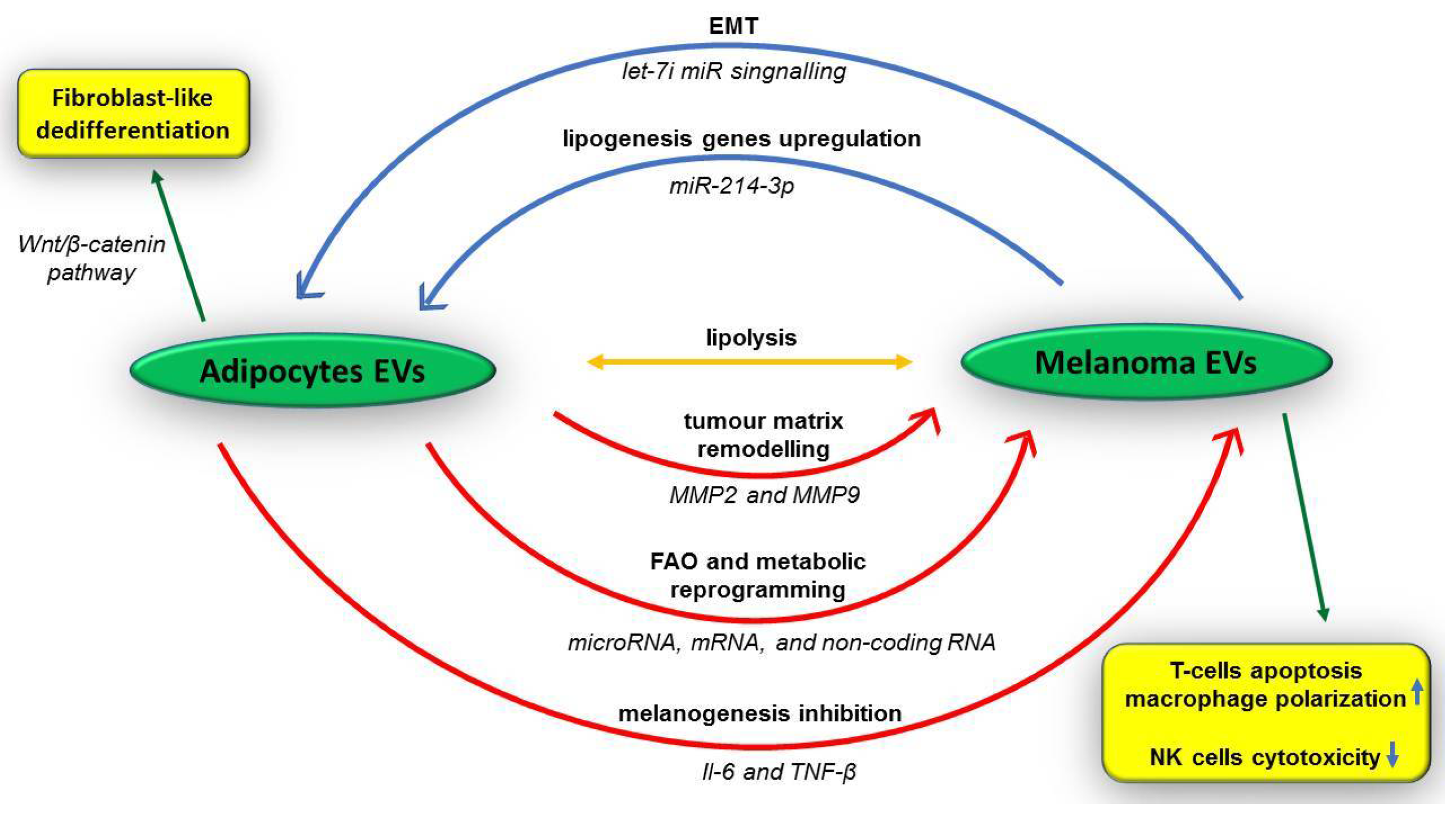
Figure 1. Cellular and extracellular components of melanoma’s microenvironment. The tumour microenvironment (TME) contains numerous cellular and extracellular components, forming together a complex, which supports melanoma development. APCs—antigen-presenting cells; CAFs—cancer-associated fibroblasts; DCs—dendritic cells; EVs—extracellular vesicles; HMGB1- high mobility group box 1 protein; MDSCs—myeloid-derived suppressor cells; miRNAs—microRNAs; PMNs—neutrophils; TCRs—T cells receptors; Tregs—regulatory T cells; ↑—increased level; ↓—decreased level.
Medicina 58 00365 g001
Figure 2. Adipocytes and melanoma cells EVs ’’dialogue’’ in tumour microenvironment. Adipocytes and melanoma cells secrete EVs, which promote tumour progression and metastasis by tumour matrix remodelling, melanogenesis inhibition, fatty acid oxidation (FAO), and metabolic reprogramming into melanoma cells, along with epithelial-mesenchymal transition (EMT) induction, lipolysis, and lipogenesis genes upregulation in fat cells. EVs—extracellular vesicles; EMT—epithelial-mesenchymal transition; FAO—fatty acid oxidation; IL-6—interleukin 6; miRNA—microRNA; MMP—matrix metalloproteinase; NK cells—natural killer cells; T-cells—T lymphocytes; TNF-β—tumour necrosis factor β; ↑—increased level; ↓—decreased level.
Medicina 58 00365 g002
Table 1. Malignant melanoma classification according to sun exposure, general features, and types of gene mutations.
MM Associated with UV ExposureMM Not Consistently Associated with UV Exposure
Low-CDS
(intermittent sun exposure)
High-CSD
(chronic sun exposure)
mucosal melanoma;
melanoma arising in congenital nevi;
melanoma arising in blue nevi;
Spitz melanoma;
acral melanoma;
uveal melanoma;
nevoid melanoma;
nodular melanoma
superficial spreading melanomasubset of nodular melanomalentigo maligna melanomadesmoplastic melanomasubset of nodular melanoma
RGP;
young age;
precursor lesion (nevi)
VGP;
young age;
precursor lesion (nevi)
RGP;
old age;
precursor lesion (MM in situ)
VGP;
old age;
precursor lesion (nevi);
de novo
VGP;
young age;
precursor lesion (nevi)
VGP and RGP;
all ages;
precursor lesion (nevi)
GENOMIC
BRAF (>50%);
NRAS (25%);
other mutations (rare)
NF1;
NRAS;
KIT
NF1;
NFKBIE;
MAPK
NRAS;
KIT;
BRAF;
CDKN2A;
TP53;
TERT
HRAS;
ROS1;
NTRK1 and NTRK3;
CCND1;
TERT;
GNAQ;
BRAF (10%);
KIT (20%);
NRAS (15–20%)
BRAF—B-Raf proto-oncogene, serine/threonine kinase; CCND1— cyclin D1; CDKN2A—cyclin-dependent kinase in-hibitor 2A; CSD—cumulative sun damage; GNAQ—G protein subunit α Q; HRAS—HRas Proto-Oncogene, GTPase; KIT—receptor tyrosine kinase; MAPK—mitogen-activated protein kinase; MM—malignant melanoma; NF1—neurofibromin 1; NRAS—neuroblastoma RAS viral oncogene homolog; NTRK1/3—neurotrophic receptor tyrosine kinase 1/3; RGB—radial growth pattern; ROS1—proto-oncogene tyrosine-protein kinase; VGP—vertical growth pattern; TERT—telomerase reverse transcriptase (TERT) promoter region; TP53—tumour protein 53; UV—ultraviolet light.
Table 2. Immunohistochemical markers useful in malignant melanoma diagnosis.
Marker/ProteinLocationsIHC FeaturesFunctions
Melan A (MART1)
  • normal skin
  • retina pigmented epithelium melanocytes
  • most melanomas
  • high sensitivity and specificity for primary and secondary melanoma
  • positive immunoexpression in clear cell sarcomas, PEComas or angiomyolipomas
  • melanocyte differentiation antigen
  • Pmel expression, processing, traffic, and stability
MITF
  • dynamic subcellular location
  • associated with variable growth and differentiation cell programs
  • high sensitivity in differentiation of melanoma from nonmelanocytic tumours
  • controversial specificity in spindle cells melanoma
  • positive immunoexpression in neurothekeoma cells, histiocytes, and mast cells
  • member of MiT family
  • melanocyte development and differentiation
  • Melan-A, Pmel, and tyrosinase transcription
HMB45
  • recognizes the Silver locus product Pmel17 located in pre-melanosomal vesicles
  • positive immunoexpression for melanoma and junctional nevus cells
  • low sensitivity for metastatic melanoma
  • positive immunoexpression in clear cell sarcomas, PEComas
  • or angiomyolipomas
  • Eumelanin polymerization
SOX10
  • nuclei of melanocytes
  • nuclei of breast myoepithelial cells
  • positive immunoexpression in melanoma, nevi, and focal positivity in desmoplastic melanoma
  • differentiates melanoma in situ from actinic keratosis with melanocytic hyperplasia (along with MITF1)
  • positive immunostaining of salivary and sweat glands, adenoid cystic carcinoma, atypical fibroxanthoma, granular cell tumour, and dermatofibrosarcoma protuberans
  • member of a family of 24 proteins involved in inflammation, cell transcription, differentiation, growth, cell cycle regulation, and calcium homeostasis
  • transcription factor
  • specification of the neural crest derivatives
  • melanocytes and Schwann cells maintenance
S-100
  • melanocytes
  • Langerhans cells
  • chondrocytes
  • glial cells
  • Schwann cells
  • high sensitivity and low specificity for melanoma
  • inflammation
  • cell transcription
  • differentiation
  • growth
  • cell cycle regulation
  • calcium homeostasis
HMB45—Human Melanoma Black; IHC—immunohistochemistry; MART1—melanoma antigen recognized by T cells 1; MelanA—protein melan-A; MiT—microphthalmia transcription factor; MITF—microphtalmia-associated transcription factor; PEComa—perivascular epithelioid cell tumours; Pmel—premelanosomal protein; Pmel17—premelanosomal protein17; S-100—protein S100; SOX10—SRY-related HMG-box 10 protein.
Table 3. Main genomic alterations in malignant melanoma.
GenesIncidencePathwayActionsMMs Type
BRAF
(BRAFV600E
BRAFV600
BRAFV600K
BRAFK601E)
45%
  • MAPK/ERK signalling
  • melanocytes cellular cycle, differentiation, and apoptosis
  • sun exposed cutaneous melanoma
<10%
  • mucosal melanoma
RAS15–30%
  • activation of downstream signalling
  • melanocytes proliferation, differentiation, and survival
  • sun-exposed cutaneous melanoma
NRAS15%
  • MAPK/PI3K signalling
  • melanocytes proliferation, differentiation, and survival
  • sun-exposed cutaneous melanoma
c-KIT (CD117)<3%
  • activation of MAPK and PI3K/AKT pathways
  • melanocytes proliferation and survival regulation
  • sun-exposed cutaneous melanoma
40%
  • mucosal melanoma
ATRX9.11%
  • regulation of chromosomal segregation in mitosis
  • melanoma progression
  • mucosal and
  • cutaneous melanoma
ARID213.32%
  • gene transcription mechanism promoter
  • tumour immunity regulation
  • mucosal melanoma
SETD29.48%
  • methylation of histone H3 lysine 36
  • aberrant differentiation or proliferation of melanocytes
  • mucosal melanoma
GNAQ/GNA1180–90%
  • MAPK signalling
  • melanocytes proliferation
  • uveal melanoma
  • rare in cutaneous melanoma
BAP16.13%
  • ubiquitin-proteasome system and DNA damage response
  • melanocytes growth and proliferation regulation
  • spitzoid tumour
  • uveal melanoma
SF3B133%
  • RNA splicing
  • tumourigenesis
  • mucosal melanoma
NF110–15%
  • downregulation of Rat Sarcoma (RAS) proteins and MAPK/PI3K signalling
  • melanocytes growing and survival
  • sun-exposed cutaneous melanomas
RAC19.2%
  • activated MAPK signalling
  • increased melanocytes proliferation and altered cell migration
  • sun-exposed cutaneous melanoma
TERT14%
  • chromosomal telomere length maintenance
  • melanocytes survival support
  • cutaneous melanoma
KRAS2.9%
  • GTPase activity
  • melanocytes proliferation and survival
  • cutaneous melanomas
ERBB2/43.29%
  • tyrosine kinases signalling
  • melanocytes proliferation and survival
  • cutaneous melanomas
CDKN2A25–35%
  • RB pathway
  • apoptosis and melanocytes survival
  • cutaneous melanomas
TP5315%
  • tumour suppressor and transcriptional activator/repressor of several downstream genes
  • increased melanocytes proliferation and reduced apoptosis
  • sun-exposed cutaneous melanomas
PTEN14%
  • PI3K signalling
  • increased mitogen signalling and cell survival
  • cutaneous melanoma
MAP2K1/210%
  • MAPK signalling
  • melanocytes proliferation
  • cutaneous melanoma
ARID2—AT rich interactive domain 2; ATRX—alpha thalassemia/mental retardation syndrome X-linked; BAP1—BRCA1 associated protein 1; BRAF—B-Raf proto-oncogene, serine/threonine kinase; CD117—cluster of differentiation 117; CDKN2A—cyclin-dependent kinase inhibitor 2A; c-kit—mast/stem cell growth factor receptor kit; DNA—deoxyribonucleic acid; ERBB2/4—Erb-b2 receptor tyrosine kinase 2/4; GNAQ—G protein subunit α Q; GNA11—G-protein subunit α11; GTPase—nucleotide guanosine triphosphatase; KRAS—Kirsten rat sarcoma viral oncogene homolog; MAP2K1/2—mitogen-activated protein kinase kinase 1 and 2; MAPK/ERK—mitogen-activated protein kinase/extracellular signal-regulated kinase; MM—malignant melanoma; NF1—neurofibromin 1; NRAS—neuroblastoma RAS viral oncogene homolog; PI3K—phosphoinositide 3-kinases; PTEN—phosphatase and tensin homolog; RAC1—Ras-related C3 botulinum toxin substrate 1; RAS—rat sarcoma virus; RB—retinoblastoma tumour suppressor; SETD2—SET domain containing 2, histone lysine methyltransferase; SF3B1—splicing factor 3b subunit 1; TERT—telomerase reverse transcriptase (TERT) promoter region; TP53—tumour protein 53.
Table 4. Putative malignant melanoma biomarkers.
Type of MoleculesMarker
Enzymes
  • Cox-2
  • LDH
  • Tyrosinase
  • MMPs
  • TIMP-1
  • Cathepsin K
  • CD10
  • IDO
  • Legumain
Soluble proteins and/or antigens
  • VEGF
  • VEGFR-3
  • CRP
  • Galectin-3
  • Osteopontin
  • Heparin- and chitin-binding lectin YKL-40
  • MIA
  • sICAM-1
  • sVCAM-1
  • CEACAM
  • CYT-MAA
  • MART-1
  • MAGE
  • TA90
  • S100 proteins
  • SOX
Melanin-related metabolites
  • L-DOPA/L-tyrosine
  • 6H5MI2C
  • 5-S-cysteinyl-DOPA
Circulating cell-free nucleic acids
  • miRNA-29c
  • miRNA-221
6H5MI2C—6-hydroxy-5-methoxyindole-2-carboxylic acid; CEACAM—carcinoembryonic antigen-related cell adhesion molecule 1; Cox-2—cyclooxygenase-2; CRP—C-reactive protein; CYT-MAA—cytoplasmic melanoma-associated antigen; IDO—indoleamine-2,3-dioxygenase; LDH—lactate dehydrogenase; L-DOPA—L-3,4-dihydroxyphenylalanine; MAGE—melanoma-associated antigen-1; MIA—melanoma inhibitory activity; miRNA—microRNA; MMPs—matrix metalloproteinases; sICAM-1—soluble intercellular adhesion molecule 1; sVCAM-1—soluble vascular cell adhesion molecule 1; TA90—tumour-associated antigen 90; TIMP-1- tissue inhibitor of metalloproteinase-1; VEGF—vascular endothelial growth factor; VEGFR-3—vascular endothelial growth factor receptor 3.
Table 5. Melanoma CSCs markers.
Marker/PathwayFunction/Advantage
Nodal embryonic signalling
  • embryonic morphogen
  • pluripotency maintenance
  • enhanced tumourigenicity and metastatic ability
  • melanoma plasticity
  • activation due to absence of its inhibitor, Lefty
  • putative prognostic marker
Nestin
  • intermediate filament
  • associated with advanced stage
hTERT
  • telomerase activation by its transcriptional regulation
SOX2
  • regulators of CSC fate
  • nuclear transcription factors involved in neural crest cells differentiation into melanocytes
  • detect positive sentinel lymph nodes
SOX10
  • neural crest stem cell transcription factor
  • regulation of SOX10-MITF pathway
  • tumour cell survival, proliferation, and metastasis
CD20 (MS4A1)
  • self-renewal
  • highly enrich in melanospheres
  • tumourigenesis
CD44
  • EMT
  • tumour invasion
  • tumour metastasis
CD49d/CD29 (α4β1 integrin heterodimer)
  • transmembrane proteins
  • promoter of cell proliferation and migration
  • tumourigenesis
CD49f (Integrin α6)
CD54/ICAM-1
  • cell–cell interaction
CD57/HNK-1
  • tumour cells adhesion, migration, and invasion
CD86/B7-2
  • downregulation of immune response
CD117 (c-KIT)
  • growth and survival of tumour cells
CD133 (prominin-1)
  • p38 MAPK pathway activation
  • long-term tumourigenic potential
  • chemoresistance
  • metastasis induction
  • angiogenesis
CD144 (vascular-endothelial VE-cadherin)
  • cell surface glycoprotein which binds to hyaluronic acid
  • promoter of EGFR-mediated pathways
  • tumour initiation and metastasis
  • chemotherapy resistance
CD146/MCAM
  • signalling receptor
  • tumour progression
CD166/ALCAM
  • tumour cell invasion and metastasis
CD271/NGFR
  • long-term tumour growth
  • metastasis
  • tumour heterogeneity
N-cadherin
  • potentiator of tumour cells invasiveness
miR-10b
miR-21
miR200c
miR-373
miR-520c
  • miRNA
  • target different signalling pathways
MDR1
  • co-expressed with ABC transporters ABCB5 and ABCC2
  • self-renewal stimulation
  • ability to form melanospheres
L1CAM
  • metastasis
ALDH1A1/A2/A3
  • self-renewal
  • high tumourigenesis
  • differentiation
  • chemoresistance
ABC transporters
(ABCB5, ABCG2/BCRP,
ABCC1/MRP, ABCC2, ABCC6)
  • regulation of transport and drug exclusion from tumour cells
  • development of multidrug resistance
  • tumour initiation
PD-1
PDL-1
  • evasion of tumour immunity
  • high tumourigenesis
CTLA-4
  • induces tumour proliferation
  • suppressor of tumour cell apoptosis
  • tumourigenesis
RANK
  • inducer of tumour growth and metastasis
HIF-1
  • promoter of tumour cells self-renewal
  • regulator of tumour microenvironment
Snail
  • transcription factor
Notch4
  • specific signalling
γ-secretase
  • Notch signalling activation
Bcl-2
  • anti-apoptotic
GLI (Hh/Glimo)
  • SOX2 regulation
DDX3X
  • translation reprogramming
  • metastasis
VEGFR-1
  • co-expression with ABCB5
  • vasculogenic
  • tumour growth
VEGFR-2
VEGF
Ang1/2
Tie2
  • angiogenic
CXCR6
  • high tumourigenic
  • self-renewal
JARID1B
  • self-renewal
  • high proliferative progeny
  • tumour growth
  • metastasis
  • Jagged1/Notch1 signalling regulation
EZH2
  • epigenetic modifier
  • promoter of tumour progression
Histone marks
H3K4me2
H3K27me3
H3K9ac
  • epigenetic modifier
  • gene activation/inactivation
  • tumour progression
Hn (heterogeneous ribonucleoproteins) hnRNPs
hnRNP A2B1
hnRNP I
hnRNP L
  • splicing repressors by inhibiting splicing sites via binding to intronic or exonic site
  • tumour progression
  • promote the expression of anti-apoptosis genes DOCK2, TPPP3, and EIF3H
ABC transporters—ATP-binding cassette transporters; ABCB5—ABC sub-family B member 5; ABCC1 ABC sub-family C member 1; ABCC2—ABC sub-family C member 2; ABCC6—ABC sub-family C member 6; ABCG2—ABC sub-family G member 2; ALCAM—activated leukocyte cell adhesion molecule; ALDH—aldehyde dehydrogenase; Ang1/2—angiopoietin 1/2; Bcl-2—B-cell lymphoma 2; BCRP—breast cancer resistant protein; CD—cluster of differentiation; c-KIT—mast/stem cell growth factor receptor kit; CSC—cancer stem cell; CTLA-4—cytotoxic T lymphocyte antigen-4; CXCR6—C-X-C Motif Chemokine Receptor 6; DDX3X—DEAD-Box Helicase 3 X-Linked; DOCK 2—dedicator of cytokinesis 2; EGFR—epidermal growth factor receptor; EIF3H—eukaryotic translation initiation factor 3 subunit H; EMT—epithelial-mesenchymal transition; EZH2—enhancer of zeste homolog 2; GLI—Hh/Glimo; H3K27me2/3—trimethylates lysine 27 of histone H2/3; H3K9—histone H3 lysine 9 acetylation; HIF-1—hypoxia inducible factor 1; HNK-1—human natural killer 1; hnRNP—heterogeneous ribonucleoproteins; hTERT—human telomerase reverse transcriptase; ICAM-1—intercellular adhesion molecule 1; JARID1B—lysine-specific demethylase 5B; L1CAM—L1 adhesion molecular; MAPK—mitogen-activated protein kinase; MCAM- melanoma cell adhesion molecule; MDR1—multidrug-resistance gene product 1; miR—microRNA gene; MS4A1- membrane spanning 4-domains A1; NGFR—nerve growth factor receptor; Notch4—Notch Receptor 4; PD-1—programmed cell death protein 1; PDL-1—programmed death-ligand 1; RANK—receptor activator of NF-κB; Snail—Zinc finger protein SNAI1; SOX—SRY-Box transcription factor 2; Tie2—tyrosine-protein kinase receptor Tie-2; TPPP3—tubulin polymerization promoting protein family member 3; VEGF—vascular endothelial growth factor; VEGFR 1/2- vascular endothelial growth factor receptor 1/2.
Publisher’s Note: MDPI stays neutral with regard to jurisdictional claims in published maps and institutional affiliations.

