
Combined Ileoileal and Ileocolic Intussusception Secondary to Inflammatory Fibroid Polyp in an Adult: A Case Report

 , , and
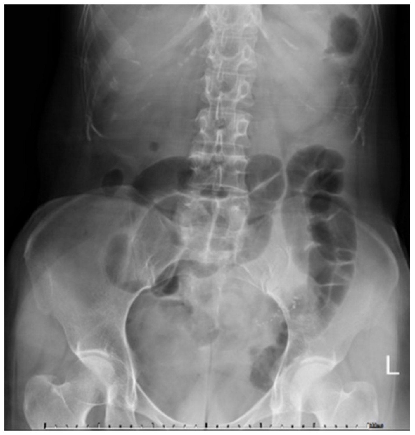
Abstract
:1. Introduction
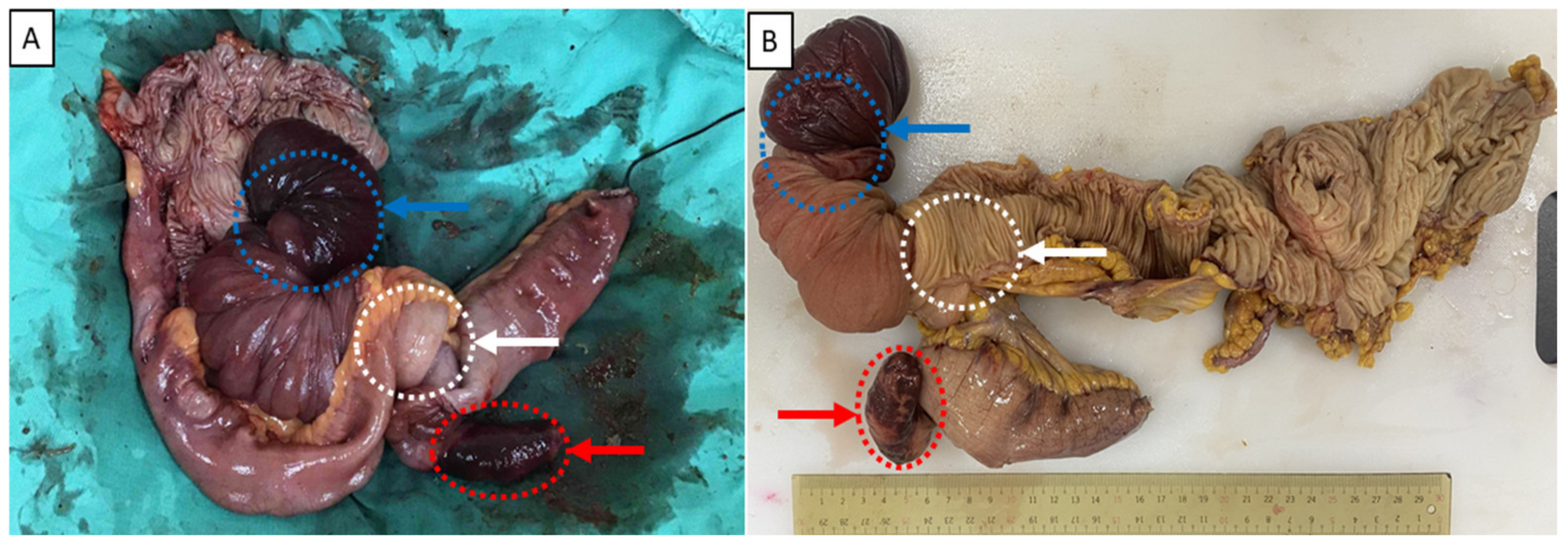
2. Case Report
3. Discussion
4. Conclusions
Author Contributions
Funding
Institutional Review Board Statement
Informed Consent Statement
Data Availability Statement
Conflicts of Interest
References
- Vanek, J. Gastric submucosal granuloma with eosinophilic infiltration. Am. J. Pathol. 1949, 25, 397–411. [Google Scholar] [PubMed]
- Hong, K.D.; Kim, J.; Ji, W.; Wexner, S.D. Adult intussusception: A systematic review and meta-analysis. Tech. Coloproctol. 2019, 23, 315–324. [Google Scholar] [CrossRef] [PubMed]
- Begos, D.G.; Sandor, A.; Modlin, I.M. The diagnosis and management of adult intussusception. Am. J. Surg. 1997, 173, 88–94. [Google Scholar] [CrossRef]
- Tan, K.Y.; Tan, S.M.; Tan, A.G.; Chen, C.Y.; Chng, H.C.; Hoe, M.N. Adult intussusception: Experience in Singapore. ANZ J. Surg. 2003, 73, 1044–1047. [Google Scholar] [CrossRef] [PubMed]
- Wang, L.T.; Wu, C.C.; Yu, J.C.; Hsiao, C.W.; Hsu, C.C.; Jao, S.W. Clinical entity and treatment strategies for adult intussusceptions: 20 years’ experience. Dis. Colon Rectum 2007, 50, 1941–1949. [Google Scholar] [CrossRef] [PubMed]
- Yakan, S.; Caliskan, C.; Makay, O.; Denecli, A.G.; Korkut, M.A. Intussusception in adults: Clinical characteristics, diagnosis and operative strategies. World J. Gastroenterol. 2009, 15, 1985–1989. [Google Scholar] [CrossRef] [PubMed]
- Schildhaus, H.U.; Cavlar, T.; Binot, E.; Büttner, R.; Wardelmann, E.; Merkelbach-Bruse, S. Inflammatory fibroid polyps harbour mutations in the platelet-derived growth factor receptor alpha (PDGFRA) gene. J. Pathol. 2008, 216, 176–182. [Google Scholar] [CrossRef] [PubMed]
- Huss, S.; Wardelmann, E.; Goltz, D.; Binot, E.; Hartmann, W.; Merkelbach-Bruse, S.; Büttner, R.; Schildhaus, H.U. Activating PDGFRA mutations in inflammatory fibroid polyps occur in exons 12, 14 and 18 and are associated with tumour localization. Histopathology 2012, 61, 59–68. [Google Scholar] [CrossRef] [PubMed]
- Liu, T.C.; Lin, M.T.; Montgomery, E.A.; Singhi, A.D. Inflammatory fibroid polyps of the gastrointestinal tract: Spectrum of clinical, morphologic, and immunohistochemistry features. Am. J. Surg. Pathol. 2013, 37, 586–592. [Google Scholar] [CrossRef] [PubMed]
- Kao, Y.K.; Chen, J.H. Adult Jejuno-jejunal intussusception due to inflammatory fibroid polyp: A case report and literature review. Medicine 2020, 99, e22080. [Google Scholar] [CrossRef] [PubMed]


Publisher’s Note: MDPI stays neutral with regard to jurisdictional claims in published maps and institutional affiliations. |
© 2022 by the authors. Licensee MDPI, Basel, Switzerland. This article is an open access article distributed under the terms and conditions of the Creative Commons Attribution (CC BY) license (https://creativecommons.org/licenses/by/4.0/).
Share and Cite
Chiu, H.-T.; Yen, H.; Weng, Y.-S.; Chen, C.-Y.; Lin, K.-H.; Chen, P.-H.; Jhou, H.-J.; Pu, T.-W. Combined Ileoileal and Ileocolic Intussusception Secondary to Inflammatory Fibroid Polyp in an Adult: A Case Report. Medicina 2022, 58, 310. https://doi.org/10.3390/medicina58020310
Chiu H-T, Yen H, Weng Y-S, Chen C-Y, Lin K-H, Chen P-H, Jhou H-J, Pu T-W. Combined Ileoileal and Ileocolic Intussusception Secondary to Inflammatory Fibroid Polyp in an Adult: A Case Report. Medicina. 2022; 58(2):310. https://doi.org/10.3390/medicina58020310
Chicago/Turabian StyleChiu, Hao-Tse, Hao Yen, Yu-Shiou Weng, Chao-Yang Chen, Kuan-Hsun Lin, Po-Huang Chen, Hong-Jie Jhou, and Ta-Wei Pu. 2022. "Combined Ileoileal and Ileocolic Intussusception Secondary to Inflammatory Fibroid Polyp in an Adult: A Case Report" Medicina 58, no. 2: 310. https://doi.org/10.3390/medicina58020310