Article

Identification and Characterization of Novel Mutations in Chronic Kidney Disease (CKD) and Autosomal Dominant Polycystic Kidney Disease (ADPKD) in Saudi Subjects by Whole-Exome Sequencing

by
Othman R. Alzahrani
1,2,
Hanan E. Alatwi
1,2,
Amnah A. Alharbi
2,3,
Abdulrahman H. Alessa
1,2,
Osama M. Al-Amer
2,4,
Abeer F. R. Alanazi
5,
Anwar M. Shams
6,
Esra’a Alomari
7,
Abdallah Y. Naser
7,
Faisal a. Alzahrani
8,
Salman Hosawi
9,10,
Saeed M. Alghamdi
11,
Wed A. Abdali
12,
Imadeldin Elfaki
2,3 and
Yousef M. Hawsawi
12,13,*
1
Department of Biology, Faculty of Sciences, University of Tabuk, Tabuk 71491, Saudi Arabia
2
Genome and Biotechnology Unit, Faculty of Sciences, University of Tabuk, Tabuk 71491, Saudi Arabia
3
Department of Biochemistry, Faculty of Sciences, University of Tabuk, Tabuk 71491, Saudi Arabia
4
Department of Medical Laboratory Technology, Faculty of Applied Medical Sciences, University of Tabuk, Tabuk 71491, Saudi Arabia
5
Department of Pharmaceutical and Biological Sciences, UCL School of Pharmacy, London WC1E 6BT, UK
6
Department of Pharmacology, College of Medicine, Taif University, P.O. Box 11099, Taif 21944, Saudi Arabia
7
Department of Applied Pharmaceutical Sciences and Clinical Pharmacy, Faculty of Pharmacy, Isra University, Amman 11622, Jordan
8
Department of Biochemistry, Faculty of science, Embryonic Stem Cell Unit, King Fahad Center for Medical Research, King Abdulaziz University, Jeddah 21589, Saudi Arabia
9
Department of Biochemistry, Faculty of Science, King AbdulAziz University, Jeddah 21589, Saudi Arabia
10
Artificial Intelligence in Systems Biology Research Unit, The King Abdulaziz University and The University of Oxford Center for Artificial Intelligence for Precision Medicine, Jeddah 21589, Saudi Arabia
11
Department of Medicine, King Faisal Specialist Hospital and Research Centre, P.O. Box 40047, Jeddah 21499, Saudi Arabia
12
Research Center, King Faisal Specialist Hospital and Research Center, MBC J04, P.O. Box 40047, Jeddah 21499, Saudi Arabia
13
College of Medicine, Al-Faisal University, P.O. Box 50927, Riyadh 11533, Saudi Arabia
*
Author to whom correspondence should be addressed.
Medicina 2022, 58(11), 1657; https://doi.org/10.3390/medicina58111657
Submission received: 7 October 2022 / Revised: 12 November 2022 / Accepted: 13 November 2022 / Published: 16 November 2022
(This article belongs to the Special Issue Genetics and Inherited Diseases)

Abstract

:
Background: Autosomal dominant polycystic kidney disease (ADPKD) is a condition usually caused by a single gene mutation and manifested by both renal and extrarenal features, eventually leading to end-stage renal disease (ESRD) by the median age of 60 years worldwide. Approximately 89% of ADPKD patients had either PKD1 or PKD2 gene mutations. The majority (85%) of the mutations are in the PKD1 gene, especially in the context of family history. Objectives: This study investigated the genetic basis and the undiscovered genes that are involved in ADPKD development among the Saudi population. Materials and Methods: In this study, 11 patients with chronic kidney disease were enrolled. The diagnosis of ADPKD was based on history and diagnostic images: CT images include enlargement of renal outlines, renal echogenicity, and presence of multiple renal cysts with dilated collecting ducts, loss of corticomedullary differentiation, and changes in GFR and serum creatinine levels. Next-generation whole-exome sequencing was conducted using the Ion Torrent PGM platform. Results: Of the 11 Saudi patients diagnosed with chronic kidney disease (CKD) and ADPKD, the most common heterozygote nonsynonymous variant in the PKD1 gene was exon15: (c.4264G > A). Two missense mutations were identified with a PKD1 (c.1758A > C and c.9774T > G), and one patient had a PKD2 mutation (c.1445T > G). Three detected variants were novel, identified at PKD1 (c.1758A > C), PKD2L2 (c.1364A > T), and TSC2 (deletion of a’a at the 3’UTR, R1680C) genes. Other variants in PKD1L1 (c.3813_381 4delinsTG) and PKD1L2 (c.404C > T) were also detected. The median age of end-stage renal disease for ADPK patients in Saudi Arabia was 30 years. Conclusion: This study reported a common variant in the PKD1 gene in Saudi patients with typical ADPKD. We also reported (to our knowledge) for the first time two novel missense variants in PKD1 and PKD2L2 genes and one indel mutation at the 3’UTR of the TSC2 gene. This study establishes that the reported mutations in the affected genes resulted in ADPKD development in the Saudi population by a median age of 30. Nevertheless, future protein–protein interaction studies to investigate the influence of these mutations on PKD1 and PKD2 functions are required. Furthermore, large-scale population-based studies to verify these findings are recommended.

1. Introduction

Monogenic cystic kidney disease is a cilia-associated disorder that is composed of two forms: autosomal recessive polycystic kidney disease (ARPKD) and autosomal dominant polycystic kidney disease (ADPKD) [1]. ARPKD is a rare and severe form of the disease that mainly presents in children, while ADPKD is the most common hereditary genetic renal disorder that occurs primarily in adulthood [2]. Clinically, the two forms of the disease can be distinguished based on the presence of many criteria, such as kidney morphology, location of the cyst, hepatic fibrosis, arterial hypertension, and age at the presentation of the symptoms [3]. The majority of ADPKD patients presented with hypertension and deterioration in the glomerular filtration rate (GFR) [4], which might require renal dialysis or transplantation in the advanced stages [5]. Based on several clinical studies, ADPKD is the most prevalent genetic renal disorder, with estimated cases of 1 in 500-2500 individuals [6] anticipated to affect over 10 million individuals globally from various ethnicities; thus, it is one of the significant clinical concerns [1].
Numerous biological and clinical studies have been conducted to delineate the genetic mechanism of ADPKD [2,7,8,9]. Mutation in the genes PKD1 (78% of cases) or PKD2 (15% of cases) accounts for the most common genetic alterations involved in the pathogenesis of ADPKD [10]. The PKD1 gene is located on chromosome 16 and encodes polycystin-1 protein (PC1), a large transmembrane integral glycoprotein [10]. The PKD1 gene consists of 46 exons with a large, duplicated region from exons 1-33 that shares a high degree of sequence identity with the other six pseudogenes near PKD1 on the same chromosome (16p) [11]. The PKD2 gene is located on chromosome 4 and encodes polycystin-2 protein (PC2), a transmembrane protein. PKD2 is smaller than PKD1 and has only 15 exons [11]. Although mutations of PKD1 and PKD2 genes are fully penetrant, their expressivity is associated with different cases of variable severity in members of the same family, suggesting the presence of additional gene modifiers [12].
Clinical information combined with imaging-based strategies, preferentially ultrasonography, is the primary approach to diagnosing patients with ADPKD [13,14]. MRI or CT scans with contrast provide highly specific and sensitive modalities in calculating the number and diameter of the cysts, especially in high-risk younger people with positive family history [15]. Aside from imaging techniques, genetic testing is required to confirm the definitive diagnosis of ADPKD, particularly in young members with few cysts and no family history or when the diagnosis is vague [8,16,17]. Indeed, genetic testing affords a valuable diagnostic and prognostic tool that might further improve clinical management and outcomes in cystic kidney disease patients [2,17,18]. Over 1500 mutations with high allelic heterogeneity of PKD1 and PKD2 genes have been indicated in the ADPKD dataset [1]. Nevertheless, the detection of PKD1 mutation requires comprehensive analysis because of its large size, complex genomic structure, and expensive technique [19].
Transmission of genetic susceptibility diseases, such as inherited renal disorders, was found to be more frequent in areas where consanguineous marriages are common, such as in the Kingdom of Saudi Arabia (KSA) [20]. Since the rate of consanguinity marriages in KSA was reported to reach up to 55%, homozygous mutations in the PKD1 gene and other genes related to cystic kidneys would be more anticipated [21]. Furthermore, allelic and genetic heterogeneity for many Mendelian disorders has evolved in KSA [22]. An observational study conducted in Saudi Arabia reported a high incidence of genetically inherited kidney diseases such as polycystic kidney disease (PKD), familial juvenile nephronophthisis, congenital urological anomalies, and familial nephrotic syndrome among Saudi children [23]. Moreover, antenatal analysis using an ultrasonography test along with Sanger sequencing of the PKD1 gene was employed among four consanguineous Saudi families to investigate the genetic background of the early-onset PKD [24]. Additionally, homozygous and compound heterozygous hypomorphic PKD1 missense alleles mutation was detected in two families and resulted in the early onset of an aggressive disease yet was compatible with life [24]. Al-Muhanna et al. performed the exome sequencing using WES technology on 16 Saudi ADPKD patients and reported that variants related to PKD1 and PKD2 genes are the most frequently detected mutations in Saudi patients with typical ADPKD. On the other hand, a specific form of ADPKD was associated with rare pathogenic mutations in cytogenic genes such as PKHD1, PKD1L3, EGF, CFTR, and TSC2 [25].
ADPKD is considered a critical health issue in Saudi Arabia, and progression to end-stage kidney disease (ESKD) is inevitable. Numerous reports have exemplified the genetic elements involved in the development of PKD/ADPKD among the Saudi population [20,21,23,24,25,26,27]. Yet the extent to which these genetic variants are involved in the pathogenesis, erraticism in clinical scenarios, and disease severity remain poorly understood. Previously, we reported several mutations that are associated with diseases in Saudi Arabia [27,28,29,30,31]. Therefore, the current study aims to examine the genetic basis and the hitherto undiscovered genes that might be involved in developing ADPKD among the Saudi population.

2. Methods, Materials, and Subjects

2.1. Patients and Family Recruitment

This study included 11 patients from different families, diagnosed with ADPKD and chronic kidney disease (CKD) probands ascertained in the nephrology clinic at the King Faisal Specialist Hospital and Research Center Jeddah (KFSH & RC); participants were enrolled in the western province of Saudi Arabia. All subjects signed the informed consent and documentation of family history forms. The diagnosis of ADPKD was achieved based on different criteria, and measurements comprised (1) medical history: the patient’s clinical manifestations; in addition to the presence of positive family history, (2) diagnostic images: CT images include enlargement of renal outlines, renal echogenicity, presence of multiple renal cysts with dilated collecting ducts, and loss of corticomedullary differentiation; and (3) laboratory data: evaluation of glomerular function (GFR) and creatinine level in the serum. Demographic data retrieved from the patients’ medical reports, including age, gender, race, ethnicity, and previous and current medical history (hypertension status, CKD stage, eGFR, affected family member, and presence of other medical conditions), are presented in Table 1. Patients were classified as G1–G5 based on the eGFR and A1–A3 based on the albumin:creatinine ratio (ACR).

2.2. Ethical Approval and Participants’ Consent

This study was approved by the Institutional Review Board of King Faisal Specialist Hospital and Research Center Jeddah (IRB number 2018-36) on 18 September 2018. Informed consent for the retention and use of patient data for research purposes was routinely obtained at the time of obtaining consent for the procedures. Before blood sample collection, all patients who met the inclusion and exclusion criteria completed the informed consent (ICF) and case report form (CRF).

2.3. DNA Extraction

Peripheral blood samples were collected from each patient (approximately 2–3 cc). Subsequently, the genomic DNA was extracted from all blood samples using a Gentra Puregene Blood Kit (Qiagen, Cat No: 158389, Düsseldorf, Germany) according to the manufacturer’s instructions. Briefly, 20 µL of proteinase K was added to 200 µL of whole blood, mixed, and incubated for 1 min at room temperature (15–25 °C), followed by adding 200 µL of buffer to the sample. Then, the mixture was incubated in a water bath at 56 °C for 10 min and spun for 2 min at 2000× g, and 200 μL of ethanol (96–100%) was added to the sample, followed by mixing by pulse-vertexing for 15 s. Finally, 100 µL of the DNA Hydration Solution was added, and it was vortexed for 5 s and incubated at 65 °C for 5 min. The quantity and quality of extracted DNA were evaluated by using the Qubit 2.0 Fluorometer (Life Technologies, Waltham, MA, USA) with the Qubit dsDNA High Sensitivity Assay Kit (Life Technologies, Cat No Q32851, Waltham, MA, USA) and NanoDrop 2000 system (Thermo Scientific, Waltham, MA, USA), respectively, according to the manufacturer’s instructions.

2.4. DNA Library Preparation

Extracted DNA (100 ng per sample) was used for library preparation using the Ion AmpliSeq Library Kit Plus (Life Technologies, Cat # 4488990, Waltham, MA, USA) with the Ion AmpliSeq Exome RDY Kit (Life Technology, cat # 8849838, Waltham, MA, USA) according to the manufacturer’s instructions.

2.5. Next-Generation Sequencing (NGS) Technology

DNA was sequenced by utilizing massive parallel sequencing via Ion Torrent PGM with the Ion PI Hi-Q Sequencing 200 Kit (Life Technologies, Cat# 4488651, Waltham, MA, USA) and the Ion PI Chip Kit v3 (Life Technologies, Cat# A26770, Waltham, MA, USA) according to the manufacturer’s instructions. Briefly, the prepared DNA was loaded in Ion Torrent semiconductor chips and loaded into the Ion Torrent PGM machine.

2.6. Variant Filtration

The reads of the Ion Torrent sequencing machine (Life Technologies, Waltham, MA, USA) were aligned to the hg19 reference genome through the tMap program. The aligned reads were investigated for variant calling through the Torrent Suite Variant Caller TVC program. The variants were annotated by using public and in-house databases and the Saudi Human Genome program

2.7. In Silico Analysis

Multiple in silico algorithm software programs were used to determine the structural function and to evaluate the pathogenic effect and the impact of the specific mutation on the biological processes of the protein, such as Mutation taster [32], Polyphen-2.0 [33], SIFT [34], and PROVEAN [35].
Polymorphism Phenotyping v2 (PolyPhen-) was used as a tool to predict the possible impact of an amino acid substitution on the structure and function of the reported protein using straightforward physical and comparative considerations (http://genetics.bwh.harvard.edu/pph2/ (accessed on 18 January 2022)

2.8. Bioinformatics

The mapping of the mutations to protein domains was performed manually using the NCBI Nucleotide database and UNIPROT (https://www.uniprot.org (accessed on 18 January 2022)). The protein–protein interaction analysis was performed using the STRING database (https://string-db.org (accessed on 18 January 2022)) and Cytoscape (https://cytoscape.org (accessed on 18 January 2022)) for data retrieval and merging, respectively. The gene ontology [36,37] and KEGG (https://www.genome.jp/kegg/pathway.html (accessed on 18 January 2022)) pathway analyses were performed using STRING enrichment. All networks retrieved from STRING in this work had a confidence score of 0.4 and included all types of evidence available in the database.

2.9. Statistical Analysis

Statistical analysis was conducted via SPSS version 22.0 statistical package software (SPSS, Chicago, IL, USA). All probability values (p-values) below 0.05 were considered statistically significant. The ORs and 95% CIs were calculated using the chi-squared test to study the comparison of genetic variations.

3. Results

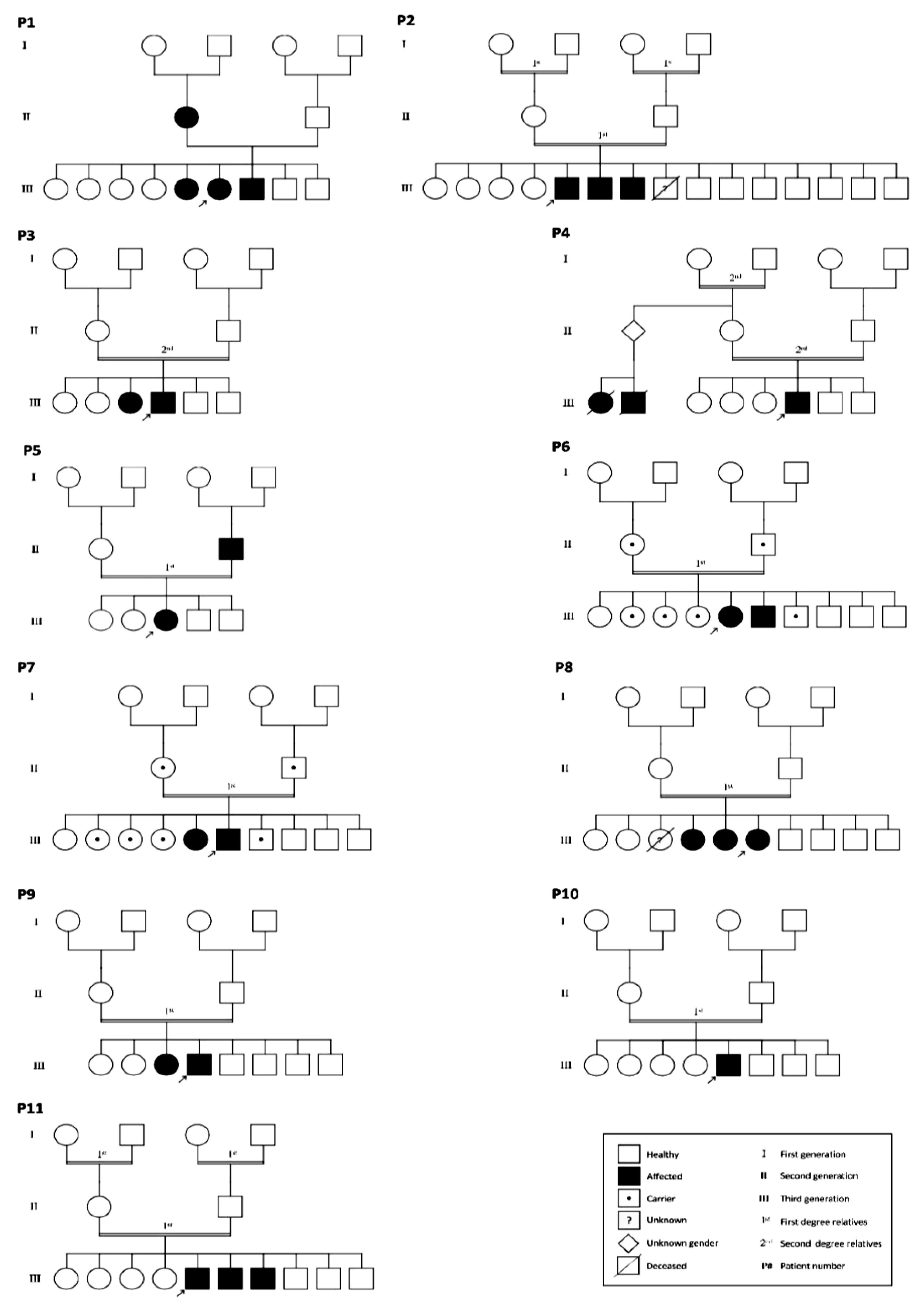
3.1. Basic Clinical Characteristics and Genetic Analysis of the Studied Subjects

In this study, 11 patients clinically diagnosed with ADPKD and CKD were selected from the King Faisal Specialist Hospital and Research Center from 2002 to 2019 to participate in this work.
As shown in Table 1, patients’ ages were distributed among young and middle-aged categories (18–46 years old), which included four females and seven males. All patients presented with different stages of CKD and abnormally low eGFR values and were diagnosed with the presence of other kidney disorders. Hypertension was detected in 63.6% (7/11) of the participants. Positive family history was detected in all participants except one patient (90.90%). We excluded from the study all patients with syndromic causes of multiple renal cysts, such as von Hippel–Lindau disease, tuberous sclerosis, and familial polythelia with multiple renal cysts. Furthermore, the family history including affected and carrier family members with ADPKD is presented in the familial pedigree for each patient (Figure 1).

3.2. Mutations Identified in Patients with ADPKD

To characterize the genetic alteration that might be engaged in the pathogenesis of ADPKD in the selected individuals, we used NGS technology on the extracted DNAs. Among eleven cases, five missense mutations (45.45%) were identified in the PKD1 gene. Three out of these five mutations were identified in exon15: c.4264G > A: p.A1422T, including the following patients P2, P8, and P11. In the following patients, the other two mutations were detected in exon9: c.1758A > C: p.E586D and exon29: c.9774T > G: p.F3258L, P4 and P9, respectively. One missense mutation (9.09%) in the PKD1L2 gene was identified in exon2: c.404C > T: p.P135L in P5, and another missense mutation (9.09%) in the PKD2L2 gene was identified in exon9: c.1364A > T: p.N455I in P6. Additionally, one missense mutation (9.09%) in the PKD2 gene was identified in exon6: c.1445T > G: p.F482C in the third patient (P3). The first patient showed one non-frame-shift substitution in the PKD1L1 gene in exon 24: c.3813_3814delinsTG. In the seventh patient, we detected exonic alteration in exon 39: c.5038C > T: p.R1680C in the TSC2 gene. The last patient (P10) shows no genetic abnormalities or positive family history (Table 1 and Table 2). To further determine the contribution of the identified mutations to the development and predisposition to ADPKD, we used the predictable bioinformatics tools, including Mutation taster, Polyphen-2.0, SIFT, and PROVEAN, to estimate the pathogenicity score for each detected genetic alteration. The pathogenic effects were observed when the mutation variants happened in the PKD1 gene involving exons 9 and 29, PKD2 gene in exon3, PKD2L2 in exon9, and TSC2 gene in exon39 (showed likely pathogenicity). To provide insight into the impact of these identified mutations on the proteins’ stability and functions, we manually mapped each genetic mutation to its corresponding protein using the NCBI Nucleotide database and UNIPROT software. Our analyses revealed that two PKD1 gene mutations did not localize within any of its known protein domains or post-translational modification (PTM) sites; these mutations were detected in exon9: c.1758A > C: p.E586D and exon29: c.9774T > G: p.F3258L. On the other hand, we found that the third mutation of the PDK1 gene (exon15: c.4264G > A: p.A1422T) is located within the PKD9 domain of PC-1 protein. Furthermore, the missense mutations of PKD1L2 and PKD2L2 genes located on exon2: c.404C > T: p.P135L and exon9: c.1364A > T: p.N455I were found to be positioned within two different motifs of PC-1 protein: the C-type lectin domain and the receptor for egg jelly (REJ), respectively. The missense mutation of the PKD2 gene (exon6: c.1445T > G: p.F482C) was not placed on any known protein domains of PTM sites of that protein. Finally, the mutation (exon39: c.5038C > T: p.R1680C) in the TSC2 gene was localized within its Rap-GAP domain.

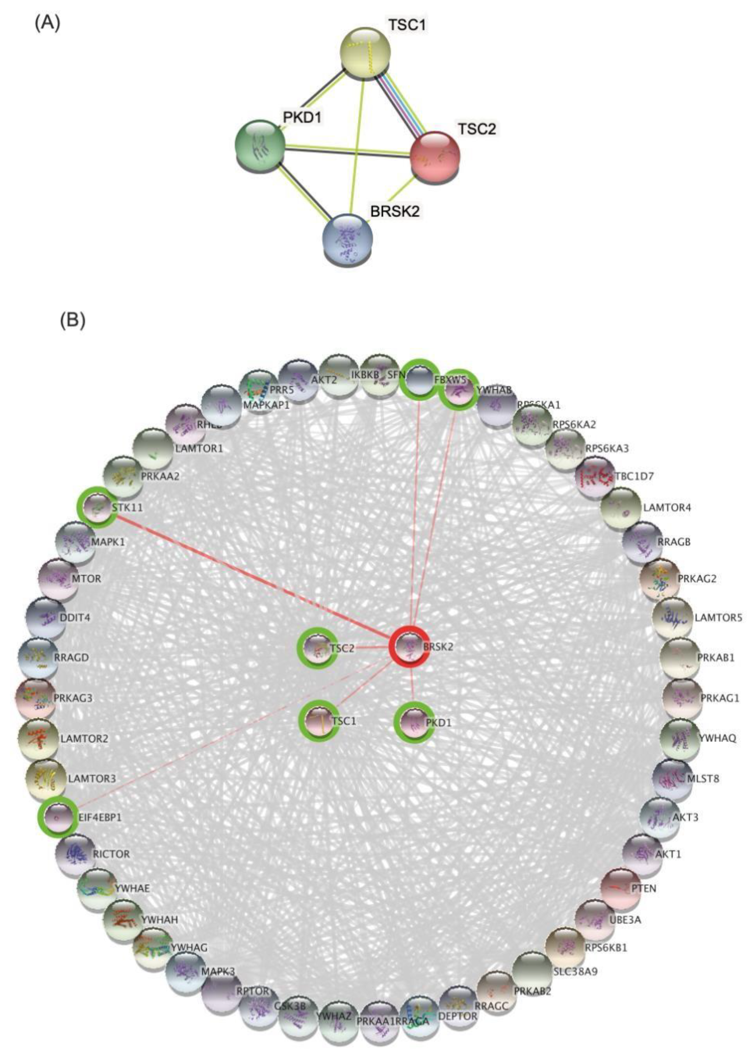
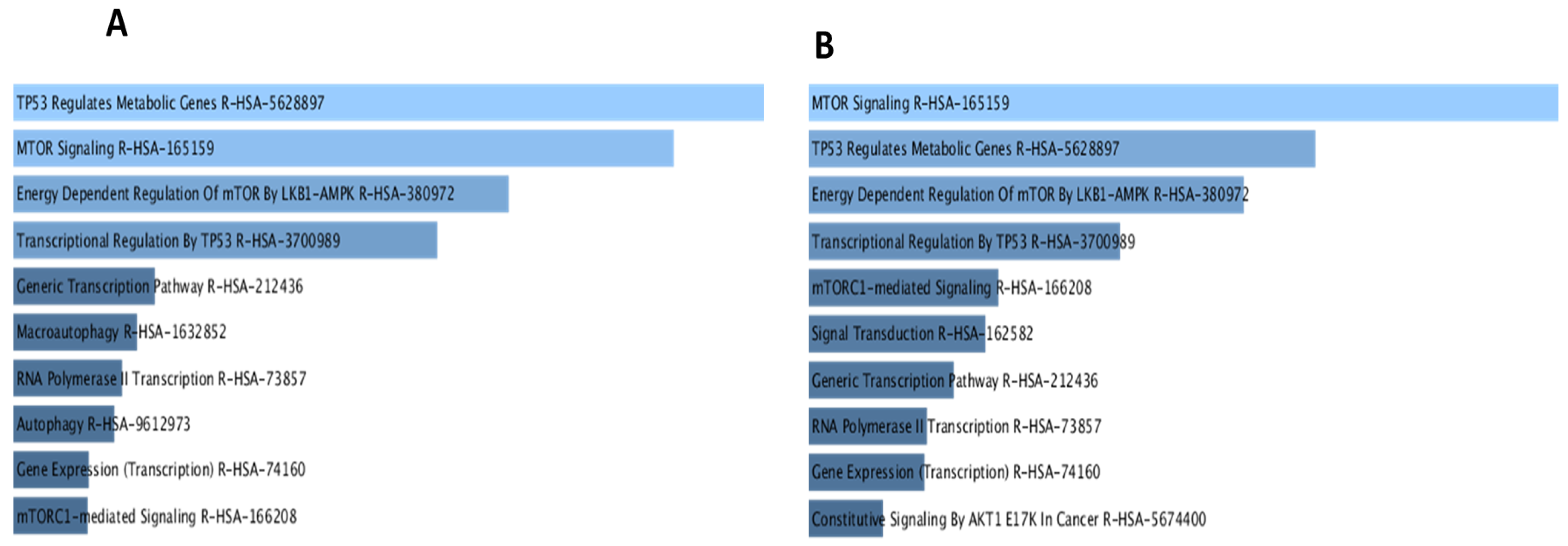
3.3. Bioinformatics Analysis and Data Mining Highlight Potential Proteins Interactions between PKD1 and TSC2 via BRSK2

To investigate potential interactions between the different proteins possessing the identified mutations that might eventually contribute to the pathogenesis of ADPKD, the protein–protein interaction (PPI) network for each of the targets (PKD1, PKD2, TSC2, and PKD1L2) was retrieved from the STRING database. As shown in Figure 2A, a map composed of four different proteins (PKD1, TSC1, TSC2, and BRSK2) resulted from merging various PPI networks. Each protein represents a common intersecting point of these merging networks. To gain further insight into the different biological activities of these four proteins, we expanded the PPI network of the four proteins to include 50 more interactions with medium interaction confidence scores of 0.4. Next, we applied gene ontology (GO) and KEGG pathway enrichment analyses for the entire network (Figure 2B). Most top nodes were influenced by TSC1, TSC2, and PKD1, while BRSK2 was found among significant nodes but at a lower ranking. To further elucidate the contribution of BRSK2, a subnetwork from Figure 2B was created by selecting the first neighbors of BRSK2, including TSC2, PKD1, TSC1, FBXW5, EIF4EBP1, STK11, and YWHAB (Figure 3) for assessment by GO and KEGG pathway analyses. The generated map revealed similar interacting nodes to that one obtained in the primary network (Figure 2B). Yet, the new network showed different arrangements of the nodes based on significance, such as the PI3K and AMPK signaling pathways. Next, we aimed to explore the potential role of BRSK2 in ADPKD pathogenesis. Therefore, a search process on PubMed was conducted, where BRSK2 has searched along with different terminologies such as ADPKD, ARPKD, PKD, Cyst, and kidney. Interestingly, searching BRSK2 against mTOR yielded several hits describing the involvement of the BRSK2 protein and TSC2 in the regulation of mTOR signaling in different contexts, including cancer cell survival and development [38]. Moreover, the PI3K/AKT/mTOR pathway was reported to encourage the growth of renal cysts and increase their size and proliferation [39].

3.4. Mutation and Allele: Frequency

For the missense SNP (rs140980374), G/A/Ancestral: G|MAF: < 0.01 (A), highest population MAF: < 0.01. For the missense variant in SNP (rs201455881), The 1000 Genomes Project Phase 3 allele frequencies showed allele frequencies of G: 0.9998 and A: 0.0001. The genotype frequency is G/G: 0.9996 and A/G: 0.00039 in all populations.

4. Discussion

4.1. Identification of Novel Variants with a Potential Contribution to the Evolution and Pathogenesis of ADPKD in Saudi Patients

ADPKD accounts for the most common genetic kidney diseases worldwide and is considered a significant health concern and clinical challenge [2,6]. Among the Saudi population, ADPKD is one of the main hereditary medical conditions due to the high level of consanguineous marriages, a suitable setting that encourages the transmission of inherited disorders [21,22]. Furthermore, the incidence of novel mutations that might contribute to the early onset of an aggressive form of ADPKD was also reported to be frequent among the Saudi population [24,25,26,40]. We previously identified several variants that are associated with diseases in the Saudi population [41,42,43,44,45]
Therefore, in the current work, we aimed to expand our knowledge and further understand the genetic background engaged in ADPKD evolution, pathogenesis, and progression, thus facilitating the insinuation of appropriate, timely management. To achieve this goal, we used NGS technology to screen the whole-exome variants of 11 clinically diagnosed ADPKD patients belonging to different Saudi families. Our analysis revealed that most of the obtained mutations in the selected patients were detected in PKD1 or PKD2 genes (54.54%), in agreement with the previous Saudi reports indicating that most individuals with ADPKD (89.1%) had mutations in the PKD1 or PKD2 genes [25,26,41]. Moreover, our results also showed that patients who harbor either PKD1 or PKD2 gene mutations showed an earlier age of diagnosis (18–37 years) with a median of 28 years, suggesting the substantial involvement of positive family history in the occurrence of the disease [46,47]. Additionally, 50% of the studied PKD1/PKD2 affected individuals reached a high disease grade (G4/5) or end-stage renal disease (ESRD).
Several reports have shown that the detection of various types of mutations in PKD1 and PKD2 genes was associated with rapidly progressive disease and early onset of renal dialysis, eventually yielding ESRD [48,49,50,51,52,53]. Moreover, individuals with PKD1 mutation developed ESRD at a median age of 58, while PKD2 mutation patients showed better prognosis and delayed onset of ESRD at around 80 years [54,55]. Interestingly, our finding documented three novel mutations in the following genes PKD1, PKD2L2, and TSC2 of the Saudi ADPKD patients.
The first novel missense variant was detected in patient P4 at position p.E586D of the PKD1 protein, and it was predicted by SIFT and PolyPhen programs, with pathogenic effects on the structure and function of the protein. The second novel missense variant was located at position p.N455I of the PKD2L2 protein in patient P6. Its pathogenic effect was estimated to be of uncertain significance using SIFT and PolyPhen programs. It was found that protein variants occurring at promoter regions or the 3’UTR could affect gene splicing, transcription factor binding, or interfere with microRNA binding sites, which could produce an abnormal amount of protein [56]. Here, we also documented a third novel variant residing at the 3’UTR of the TSC2 gene in patient P7 at the position of 2138671–2138672 on chromosome 16, which corresponds to the protein site p.R1680C, which resulted in amino acid deletion and showed likely pathogenetic effects. TSC2, the tuberous sclerosis complex, the most common gene for an autosomal dominant genetic disease, is featured by the formation of benign tumors (hamartomas) in different organs [57]. Tuberous sclerosis manifests in the presence of CNS/mental, psychological, and skin manifestations and frequent renal cysts [58,59]. TSC2 and PKD1 genes were demonstrated to lie adjacent at chromosome 16p13.3, highlighting a shared cross-talk by both genes [58]. High-throughput genetic tools allowed the discovery of a broad range of mutations in TSC1 or TSC2 genes [59]. A study found that large mutational deletion of the TSC2 gene is correlated with the development of early-onset polycystic kidney disease in which the adjacent PKD1 gene was also affected by this deletion [60]. Likewise, Consugar et al. reported that large genomic deletions could also cause PDK1/TSC2 contiguous gene deletion syndrome that is characterized by the presence of various degrees of cystic kidney with early onset of ESRD, hypertension, neurological, behavioral, facial, and skin abnormalities [57,61,62]. Additionally, individuals with a severe and early diagnosed form of the renal cystic disease were also found to display TSC2 gene mutation [58].

4.2. Potential Damaging Impacts of the Detected Mutations on the Protein Structure and Function

To further broaden our understanding of the detrimental influences of the identified mutations on the protein functional efficiency, we calculated the pathogenicity score and then elucidated the positions at which these different mutations are situated within each protein. Five patients with PKD1 gene mutations were identified; three of them (P2, P8, and P11) display a missense variant (exon15: c.4264G > A: p.A1422T) in which the alanine (a very small hydrophobic) is replaced by threonine (a small neural amino acid). This missense variant (exon15: c.4264G > A: p.A1422T) showed an uncertain pathogenic effect and was estimated to locate within the PKD9 domain of the PC-1 protein. PKD1 gene encodes polycystin-1 (PC-1), a large cell surface glycoprotein. A 16-PKD-repeat region (or Ig-like domain) was identified within the extracellular region of the PC-1 protein [63]. The PKD domain showed a ligand binding motif for potential regulation of extracellular signaling involved in protein–protein and protein–carbohydrate interactions and cellular adhesive process [64,65]. Indeed, antibody targeting of the PKD domain in an in vitro model interfered with cellular interactions, thus suggesting that maintaining an intact PKD domain is an important step in preventing cystic generation in ADPKD [66].
The other two patients, P4 and P9, showed missense mutations in exon9: c.1758A > C: p.E586D and exon29: c.9774T > G: p.F3258L, respectively. In the first mutation c.1758A > C: p.E586D, the glutamic acid (medium, hydrophilic, and negatively charged) is replaced by aspartic acid (small, hydrophilic, negatively charged). In the second mutation, c.9774T > G: p.F3258L, the phenylalanine (very large, hydrophobic) is replaced by leucine (large, hydrophobic). These two variants were calculated to possess a pathogenic impact on protein function, yet they were not situated at any predetermined domain or PTM site of the protein. Along with this, the missense mutation of the PKD2 gene (exon6: c.1445T > G: p.F482C) revealed the same findings, and this mutation was also not found to be located on any known protein domains of PTM sites. The physiological roles of ADPKD proteins, PC-1, and PC-2, are regulated by several PTM processes, including phosphorylation, glycosylation, and proteolytic cleavage [67].
Our results also revealed the uncertain pathogenic significance of both mutations of PKD1L2 and PKD2L2 (exon2: c.404C > T: p.P135L and exon9: c.1364A > T: p.N455I). In the mutation of exon2 c.404C > T: p.P135L, the proline (small neutral) is replaced by leucine (large, hydrophobic), and in exon9: c.1364A > T: p.N455I, the asparagine (small, hydrophilic) is replaced by isoleucine (large and hydrophobic). These variants were found to be positioned within two distinct protein domains, the C-type lectin and REJ domains of PC-1 protein on its ECD, respectively. Additionally, C-type lectins were found to modulate cell–cell adhesions and various immunological responses to conduct intracellular signaling [68,69]. The C-type lectin domain, through an auto-proteolytic process, can be fragmented from the PC-1 protein and cell membrane to act as secreted ligand to activate the polycystine channels in the cilia and plasma membrane [70]. Thus, the mutation of PKD1L2 (exon2: c.404C > T: p.P135L) at the C-type lectin domain could affect this motif’s function and consequently contribute to the disease development. Numerous mutations, including missense mutations, have been identified in this motif, with the majority of these variants predicted to deliver detrimental consequences [71]. Variants, including the REJ region, disrupt the cleavage ability and block the activation of the JAK-STAT pathway by PC-1 protein and inhibit tubular formation, a cardinal feature of PC-1. Indeed, an in vitro model of transfected cells with the mutated REJ domain produced spherical cyst-like structures, thus highlighting the substantial involvement of this motif in ADPKD pathogenesis [72]. Together, these results indicate the indispensable role of PC-1 protein in preserving functional kidneys and that its N-terminal domain (ECD) is a hotspot for various mutations that engaged in ADPKD development.
The TSC1 C-domain interacts with the TSC2 N-domain to generate the TSC complex with a gene product of hamartin and tuberin, respectively [73]. Interestingly, PC-1 protein was also found through its cytoplasmic C-terminal to inhibit mTOR activity by direct interaction with TCS2 and altering its cellular localization, thus preventing TSC2 inactivation by AKT phosphorylation. Additionally, PC-1/C-domain enhances the interaction of TSC2 with its partner TSC1 which further suppresses the mTOR cascade [74]. Immunohistochemistry studies showed low expression of tuberin (TSC2) in angiomyolipoma, which contains activated mTOR signals, compared to healthy kidneys, authenticating the suppression role of TSC2 on mTOR activity [75]. Additionally, compelling evidence has shown that the overactivation of mTOR signaling encourages cyst growth and proliferation in PKD [40] and that epithelial cells of cystic kidneys in ADPKD express elevated levels of mTOR activity [76]. Rapamycin, an mTOR inhibitor, showed promising results in reducing kidney volume by up to 25% in ADPKD patients [77]. Likewise, dual blocking of mTOR using NVP-BEZ235 treatment resulted in normalizing renal function and morphology in ADPKD animal models [78]. The mutation described in the current study of the TSC2 gene (exon39: c.5038C > T: p.R1680C) was estimated to be placed within TSC2/RAP-GAP domain and was predicted to show a potential pathogenic effect. This variant potentially plays a significant role alongside other mutations in the pathogenesis and progression of ADPKD disease.

4.3. Prediction of Protein–Protein Interactions Demonstrated a Common Pathogenic Pathway

Our attempts to understand the impact of the different mutations on the functions and interactions of proteins have revealed some interesting potential targets, one of which is the brain-specific serine/threonine-protein kinase BRSK2 of the AMPK family. This protein is not heavily studied, and existing studies have linked BRSK2 protein to neuron development; insulin secretion; and other biological processes such as apoptosis, cytoskeletal remodeling, and mTOR signaling [79,80,81]. The GO and KEGG pathway analysis of the first network, as revealed in Figure 4A, highlighted several significant findings that provided us with confidence in the notion that mTOR is a common pathway shared by PKD1, PKD2, TSC2, and BRSK2. Indeed, overactivation of the PI3K/Akt/mTOR pathway was found to encourage hyperproliferation in cancer and kidney cysts [40]. Furthermore, the GO and KEGG pathway results associated with the subnetwork focused on BRSK2 displayed the mTOR pathway as the most significant player, followed by some interesting findings such as autophagy, PI3K pathway, and cell cycle (Figure 4B). These findings suggest that mTOR and possibly other metabolic pathways, including PI3K and AMPK, as well as autophagy, may play an essential role in the disease pathogenesis and progression. Therefore, the newly recognized mutations in this work could tip the balance of these different cellular activities. The evidence used in STRING linking BRSK2 to TSC2 and PKD1 was based on text mining or co-expression; however, we conducted a text mining search using terminologies that are related to our work. Indeed, physical interactions between the examined proteins remain to be demonstrated in protein–protein interaction (PPI) studies [82]. Yet, following a similar strategy, we were able to obtain several encouraging data indicating BRSK2 is involved in the TOR pathway. Consequently, we hypothesized that the proteins TSC2, PKD1, and BRSK2 could be involved in the regulation of the mTOR pathway. Nevertheless, the extent to which this interaction is significant in the context of ADPKD pathogenesis or progression requires further experimental validation. Limitations of this study include the small sample size used and that protein biochemistry studies were not conducted. This study, along with other reports, is another authentication of ADPKD severity and incompatibility with life when both alleles of the PKD gene are affected [25]. The reported mutations were pathogenic and considered a causative mutation for the diseases.

5. Conclusions

Places that are known for high consanguineous marriage rates deliver unique populations for investigating genetically transmitted diseases, particularly autosomal recessive transmission. Herein, exome sequencing of DNA obtained from Saudi patients who presented with typical ADPKD revealed that mutations in PKD1 and PKD2 are the most common cause of typical ADPKD in this population. These patients displayed positive family history, early onset, and aggressive form of the disease. We also demonstrated other variants that presented with variable levels of disease severity. Patients with a positive family history of ADPKD might eventually receive substantial benefits from the advances in genetic screening tools to detect early-onset disease and overcome unpleasant consequences. Moreover, establishing early disease biomarkers that usually associate with an aggressive course can answer unresolved questions related to disease pathogenesis, prognosis, and therapeutic modalities. Genetic testing for ADPKD can improve diagnostic precision and prognosis as well as support family planning and genetic counseling. Further PPI and protein biochemistry studies to investigate the influence of these mutations on the PKD1 and PKD2 functions are required. In addition, large-scale population-based studies to verify these findings are recommended.

Author Contributions

O.R.A. and A.H.A. obtained the ethical approval and funded the project and overall project administration. Y.M.H., A.F.R.A., A.M.S. and W.A.A. recruited the patients, obtained the IRB approval, collected clinical data, and conducted the lab work. F.a.A. conducted the genetic consultation. S.H., E.A. and O.M.A.-A. performed the bioinformatics data analysis. A.Y.N. and O.R.A. performed the data interpretations and created the figures. S.M.A. performed the clinical examination for patients. O.R.A., A.A.A. and H.E.A. designed the family pedigree chart. A.M.S., O.R.A. and I.E. wrote, revised, and edited the final draft of the paper and performed data analysis. All authors have read and agreed to the published version of the manuscript.

Funding

The project is supported by both the Saudi human genome program (SHGP), King Abdulaziz City for Science Technology, under grant No. 2018-36, and the Deanship of Scientific Research (DSR), University of Tabuk, Tabuk, under grant No. S-1441-0080.

Institutional Review Board Statement

This study was approved by the Institutional Review Board of King Faisal Specialist Hospital and Research Center Jeddah (IRB number 2018-36).

Informed Consent Statement

Not applicable.

Data Availability Statement

All data generated or analyzed during this study are included in this published article.

Acknowledgments

The authors would like to thank the families for their participation and the sequencing core facilities at KFSHRC-Jeddah for their technical help (IRB approval No. 2018-36). The project is supported by both the Saudi Human Genome Program (SHGP), King Abdulaziz City for Science Technology, and the Deanship of Scientific Research (DSR), University of Tabuk, Tabuk, under grant No. (S-1441-0080). The authors, therefore, gratefully, acknowledge the SHGP and DSR for financial support.

Conflicts of Interest

The authors have declared that no conflict of interest exists.

References

  1. Igarashi, P.; Somlo, S. Polycystic kidney disease. J. Am. Soc. Nephrol. 2007, 18, 1371–1373. [Google Scholar] [CrossRef] [PubMed] [Green Version]
  2. Bergmann, C.; Guay-Woodford, L.M.; Harris, P.C.; Horie, S.; Peters, D.J.M.; Torres, V.E. Polycystic kidney disease. Nat. Rev. Dis. Primers 2018, 4, 50. [Google Scholar] [CrossRef] [PubMed]
  3. Bergmann, C. ARPKD and early manifestations of ADPKD: The original polycystic kidney disease and phenocopies. Pediatr. Nephrol. 2015, 30, 15–30. [Google Scholar] [CrossRef] [PubMed] [Green Version]
  4. Rahbari-Oskoui, F.; Williams, O.; Chapman, A. Mechanisms and management of hypertension in autosomal dominant polycystic kidney disease. Nephrol. Dial. Transplant. 2014, 29, 2194–2201. [Google Scholar] [CrossRef] [Green Version]
  5. Levey, A.S.; Atkins, R.; Coresh, J.; Cohen, E.P.; Collins, A.J.; Eckardt, K.U.; Nahas, M.E.; Jaber, B.L.; Jadoul, M.; Levin, A.; et al. Chronic kidney disease as a global public health problem: Approaches and initiatives—A position statement from Kidney Disease Improving Global Outcomes. Kidney Int. 2007, 72, 247–259. [Google Scholar] [CrossRef] [Green Version]
  6. Lanktree, M.B.; Haghighi, A.; Guiard, E.; Iliuta, I.-A.; Song, X.; Harris, P.C.; Paterson, A.D.; Pei, Y. Prevalence estimates of polycystic kidney and liver disease by population sequencing. J. Am. Soc. Nephrol. 2018, 29, 2593–2600. [Google Scholar] [CrossRef] [Green Version]
  7. Hopp, K.; Ward, C.J.; Hommerding, C.J.; Nasr, S.H.; Tuan, H.-F.; Gainullin, V.G.; Rossetti, S.; Torres, V.E.; Harris, P.C. Functional polycystin-1 dosage governs autosomal dominant polycystic kidney disease severity. J. Clin. Investig. 2012, 122, 4257–4273. [Google Scholar] [CrossRef] [Green Version]
  8. Lanktree, M.B.; Haghighi, A.; di Bari, I.; Song, X.; Pei, Y. Insights into autosomal dominant polycystic kidney disease from genetic studies. Clin. J. Am. Soc. Nephrol. 2021, 16, 790–799. [Google Scholar] [CrossRef]
  9. Harris, P.C.; Torres, V.E. Genetic mechanisms and signaling pathways in autosomal dominant polycystic kidney disease. J. Clin. Investig. 2014, 124, 2315–2324. [Google Scholar] [CrossRef] [Green Version]
  10. Rossetti, S.; Consugar, M.B.; Chapman, A.B.; Torres, V.E.; Guay-Woodford, L.M.; Grantham, J.J.; Bennett, W.M.; Meyers, C.M.; Walker, D.L.; Bae, K.; et al. Comprehensive molecular diagnostics in autosomal dominant polycystic kidney disease. J. Am. Soc. Nephrol. 2007, 18, 2143–2160. [Google Scholar] [CrossRef] [Green Version]
  11. Grieben, M.; Pike, A.C.; Shintre, C.A.; Venturi, E.; El-Ajouz, S.; Tessitore, A.; Shrestha, L.; Mukhopadhyay, S.; Mahajan, P.; Chalk, R.; et al. Structure of the polycystic kidney disease TRP channel Polycystin-2 (PC2). Nat. Struct. Mol. Biol. 2017, 24, 114–122. [Google Scholar] [CrossRef] [PubMed]
  12. Audrezet, M.P.; Cornec-Le Gall, E.; Chen, J.M.; Redon, S.; Quere, I.; Creff, J.; Benech, C.; Maestri, S.; Le Meur, Y.; Ferec, C. Autosomal dominant polycystic kidney disease: Comprehensive mutation analysis of PKD1 and PKD2 in 700 unrelated patients. Hum. Mutat. 2012, 33, 1239–1250. [Google Scholar] [CrossRef] [PubMed]
  13. Cornec-Le Gall, E.; Blais, J.D.; Irazabal, M.V.; Devuyst, O.; Gansevoort, R.T.; Perrone, R.D.; Chapman, A.B.; Czerwiec, F.S.; Ouyang, J.; Heyer, C.M. Can we further enrich autosomal dominant polycystic kidney disease clinical trials for rapidly progressive patients? Application of the PROPKD score in the TEMPO trial. Nephrol. Dial. Transplant. 2018, 33, 645–652. [Google Scholar] [CrossRef] [PubMed] [Green Version]
  14. Gimpel, C.; Bergmann, C.; Bockenhauer, D.; Breysem, L.; Cadnapaphornchai, M.A.; Cetiner, M.; Dudley, J.; Emma, F.; Konrad, M.; Harris, T. International consensus statement on the diagnosis and management of autosomal dominant polycystic kidney disease in children and young people. Nat. Rev. Nephrol. 2019, 15, 713–726. [Google Scholar] [CrossRef] [Green Version]
  15. Hwang, Y.-H.; Barua, M.; McNaught, A.; Khalili, K.; Pei, Y. Imaging-Based Diagnosis of Autosomal Dominant Polycystic Kidney Disease. In Polycystic Kidney Disease; Springer: Berlin/Heidelberg, Germany, 2018; pp. 133–142. [Google Scholar]
  16. Iliuta, I.-A.; Kalatharan, V.; Wang, K.; Cornec-Le Gall, E.; Conklin, J.; Pourafkari, M.; Ting, R.; Chen, C.; Borgo, A.C.; He, N. Polycystic kidney disease without an apparent family history. J. Am. Soc. Nephrol. 2017, 28, 2768–2776. [Google Scholar] [CrossRef] [PubMed] [Green Version]
  17. Chebib, F.T.; Torres, V.E. Recent advances in the management of autosomal dominant polycystic kidney disease. Clin. J. Am. Soc. Nephrol. 2018, 13, 1765–1776. [Google Scholar] [CrossRef] [PubMed] [Green Version]
  18. Lanktree, M.B.; Chapman, A.B. New treatment paradigms for ADPKD: Moving towards precision medicine. Nat. Rev. Nephrol. 2017, 13, 750–768. [Google Scholar] [CrossRef]
  19. Bullich, G.; Domingo-Gallego, A.; Vargas, I.; Ruiz, P.; Lorente-Grandoso, L.; Furlano, M.; Fraga, G.; Madrid, Á.; Ariceta, G.; Borregán, M. A kidney-disease gene panel allows a comprehensive genetic diagnosis of cystic and glomerular inherited kidney diseases. Kidney Int. 2018, 94, 363–371. [Google Scholar] [CrossRef]
  20. Bondagji, N.S. Antenatal diagnosis, prevalence and outcome of congenital anomalies of the kidney and urinary tract in Saudi Arabia. Urol. Ann. 2014, 6, 36. [Google Scholar] [CrossRef]
  21. El-Hazmi, M.; Al-Swailem, A.; Warsy, A.; Al-Swailem, A.; Sulaimani, R.; Al-Meshari, A. Consanguinity among the Saudi Arabian population. J. Med. Genet. 1995, 32, 623–626. [Google Scholar] [CrossRef] [Green Version]
  22. Alkuraya, F.S. Autozygome decoded. Genet. Med. 2010, 12, 765–771. [Google Scholar] [CrossRef] [PubMed]
  23. Mattoo, T.K. Genetically transmitted renal diseases in children: A saudi perspective. Saudi J. Kidney Dis. Transplant. 1998, 9, 105. [Google Scholar]
  24. Al-Hamed, M.H.; Alsahan, N.; Rice, S.J.; Edwards, N.; Nooreddeen, E.; Alotaibi, M.; Kurdi, W.; Alnemer, M.; Altaleb, N.; Ali, W. Bialleleic PKD1 mutations underlie early-onset autosomal dominant polycystic kidney disease in Saudi Arabian families. Pediatr. Nephrol. 2019, 34, 1615–1623. [Google Scholar] [CrossRef] [PubMed]
  25. Al-Muhanna, F.A.; Al-Rubaish, A.M.; Vatte, C.; Mohiuddin, S.S.; Cyrus, C.; Ahmad, A.; Shakil Akhtar, M.; Albezra, M.A.; Alali, R.A.; Almuhanna, A.F. Exome sequencing of Saudi Arabian patients with ADPKD. Renal Fail. 2019, 41, 842–849. [Google Scholar] [CrossRef] [PubMed] [Green Version]
  26. Al-Muhanna, F.A.; Malhotra, K.K.; Saeed, I.; Al-Mueilo, S. Autosomal dominant polycystic kidney disease: Observations from a university hospital in Saudi Arabia. Saudi J. Kidney Dis. Transplant. 1995, 6, 28. [Google Scholar]
  27. Al-Amer, O.M.; Oyouni, A.A.A.; Alshehri, M.A.; Alasmari, A.; Alzahrani, O.R.; Aljohani, S.A.S.; Alasmael, N.; Theyab, A.; Algahtani, M.; Al Sadoun, H.; et al. Association of SNPs within TMPRSS6 and BMP2 genes with iron deficiency status in Saudi Arabia. PLoS ONE 2021, 16, e0257895. [Google Scholar] [CrossRef]
  28. Alanazi, I.O.; Shaik, J.P.; Parine, N.R.; Azzam, N.A.; Alharbi, O.; Hawsawi, Y.M.; Oyouni, A.A.A.; Al-Amer, O.M.; Alzahrani, F.; Almadi, M.A.; et al. Association of HER1 and HER2 Gene Variants in the Predisposition of Colorectal Cancer. J. Oncol. 2021, 2021, 6180337. [Google Scholar] [CrossRef]
  29. Alrefaei, A.F.; Hawsawi, Y.M.; Almaleki, D.; Alafif, T.; Alzahrani, F.A.; Bakhrebah, M.A. Genetic data sharing and artificial intelligence in the era of personalized medicine based on a cross-sectional analysis of the Saudi human genome program. Sci. Rep. 2022, 12, 1405. [Google Scholar] [CrossRef]
  30. Alsohime, F.; Almaghamsi, T.; Basha, T.A.; Alardati, H.; Alghamdi, M.; Hawsawi, Y.M. Unusual Prominent Pulmonary Involvement in a Homozygous PRF1 Gene Variant in a Female Patient. J. Clin. Immunol. 2021, 41, 217–220. [Google Scholar] [CrossRef]
  31. Rath, S.; Hawsawi, Y.M.; Alzahrani, F.; Khan, M.I. Epigenetic regulation of inflammation: The metabolomics connection. Semin. Cell Dev. Biol. 2022. [Google Scholar] [CrossRef]
  32. Schwarz, J.M.; Cooper, D.N.; Schuelke, M.; Seelow, D. MutationTaster2: Mutation prediction for the deep-sequencing age. Nat. Methods 2014, 11, 361–362. [Google Scholar] [CrossRef] [PubMed]
  33. Adzhubei, I.A.; Schmidt, S.; Peshkin, L.; Ramensky, V.E.; Gerasimova, A.; Bork, P.; Kondrashov, A.S.; Sunyaev, S.R. A method and server for predicting damaging missense mutations. Nat. Methods 2010, 7, 248–249. [Google Scholar] [CrossRef] [PubMed]
  34. Kumar, P.; Henikoff, S.; Ng, P.C. Predicting the effects of coding non-synonymous variants on protein function using the SIFT algorithm. Nat. Protocols 2009, 4, 1073–1081. [Google Scholar] [CrossRef] [PubMed]
  35. Choi, Y.; Sims, G.E.; Murphy, S.; Miller, J.R.; Chan, A.P. Predicting the functional effect of amino acid substitutions and indels. PLoS ONE 2012, 7, e46688. [Google Scholar] [CrossRef] [PubMed] [Green Version]
  36. Ashburner, M.; Ball, C.A.; Blake, J.A.; Botstein, D.; Butler, H.; Cherry, J.M.; Davis, A.P.; Dolinski, K.; Dwight, S.S.; Eppig, J.T. Gene ontology: Tool for the unification of biology. Nat. Genet. 2000, 25, 25–29. [Google Scholar] [CrossRef] [PubMed] [Green Version]
  37. Mir, R.; Elfaki, I.; Duhier, F.M.A.; Alotaibi, M.A.; AlAlawy, A.I.; Barnawi, J.; Babakr, A.T.; Mir, M.M.; Mirghani, H.; Hamadi, A.; et al. Molecular Determination of mirRNA-126 rs4636297, Phosphoinositide-3-Kinase Regulatory Subunit 1-Gene Variability rs7713645, rs706713 (Tyr73Tyr), rs3730089 (Met326Ile) and Their Association with Susceptibility to T2D. J. Pers. Med. 2021, 11, 861. [Google Scholar] [CrossRef]
  38. Saiyin, H.; Na, N.; Han, X.; Fang, Y.; Wu, Y.; Lou, W.; Yang, X. BRSK2 induced by nutrient deprivation promotes Akt activity in pancreatic cancer via downregulation of mTOR activity. Oncotarget 2017, 8, 44669–44681. [Google Scholar] [CrossRef] [Green Version]
  39. Margaria, J.P.; Campa, C.C.; De Santis, M.C.; Hirsch, E.; Franco, I. The PI3K/Akt/mTOR pathway in polycystic kidney disease: A complex interaction with polycystins and primary cilium. Cell Signal. 2020, 66, 109468. [Google Scholar] [CrossRef]
  40. Waters, A.M.; Beales, P.L. Ciliopathies: An expanding disease spectrum. Pediatr. Nephrol. 2011, 26, 1039–1056. [Google Scholar] [CrossRef] [Green Version]
  41. Alzahrani, F.A.; Ahmed, F.; Sharma, M.; Rehan, M.; Mahfuz, M.; Baeshen, M.N.; Hawsawi, Y.; Almatrafi, A.; Alsagaby, S.A.; Kamal, M.A.; et al. Investigating the pathogenic SNPs in BLM helicase and their biological consequences by computational approach. Sci. Rep. 2020, 10, 12377. [Google Scholar] [CrossRef]
  42. Alzahrani, F.A.; Hawsawi, Y.M.; Altayeb, H.N.; Alsiwiehri, N.O.; Alzahrani, O.R.; Alatwi, H.E.; Al-Amer, O.M.; Alomar, S.; Mansour, L. In silico modeling of the interaction between TEX19 and LIRE1, and analysis of TEX19 gene missense SNPs. Mol. Genet. Genom. Med. 2021, 9, e1707. [Google Scholar] [CrossRef] [PubMed]
  43. El-Kattawy, A.M.; Algezawy, O.; Alfaifi, M.Y.; Noseer, E.A.; Hawsawi, Y.M.; Alzahrani, O.R.; Algarni, A.; Kahilo, K.A.; El-Magd, M.A. Therapeutic potential of camel milk exosomes against HepaRG cells with potent apoptotic, anti-inflammatory, and anti-angiogenesis effects for colostrum exosomes. Biomed. Pharmacother. 2021, 143, 112220. [Google Scholar] [CrossRef] [PubMed]
  44. Semlali, A.; Almutairi, M.; Rouabhia, M.; Reddy Parine, N.; Al Amri, A.; Al-Numair, N.S.; Hawsawi, Y.M.; Saud Alanazi, M. Novel sequence variants in the TLR6 gene associated with advanced breast cancer risk in the Saudi Arabian population. PLoS ONE 2018, 13, e0203376. [Google Scholar] [CrossRef] [PubMed]
  45. Semlali, A.; Parine, N.R.; Al-Numair, N.S.; Almutairi, M.; Hawsawi, Y.M.; Amri, A.A.; Aljebreen, A.M.; Arafah, M.; Almadi, M.A.; Azzam, N.A.; et al. Potential role of Toll-like receptor 2 expression and polymorphisms in colon cancer susceptibility in the Saudi Arabian population. Onco Targets Ther. 2018, 11, 8127–8141. [Google Scholar] [CrossRef] [Green Version]
  46. Elisakova, V.; Merta, M.; Reiterova, J.; Baxova, A.; Kotlas, J.; Hirschfeldova, K.; Obeidova, L.; Tesar, V.; Stekrova, J. Bilineal inheritance of pathogenic PKD1 and PKD2 variants in a Czech family with autosomal dominant polycystic kidney disease—A case report. BMC Nephrol. 2018, 19, 163. [Google Scholar] [CrossRef]
  47. Fick, G.M.; Johnson, A.M.; Strain, J.D.; Kimberling, W.J.; Kumar, S.; Manco-Johnson, M.L.; Duley, I.T.; Gabow, P.A. Characteristics of very early onset autosomal dominant polycystic kidney disease. J. Am. Soc. Nephrol. 1993, 3, 1863–1870. [Google Scholar] [CrossRef]
  48. Chang, M.-Y.; Chen, H.-M.; Jenq, C.-C.; Lee, S.-Y.; Chen, Y.-M.; Tian, Y.-C.; Chen, Y.-C.; Hung, C.-C.; Fang, J.-T.; Yang, C.-W.; et al. Novel PKD1 and PKD2 mutations in Taiwanese patients with autosomal dominant polycystic kidney disease. J. Hum. Genet. 2013, 58, 720–727. [Google Scholar] [CrossRef] [Green Version]
  49. Liu, B.; Chen, S.C.; Yang, Y.M.; Yan, K.; Qian, Y.Q.; Zhang, J.Y.; Hu, Y.T.; Dong, M.Y.; Jin, F.; Huang, H.F.; et al. Identification of novel PKD1 and PKD2 mutations in a Chinese population with autosomal dominant polycystic kidney disease. Sci. Rep. 2015, 5, 17468. [Google Scholar] [CrossRef] [Green Version]
  50. Boucher, C.; Sandford, R. Autosomal dominant polycystic kidney disease (ADPKD, MIM 173900, PKD1 and PKD2 genes, protein products known as polycystin-1 and polycystin-2). Eur. J. Hum. Genet. 2004, 12, 347–354. [Google Scholar] [CrossRef] [Green Version]
  51. Vouk, K.; Strmecki, L.; Stekrova, J.; Reiterova, J.; Bidovec, M.; Hudler, P.; Kenig, A.; Jereb, S.; Zupanic-Pajnic, I.; Balazic, J.; et al. PKD1 and PKD2 mutations in Slovenian families with autosomal dominant polycystic kidney disease. BMC Med. Genet. 2006, 7, 6. [Google Scholar] [CrossRef] [Green Version]
  52. Ali, H.; Hussain, N.; Naim, M.; Zayed, M.; Al-Mulla, F.; Kehinde, E.O.; Seaburg, L.M.; Sundsbak, J.L.; Harris, P.C. A novel PKD1 variant demonstrates a disease-modifying role in trans with a truncating PKD1 mutation in patients with autosomal dominant polycystic kidney disease. BMC Nephrol. 2015, 16, 26. [Google Scholar] [CrossRef] [PubMed] [Green Version]
  53. Cornec-Le Gall, E.; Audrézet, M.-P.; Chen, J.-M.; Hourmant, M.; Morin, M.-P.; Perrichot, R.; Charasse, C.; Whebe, B.; Renaudineau, E.; Jousset, P. Type of PKD1 mutation influences renal outcome in ADPKD. J. Am. Soc. Nephrol. 2013, 24, 1006–1013. [Google Scholar] [CrossRef] [PubMed]
  54. Cornec-Le Gall, E.; Audrézet, M.-P.; Rousseau, A.; Hourmant, M.; Renaudineau, E.; Charasse, C.; Morin, M.-P.; Moal, M.-C.; Dantal, J.; Wehbe, B. The PROPKD score: A new algorithm to predict renal survival in autosomal dominant polycystic kidney disease. J. Am. Soc. Nephrol. 2016, 27, 942–951. [Google Scholar] [CrossRef] [PubMed] [Green Version]
  55. Hateboer, N.; Dijk, M.A.; Bogdanova, N.; Coto, E.; Saggar-Malik, A.K.; San Millan, J.L.; Torra, R.; Breuning, M.; Ravine, D.; Group, E.P.-P.S. Comparison of phenotypes of polycystic kidney disease types 1 and 2. Lancet 1999, 353, 103–107. [Google Scholar] [CrossRef]
  56. Moszyńska, A.; Gebert, M.; Collawn, J.F.; Bartoszewski, R. SNPs in microRNA target sites and their potential role in human disease. Open Biol. 2017, 7, 170019. [Google Scholar] [CrossRef]
  57. Consugar, M.B.; Wong, W.C.; Lundquist, P.A.; Rossetti, S.; Kubly, V.J.; Walker, D.L.; Rangel, L.J.; Aspinwall, R.; Niaudet, W.P.; Özen, S. Characterization of large rearrangements in autosomal dominant polycystic kidney disease and the PKD1/TSC2 contiguous gene syndrome. Kidney Int. 2008, 74, 1468–1479. [Google Scholar] [CrossRef] [Green Version]
  58. Sampson, J.R.; Maheshwar, M.M.; Aspinwall, R.; Thompson, P.; Cheadle, J.P.; Ravine, D.; Roy, S.; Haan, E.; Bernstein, J.; Harris, P.C. Renal cystic disease in tuberous sclerosis: Role of the polycystic kidney disease 1 gene. Am. J. Hum. Genet. 1997, 61, 843–851. [Google Scholar] [CrossRef] [Green Version]
  59. Rosset, C.; Netto, C.B.O.; Ashton-Prolla, P. TSC1 and TSC2 gene mutations and their implications for treatment in Tuberous Sclerosis Complex: A review. Genet. Mol. Biol. 2017, 40, 69–79. [Google Scholar] [CrossRef] [Green Version]
  60. Brook-Carter, P.T.; Peral, B.; Ward, C.J.; Thompson, P.; Hughes, J.; Maheshwar, M.M.; Nellist, M.; Gamble, V.; Harris, P.C.; Sampson, J.R. Deletion of the TSC2 and PKD1 genes associated with severe infantile polycystic kidney disease—A contiguous gene syndrome. Nat. Genet. 1994, 8, 328–332. [Google Scholar] [CrossRef]
  61. Cabrera-Lopez, C.; Bullich, G.; Marti, T.; Catala, V.; Ballarin, J.; Bissler, J.J.; Harris, P.C.; Ars, E.; Torra, R. Insight into response to mTOR inhibition when PKD1 and TSC2 are mutated. BMC Med. Genet. 2015, 16, 39. [Google Scholar] [CrossRef] [Green Version]
  62. Cornec-Le Gall, E.; Audrézet, M.-P.; Renaudineau, E.; Hourmant, M.; Charasse, C.; Michez, E.; Frouget, T.; Vigneau, C.; Dantal, J.; Siohan, P. PKD2-related autosomal dominant polycystic kidney disease: Prevalence, clinical presentation, mutation spectrum, and prognosis. Am. J. Kidney Dis. 2017, 70, 476–485. [Google Scholar] [CrossRef] [PubMed] [Green Version]
  63. Van Adelsberg, J. Peptides from the PKD repeats of polycystin, the PKD1 gene product, modulate pattern formation in the developing kidney. Dev. Genet. 1999, 24, 299–308. [Google Scholar] [CrossRef]
  64. Bycroft, M.; Bateman, A.; Clarke, J.; Hamill, S.J.; Sandford, R.; Thomas, R.L.; Chothia, C. The structure of a PKD domain from polycystin-1: Implications for polycystic kidney disease. EMBO J. 1999, 18, 297–305. [Google Scholar] [CrossRef] [Green Version]
  65. Weston, B.S.; Malhas, A.N.; Price, R.G. Structure–function relationships of the extracellular domain of the autosomal dominant polycystic kidney disease-associated protein, polycystin-1. FEBS Lett. 2003, 538, 8–13. [Google Scholar] [CrossRef] [Green Version]
  66. Ibraghimov-Beskrovnaya, O.; Bukanov, N.O.; Donohue, L.C.; Dackowski, W.R.; Klinger, K.W.; Landes, G.M. Strong homophilic interactions of the Ig-like domains of polycystin-1, the protein product of an autosomal dominant polycystic kidney disease gene, PKD1. Hum. Mol. Genet. 2000, 9, 1641–1649. [Google Scholar] [CrossRef] [PubMed]
  67. Streets, A.; Ong, A. Post-translational modifications of the polycystin proteins. Cell Signal. 2020, 72, 109644. [Google Scholar] [CrossRef]
  68. Drickamer, K.; Taylor, M.E. Recent insights into structures and functions of C-type lectins in the immune system. Curr. Opin. Struct. Biol. 2015, 34, 26–34. [Google Scholar] [CrossRef] [Green Version]
  69. Cummings, R.D.; McEver, R.P. C-Type Lectins. In Essentials of Glycobiology; Varki, A., Cummings, R.D., Esko, J.D., Stanley, P., Hart, G.W., Aebi, M., Darvill, A.G., Kinoshita, T., Packer, N.H., Prestegard, J.H., et al., Eds.; Cold Spring Harbor Laboratory Press: Cold Spring Harbor, NY, USA, 2015; pp. 435–452. [Google Scholar]
  70. Ward, M.; Hughes, C.F.; Strain, J.J.; Reilly, R.; Cunningham, C.; Molloy, A.M.; Horigan, G.; Casey, M.; McCarroll, K.; O’Kane, M.; et al. Impact of the common MTHFR 677C-->T polymorphism on blood pressure in adulthood and role of riboflavin in modifying the genetic risk of hypertension: Evidence from the JINGO project. BMC Med. 2020, 18, 318. [Google Scholar] [CrossRef]
  71. Xu, M.; Ma, L.; Bujalowski, P.J.; Qian, F.; Sutton, R.B.; Oberhauser, A.F. Analysis of the REJ Module of Polycystin-1 Using Molecular Modeling and Force-Spectroscopy Techniques. J. Biophys. 2013, 2013, 525231. [Google Scholar] [CrossRef] [Green Version]
  72. Qian, F.; Boletta, A.; Bhunia, A.K.; Xu, H.; Liu, L.; Ahrabi, A.K.; Watnick, T.J.; Zhou, F.; Germino, G.G. Cleavage of polycystin-1 requires the receptor for egg jelly domain and is disrupted by human autosomal-dominant polycystic kidney disease 1-associated mutations. Proc. Natl. Acad. Sci. USA 2002, 99, 16981–16986. [Google Scholar] [CrossRef] [Green Version]
  73. European Chromosome 16 Tuberous Sclerosis Consortium. Identification and characterization of the tuberous sclerosis gene on chromosome 16. Cell 1993, 75, 1305–1315. [Google Scholar] [CrossRef]
  74. Dere, R.; Wilson, P.D.; Sandford, R.N.; Walker, C.L. Carboxy terminal tail of polycystin-1 regulates localization of TSC2 to repress mTOR. PLoS ONE 2010, 5, e9239. [Google Scholar] [CrossRef] [PubMed]
  75. El-Hashemite, N.; Zhang, H.; Henske, E.P.; Kwiatkowski, D.J. Mutation in TSC2 and activation of mammalian target of rapamycin signalling pathway in renal angiomyolipoma. Lancet 2003, 361, 1348–1349. [Google Scholar] [CrossRef]
  76. Shillingford, J.M.; Murcia, N.S.; Larson, C.H.; Low, S.H.; Hedgepeth, R.; Brown, N.; Flask, C.A.; Novick, A.C.; Goldfarb, D.A.; Kramer-Zucker, A.; et al. The mTOR pathway is regulated by polycystin-1, and its inhibition reverses renal cystogenesis in polycystic kidney disease. Proc. Natl. Acad. Sci. USA 2006, 103, 5466–5471. [Google Scholar] [CrossRef] [PubMed] [Green Version]
  77. Mostov, K.E. mTOR is out of control in polycystic kidney disease. Proc. Natl. Acad. Sci. USA 2006, 103, 5247–5248. [Google Scholar] [CrossRef] [PubMed] [Green Version]
  78. Liu, Y.; Pejchinovski, M.; Wang, X.; Fu, X.; Castelletti, D.; Watnick, T.J.; Arcaro, A.; Siwy, J.; Mullen, W.; Mischak, H.; et al. Dual mTOR/PI3K inhibition limits PI3K-dependent pathways activated upon mTOR inhibition in autosomal dominant polycystic kidney disease. Sci. Rep. 2018, 8, 5584. [Google Scholar] [CrossRef] [PubMed] [Green Version]
  79. Hiatt, S.M.; Thompson, M.L.; Prokop, J.W.; Lawlor, J.M.J.; Gray, D.E.; Bebin, E.M.; Rinne, T.; Kempers, M.; Pfundt, R.; van Bon, B.W.; et al. Deleterious Variation in BRSK2 Associates with a Neurodevelopmental Disorder. Am. J. Hum. Genet. 2019, 104, 701–708. [Google Scholar] [CrossRef] [Green Version]
  80. Nie, J.; Han, X.; Shi, Y. SAD-A and AMPK kinases: The “yin and yang” regulators of mTORC1 signaling in pancreatic beta cells. Cell Cycle 2013, 12, 3366–3369. [Google Scholar] [CrossRef] [Green Version]
  81. Wang, Y.; Wan, B.; Li, D.; Zhou, J.; Li, R.; Bai, M.; Chen, F.; Yu, L. BRSK2 is regulated by ER stress in protein level and involved in ER stress-induced apoptosis. Biochem. Biophys. Res. Commun. 2012, 423, 813–818. [Google Scholar] [CrossRef]
  82. Lu, H.; Zhou, Q.; He, J.; Jiang, Z.; Peng, C.; Tong, R.; Shi, J. Recent advances in the development of protein-protein interactions modulators: Mechanisms and clinical trials. Signal. Transduct. Target. Ther. 2020, 5, 213. [Google Scholar] [CrossRef]
Figure 1. Familial pedigree of the subjects. Three-generation pedigrees for 11 patients diagnosed with ADPKD (P1–11). Affected individuals are indicated by black; index cases are characterized by a black arrow, square (male), and circle (female). Pedigree Chart Designer was used for this figure.
Medicina 58 01657 g001
Figure 2. Prediction of protein–protein interaction (PPI) network analysis of proteins harboring the identified mutations (A) the PPI network of the mutated protein. (B) The expansion of the network to contain up to 50 more partners based on the various interactions offered by STRING with a confidence score of 0.4.
Medicina 58 01657 g002
Figure 3. Protein-protein interaction network analysis of the subnetwork focused on BRSK2. The first neighbors of BRSK2 from the main network (Figure 2B) included 7 proteins that were used to form different levels of interactions, which was later expanded to include 50 more interactions in the second level. These interactions are based on the various interactions offered by STRING and a confidence score of 0.4.
Medicina 58 01657 g003
Figure 4. Gene ontology and KEGG pathway analysis of the different PPI networks. (A) Top 10 KEGG pathways of the expanded network based on the identified PPI network for the mutated proteins (PKD1, PKD2, TSC2, and BRSK2). Terms are ordered based on significance (p-value). (B) Top 10 KEGG pathways of the expanded network based on the first neighbors of BRSK2 derived from the main PPI network. Terms are ordered based on significance (p-value).
Medicina 58 01657 g004
Table 1. Demographic and clinical characteristics of the cases.
Patient IDAgeSexNationalityEthnicityHypertensioneGFR (mL/min/1.73m2)CKD stageAffected Family MembersOther Diseases
P146FSAArabYesNAG4/A33PKD
P237MSAArabNo60–89 mL/min/1.73G2/A12Hereditary nephritis and CKD
P319MSAArabNo15 mL/min/1.73G5/A31CKD
P418MSAArabYes15G5/A32Hereditary nephritis
CKD
P540FSAArabYes60G2/A31CKD
P633FSAArabYes5G5/A31 and 6 carrierCKD
ESRD
P730MSAArabYes60G2/A21 and 6 carrierCKD
P830FSAArabYes60–89G2/A32CKD
P926MSAArabNo49G3a/A21CKD
P1026MSAArabYesNAG4/A30CKD
P1131MSAArabNoNAG5/A32CKD
ESRD
Patients are classified as G1-G5 based on the eGFR and A1-A3 based on the albumin:creatinine ratio (ACR). CKD, chronic kidney disease. PKD: Polycystic kidney disease. ESRD: End-Stage Renal Disease. GFR, glomerular filtration rate. SA, Saudi Arabia. eGFR: in males, 100–130 mL/min/1.73 m2; in females, 90–120 mL/min/1.73 m2.
Table 2. The mutations identified in all patients diagnosed with ADPKD. Table contains the mutated genes, mutations on the cDNA level and protein level, type of mutation, and exome. It also provides information about the zygosity and pathogenicity.
PatientsGenecDNA ChangeProtein ChangeMutation
Type
Exome No.ZygosityPathogenicitySNP ID
P1PKD1L1c.3813_3814delinsTG Non-frameshift substitution24HeteroUncertain significance
P2PKD1c.4264G > Ap.A1422TMissense Variant15HeteroUncertain significancers140980374
P3PKD2c.1445T > Gp.F482CMissense Variant6HomoPathogenicrs75762896
P4PKD1c.1758A > Cp.E586DMissense Variant9HeteroPathogenic
P5PKD1L2c.404C > Tp.P135LMissense Variant2HeteroUncertain significancers201455881
P6PKD2L2c.1364A > Tp.N455IMissense Variant9HeteroUncertain significance
P7TSC2c.5038C > Tp.R1680CN/A39ExonicLikely pathogenicrs45517423
P8PKD1c.4264G > Ap.A1422TMissense Variant15HomoUncertain significancers140980374
P9PKD1c.9774T > Gp.F3258LMissense Variant29HeteroPathogenicN/A
P10None N/AN/AN/AN/AN/AN/AN/A
P11PKD1c.4264G > Ap.A1422TMissense Variant15HeteroUncertain significancers140980374
Publisher’s Note: MDPI stays neutral with regard to jurisdictional claims in published maps and institutional affiliations.

