
Encapsulated Omental Necrosis as an Unexpected Postoperative Finding: A Case Report

 , ,
, , 
Abstract
:1. Introduction
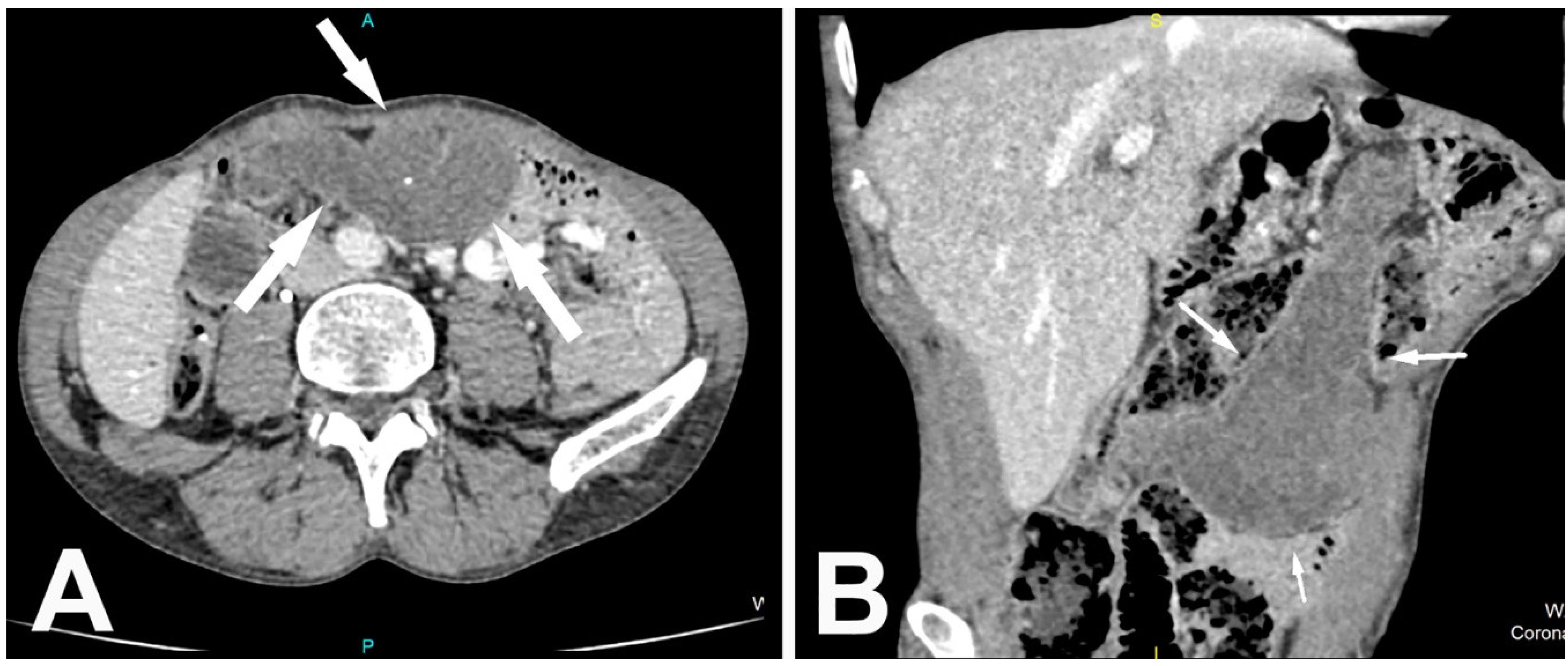
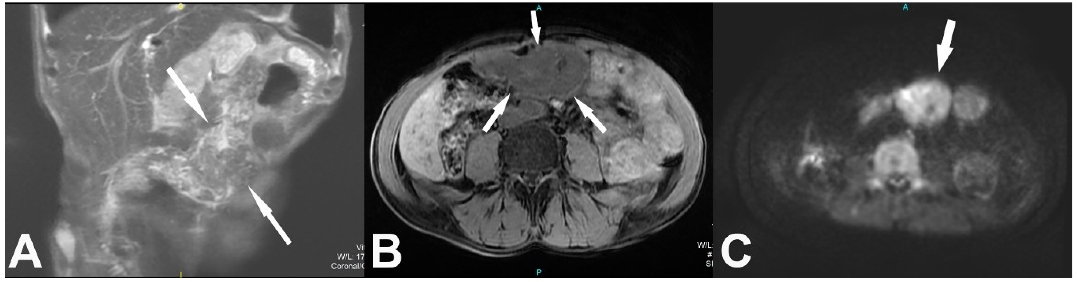
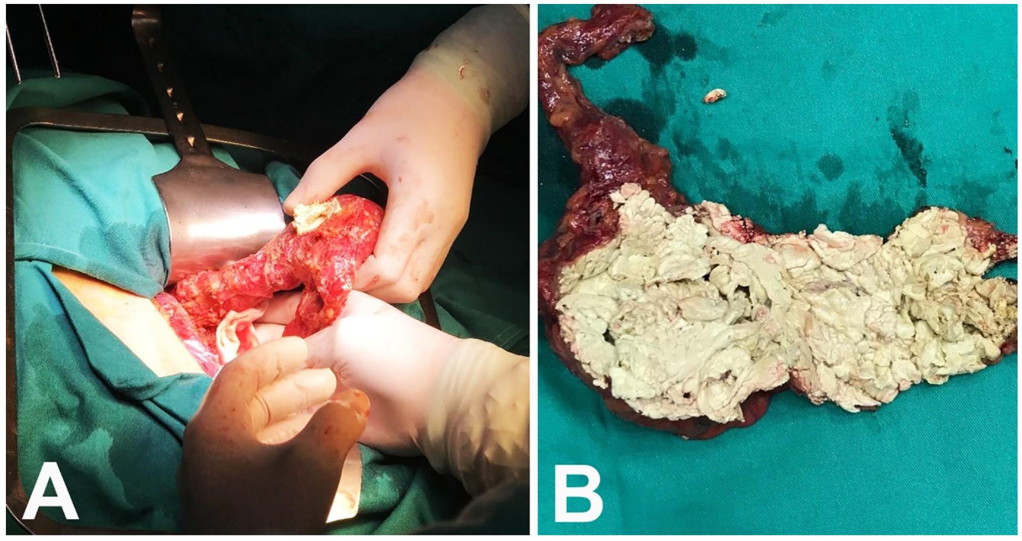
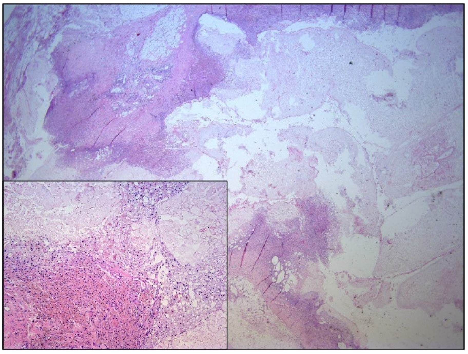
2. Case Report
3. Discussion
4. Conclusions
Author Contributions
Funding
Institutional Review Board Statement
Informed Consent Statement
Data Availability Statement
Conflicts of Interest
References
- Kamaya, A.; Federle, M.P.; Desser, T.S. Imaging manifestations of abdominal fat necrosis and its mimics. Radiographics 2011, 31, 2021–2034. [Google Scholar] [CrossRef] [Green Version]
- Chan, L.P.; Gee, R.; Keogh, C.; Munk, P.L. Imaging features of fat necrosis. Am. J. Roentgenol. 2003, 181, 955–959. [Google Scholar] [CrossRef]
- Kerr, S.F.; Hyland, R.; Rowbotham, E.; Chalmers, A.G. Postoperative omental infarction following colonic resection. Clin. Radiol. 2012, 67, 134–139. [Google Scholar] [CrossRef] [PubMed]
- Schwartzman, G.J.; Jacobsm, J.E.; Birnbaum, B.A. Omental infarction as a delayed complication of abdominal surgery. Clin. Imaging 2001, 25, 341–343. [Google Scholar] [CrossRef]
- Wiesner, W.; Kaplan, V.; Bongartz, G. Omental infarction associated with right-sided heart failure. Eur. Radiol. 2000, 10, 1130–1132. [Google Scholar] [CrossRef]
- Shera, T.; Choh, N.; Jabeen, S.; Ashraf, O.; Khan, A.; Shaheen, F.; Wani, G.M.; Wani, M.; Shah, M.; Gojwari, T.; et al. Primary and secondary omental infarction: A 5-year experience in a tertiary care hospital. Saudi Surg. J. 2017, 5, 77. [Google Scholar] [CrossRef]
- Trovato, P.; Simonetti, I.; Verde, F.; Lomoro, P.; Vinci, G.; Tarotto, L.; Corvino, F.; Corvino, A. Acute epiploic appendagitis: Ultrasound and computed tomography findings of a rare case of acute abdominal pain and the role of other imaging techniques. Pol. J. Radiol. 2020, 85, e178–e182. [Google Scholar] [CrossRef] [PubMed]
- Oh, J.Y.; Cho, J.H.; Kang, M.J.; Lee, J.H.; Kwon, H.J.; Nam, K.J.; Kim, M.C.; Choi, H. Omental infarction caused by laparoscopy-assisted gastrectomy for gastric cancer: CT findings. Clin. Radiol. 2011, 66, 966–973. [Google Scholar] [CrossRef] [PubMed]
- Takao, H.; Yamahira, K.; Watanabe, T. Encapsulated Fat Necrosis Mimicking Abdominal Liposarcoma. J. Comput. Assist. Tomogr. 2004, 28, 193–194. [Google Scholar] [CrossRef] [PubMed]
- Davidson, T.; Lotan, E.; Klang, E.; Nissan, J.; Goldstein, J.; Goshen, E.; Ben-Haim, S.; Apter, S.; Chikman, B. Fat necrosis after abdominal surgery: A pitfall in interpretation of FDG-PET/CT. Eur. Radiol. 2017, 28, 2264–2272. [Google Scholar] [CrossRef] [PubMed]
- Aho, H.J.; Sternby, B.; Nevalainen, T.J. Fat Necrosis in Human Acute Pancreatitis. Acta Pathol. Microbiol. Scand. Ser. A Pathol. 2009, 94A, 101–105. [Google Scholar] [CrossRef] [PubMed]
- Vardar, B.; Kızılcı, S. Incidence of lipohypertrophy in diabetic patients and a study of influencing factors. Diabetes Res. Clin. Pract. 2007, 77, 231–236. [Google Scholar] [CrossRef] [PubMed]


Publisher’s Note: MDPI stays neutral with regard to jurisdictional claims in published maps and institutional affiliations. |
© 2021 by the authors. Licensee MDPI, Basel, Switzerland. This article is an open access article distributed under the terms and conditions of the Creative Commons Attribution (CC BY) license (https://creativecommons.org/licenses/by/4.0/).
Share and Cite
Mitrovic, M.; Velickovic, D.; Micev, M.; Sljukic, V.; Djuric, P.; Tadic, B.; Skrobic, O.; Djokic Kovac, J. Encapsulated Omental Necrosis as an Unexpected Postoperative Finding: A Case Report. Medicina 2021, 57, 865. https://doi.org/10.3390/medicina57090865
Mitrovic M, Velickovic D, Micev M, Sljukic V, Djuric P, Tadic B, Skrobic O, Djokic Kovac J. Encapsulated Omental Necrosis as an Unexpected Postoperative Finding: A Case Report. Medicina. 2021; 57(9):865. https://doi.org/10.3390/medicina57090865
Chicago/Turabian StyleMitrovic, Milica, Dejan Velickovic, Marjan Micev, Vladimir Sljukic, Petar Djuric, Boris Tadic, Ognjan Skrobic, and Jelena Djokic Kovac. 2021. "Encapsulated Omental Necrosis as an Unexpected Postoperative Finding: A Case Report" Medicina 57, no. 9: 865. https://doi.org/10.3390/medicina57090865