
The Role of Chemokines in Psoriasis—An Overview

 ,
,
Abstract
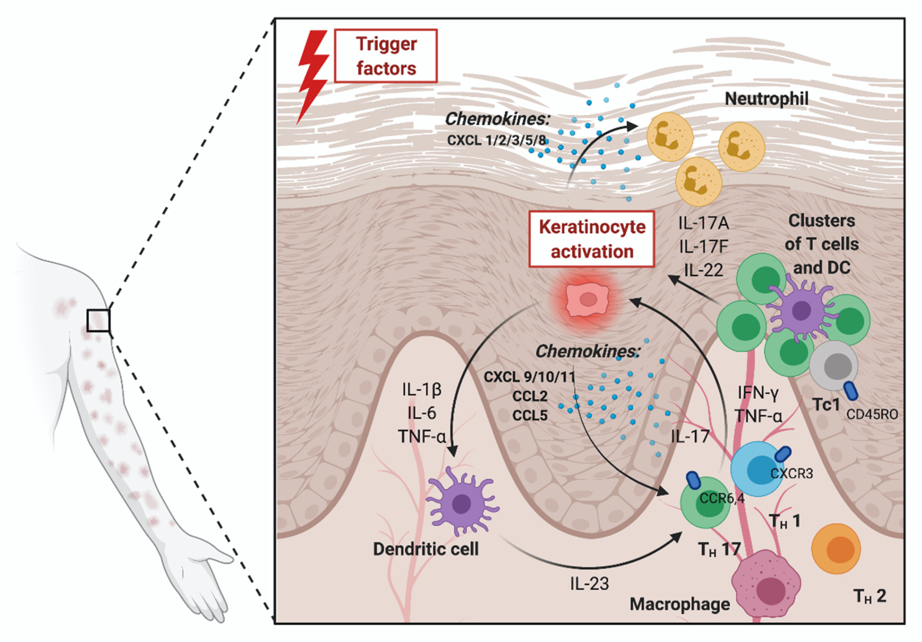
:1. Introduction
Systemic Treatment of Psoriasis and Its Impact on Chemokines
2. Conclusions
Author Contributions
Funding
Institutional Review Board Statement
Informed Consent Statement
Data Availability Statement
Conflicts of Interest
References
- Dai, Y.-J.; Li, Y.-Y.; Zeng, H.-M.; Liang, X.-A.; Xie, Z.-J.; Zheng, Z.-A.; Pan, Q.-H.; Xing, Y.-X. Effect of pharmacological intervention on MIP-1α, MIP-1β and MCP-1 expression in patients with psoriasis vulgaris. Asian Pac. J. Trop. Med. 2014, 7, 582–584. [Google Scholar] [CrossRef] [Green Version]
- Mabuchi, T.; Chang, T.W.; Quinter, S.; Hwang, S.T. Chemokine receptors in the pathogenesis and therapy of psoriasis. J. Dermatol. Sci. 2012, 65, 4–11. [Google Scholar] [CrossRef] [PubMed]
- Palomino, D.C.T.; Marti, L.C. Chemokines and immunity. Einstein 2015, 13, 469–473. [Google Scholar] [CrossRef] [Green Version]
- Nestle, F.O.; Kaplan, D.H.; Barker, J. Psoriasis. N. Engl. J. Med. 2009, 361, 496–509. [Google Scholar] [CrossRef] [PubMed]
- Hawkes, J.E.; Chan, T.C.; Krueger, J.G. Psoriasis pathogenesis and the development of novel targeted immune therapies. J. Allergy Clin. Immunol. 2017, 140, 645–653. [Google Scholar] [CrossRef] [PubMed] [Green Version]
- Fujisawa, T.; Murase, K.; Kanoh, H.; Takemura, M.; Ohnishi, H.; Seishima, M. Adsorptive Depletion of CD14+CD16+Proinflammatory Monocyte Phenotype in Patients with Generalized Pustular Psoriasis: Clinical Efficacy and Effects on Cytokines. Ther. Apher. Dial. 2012, 16, 436–444. [Google Scholar] [CrossRef] [PubMed]
- Abji, F.; A Pollock, R.; Liang, K.; Chandran, V.; Gladman, D.D. Th17 gene expression in psoriatic arthritis synovial fluid and peripheral blood compared to osteoarthritis and cutaneous psoriasis. Clin. Exp. Rheumatol. 2017, 36, 486–489. [Google Scholar]
- Abdelaal, N.H.; Elhefnawy, N.G.; Abdulmonem, S.R.; Sayed, S.; Saleh, N.A.; Saleh, M.A. Evaluation of the expression of the stromal cell-derived factor-1 alpha (CXCL 12) in psoriatic patients after treatment with Methotrexate. J. Cosmet. Dermatol. 2019, 19, 253–258. [Google Scholar] [CrossRef] [PubMed]
- Moadab, F.; Khorramdelazad, H.; Abbasifard, M. Role of CCL2/CCR2 axis in the immunopathogenesis of rheumatoid arthritis: Latest evidence and therapeutic approaches. Life Sci. 2021, 269, 119034. [Google Scholar] [CrossRef]
- Garshick, M.S.; Barrett, T.; Wechter, T.; Azarchi, S.; Scher, J.U.; Neimann, A.; Katz, S.; Fuentes-Duculan, J.; Cannizzaro, M.V.; Jelic, S.; et al. Inflammasome Signaling and Impaired Vascular Health in Psoriasis. Arter. Thromb. Vasc. Biol. 2019, 39, 787–798. [Google Scholar] [CrossRef]
- Kouassi, K.T.; Gunasekar, P.; Agrawal, D.K.; Jadhav, G.P. TREM-1; Is It a Pivotal Target for Cardiovascular Diseases? J. Cardiovasc. Dev. Dis. 2018, 5, 45. [Google Scholar] [CrossRef] [Green Version]
- Vaughan, D.E. PAI-1 and atherothrombosis. J. Thromb. Haemost. 2005, 3, 1879–1883. [Google Scholar] [CrossRef]
- Barale, C.; Russo, I. Influence of Cardiometabolic Risk Factors on Platelet Function. Int. J. Mol. Sci. 2020, 21, 623. [Google Scholar] [CrossRef] [Green Version]
- Van Der Zalm, I.J.B.; Van Der Valk, E.S.; Wester, V.L.; Nagtzaam, N.M.A.; Van Rossum, E.F.C.; Leenen, P.J.M.; Dik, W.A. Obesity-associated T-cell and macrophage activation improve partly after a lifestyle intervention. Int. J. Obes. 2020, 44, 1–13. [Google Scholar] [CrossRef] [PubMed]
- Kim, H.O.; Cho, S.I.; Chung, B.Y.; Ahn, H.K.; Park, C.W.; Lee, C.H. Expression of CCL1 and CCL18 in atopic dermatitis and psoriasis. Clin. Exp. Dermatol. 2012, 37, 521–526. [Google Scholar] [CrossRef]
- Canavese, M.; Altruda, F.; Silengo, L. Therapeutic Efficacy and Immunological Response of CCL5 Antagonists in Models of Contact Skin Reaction. PLoS ONE 2010, 5, e8725. [Google Scholar] [CrossRef] [PubMed]
- Brunner, P.M.; Suárez-Fariñas, M.; He, H.; Malik, K.; Wen, H.-C.; Gonzalez, J.; Chan, T.C.-C.; Estrada, Y.; Zheng, X.; Khattri, S.; et al. The atopic dermatitis blood signature is characterized by increases in inflammatory and cardiovascular risk proteins. Sci. Rep. 2017, 7, 1–12. [Google Scholar] [CrossRef]
- Ferrari, S.M.; Ruffilli, I.; Colaci, M.; Antonelli, A.; Ferri, C.; Fallahi, P. CXCL10 in psoriasis. Adv. Med Sci. 2015, 60, 349–354. [Google Scholar] [CrossRef] [PubMed]
- Krause, K.; Sabat, R.; Witte-Händel, E.; Schulze, A.; Puhl, V.; Maurer, M.; Wolk, K. Association of CCL2 with systemic inflammation in Schnitzler syndrome. Br. J. Dermatol. 2019, 180, 859–868. [Google Scholar] [CrossRef]
- Dembic, Z. Cytokines of the Immune System. In The Cytokines of the Immune System; Academic Press: Cambridge, MA, USA, 2015. [Google Scholar] [CrossRef]
- Behfar, S.; Hassanshahi, G.; Nazari, A.; Khorramdelazad, H. A brief look at the role of monocyte chemoattractant protein-1 (CCL2) in the pathophysiology of psoriasis. Cytokine 2018, 110, 226–231. [Google Scholar] [CrossRef] [PubMed]
- Gao, M.; Wang, A. Effect of NB-UVB on levels of MCP-1 and CCR6 mRNA in patients with psoriasis vulgaris. Genet. Mol. Res. 2015, 14, 12137–12144. [Google Scholar] [CrossRef]
- Pohl, D.; Andrýs, C.; Borska, L.; Fiala, Z.; Hamáková, K.; Ettler, K.; Krejsek, J. CC and CXC Chemokines Patterns in Psoriasis Determined by Protein Array Method Were Influenced by Goeckerman’s Therapy. Acta Med. (Hradec Kralove) 2009, 52, 9–13. [Google Scholar] [CrossRef] [PubMed] [Green Version]
- Lembo, S.; Capasso, R.; Balato, A.; Cirillo, T.; Flora, F.; Zappia, V.; Balato, N.; Ingrosso, D.; Ayala, F. MCP-1 in psoriatic patients: Effect of biological therapy. J. Dermatol. Treat. 2013, 25, 83–86. [Google Scholar] [CrossRef] [PubMed]
- Nedoszytko, B.; Sokołowska-Wojdyło, M.; Ruckemann-Dziurdzińska, K.; Roszkiewicz, J.; Nowicki, R.J. Chemokines and cytokines network in the pathogenesis of the inflammatory skin diseases: Atopic dermatitis, psoriasis and skin mastocytosis. Postep. Dermatol. Alergol. 2014, 2, 84–91. [Google Scholar] [CrossRef] [PubMed]
- De Groot, M.; Teunissen, M.B.M.; Ortonne, J.P.; Lambert, J.R.; Naeyaert, J.M.; Picavet, D.I.; Arreaza, M.G.; Simon, J.S.; Kraan, M.; Bos, J.D.; et al. Expression of the chemokine receptor CCR5 in psoriasis and results of a randomized placebo controlled trial with a CCR5 inhibitor. Arch. Dermatol. Res. 2007, 299, 305–313. [Google Scholar] [CrossRef] [Green Version]
- Rateb, A.A.; Fawzi, M.M.; Hay, R.A.; Mohammed, F.N.; Amr, K. Reduction of RANTES expression in lesional psoriatic skin after narrow band ultraviolet therapy: A possible marker of therapeutic efficacy. Eur. J. Dermatol. 2012, 22, 481–487. [Google Scholar] [CrossRef]
- Schottelius, A.J.G.; Moldawer, L.L.; Dinarello, C.A.; Asadullah, K.; Sterry, W.; Edwards, C.K. Biology of tumor necrosis factor-alpha- implications for psoriasis. Exp. Dermatol. 2004, 13, 193–222. [Google Scholar] [CrossRef]
- Myśliwiec, H.; Flisiak, I.; Baran, A.; Gorska, M.; Chodynicka, B. Evaluation of CD40, its ligand CD40L and Bcl-2 in psoriatic patients. Folia Histochem. Cytobiol. 2012, 50, 75–79. [Google Scholar] [CrossRef] [Green Version]
- Venerito, V.; Natuzzi, D.; Bizzoca, R.; Lacarpia, N.; Cacciapaglia, F.; Lopalco, G.; Iannone, F. Serum sCD40L levels are increased in patients with psoriatic arthritis and are associated with clinical response to apremilast. Clin. Exp. Immunol. 2020, 201, 200–204. [Google Scholar] [CrossRef]
- Lian, S.; Zhai, X.; Wang, X.; Zhu, H.; Zhang, S.; Wang, W.; Wang, Z.; Huang, J. Elevated expression of growth-regulated oncogene-alpha in tumor and stromal cells predicts unfavorable prognosis in pancreatic cancer. Medicine 2016, 95, e4328. [Google Scholar] [CrossRef]
- Kanayama, Y.; Torii, K.; Ikumi, K.; Morita, A. Bath-PUVA therapy suppresses keratinocyte-derived chemokines in pathoge-netically relevant cells. JID Innov. 2021, 1, 100027. [Google Scholar] [CrossRef]
- Stoof, T.; Flier, J.; Sampat, S.; Nieboer, C.; Tensen, C.; Boorsma, D. The antipsoriatic drug dimethylfumarate strongly suppresses chemokine production in human keratinocytes and peripheral blood mononuclear cells. Br. J. Dermatol. 2001, 144, 1114–1120. [Google Scholar] [CrossRef]
- Abji, F.; Lee, K.; Pollock, R.; Machhar, R.; Cook, R.; Chandran, V.; Gladman, D. Declining levels of serum chemokine (C-X-C motif) ligand 10 over time are associated with new onset of psoriatic arthritis in patients with psoriasis: A new biomarker? Br. J. Dermatol. 2020, 183, 920–927. [Google Scholar] [CrossRef]
- Antonelli, A.; Ferrari, S.M.; Giuggioli, D.; Ferrannini, E.; Ferri, C.; Fallahi, P. Chemokine (C–X–C motif) ligand (CXCL)10 in autoimmune diseases. Autoimmun. Rev. 2014, 13, 272–280. [Google Scholar] [CrossRef]
- Gottlieb, A.B.; Chamian, F.; Masud, S.; Cardinale, I.; Abello, M.V.; A Lowes, M.; Chen, F.; Magliocco, M.; Krueger, J.G. TNF Inhibition Rapidly Down-Regulates Multiple Proinflammatory Pathways in Psoriasis Plaques. J. Immunol. 2005, 175, 2721–2729. [Google Scholar] [CrossRef] [Green Version]
- Schafer, P.H.; Parton, A.; Gandhi, A.K.; Capone, L.; Adams, M.; Wu, L.; Bartlett, J.B.; A Loveland, M.; Gilhar, A.; Cheung, Y.-F.; et al. Apremilast, a cAMP phosphodiesterase-4 inhibitor, demonstrates anti-inflammatory activity in vitro and in a model of psoriasis. Br. J. Pharmacol. 2009, 159, 842–855. [Google Scholar] [CrossRef] [Green Version]
- Petkovic, V.; Moghini, C.; Paoletti, S.; Uguccioni, M.; Gerber, B. I-TAC/CXCL11 is a natural antagonist for CCR. J. Leukoc. Biol. 2004, 76, 701–708. [Google Scholar] [CrossRef]
- Flier, J.; Boorsma, D.M.; Van Beek, P.J.; Nieboer, C.; Stoof, T.J.; Willemze, R.; Tensen, C.P. Differential expression of CXCR3 targeting chemokines CXCL10, CXCL9, and CXCL11 in different types of skin inflammation. J. Pathol. 2001, 194, 398–405. [Google Scholar] [CrossRef]
- Glickman, J.W.; Dubin, C.; Renert-Yuval, Y.; Dahabreh, D.; Kimmel, G.W.; Auyeung, K.; Estrada, Y.D.; Singer, G.; Krueger, J.G.; Pavel, A.B.; et al. Cross-sectional study of blood biomarkers of patients with moderate to severe alopecia areata reveals systemic immune and cardiovascular biomarker dysregulation. J. Am. Acad. Dermatol. 2021, 84, 370–380. [Google Scholar] [CrossRef] [PubMed]
- Karin, N. The multiple faces of CXCL12 (SDF-1α) in the regulation of immunity during health and disease. J. Leukoc. Biol. 2010, 88, 463–473. [Google Scholar] [CrossRef] [PubMed]
- Zgraggen, S.; Huggenberger, R.; Kerl, K.; Detmar, M. An Important Role of the SDF-1/CXCR4 Axis in Chronic Skin Inflammation. PLoS ONE 2014, 9, e93665. [Google Scholar] [CrossRef] [PubMed]
- Ghosh, A.K.; Vaughan, D.E. PAI-1 in tissue fibrosis. J. Cell. Physiol. 2012, 227, 493–507. [Google Scholar] [CrossRef] [Green Version]
- Teixeira, G.G.; Mari, N.L.; De Paula, J.C.C.; De Alcantara, C.C.; Flauzino, T.; Lozovoy, M.A.B.; Martin, L.M.M.; Reiche, E.M.V.; Maes, M.; Dichi, I.; et al. Cell adhesion molecules, plasminogen activator inhibitor type 1, and metabolic syndrome in patients with psoriasis. Clin. Exp. Med. 2020, 20, 39–48. [Google Scholar] [CrossRef]
- Sigurdardottir, G.; Ekman, A.-K.; Ståhle, M.; Bivik, C.; Enerbäck, C. Systemic treatment and narrowband ultraviolet B differentially affect cardiovascular risk markers in psoriasis. J. Am. Acad. Dermatol. 2014, 70, 1067–1075. [Google Scholar] [CrossRef] [PubMed] [Green Version]
- Hyder, L.A.; Gonzalez, J.; Harden, J.L.; Johnson-Huang, L.M.; Zaba, L.C.; Pierson, K.C.; Eungdamrong, N.J.; Lentini, T.; Gulati, N.; Fuentes-Duculan, J.; et al. TREM-1 as a potential therapeutic target in psoriasis. J. Investig. Dermatol. 2013, 133, 1742–1751. [Google Scholar] [CrossRef] [PubMed] [Green Version]
- Quan, C.; Cho, M.K.; Shao, Y.; Mianecki, L.E.; Liao, E.; Perry, D.; Quan, T. Dermal fibroblast expression of stromal cell-derived factor-1 (SDF-1) promotes epidermal keratinocyte proliferation in normal and diseased skin. Protein Cell 2015, 6, 890–903. [Google Scholar] [CrossRef] [Green Version]
- Abji, F.; Pollock, R.A.; Liang, K.; Chandran, V.; Gladman, D.D. Brief Report: CXCL10 Is a Possible Biomarker for the Development of Psoriatic Arthritis Among Patients with Psoriasis. Arthritis Rheumatol. 2016, 68, 2911–2916. [Google Scholar] [CrossRef] [Green Version]
- Shih, C.-M.; Hsieh, C.-K.; Huang, C.-Y.; Huang, C.-Y.; Wang, K.-H.; Fong, T.-H.; Trang, N.T.T.; Liu, K.-T.; Lee, A.-W. Lycopene Inhibit IMQ-Induced Psoriasis-Like Inflammation by Inhibiting ICAM-1 Production in Mice. Polymers 2020, 12, 1521. [Google Scholar] [CrossRef]
- Bressan, A.L.; Picciani, B.L.S.; Azulay-Abulafia, L.; Fausto-Silva, A.K.; Almeida, P.N.; Cunha, K.S.G.; Dias, E.P.; Carneiro, S. Evaluation of ICAM-1 expression and vascular changes in the skin of patients with plaque, pustular, and erythrodermic psoriasis. Int. J. Dermatol. 2018, 57, 209–216. [Google Scholar] [CrossRef] [PubMed]
- Gerkowicz, A.; Pietrzak, A.; Szepietowski, J.C.; Radej, S.; Chodorowska, G. Biochemical markers of psoriasis as a metabolic disease. Folia Histochem. Cytobiol. 2012, 50, 155–170. [Google Scholar] [CrossRef] [Green Version]


| Molecule | Receptor | Function | Role in Psoriasis | Presence in Psoriasis Comorbidities | Impact of Psoriasis Treatment |
|---|---|---|---|---|---|
| CCL1/l-309 | CCR8 |
|
|
| |
| CCL2/MCP-1 | CCR2 CCR4 |
| |||
| MIP-1α/MIP-1β (CCL3/CCL4) | CCR1 CCR5 |
|
|
| |
| CCL5/RANTES | CCR1 CCR3 CCR5 |
|
| ||
| CD40L/TNFSF5 | CD40 |
|
|
| |
| CXCL1/GRO-α | CXCR2 |
|
|
| |
| CXCL10/IP-10 | CXCR3 |
|
| ||
| CXCL11/I-TAC | CXCR3 |
|
|
| |
| CXCL12/SDF-1 | CXCR4 CXCR7 |
|
|
| |
| Serpin E1/PAI-1 | tPA uPA |
|
| ||
| TREM-1 | n/a |
|
|
|
Publisher’s Note: MDPI stays neutral with regard to jurisdictional claims in published maps and institutional affiliations. |
© 2021 by the authors. Licensee MDPI, Basel, Switzerland. This article is an open access article distributed under the terms and conditions of the Creative Commons Attribution (CC BY) license (https://creativecommons.org/licenses/by/4.0/).
Share and Cite
Zdanowska, N.; Kasprowicz-Furmańczyk, M.; Placek, W.; Owczarczyk-Saczonek, A. The Role of Chemokines in Psoriasis—An Overview. Medicina 2021, 57, 754. https://doi.org/10.3390/medicina57080754
Zdanowska N, Kasprowicz-Furmańczyk M, Placek W, Owczarczyk-Saczonek A. The Role of Chemokines in Psoriasis—An Overview. Medicina. 2021; 57(8):754. https://doi.org/10.3390/medicina57080754
Chicago/Turabian StyleZdanowska, Natalia, Marta Kasprowicz-Furmańczyk, Waldemar Placek, and Agnieszka Owczarczyk-Saczonek. 2021. "The Role of Chemokines in Psoriasis—An Overview" Medicina 57, no. 8: 754. https://doi.org/10.3390/medicina57080754