Article

Impact of Changing Surgical Strategies on Clinical Outcomes in Patients with Parotid Carcinoma: A 53-Year Single-Institution Experience

1
Department of Otolaryngology, Tenri Hospital, 200 Mishima-cho, Tenri, Nara 632-8552, Japan
2
Department of Otolaryngology, Shizuoka City Hospital, 10-93 Otte-cho, Aoi-ku, Shizuoka 420-8630, Japan
3
Department of Otolaryngology—Head and Neck Surgery, School of Medicine, Fujita Health University, 1-98 Dengakugakubo, Kutsukake-cho, Toyoake 470-1192, Aichi, Japan
*
Author to whom correspondence should be addressed.
Medicina 2021, 57(8), 745; https://doi.org/10.3390/medicina57080745
Submission received: 1 June 2021 / Revised: 18 July 2021 / Accepted: 21 July 2021 / Published: 23 July 2021
(This article belongs to the Special Issue Metastatic Head and Neck Malignancy)

Abstract

:
Background and Objectives: We investigated the clinical outcomes of patients who underwent surgery for parotid carcinoma in a single institution during a 53-year period. This study aimed to estimate the impact of changing the surgical approach to parotid carcinoma on clinical outcomes including the incidence rate of the facial nerve palsy. Materials and Methods: Sixty-seven patients with parotid carcinoma who underwent surgery between 1966 and 2018 were retrospectively reviewed. Group A consisted of 29 patients who underwent surgery from 1966 to 2002, and Group B consisted of 38 patients from 2002 to 2018. Treatment outcomes were estimated. Additionally, candidate prognostic factors of Group B, the current surgical approach group, were evaluated. Results: Partial parotidectomy and total parotidectomy were performed in 35 and 32 patients, respectively. Partial parotidectomy was performed in 4 patients in Group A and 31 patients in Group B, with a predominant increase in Group B. The facial nerve was preserved in 43 patients, among whom 8 in Group A (8/17; 47.1%) and 7 in Group B (7/26; 26.9%) had temporary postoperative facial nerve palsy. Postoperative radiotherapy was performed on 35 patients. The 5-year OS, DSS, and DFS rates for Group A were 77.1%, 79.9%, and 71.5%, respectively. The 5-year OS, DSS, and DFS rates for Group B were 77.1%, 77.1%, and 72.4%, respectively. Clinical T4 stage, clinical N+ stage, stage IV disease, and tumor invasion of the facial nerve were independent prognostic factors in Group B. Conclusions: The incidence of facial nerve palsy in the current surgical approach group decreased compared with that in the previous surgical approach group. The current surgical management and treatment policies for parotid carcinoma have led to improved outcomes.

1. Introduction

Parotid carcinoma is a relatively rare disease that accounts for less than 5% of head and neck malignancies worldwide [1]. In Japan, the annual incidence of parotid carcinoma is approximately 3 out of every 1,000,000 people [2]. The World Health Organization (WHO) recognizes at least 24 histological types of salivary gland carcinomas [3]. Their rarity and histological variety have prevented the development of a definitive surgical policy and prognostic prediction method for this disease. Treatment strategies for parotid carcinoma include surgical resection, radiotherapy, and chemotherapy. Surgical resection is the primary treatment for operable parotid carcinoma; in addition, postoperative radiotherapy is used to reduce the incidence of local and cervical lymph node recurrence in high-risk patients [4]. However, the institutional policy and surgeon’s preference are the main determinants of the surgical method used for treating parotid carcinoma, area of resection of the parotid gland, indications for neck dissection in clinical N0 (cN0) cases, and treatment of the facial nerve.
In the past, total parotidectomy was the standard treatment of choice for parotid gland carcinoma with a poor prognosis [5]. Total parotidectomy involves the unnecessary removal of normal parotid tissue, which may result in postoperative impairment of the salivary glands and facial nerve function [6]. Therefore, partial parotidectomy has emerged as a more conservative approach over the past 2 decades [7,8]. In partial parotidectomy, only the tumor-bearing area of the parotid parenchyma is removed. The main trunk of the facial nerve is identified, and the facial nerve branch adjacent to the tumor is dissected and preserved. Compared to total parotidectomy, partial parotidectomy is associated with a low incidence of surgical complications, such as facial palsy, loss of sensation of the ear, poor aesthetics, and Frey’s syndrome [9]. Total parotidectomy is not necessary for low-grade cancers if the tumor is completely resectable, as alternative approaches can be used [6,10]. In terms of high-grade cancers, some reports have uniformly recommended total parotid resection [11], while others have suggested that lobectomy is sufficient for T1 and T2 disease without facial nerve involvement [6,12]. The indications for prophylactic neck dissection for cN0 parotid carcinoma are unclear. While several authors recommend prophylactic neck dissection in all patients with parotid carcinoma, others recommend postoperative radiotherapy or observation [13,14,15,16].
At our institute, the surgical strategy for parotid carcinoma shifted from total parotidectomy to partial parotidectomy in 2002. Partial parotidectomy was preferred because it preserves the facial nerve as much as possible in patients without preoperative facial palsy. In addition, the neck management for cN0 parotid carcinoma was changed from prophylactic neck dissection at cT3 and T4 cases before 2002 to no prophylactic neck dissection for cases after 2002. These changes in the surgical management and treatment policies may have improved treatment outcomes, such as postoperative facial nerve function. This study aimed to assess the changes in clinical outcomes of patients treated for parotid carcinoma at our institution due to changes in surgical strategy before/after 2002. To the best of our knowledge, this is the first study to compare postoperative outcomes before and after implementing specific surgical management policy changes for parotid carcinoma at a single institution.

2. Materials and Methods
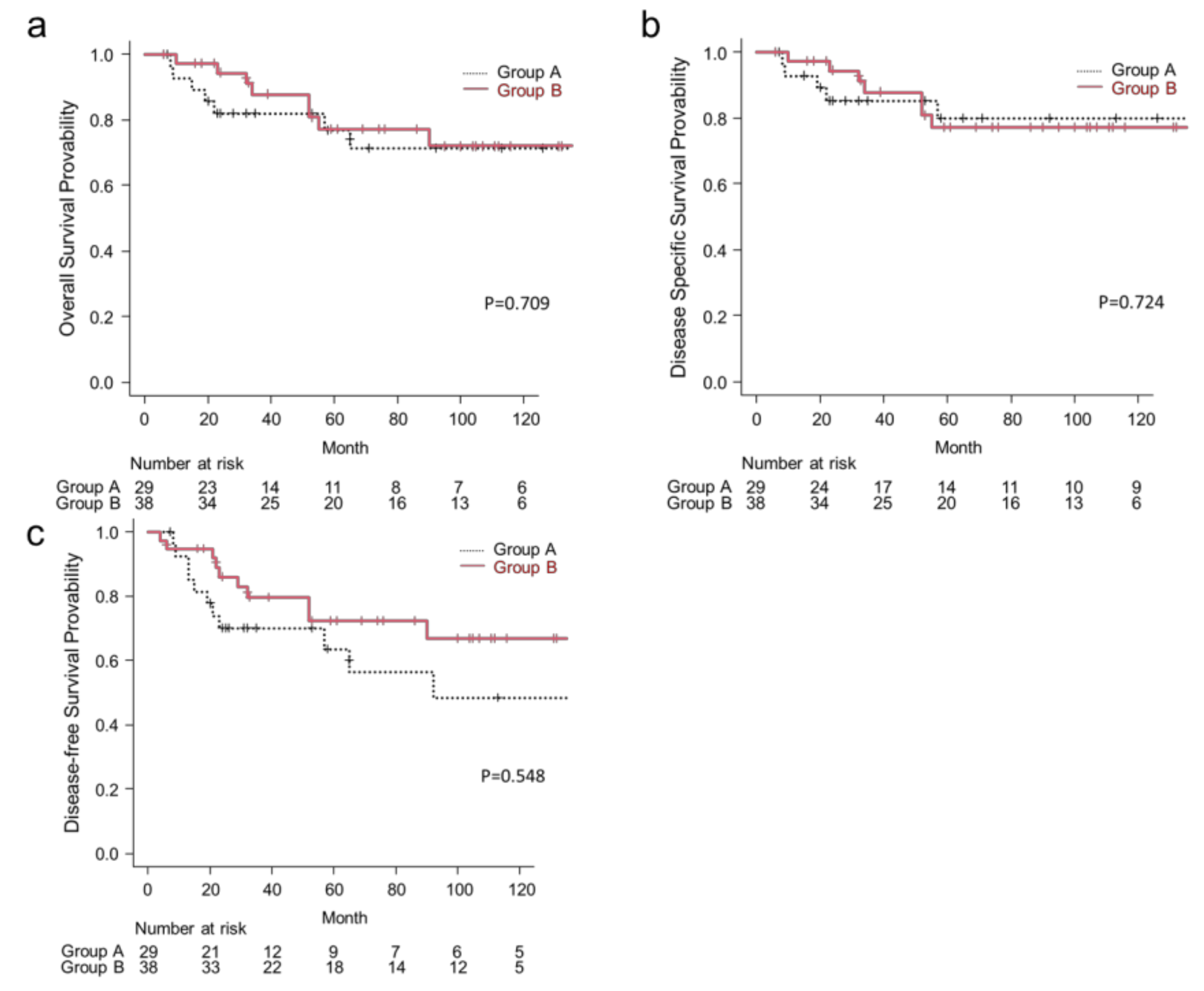
We retrospectively analyzed 67 patients with primary parotid carcinoma who underwent surgical treatment at the Tenri Hospital between January 1966 and December 2018. Patients who received palliative treatment were excluded from this study. Patients were classified into two groups based on the date of surgery. Group A, the previous surgical approach group, consisted of 29 patients who underwent surgery from January 1966 to December 2002. During this period, total parotidectomy was chosen as the main surgical method, and prophylactic neck dissection was performed for cN0 patients with cT3 or T4. Group B, the current surgical approach group, consisted of 38 patients who underwent surgery from January 2002 to December 2018. In the current surgical approach group, the surgical technique was changed to a conservative approach, such as partial parotidectomy or no prophylactic neck dissection in cN0 cases with cT3 or T4. All patients received a preoperative physical examination, ultrasonography (US), contrast-enhanced head and neck computed tomography (CT), and/or magnetic resonance imaging (MRI). CT and/or MRI were performed in order to find the location, extent, and invasion of the primary tumor as well as the presence of nodal metastasis. Ultrasound-guided fine-needle aspiration biopsy was performed for the primary tumor and for lymph nodes suspected to be metastatic by the imaging studies. In cases of suspected distant metastasis, bone scintigraphy or FDG-PET was conducted.
The personal records of all patients were reviewed, and age, sex, clinical, surgical, pathological, and follow-up data were extracted. The clinical stage was classified based on the 8th edition of the tumor–node–metastasis (TNM) classification [17]. The histological type was classified based on the 4th edition of the WHO classification [3]. Treatment outcomes of the two groups including overall survival (OS), disease-specific survival (DSS), disease-free survival (DFS), and the incidence rate of the facial nerve palsy were examined. The following candidate prognostic factors were assessed: age, sex, cT stage, cN stage, intraoperative facial nerve invasion, histological grade, and postoperative radiotherapy. We analyzed local, cervical, and distant recurrences in all patients; we also analyzed cervical lymph node recurrence in cN0 patients and recurrences in patients treated with postoperative radiotherapy.
In terms of the surgical management and treatment policy for parotid carcinoma, total parotidectomy was chosen as the main surgical method in Group A. On the other hands, partial parotidectomy was chosen as the main surgical method in Group B, and total parotidectomy was chosen in cases in which almost all the nerves were involved in a large tumor. The facial nerve was resected in patients with preoperative facial nerve palsy in both groups. In patients without preoperative facial nerve palsy, if it was found to be in contact with the tumor intraoperatively, the nerve was sacrificed in group A, while the nerve was detached from the tumor and preserved in group B. Postoperative facial nerve function was evaluated on the first postoperative day by clinical examination and graded according to the Yanagihara 40-point system (Yanagihara score) [18]. Until 2002, prophylactic neck dissection was performed in patients with advanced parotid carcinomas (clinical T3 or T4), even in the cN0 cases. Since 2002, prophylactic neck dissection was not performed in patients with cN0 parotid carcinoma. In addition, postoperative radiotherapy was administered to patients with high-grade malignancy, T4 tumor, perineural or lympho-vascular invasion, positive resection margin on histopathological evaluation, or multiple cervical lymph node metastases. In patients without cervical lymph node metastasis, the irradiation field (50 Gy) was limited to the parotid gland. In patients with cervical lymph node metastasis, the irradiation field (50 Gy) was extended to the lateral neck, and chemoradiation therapy was performed.
Group A patients were followed up until the time of death or until 2002, whereas Group B patients were followed up until the time of death or until February 2020. US, CT, and/or MRI were performed every 3 months during the first year and every 6 months in the subsequent years for the evaluation of regional and cervical lymph nodes and distant metastases.
Categorical variables were compared using the chi-square test, and continuous variables were compared using the Student’s t-test. The treatment outcomes, including OS, DSS, and DFS, were evaluated using the Kaplan–Meier method, and groups were compared using the log-rank test. A p-value < 0.05 was considered statistically significant. All statistical analyses were performed with EZR (Saitama Medical Centre, Jichi Medical University), which is a graphical user interface for R (The R Foundation for Statistical Computing, Vienna, Austria, version 2.13.0) [19].

3. Results

3.1. Patient Characteristics

The patient characteristics of the two groups, including age, sex, cT and cN classifications, disease stage, surgical methods, and preoperative/postoperative facial nerve palsy, postoperative radiotherapy, and recurrence rate are presented in Table 1. No statistically significant differences in sex, cT and cN classification, disease stage, facial nerve resection, preoperative/postoperative facial nerve palsy, and postoperative radiotherapy were noted between the two groups. Group B had a significantly higher median age. Partial parotidectomy was performed in 4 patients in Group A, and 31 patients in Group B, with a predominant increase in Group B. Prophylactic neck dissection was performed in 5 patients in Group A (2 on type modified radical, and 3 on type selective), among which only 1 patient had occult cervical neck metastasis. Therapeutic neck dissection was performed in 7 patients in Group A (3 on type radical, and 4 on type modified radical) and 2 patients in Group B (both on type selective) with clinically positive nodes.
Regarding facial nerve function and surgical treatment for facial nerve, 3 patients in Group A and 1 patient in Group B exhibited preoperative facial palsy, and the facial nerve of these patients was sacrificed. There were 26 patients and 37 patients without preoperative facial nerve palsy in Group A and B, respectively. Of the 26 patients without preoperative facial nerve palsy in Group A, 9 sacrificed the facial nerve and 17 preserved the facial nerve during surgery. Of the 37 patients without preoperative facial nerve palsy in Group B, 11 sacrificed the facial nerve and 26 patients preserved the facial nerve during surgery. Among the 17 patients with facial nerve preservation in Group A, 8 (47.1%; 8/17) exhibited temporary postoperative facial nerve palsy (each Yanagihara score was less than 2 points), and among the 26 patients with facial nerve preservation in Group B, 7 (26.9%; 7/26) exhibited temporary postoperative facial nerve palsy. As a result, the incidence of postoperative facial nerve palsy in patients with preoperative facial palsy was 65.4% (17/26) in Group A and 48.6% (18/37) in Group B. Postoperative radiotherapy was performed in 35 patients, including 25 patients with high-grade carcinoma and 10 patients with low/intermediate-grade carcinoma (6 due to a T4 tumor and 3 due to perineural invasion).

3.2. Histopathologic Results

Histological grades and histologies in our study are shown in Table 2. Mucoepidermoid carcinoma was the most common histological type (34.3%), followed by acinic cell carcinoma (22.4%), carcinoma ex pleomorphic adenoma (17.9%), adenocystic carcinoma(9.0%), squamous cell carcinoma (6.0%), and others (10.4%). Thirty-three, 4, and 30 patients had low-grade, intermediate-grade, and high-grade carcinomas, respectively. Among the 23 patients with mucoepidermoid carcinoma, 14 had low-grade carcinomas, and 9 had high-grade carcinomas. Among the 12 patients with carcinoma ex pleomorphic adenoma, 1 had low-grade carcinomas, and 11 had high-grade carcinomas. Among the 6 patients with adenocystic carcinoma, 4 had intermediate-grade carcinomas, and 2 had high-grade carcinomas.

3.3. Treatment Outcomes

Median follow-up for patients was 61 months for the entire cohort, 58 months for Group A, and 65 months for Group B. The Kaplan–Meier curves of treatment outcomes of the two groups, including OS, DSS, and DFS, are shown in Figure 1. At the time of analysis, there were 17 deaths (25.3% of patients). The 5-year OS rates for Group B vs. Group A patients were 77.1% vs. 74.5%, respectively (p = 0.709) (Figure 1a). The 5-year DSS rates for Group B vs. Group A patients were 77.1% vs. 77.6%, respectively (p = 0.724) (Figure 1b). The 5-year DFS rates for Group B vs. Group A patients were 72.4% vs. 71.0%, respectively (p = 0.548) (Figure 1c). The results of the univariate analysis of clinical factors of Group A and Group B affecting the 5-year OS, DSS, and DFS rates are listed in Table 3. In Group A, candidate factors that showed a significant association with 5-year OS, DSS, and DFS included sex, cT classification, and disease stage. Clinical N classification was significantly associated with 5-year DSS and DFS, and histology grade was significantly associated only with 5-year DSS. In Group B, candidate factors that showed a significant association with 5-year OS, DSS, and DFS included cT classification, cN classification, disease stage, and facial nerve invasion. The Kaplan–Meier curves of Group B for OS stratified by these factors are shown in Figure 2. The 5-year OS rate was significantly higher in patients with cT1–3 disease than in those with cT4 disease (93.8% vs. 55.0%; p = 0.014; Figure 2a). The 5-year OS rate was significantly higher in clinically node-negative patients than in clinically node-positive patients (82.3% vs. 0%; p < 0.001; Figure 2b). The 5-year OS rate was significantly higher in patients with stage I–III disease than in those with stage IV disease (93.3% vs. 58.2%; p = 0.025; Figure 2c). Patients with intraoperative facial nerve invasion had a significantly lower 5-year OS rate than those without (50.0% vs. 89.2%; p = 0.009; Figure 2d). The remaining factors (age, sex, histological grade, and postoperative radiotherapy) were not significantly associated with the 5-year OS, DSS, or DFS; however, high-grade histology exhibited a trend for a poorer 5-year DSS (p = 0.102).

3.4. Recurrence

Details of the recurrent cases are summarized in Table 4. Group A cases in the first half of the study period had insufficient medical record review, and clinical outcomes were not fully known. Disease recurrence was observed in 17 patients (25.3%), including 11 patients with local recurrence (16.4%), 9 with cervical lymph node recurrence (13.4%), and 9 with distant metastasis (13.4%). Local recurrence was significantly lower in Group B than in Group A (1 in Group B vs. 10 in Group A, p < 0.01). From these recurrent cases, 10 deaths were noted. Histological diagnoses in patients with distant metastasis included mucoepidermoid carcinoma (4 cases), acinic cell carcinoma (2 cases), squamous cell carcinoma (1 case), adenocystic carcinoma (1 case), and carcinoma ex-pleomorphic adenoma (1 case). Cervical lymph node recurrence was observed in 5 of 58 cN0 cases (8.6%); these patients were managed with salvage surgery (in one case, the medical history could not be completed due to insufficient medical record research). Among the 35 patients who underwent postoperative radiotherapy, 9 experienced recurrences. Local recurrence and cervical lymph node recurrence were observed in 5 patients and 3 patients, respectively, all of whom underwent successful salvage surgery. Distant metastasis was observed in 5 patients; of these, 1 patient was successfully treated with salvage surgery.

4. Discussion

At our institute, total parotidectomy was performed for almost all parotid carcinomas until 2002. The facial nerve was resected during surgery in patients with preoperative facial nerve palsy or when the nerve was in contact with the tumor. Therefore, the incidence of postoperative facial nerve palsy was high until 2002: 47.1% (8/17) among patients with facial nerve preservation and 65.4% (17/26) among patients without preoperative facial nerve palsy. A few decades ago, the prevailing view was that more extensive resection at the expense of the facial nerve did not lead to improved outcomes [20], and the National Comprehensive Cancer Network guidelines recommended preservation of the facial nerve in the absence of facial nerve palsy, regardless of histological grade, unless the tumor had directly invaded and adhered to the nerve [21]. In 2002, we re-evaluated the surgical management of parotid carcinomas and identified partial parotidectomy as the primary surgical approach to reduce postoperative palsy incidence. If there was no preoperative facial palsy, the facial nerve was preserved by detaching it from the tumor, even in cases in which the nerve was in contact with the tumor. Thus, the incidence rate of facial nerve palsy in the current surgical strategy decreased to 26.9% (7/26) among patients with facial nerve preservation and 48.6% (18/37) among patients without preoperative facial nerve palsy, which was lower than that in the previous surgical strategy at our institute. Regarding local recurrence, the rate rather decreased from 35.7% (10/29) before 2002 to 2.6% (1/38) after 2002. Although the reasons are not known from the data of this study, it is possible that the skills of the surgeons have improved due to the increase in the number of head and neck cancer cases, including parotid cancer, since 2002. In addition, advances in imaging technology as well as radiation techniques (e.g., IMRT) might play an important role in staging of the cancers treated as well as the decrease of local recurrence rates. Compared to multicenter studies, single-center studies have less bias; this allowed us to compare the differences in treatment outcomes caused by changes in surgical management and treatment policies. To date, no studies have compared postoperative outcomes before and after the implementation of specific changes in the surgical management policy for parotid carcinoma at a single institution. Thus, this study provides insights into the potential impact that overarching policy changes can have on individual patient outcomes, such as the incidence of postoperative facial nerve palsy.
Until 2002, prophylactic neck dissection was performed in patients with advanced parotid carcinomas with clinical T3 or T4, even in the cN0 cases. Since 2002, prophylactic neck dissection has not been performed in patients with cN0 parotid carcinoma. Treatment policies for postoperative radiotherapy in cases of high-grade malignancy (or a tendency to infiltrate adjacent tissues, based on histological examination) remain unchanged. Until 2002, the 5-year OS rate after surgical treatment for parotid carcinoma was 74.5%. This is similar to the 5-year OS rate of 77.1% observed in the current surgical group. Some reports have suggested that prophylactic neck dissection should be considered in high-grade and T4 cases because of the high rate of occult lymph node metastasis [22]. Other studies have indicated that prophylactic neck dissection in cN0 cases does not improve survival and that the benefit of prophylactic neck dissection is limited in terms of prolonging survival [23,24,25]. In this study, only 1 of 5 cN0 patients had occult cervical lymph node metastasis in Group A, and 3 of the 36 cN0 patients showed late-onset cervical lymph node metastases in Group B, which were successfully managed by salvage neck dissection. These results suggest that prophylactic neck dissection is not always necessary and that early detection and prompt treatment of cervical lymph node metastases by means of strict postoperative follow-up are important.
In the latter period of this study, the facial nerve was preserved as much as possible if it could be detached from the tumor intraoperatively, even in cases of high-grade carcinoma. However, in such high-risk patients, the possibility of local recurrence and cervical lymph node recurrence should be considered. Many previous studies have reported a reduction in the incidence of postoperative local and cervical lymph node recurrences following postoperative radiotherapy [4], especially in cases involving T3–4 tumors, perineural invasion, and high-grade malignancy [26,27]. The median irradiation dose is reported to be 50 Gy [26,28]. In accordance with our institutional treatment policy, 35 patients with high-grade malignancy, T4 tumor, perineural invasion, positive margin on histopathological evaluation, or multiple lymph node metastases underwent postoperative adjunct radiotherapy in this study. Among the 35 patients, local recurrence was observed in 5 patients; cervical lymph node recurrence in 3 patients; and distant metastasis in 5 patients (including multiple forms of recurrence). Among patients receiving postoperative radiotherapy, local recurrence and/or cervical lymph node recurrence were observed in 3 patients who were managed with salvage surgery (in one case, the medical history could not be completed due to insufficient medical record research). Thus, postoperative radiotherapy appears to be effective in preventing local and cervical lymph node recurrence.
The 5-year OS, DSS, and DFS rates for parotid carcinoma at our institute were comparable to those mentioned in other reports [29,30,31,32]. Univariate analysis suggested that cT4 stage, cN+ stage, stage IV disease, and facial nerve invasion were prognostic factors for parotid carcinoma. In terms of histological grade, in the first half of this study, the 5-year DSS rates for low-/intermediate-grade patients and high-grade patients were 94.4% and 34.3%, respectively, which were significantly different (p = 0.026), similar to those in other reports [23,33]. Although postoperative radiotherapy is effective in preventing local and cervical lymph node recurrences in patients with a poor prognosis, distant metastasis may ultimately determine the prognosis. Therefore, the development of novel and effective therapies is required; potential examples include molecular targeted therapy and immunotherapy, which have been shown to have antitumor activity against salivary gland carcinoma [34,35].
The present study has some limitations. These include its retrospective nature from a single-institution study. Furthermore, due to the rarity of parotid carcinoma, the number of patients per year is rather low. In addition, we were unable to make statistical comparisons among the different pathological types, owing to their large variety. In order to overcome these limitations, long-term multicenter studies with larger cohorts are required.

5. Conclusions

The incidence of postoperative facial nerve palsy among patients with facial nerve preservation in the current surgical strategy was lower than that documented in the previous surgical strategy at our institute; however, the 5-year each OS rate was similar between the two studies. cT4 stage, N+ stage, stage IV disease, and facial nerve infiltration were identified as independent prognostic factors. The current surgical management and treatment policies for parotid carcinoma have led to improved rates of postoperative facial nerve palsy as well as improved rates of OS, DSS, and DFS. Regardless of the effectiveness of postoperative radiotherapy in preventing local and cervical lymph node recurrences in cases of high-grade malignancy, distant metastasis ultimately determines the patients’ prognosis. Therefore, the development of new therapies, including molecular targeted therapy and immunotherapy, is warranted.

Author Contributions

T.K., Y.O., S.O., K.H. and R.Y. contributed to the study conception and design, and supervised the study. Experimental methodology development, data collection, and analysis were performed by H.Y. The first draft of the manuscript was written by R.H. and all authors commented on subsequent versions of the manuscript. All authors have read and agreed to the published version of the manuscript.

Funding

This research received no external funding.

Institutional Review Board Statement

Ethical review and approval were waived by the local Ethics Committee of Tenri Hospital in view of the retrospective nature of the study, and all the procedures being performed were part of routine care.

Informed Consent Statement

The databases we used were all stripped of identifying information, and thus informed consent was not needed.

Data Availability Statement

Data will be made available upon request.

Conflicts of Interest

The authors declare no conflict of interest.

References

  1. Kokemueller, H.; Brueggemann, N.; Swennen, G.; Eckardt, A. Mucoepidermoid carcinoma of the salivary glands–clinical review of 42 cases. Oral. Oncol. 2005, 41, 3–10. [Google Scholar] [CrossRef]
  2. Hayashi, R.; Nibu, K.; Yoshimoto, S. Report of Head and Neck Cancer Registry of Japan Clinical Statics of Registered Patients, 2016; Japan Society of Head Neck Cancer: Tokyo, Japan, 2019. [Google Scholar]
  3. Seethala, R.R.; Stenman, G. Update from the 4th Edition of the World Health Organization Classification of Head and Neck Tumours: Tumors of the Salivary Gland. Head Neck Pathol. 2017, 11, 55–67. [Google Scholar] [CrossRef] [PubMed] [Green Version]
  4. Mendenhall, W.M.; Morris, C.G.; Amdur, R.J.; Werning, J.W.; Villaret, D.B. Radiotherapy alone or combined with surgery for salivary gland carcinoma. Cancer 2005, 103, 2544–2550. [Google Scholar] [CrossRef]
  5. Catania, A.; Falvo, L.; D’Andrea, V.; Biancafarina, A.; De Stefano, M.; De Antoni, E. Parotid gland tumours. Our experience and a review of the literature. Chir Ital. 2003, 55, 857–864. [Google Scholar]
  6. Park, G.C.; Roh, J.L.; Cho, K.J.; Jin, M.H.; Jung, Y.G.; Lee, H.W.; Kim, T.G.; Choi, S.H.; Nam, S.Y.; Kim, S.Y. Clinically Node-Negative Parotid Gland Cancers: Prognostic Factors of Survival and Surgical Extent. Oncology 2020, 98, 102–110. [Google Scholar] [CrossRef] [PubMed]
  7. Lim, Y.C.; Lee, S.Y.; Kim, K.; Lee, J.S.; Koo, B.S.; Shin, H.A.; Choi, E.C. Conservative parotidectomy for the treatment of parotid cancers. Oral Oncol. 2005, 41, 1021–1027. [Google Scholar] [CrossRef] [PubMed]
  8. Magnano, M.; Gervasio, C.F.; Cravero, L.; Machetta, G.; Lerda, W.; Beltramo, G.; Orecchia, R.; Ragona, R.; Bussi, M. Treatment of malignant neoplasms of the parotid gland. Otolaryngol. Head Neck Surg. 1999, 121, 627–632. [Google Scholar] [CrossRef]
  9. Roh, J.L.; Kim, H.S.; Park, C.I. Randomized clinical trial comparing partial parotidectomy versus superficial or total parotidectomy. Br. J. Surg. 2007, 94, 1081–1087. [Google Scholar] [CrossRef] [PubMed]
  10. Hoffman, H.T.; Karnell, L.H.; Robinson, R.A.; Pinkston, J.A.; Menck, H.R. National Cancer Data Base report on cancer of the head and neck: Acinic cell carcinoma. Head Neck 1999, 21, 297–309. [Google Scholar] [CrossRef]
  11. Jang, J.Y.; Choi, N.; Ko, Y.H.; Chung, M.K.; Son, Y.I.; Baek, C.H.; Baek, K.H.; Jeong, H.S. Treatment outcomes in metastatic and localized high-grade salivary gland cancer: High chance of cure with surgery and post-operative radiation in T1-2 N0 high-grade salivary gland cancer. BMC Cancer 2018, 18, 672. [Google Scholar] [CrossRef] [PubMed] [Green Version]
  12. Tao, L.; Zhang, D.; Zhang, M.; Zhou, L. Clinical behaviours and prognoses of high- and low-risk parotid malignancies based on histology. Eur. Arch. Otorhinolaryngol. 2019, 276, 497–503. [Google Scholar] [CrossRef]
  13. Ali, S.; Palmer, F.L.; DiLorenzo, M.; Shah, J.P.; Patel, S.G.; Ganly, I. Treatment of the neck in carcinoma of the parotid gland. Ann. Surg. Oncol. 2014, 21, 3042–3048. [Google Scholar] [CrossRef]
  14. Armstrong, J.G.; Harrison, L.B.; Thaler, H.T.; Friedlander-Klar, H.; Fass, D.E.; Zelefsky, M.J.; Shah, J.P.; Strong, E.W.; Spiro, R.H. The indications for elective treatment of the neck in cancer of the major salivary glands. Cancer 1992, 69, 615–619. [Google Scholar] [CrossRef]
  15. Kawata, R.; Koutetsu, L.; Yoshimura, K.; Nishikawa, S.; Takenaka, H. Indication for elective neck dissection for N0 carcinoma of the parotid gland: A single institution’s 20-year experience. Acta Otolaryngol. 2010, 130, 286–292. [Google Scholar] [CrossRef] [PubMed]
  16. Zbären, P.; Schüpbach, J.; Nuyens, M.; Stauffer, E. Elective neck dissection versus observation in primary parotid carcinoma. Otolaryngol. Head Neck Surg. 2005, 132, 387–391. [Google Scholar] [CrossRef]
  17. Brierley, J.D.; Gospodarowicz, M.K.; Wittekind, C. (Eds.) International Union Against Cancer (UICC): TNM Classification of Malignant Tumours, 8th ed.; Wiley-Blackwell: Oxford, UK, 2017. [Google Scholar]
  18. Yanagihara, N. Grading of facial palsy. In Proceedings of the Third International Symposium on Facial Nerve Surgery, Zurich, Switzerland, 9–12 August 1976; Aesculapius Publishing Company: Zurich, Switzerland, 1976; pp. 533–535. [Google Scholar]
  19. Kanda, Y. Investigation of the freely available easy-to-use software EZR for medical statistics. Bone Marrow Transpl. 2013, 48, 452–458. [Google Scholar] [CrossRef] [Green Version]
  20. Spiro, J.D.; Spiro, R.H. Cancer of the parotid gland: Role of 7th nerve preservation. World J. Surg. 2003, 27, 863–867. [Google Scholar] [CrossRef] [PubMed]
  21. Pfister, D.G.; Spencer, S.; Brizel, D.M.; Burtness, B.; Busse, P.M.; Caudell, J.J.; Cmelak, A.J.; Colevas, A.D.; Dunphy, F.; Eisele, D.W.; et al. Head and neck cancers, Version 2.2014. Clinical practice guidelines in oncology. J. Natl. Compr. Cancer Netw. 2014, 12, 1454–1487. [Google Scholar] [CrossRef] [Green Version]
  22. Kaura, A.; Kennedy, R.A.; Ali, S.; Odell, E.; Simo, R.; Jeannon, J.P.; Oakley, R. Utility of neck dissection for management of carcinoma of the parotid gland. Br. J. Oral Maxillofac. Surg. 2019, 57, 1039–1043. [Google Scholar] [CrossRef] [PubMed]
  23. Israel, Y.; Rachmiel, A.; Gourevich, K.; Nagler, R. Mortality rates and prognostic factors in patients with malignant salivary tumors. Med. Oncol. 2019, 36, 65. [Google Scholar] [CrossRef]
  24. Valstar, M.H.; van den Brekel, M.W.; Smeele, L.E. Interpretation of treatment outcome in the clinically node-negative neck in primary parotid carcinoma: A systematic review of the literature. Head Neck 2010, 32, 1402–1411. [Google Scholar] [CrossRef]
  25. Qian, K.; Guo, K.; Zheng, X.; Sun, W.; Sun, T.; Chen, L.; Ma, D.; Wu, Y.; Ji, Q.; Wang, Z. The limited role of elective neck dissection in patients with cN0 salivary gland carcinoma. J. Craniomaxillofac. Surg. 2019, 47, 47–52. [Google Scholar] [CrossRef]
  26. Terhaard, C.H.; Lubsen, H.; Rasch, C.R.; Levendag, P.C.; Kaanders, H.H.; Tjho-Heslinga, R.E.; van Den Ende, P.L.; Burlage, F. The role of radiotherapy in the treatment of malignant salivary gland tumors. Int. J. Radiat. Oncol. Biol. Phys. 2005, 61, 103–111. [Google Scholar] [CrossRef]
  27. Sood, S.; McGurk, M.; Vaz, F. Management of Salivary Gland Tumours: United Kingdom National Multidisciplinary Guidelines. J. Laryngol. Otol. 2016, 130, S142–S149. [Google Scholar] [CrossRef] [PubMed]
  28. Chen, A.M.; Garcia, J.; Bucci, M.K.; Quivey, J.M.; Eisele, D.W. The role of postoperative radiation therapy in carcinoma ex pleomorphic adenoma of the parotid gland. Int. J. Radiat. Oncol. Biol. Phys. 2007, 67, 138–143. [Google Scholar] [CrossRef] [PubMed]
  29. Stodulski, D.; Mikaszewski, B.; Stankiewicz, C. Are all prognostic factors in parotid gland carcinoma well recognized? Eur. Arch. Otorhinolaryngol. 2012, 269, 1019–1025. [Google Scholar] [CrossRef] [Green Version]
  30. Chang, J.W.; Hong, H.J.; Ban, M.J.; Shin, Y.S.; Kim, W.S.; Koh, Y.W.; Choi, E.C. Prognostic Factors and Treatment Outcomes of Parotid Gland Cancer: A 10-Year Single-Center Experience. Otolaryngol. Head Neck Surg. 2015, 153, 981–989. [Google Scholar] [CrossRef] [PubMed]
  31. Nakano, T.; Yasumatsu, R.; Kogo, R.; Hashimoto, K.; Asai, K.; Ohga, S.; Yamamoto, H.; Nakashima, T.; Nakagawa, T. Parotid gland carcinoma: 32 years’ experience from a single institute. J. Laryngol. Otol. 2019, 133, 604–609. [Google Scholar] [CrossRef]
  32. Kandaz, M.; Soydemir, G.; Bahat, Z.; Canyilmaz, E.; Yoney, A. Prognostic Factors and Clinical Outcome in Parotid Gland Tumors: A Single Institution Experience from the Eastern Black Sea Region of Turkey. Asian Pac. J. Cancer Prev. 2016, 17, 1169–1174. [Google Scholar] [CrossRef] [Green Version]
  33. Parikh, A.S.; Khawaja, A.; Puram, S.V.; Srikanth, P.; Tjoa, T.; Lee, H.; Sethi, R.K.V.; Bulbul, M.; Varvares, M.A.; Rocco, J.W.; et al. Outcomes and prognostic factors in parotid gland malignancies: A 10-year single center experience. Laryngoscope Investig. Otolaryngol. 2019, 4, 632–639. [Google Scholar] [CrossRef] [PubMed] [Green Version]
  34. Cohen, R.B.; Delord, J.P.; Doi, T.; Piha-Paul, S.A.; Liu, S.V.; Gilbert, J.; Algazi, A.P.; Damian, S.; Hong, R.L.; Le Tourneau, C.; et al. Pembrolizumab for the Treatment of Advanced Salivary Gland Carcinoma: Findings of the Phase 1b KEYNOTE-028 Study. Am. J. Clin. Oncol. 2018, 41, 1083. [Google Scholar] [CrossRef] [PubMed]
  35. Takemoto, K.; Miyahara, N.; Chikuie, N.; Hamamoto, T.; Ishino, T.; Ueda, T.; Takeno, S. Efficacy of anti-PD-1 therapy in a patient with brain metastasis of parotid carcinoma: A case report. Auris Nasus Larynx 2019, 46, 813–817. [Google Scholar] [CrossRef] [PubMed]
Figure 1. Kaplan–Meier curves of Group A and Group B ((a) OS; (b) DSS; (c) DFS). OS, overall survival; DSS, disease-specific survival; DFS, disease-free survival.
Medicina 57 00745 g001
Figure 2. Kaplan–Meier curves of OS of Group B stratified by (a) clinical T stage, (b) clinical N stage, (c) disease stage, and (d) intraoperative facial nerve invasion.
Medicina 57 00745 g002
Table 1. Patient characteristics (n = 67).
VariableGroup A (n = 29)Group B (n = 38)p Value
Median age (range), years54 (15–74)62 (20–89)0.040 *
Sex 0.93
 Male1822
 Female1116
Preoperative facial nerve palsy 0.42
 No2637
 Yes31
Clinical T classification 0.075
 T129
 T2159
 T346
 T4814
Clinical N classification
 N022360.090
 N130
 N2b32
 N2c10
Disease stage 0.16
 I29
 II128
 III46
 IV1115
Surgery
 Parotid resection <0.001 *
 Partial431
 Total257
Facial nerve resection 0.0291 *
 Preserved1726
 Partial18
 Total114
Neck dissection <0.001 *
 Yes122
 No1736
Postoperative facial nerve palsy 0.15
 No1021
 Yes1917
Postoperative radiotherapy 0.75
 Yes1421
 No1517
*: p < 0.05.
Table 2. Histological grades and Histology.
Histological GradeHistologyGroup A
(n = 29)
Group B
(n = 38)
Total
Low 161733
Mucoepidermoid carcinoma, low grade8614
Acinic cell carcinoma8715
Epithelial-myoepithelial carcinoma022
Carcinoma ex-plemorphic adenoma, non- invasive011
Basal cell adenocarcinoma011
Intermediate 314
Adenocystic carcinoma, tubular type112
Adenocystic carcinoma, cribriform type202
High 102030
Mucoepidermoid carcinoma, high grade459
Carcinoma ex-plemorphic adenoma, invasive3811
Squamous cell carcinoma134
Adenocystic carcinoma, solid type112
Adenocarcinoma, NOS011
Salivary duct carcinoma112
Undifferentiated carcinoma011
Table 3. Univariate analysis of clinicopathological factors for 5-year OS, DSS, and DFR.
(a) Group A
Variable (Number)OS (5 Years)pDSS (5 Years)pDFS (5 Years)p
Age (years) 0.604 0.608 0.508
 <60 (22)73.9% 78.1% 56.2%
 60 or higher (7)85.7% 85.7% 85.7%
Sex 0.007 * 0.013 * 0.006 *
 Male (18)61.3% 65.7% 41.4%
 Female (11)100% 100% 100%
Clinical T classification <0.001 * <0.001 * <0.001 *
 T1–3 (24)91.3% 95.5% 81.8%
 T4 (5)0.0% 0.0% 0.0%
Clinical N classification 0.208 0.043 * 0.009 *
 N0 (22)84.0% 88.2% 76.5%
 N+ (7)53.6% 53.6% 19.0%
Disease stage <0.001 * <0.001 * <0.001 *
 I–III (23)95.5% 100.0% 85.7%
 IV (6)0.0% 0.0% 0.0%
Facial nerve invasion 0.348 0.075 0.17
 No (17)87.5% 93.3% 80.0%
 Yes (12)55.6% 55.6% 37.5%
Histology grade 0.147 0.026 * 0.274
 Low/intermediate (19)88.9% 94.4% 76.5%
 High (10)34.3% 34.3% 28.6%
Postoperative radiotherapy 0.788 0.218 0.96
 Yes (14)62.3% 62.3% 53.0%
 No (15)85.7% 92.9% 69.2%
(b) Group B
Variable (Number)OS (5 Years)pDSS
(5 Years)
pDFS (5 Years)p
Age (years) 0.770 0.580 0.997
 <60 (13)73.3% 73.3% 75.5%
 60 or higher (25)80.6% 86.3% 70.2%
Sex 0.481 0.599 0.771
 Male (22)72.5% 78.5% 74.1%
 Female (16)83.9% 83.9% 69.9%
Clinical T classification 0.014 * 0.002 * 0.006 *
 T1–3 (24)93.8% 100.0% 87.5%
 T4 (14)55.0% 55.0% 50.0%
Clinical N classification <0.001 * <0.001 * <0.001 *
 N0 (36)82.3% 85.8% 76.9%
 N+ (2)0.0% 0.0% 0.0%
Disease stage 0.025 * 0.004 * 0.013 *
 I–III (23)93.3% 100.0% 86.7%
 IV (15)58.2% 58.2% 53.3%
Facial nerve invasion 0.009 * 0.001 * 0.003 *
 No (28)89.2% 94.1% 85.7%
 Yes (10)50.0% 50.0% 40.0%
Histology grade 0.483 0.102 0.214
 Low/intermediate (18)84.6% 91.7% 86.6%
 High (20)70.9% 70.9% 60.7%
Postoperative radiotherapy 0.633 0.720 0.927
 Yes (21)81.9% 81.9% 72.6%
 No (17)70.8% 77.9% 72.8%
OS, overall survival; DSS, disease-specific survival; DFS, disease disease-free survival. *: p < 0.05.
Table 4. Recurrence cases.
NoHistologycTcNcStageGradePORT †Recurrence Site and ManagementStatus at Last Follow-Up
Group A
1Acinic cell carcinoma202Low gradeLR: salvage surgery → controlled58 months, alive
2Acinic cell carcinoma202Low gradeLR, CLNR: salvage surgery → controlled
DM: chemoradiation therapy
152 months, died
3Mucoepidermoid carcinoma4a2b4aHigh grade+LR: palliative care22 months, died
4Mucoepidermoid carcinoma213Low grade+LR: salvage surgery → controlled328 months, alive
5Acinic cell carcinoma202Low gradeLR, CLNR: unknown228 months, alive
6Adenoid cystic carcinoma4a2b4aIntermediate grade+LR, CLNR, DM: unknown8 months, died
7Carcinoma ex-plemorphic adenoma4a04aHigh grade+LR: unknown57 months, died
8Mucoepidermoid carcinoma4a2c4aHigh gradeLR, CLNR, DM: unknown23 months, alive
9Mucoepidermoid carcinoma4a2b4aHigh gradeLR, CLNR, DM: unknown9 months, died
10Mucoepidermoid carcinoma202Low gradeLR: unknown272 months, alive
11Squamous cell carcinoma202High grade+DM: unknown19 months, died
Group B
1Mucoepidermoid carcinoma4b2b4bHigh grade+DM: salvage surgery → uncontrolled52 months, died
2Mucoepidermoid carcinoma303High grade+DM: salvage surgery → controlled95 months, alive
3Carcinoma ex-plemorphic adenoma4a2b4aHigh gradeCLNR: palliative care10 months, died
4Carcinoma ex-plemorphic adenoma4a04aHigh grade+CLNR: salvage surgery → controlled146 months, alive
5Carcinoma ex-plemorphic adenoma4a04aHigh grade+LR, CLNR: salvage surgery → controlledDM: palliative care34 months, died
6Acinic cell carcinoma4b04bLow gradeCLNR: salvage surgery → controlled
DM: palliative care
55 months, died
† PORT, postoperative radiotherapy. LR, local recurrence; CLNR, cervical lymph node recurrence; DM, distant metastasis.
Publisher’s Note: MDPI stays neutral with regard to jurisdictional claims in published maps and institutional affiliations.

