
Determining Response to Treatment for Drug-Induced Bronchocentric Granulomatosis by the Forced Oscillation Technique

 , , , , and
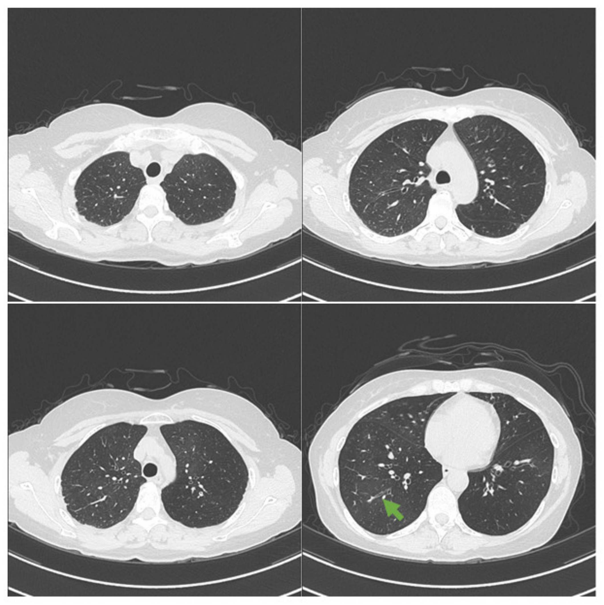
Abstract
:1. Introduction
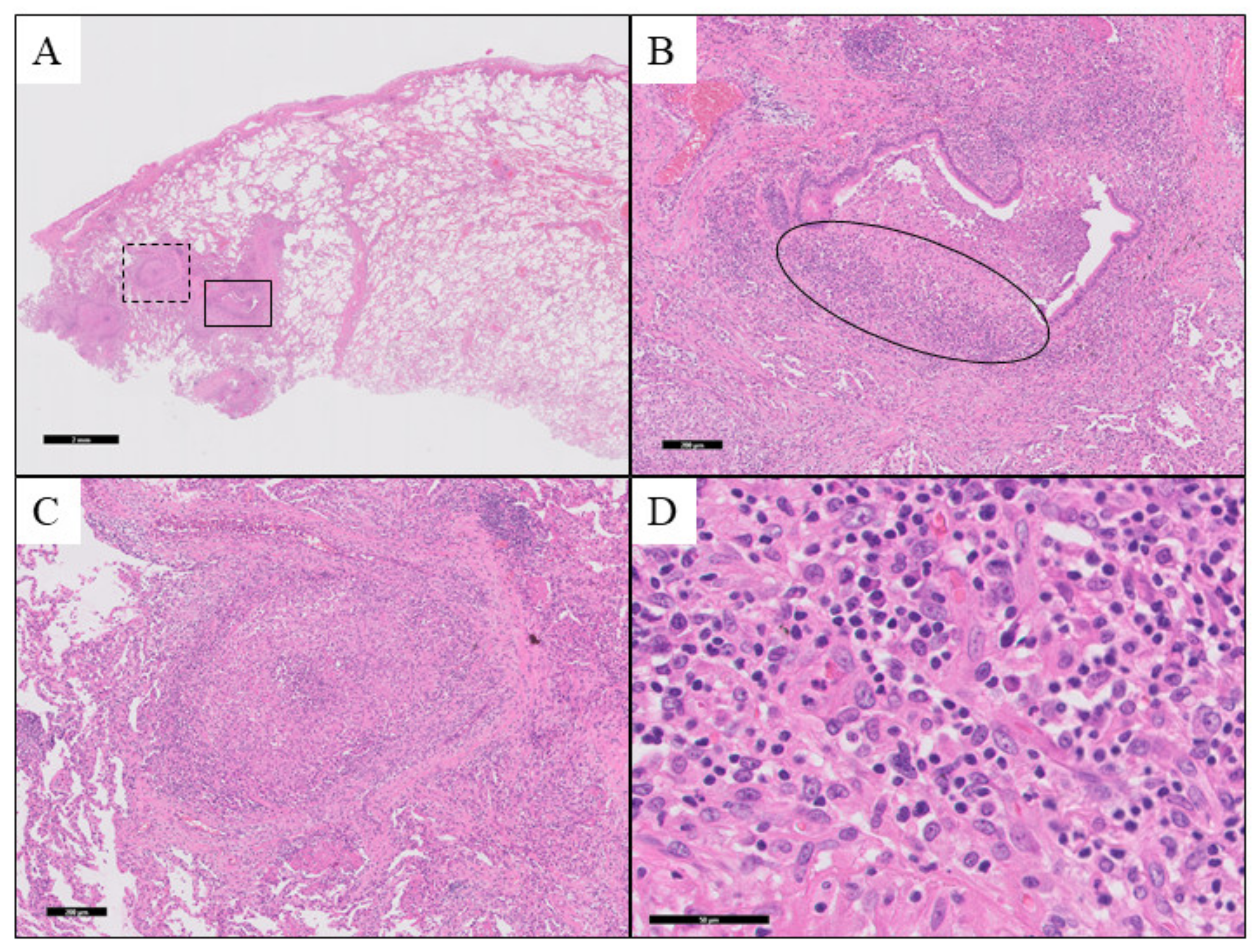
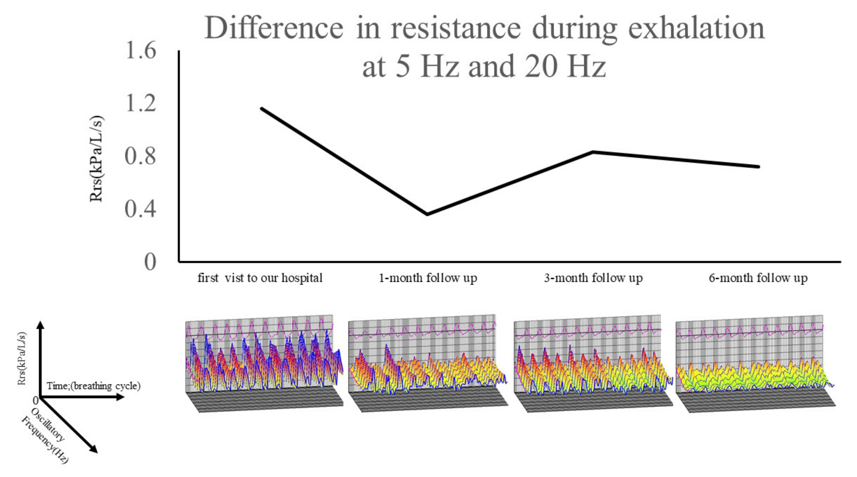
2. Case Presentation
3. Discussion
4. Conclusions
Author Contributions
Funding
Institutional Review Board Statement
Informed Consent Statement
Conflicts of Interest
References
- Tamilarasan, A.G.; Cunningham, G.; Irving, P.M.; Samaan, M.A. Recent advances in monoclonal antibody therapy in IBD: Practical issues. Frontline Gastroenterol. 2019, 10, 409–416. [Google Scholar] [CrossRef]
- Davignon, J.L.; Rauwel, B.; Degboé, Y.; Constantin, A.; Boyer, J.F.; Kruglov, A. Modulation of T-cell responses by anti-tumor necrosis factor treatments in rheumatoid arthritis: A review. Arthritis Res. 2018, 20, 229. [Google Scholar] [CrossRef] [PubMed] [Green Version]
- Sondag, M.; Verhoeven, F.; Guillot, X.; Prati, C.; Wendling, D. Efficacy of new treatments for dactylitis of psoriatic arthritis: Update of literature review. Clin. Rheumatol. 2019, 38, 591–596. [Google Scholar] [CrossRef] [PubMed]
- Crommelin, H.A.; Vorselaars, A.D.; van Moorsel, C.H.; Korenromp, I.H.; Deneer, V.H.; Grutters, J.C. Anti-TNF therapeutics for the treatment of sarcoidosis. Immunotherapy 2014, 6, 1127–1143. [Google Scholar] [CrossRef]
- Toussirot, É.; Aubin, F. Paradoxical reactions under TNF-alpha blocking agents and other biological agents given for chronic immune-mediated diseases: An analytical and comprehensive overview. RMD Open 2016, 2, e000239. [Google Scholar] [CrossRef] [PubMed] [Green Version]
- Iskandar, H.N.; Dhere, T.; Farraye, F.A. Ulcerative colitis: Update on medical management. Curr. Gastroenterol. Rep. 2015, 17, 1–11. [Google Scholar] [CrossRef]
- Daïen, C.I.; Monnier, A.; Claudepierre, P.; Constantin, A.; Eschard, J.P.; Houvenagel, E. Sarcoid-like granulomatosis in patients treated with tumor necrosis factor blockers: 10 cases. Rheumatology 2009, 48, 883–886. [Google Scholar] [CrossRef] [PubMed] [Green Version]
- Usmani, O.S.; Singh, D.; Spinola, M.; Bizzi, A.; Barnes, P.J. The prevalence of small airways disease in adult asthma: A systematic literature review. Respir. Med. 2016, 116, 19–27. [Google Scholar] [CrossRef] [Green Version]
- Postma, D.S.; Brightling, C.; Baldi, S.; Van Den Berge, M.; Fabbri, L.M.; Gagnatelli, A. Exploring the relevance and extent of small airways dysfunction in asthma (ATLANTIS): Baseline data from a prospective cohort study. Lancet Respir. Med. 2019, 7, 402–416. [Google Scholar] [CrossRef] [Green Version]
- Baughman, R.P.; Drent, M.; Kavuru, M.; Judson, M.A.; Costabel, U.; du Bois, R. Infliximab therapy in patients with chronic sarcoidosis and pulmonary involvement. Am. J. Respir. Crit. Care Med. 2006, 174, 795–802. [Google Scholar] [CrossRef] [Green Version]
- Hostettler, K.E.; Studler, U.; Tamm, M.; Brutsche, M.H. Long-term treatment with infliximab in patients with sarcoidosis. Respiration 2012, 83, 218–224. [Google Scholar] [CrossRef]
- Wendling, D.; Prati, C. Paradoxical effects of anti-TNF-alpha agents in inflammatory diseases. Expert Rev. Clin. Immunol. 2014, 10, 159–169. [Google Scholar] [CrossRef]
- Massara, A.; Cavazzini, L.; La Corte, R.; Trotta, F. Sarcoidosis appearing during anti-tumor necrosis factor alpha therapy: A new “class effect” paradoxical phenomenon. Two case reports and literature review. Semin. Arthritis Rheum. 2010, 39, 313–319. [Google Scholar] [CrossRef] [PubMed]
- Dhaille, F.; Viseux, V.; Caudron, A.; Dadban, A.; Tribout, C.; Boumier, P. Cutaneous sarcoidosis occurring during anti-TNF-alpha treatment: Report of two cases. Dermatology 2010, 220, 234–237. [Google Scholar] [CrossRef] [PubMed]
- Marseglia, G.L.; Cirillo, I.; Vizzaccaro, A.; Klersy, C.; Tosca, M.A.; La Rosa, M. Role of forced expiratory flow at 25–75% as an early marker of small airways impairment in subjects with allergic rhinitis. Allergy Asthma Proc. 2007, 28, 74–78. [Google Scholar] [CrossRef] [PubMed]
- Cirillo, I.; Klersy, C.; Marseglia, G.L.; Vizzaccaro, A.; Pallestrini, E.; Tosca, M. Role of FEF25%–75% as a predictor of bronchial hyperreactivity in allergic patients. Ann. Allergy Asthma Immunol. 2006, 96, 692–700. [Google Scholar] [CrossRef]
- Houghton, C.M.; Woodcock, A.A.; Singh, D. A comparison of plethysmography, spirometry and oscillometry for assessing the pulmonary effects of inhaled ipratropium bromide in healthy subjects and patients with asthma. Br. J. Clin. Pharm. 2005, 59, 152–159. [Google Scholar] [CrossRef]
- Borrill, Z.L.; Houghton, C.M.; Woodcock, A.A.; Vestbo, J.; Singh, D. Measuring bronchodilation in COPD clinical trials. Br. J. Clin. Pharm. 2005, 59, 379–384. [Google Scholar] [CrossRef] [PubMed] [Green Version]
- Takeda, T.; Oga, T.; Niimi, A.; Matsumoto, H.; Ito, I.; Yamaguchi, M. Relationship between small airway function and health status, dyspnea and disease control in asthma. Respiration 2010, 80, 120–126. [Google Scholar] [CrossRef] [Green Version]
- Brashier, B.; Salvi, S. Measuring lung function using sound waves: Role of the forced oscillation technique and impulse oscillometry system. Breathe 2015, 11, 57–65. [Google Scholar] [CrossRef]
- Bikov, A.; Pride, N.B.; Goldman, M.D.; Hull, J.H.; Horvath, I.; Barnes, P.J. Glottal Aperture and Buccal Airflow Leaks Critically Affect Forced Oscillometry Measurements. Chest 2015, 148, 731–738. [Google Scholar] [CrossRef] [PubMed]


Publisher’s Note: MDPI stays neutral with regard to jurisdictional claims in published maps and institutional affiliations. |
© 2021 by the authors. Licensee MDPI, Basel, Switzerland. This article is an open access article distributed under the terms and conditions of the Creative Commons Attribution (CC BY) license (https://creativecommons.org/licenses/by/4.0/).
Share and Cite
Fukahori, S.; Obase, Y.; Fukushima, C.; Takao, D.; Iriki, J.; Ozasa, M.; Zaizen, Y.; Takamura, N.; Fukuoka, J.; Ashizawa, K.; et al. Determining Response to Treatment for Drug-Induced Bronchocentric Granulomatosis by the Forced Oscillation Technique. Medicina 2021, 57, 1315. https://doi.org/10.3390/medicina57121315
Fukahori S, Obase Y, Fukushima C, Takao D, Iriki J, Ozasa M, Zaizen Y, Takamura N, Fukuoka J, Ashizawa K, et al. Determining Response to Treatment for Drug-Induced Bronchocentric Granulomatosis by the Forced Oscillation Technique. Medicina. 2021; 57(12):1315. https://doi.org/10.3390/medicina57121315
Chicago/Turabian StyleFukahori, Susumu, Yasushi Obase, Chizu Fukushima, Daisuke Takao, Jun Iriki, Mutsumi Ozasa, Yoshiaki Zaizen, Noboru Takamura, Junya Fukuoka, Kazuto Ashizawa, and et al. 2021. "Determining Response to Treatment for Drug-Induced Bronchocentric Granulomatosis by the Forced Oscillation Technique" Medicina 57, no. 12: 1315. https://doi.org/10.3390/medicina57121315