
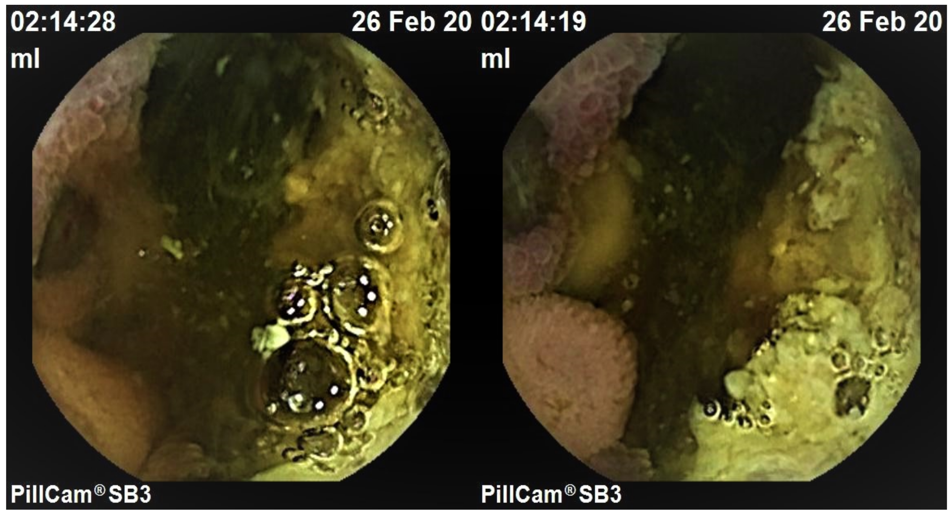
Small Bowel Metastatic Melanoma: An Emblematic “Coal-Black” Appearance at Videocapsule Endoscopy

 ,
, 
Abstract
:1. Introduction
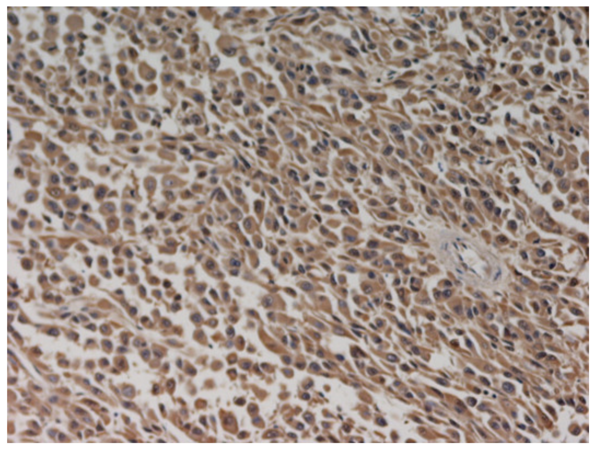
2. Case Report
3. Discussion
4. Conclusions
Supplementary Materials
Author Contributions
Funding
Institutional Review Board Statement
Informed Consent Statement
Conflicts of Interest
References
- Contaldo, A.; Losurdo, G.; Albano, F.; Iannone, A.; Barone, M.; Ierardi, E.; Di Leo, A.; Principi, M. The Spectrum of Small Intestinal Lesions in Patients with Unexplained Iron Deficiency Anemia Detected by Video Capsule Endoscopy. Medicina 2019, 55, 59. [Google Scholar] [CrossRef] [PubMed] [Green Version]
- Prakoso, E.; Selby, W.S. Capsule Endoscopy in Patients with Malignant Melanoma. Am. J. Gastroenterol. 2007, 102, 1204–1208. [Google Scholar] [CrossRef] [PubMed]
- De Francesco, V.; Stoppino, G.; Tonti, P.; D’Agnessa, M.; Castriota, M.; Panella, C.; Ierardi, E. Ileal metastasis from thoracic melanoma disclosed by video capsule endoscopy: An unusual but not extraordinary source of obscure bleeding. Endoscopy 2007, 39, E109. [Google Scholar] [CrossRef] [PubMed] [Green Version]
- Wei, S.-C.; Su, W.-C.; Chang, M.-C.; Chang, Y.-T.; Wang, C.-Y.; Wong, J.-M. Incidence, endoscopic morphology and distribution of metastatic lesions in the gastrointestinal tract. J. Gastroenterol. Hepatol. 2006, 22, 827–831. [Google Scholar] [CrossRef] [PubMed]
- Smedegaard, J.; Adamsen, S. Metastatic malignant melanoma of the small intestine: Capsule endoscopic appearance. Endoscopy 2007, 39, E209. [Google Scholar] [CrossRef] [PubMed] [Green Version]
- Urbain, D.; Aerts, M.; Reynaert, H.; Mana, F.; Neyns, B. Small-bowel metastasis of malignant melanoma: Video capsule endoscopy appearance. Endoscopy 2010, 42, E185. [Google Scholar] [CrossRef] [PubMed] [Green Version]
- Szumera-Ciećkiewicz, A.; Bosisio, F.; Teterycz, P.; Antoranz, A.; Delogu, F.; Koljenović, S.; van de Wiel, B.A.; Blokx, W.; van Kempen, L.; Rutkowski, P.; et al. SOX10 is as specific as S100 protein in detecting metastases of melanoma in lymph nodes and is recommended for sentinel lymph node assessment. Eur. J. Cancer 2020, 137, 175–182. [Google Scholar] [CrossRef] [PubMed]
- Rondonotti, E.; Pennazio, M.; Toth, E.; Menchen, P.; Riccioni, M.E.; De Palma, G.D.; Scotto, F.; De Looze, D.; Pachofsky, T.; Tacheci, I.; et al. Small-bowel neoplasms in patients undergoing video capsule endoscopy: A multicenter European study. Endoscopy 2008, 40, 488–495. [Google Scholar] [CrossRef] [PubMed] [Green Version]
- Miyazawa, H.; Yanagi, T.; Yamaguchi, Y.; Imafuku, K.; Kitamura, S.; Hata, H.; Uehara, J.; Ichikawa, N.; Ohno, Y.; Yoshida, T.; et al. Two cases of melanomas paradoxically metastasizing to the intestinal tract during nivolumab therapy. J. Dermatol. 2017, 5, 1481–1962. [Google Scholar] [CrossRef] [PubMed]


Publisher’s Note: MDPI stays neutral with regard to jurisdictional claims in published maps and institutional affiliations. |
© 2021 by the authors. Licensee MDPI, Basel, Switzerland. This article is an open access article distributed under the terms and conditions of the Creative Commons Attribution (CC BY) license (https://creativecommons.org/licenses/by/4.0/).
Share and Cite
Todeschini, A.; Loconte, I.; Contaldo, A.; Ierardi, E.; Di Leo, A.; Principi, M. Small Bowel Metastatic Melanoma: An Emblematic “Coal-Black” Appearance at Videocapsule Endoscopy. Medicina 2021, 57, 1313. https://doi.org/10.3390/medicina57121313
Todeschini A, Loconte I, Contaldo A, Ierardi E, Di Leo A, Principi M. Small Bowel Metastatic Melanoma: An Emblematic “Coal-Black” Appearance at Videocapsule Endoscopy. Medicina. 2021; 57(12):1313. https://doi.org/10.3390/medicina57121313
Chicago/Turabian StyleTodeschini, Alessia, Ilaria Loconte, Antonella Contaldo, Enzo Ierardi, Alfredo Di Leo, and Mariabeatrice Principi. 2021. "Small Bowel Metastatic Melanoma: An Emblematic “Coal-Black” Appearance at Videocapsule Endoscopy" Medicina 57, no. 12: 1313. https://doi.org/10.3390/medicina57121313