A Very Rare Case of Colosalpingeal Fistula Secondary to Diverticulitis: An Overview of Development, Clinical Features and Management

 ,
,  ,
, 
Abstract
:1. Introduction
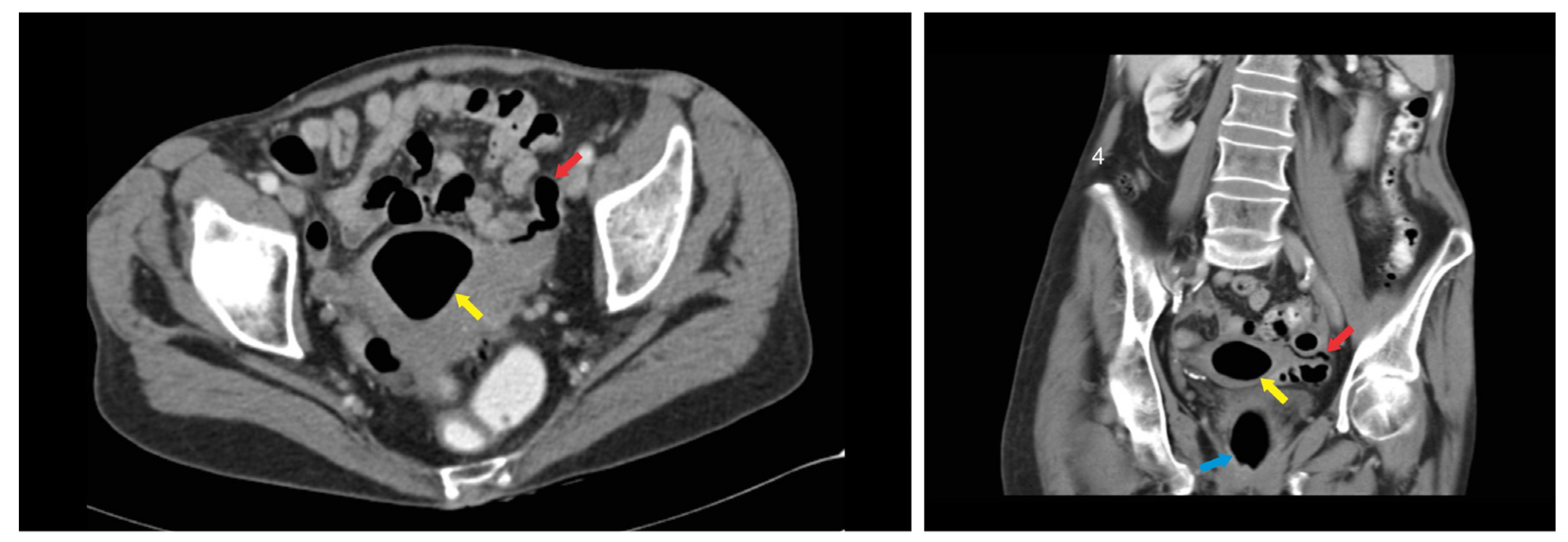
2. Case Report
3. Discussion
4. Conclusions
Author Contributions
Funding
Acknowledgments
Conflicts of Interest
References
- Vasilevsky, C.A.; Belliveau, P.; Trudel, J.L.; Stein, B.L.; Gordon, P.H. Fistulas complicating diverticulitis. Int. J. Colorect. Dis. 1998, 13, 57–60. [Google Scholar] [CrossRef] [PubMed]
- Tursi, A.; Scarpignato, C.; Strate; Lanas, A.; Kruis, W.; Lahat, A.; Danese, S. Colonic diverticular disease. Nat. Rev. Dis. Primers 2020, 6, 20. [Google Scholar] [CrossRef] [PubMed]
- Lanas, A.; Abad-Baroja, D.; Lanas-Gimeno, A. Progress and challenges in the management of diverticular disease: Which treatment? Therap. Adv. Gastroenterol. 2018, 11, 1756284818789055. [Google Scholar] [CrossRef] [PubMed] [Green Version]
- Marcucci, T.; Giannessi, S.; Giudici, F.; Riccadonna, S.; Gori, A.; Tonelli, F. Management of colovesical and colovaginal diverticular fistulas Our experience and literature reviewed. Ann. Itali. Chir. 2017, 88, 55–61. [Google Scholar]
- Raidh, B.H.S.; Khalil, B.M.; Mohamed, B.B.; Mounir, B.M.; Abdeljelil, Z. Sigmoïdite diverticulaire compliquée d’une fistule colo-tubaire survenant au cours d’une grossesse. La Tunis Médicale 2011, 89, 574–575. [Google Scholar]
- Botezatu, R.; Marian, R.; Gica, N.; Iancu, G.; Peltecu, G.; Panaitescu, A.M. Axial torsion and infarction of Meckel’s diverticulum in the 3rd trimester of pregnancy. Chirurgia. 2018, 113, 266–269. [Google Scholar] [CrossRef]
- Tancer, M.L.; Veridiano, N.P. Genital fistulas caused by diverticular disease of the sigmoid colon. Am. J. Obstet. Gynecol. 1996, 174, 1547–1550. [Google Scholar] [CrossRef]
- Maun, D.; Vine, A.; Slater, G. Ileosalpingeal fistula: An unusual complication of Crohn’s disease. Mt. Sinai J. Med. 2006, 73, 1115–1116. [Google Scholar]
- Chaikof, E.L.; Cambria, R.P.; Warshaw, A.L. Colouterine fistula secondary to diverticulitis. Dis. Colon Rectum 1985, 28, 358–360. [Google Scholar] [CrossRef]
- McDaid, J.; Reichl, C.; Hamzah, I.; Fitter, S.; Harbach, L.; Savage, A.P. Diverticular fistulation is associated with nicorandil usage. Ann. R. Coll. Surg. Engl. 2010, 92, 463–465. [Google Scholar] [CrossRef]
- Panghaal, V.S.; Chernyak, V.; Patlas, M.; Rozenblit, A.M. CT features of adnexal involvement in patients with diverticulitis. Am. J. Roentgenol. 2009, 192, 963–966. [Google Scholar] [CrossRef] [PubMed]
- Al Sinani, N.S. Bi-salpingocolonic fistula report of both fallopian tubes fistulizing with sigmoid diverticulum with literature review. J. King Abdulaziz Univ. Med. Sci. 2012, 19, 3. [Google Scholar]
- Birnbaum, B.A.; Balthazar, E.J. CT of appendicitis and diverticulitis. Radiol. Clin. N. Am. 1994, 32, 885–898. [Google Scholar] [PubMed]
- Ruiz-Tovar, J.; Gamallo, C. Pneumosalpynx caused by colosalpingeal fistula secondary to acute colonic diverticulitis. Int. J. Colorectal. Dis. 2011, 26, 1357–1358. [Google Scholar] [CrossRef]
- Williams, S.M.; Nolan, D.J. Colosalpingeal fistula: A rare complication of colonic diverticular disease. Eur. Radiol. 1999, 9, 1432–1433. [Google Scholar] [CrossRef]
- Golabek, T.; Szymanska, A.; Szopinski, T.; Bukowczan, J.; Furmanek, M.; Powroznik, J.; Chlosta, P. Enterovesical Fistulae: Aetiology, Imaging, and Management. Gastroenterol. Res. Pract. 2013, 2013, 617967. [Google Scholar] [CrossRef] [Green Version]
- Kumar, A.; Bhargava, S.K.; Mehrotra, G.; Pushkarna, R. Enterotubal fistulae secondary to tuberculosis: Report of three cases and review of literature. Clin. Radiol. 2001, 56, 858–860. [Google Scholar] [CrossRef]
- Clotteau, J.E.; Premont, M.; Belkaid, A.; Habib, E. Female genital tuberculosis. Apropos of a case of salpingo-sigmoidal fistula. Chirurgie 1983, 109, 374–377. [Google Scholar]
- Hoffer, F.A.; Ablow, R.C.; Gryboski, J.D.; Seashore, J.H. Primary appendicitis with an appendoci-tuboovarian fistula. AJR 1982, 138, 742–743. [Google Scholar] [CrossRef] [Green Version]
- Simstein, N.L. Colo-tubo-ovarian fistula as complication of pelvic inflammatory disease. S. Med. J. 1981, 74, 512–513. [Google Scholar] [CrossRef]
- Hain, J.M.; Sherick, D.G. Salpingo colonic Fistula Secondary to Diverticulitis. Am. Surg. 1996, 62, 984–988. [Google Scholar] [PubMed]
- Poulin, E.C.; Schlachta, C.M.; Mamazza, J.; Seshadri, P. Should enteric fistulas from Crohn’s disease or diverticulitis be treated laparoscopically or by open surgery? Dis. Colon Rectum 2000, 43, 621–626. [Google Scholar] [CrossRef] [PubMed]
- Zonca, P.; Jacobi, C.A.; Meyer, G.P. The current view of surgical treatment of diverticular disease. Rozhl. Chir. 2009, 88, 568–576. [Google Scholar] [PubMed]
- Cirocchi, R.; Arezzo, A.; Renzi, C.; Cochetti, G.; D’Andrea, V.; Fingerhut, A.; Mearini, E.; Binda, G.A. Is laparoscopic surgery the best treatment in fistulas complicating diverticular disease of the sigmoid colon? A systematic review. Int. J. Surg. 2015, 24, 95–100. [Google Scholar] [CrossRef]
- Garcea, G.; Majid, I.; Sutton, C.D.; Pattenden, C.J.; Thomas, W.M. Diagnosis and management of colovesical fistulae; six-year experience of 90 consecutive cases. Colorectal Dis. 2006, 8, 347–352. [Google Scholar] [CrossRef]


© 2020 by the authors. Licensee MDPI, Basel, Switzerland. This article is an open access article distributed under the terms and conditions of the Creative Commons Attribution (CC BY) license (http://creativecommons.org/licenses/by/4.0/).
Share and Cite
Darii Plopa, N.; Gica, N.; Gerard, M.; Nollevaux, M.-C.; Pavlovic, M.; Anton, E. A Very Rare Case of Colosalpingeal Fistula Secondary to Diverticulitis: An Overview of Development, Clinical Features and Management. Medicina 2020, 56, 477. https://doi.org/10.3390/medicina56090477
Darii Plopa N, Gica N, Gerard M, Nollevaux M-C, Pavlovic M, Anton E. A Very Rare Case of Colosalpingeal Fistula Secondary to Diverticulitis: An Overview of Development, Clinical Features and Management. Medicina. 2020; 56(9):477. https://doi.org/10.3390/medicina56090477
Chicago/Turabian StyleDarii Plopa, Natalia, Nicolae Gica, Marie Gerard, Marie-Cécile Nollevaux, Milenko Pavlovic, and Emil Anton. 2020. "A Very Rare Case of Colosalpingeal Fistula Secondary to Diverticulitis: An Overview of Development, Clinical Features and Management" Medicina 56, no. 9: 477. https://doi.org/10.3390/medicina56090477