Calcific Myonecrosis of the Leg: A Rare Entity

 ,
, 
 , and
, and 
Abstract
:1. Introduction
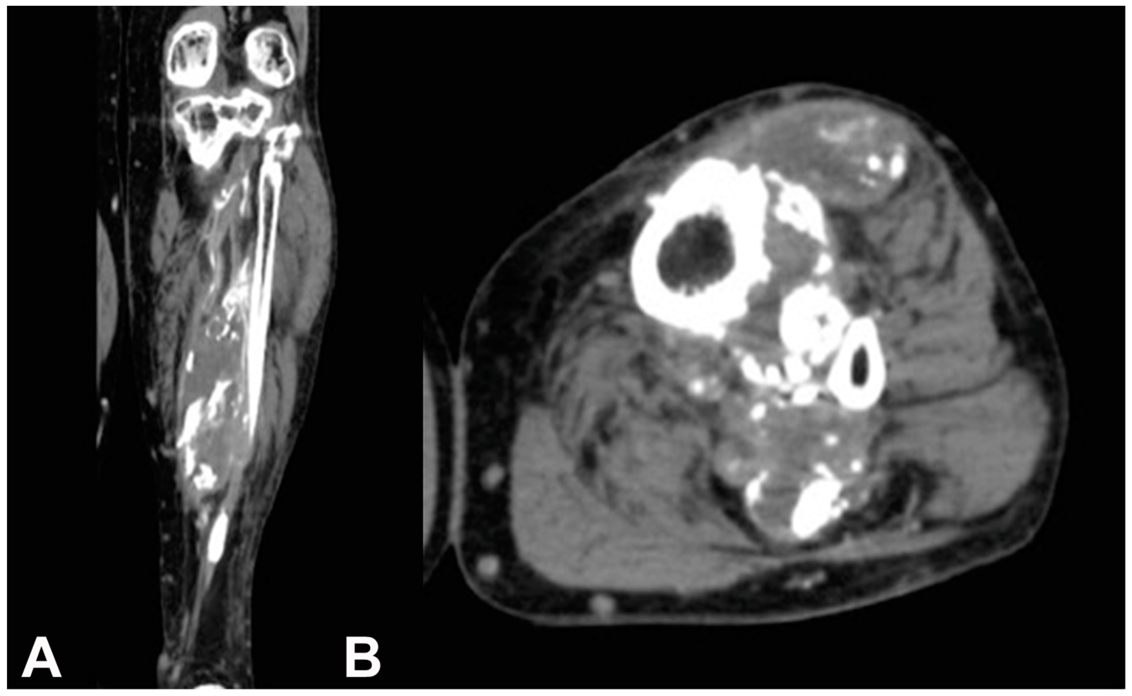
2. Case Report
3. Discussion
4. Conclusions
Author Contributions
Funding
Conflicts of Interest
References
- Gallie, W.E.; Thomson, S. Volkmann’s ischemic contracture: Two case reports with identical late sequelae. Can. J. Surg. 1960, 3, 164–166. [Google Scholar]
- Wang, J.W.; Chen, W.J. Calcific myonecrosis of the leg. Clin. Orthop. Relat. Res. 2001, 389, 185–190. [Google Scholar] [CrossRef]
- Larson, R.C.; Sierra, R.J.; Sundaram, M.; Inwards, C.; Scully, S.P. Calcific myonecrosis: A unique presentation in the upper extremity. Skelet. Radiol. 2004, 33, 306–309. [Google Scholar] [CrossRef]
- Dhillon, M.; Davies, A.M.; Benham, J.; Evans, N.; Mangham, D.C.; Grimer, R.J. Calcific myonecrosis: A report of ten new cases with an emphasis on MR imaging. Eur. Radiol. 2004, 14, 1974–1979. [Google Scholar] [CrossRef]
- Muramatsu, K.; Ihara, K.; Seki, T.; Imagama, T.; Taguchi, T. Calcific myonecrosis of the lower leg: Diagnosis and options of treatment. Arch. Orthop. Trauma. Surg. 2009, 129, 935–939. [Google Scholar] [CrossRef]
- Janzen, D.L.; Connell, D.G.; Vaisler, B.J. Calcific myonecrosis of the calf manifesting as an enlarging soft-tissue mass: Imaging features. Am. J. Roentgenol. 1993, 160, 1072–1074. [Google Scholar] [CrossRef]
- Holobinko, J.N.; Damron, T.A.; Scerpella, P.R.; Hojnowski, L. Calcific myonecrosis: Keys to early recognition. Skelet. Radiol. 2003, 32, 35–40. [Google Scholar] [CrossRef]
- Batz, R.; Sofka, C.M.; Adler, R.S.; Mintz, D.N.; DiCarlo, E. Dermatomyositis and calcific myonecrosis in the leg: Ultrasound as an aid in management. Skelet. Radiol. 2006, 35, 113–116. [Google Scholar] [CrossRef]
- O’Keefe, R.J.; O’Connell, J.X.; Temple, H.T.; Scully, S.P.; Kattapuram, S.V.; Springfield, D.S.; Rosenberg, A.E.; Mankin, H.J. Calcific myonecrosis. Clin. Orthop. Relat. Res. 1995, 318, 205–213. [Google Scholar]
- Zohman, G.L.; Pierce, J.; Chapman, M.W.; Greenspan, A.; Gandour-Edwards, R. Calcific myonecrosis mimicking an invasive soft-tissue neoplasm. A case report and review of the literature. J. Bone Joint Surg. Am. 1998, 80, 1193–1197. [Google Scholar] [CrossRef]
- Flinn, J.; Beggs, I. Case of the month: Calcified leg. Br. J. Radiol. 1996, 69, 371–372. [Google Scholar] [CrossRef]
- Ozbarlas, S.; Kalaci, A.; Ozkan, C.; Togrul, E. A previously healthy 77-year-old man with a painful mass in the calf for two months. Ann. Saudi. Med. 2007, 27, 49–50. [Google Scholar] [CrossRef]
- De Carvalho, B.R. Calcific myonecrosis: A two-patient case series. Jpn. J. Radiol. 2012, 30, 517–521. [Google Scholar] [CrossRef]
- Okada, A.; Hatori, M.; Hosaka, M.; Watanuki, M.; Itoi, E. Calcific myonecrosis and the role of imaging in the diagnosis: A case report. Ups. J. Med. Sci. 2009, 114, 178–183. [Google Scholar] [CrossRef]
- Schneider, S.; Duewell, S.; Graf, H.; Forster, A. Painful swollen leg 52 years after bimalleolar fracture. Praxis 2009, 98, 1457–1461. [Google Scholar] [CrossRef]
- Chun, Y.S.; Shim, H.S. Calcific myonecrosis of the antetibial area. Clin. Orthop. Surg. 2010, 2, 191–194. [Google Scholar] [CrossRef]
- Papanikolaou, A.; Chini, M.; Pavlakis, D.; Lioni, A.; Lazanas, M.; Maris, J. Calcific myonecrosis of the leg: Report of three patients presenting with infection. Surg. Infect. 2011, 12, 247–250. [Google Scholar] [CrossRef]
- Portabella, F.; Nárvaez, J.A.; Llatjos, R.; Cabo, J.; Maireles, M.; Serrano, C.; Pedrero, S.; Romero, E.; Pablos, O.; Saborido, A. calcific myonecrosis of the leg. Rev. Esp. Cir. Ortop. Traumatol. 2012, 56, 46–50. [Google Scholar] [CrossRef]
- O’Dwyer, H.M.; Al-Nakshabandi, N.A.; Al-Muzahmi, K.; Ryan, A.; O’Connell, J.X.; Munk, P.L. Calcific myonecrosis: Keys to recognition and management. Am. J. Roentgenol. 2006, 187, W67–W76. [Google Scholar] [CrossRef]
- Jalil, R.; Roach, J.; Smith, A.; Mukundan, C. Calcific myonecrosis: A case report and review of the literature. BMJ Case Rep. 2012, 2012, bcr2012007186. [Google Scholar] [CrossRef]
- Karkhanis, S.R.; Botchu, R.; James, S.; Evans, N. Bilateral calcific myonecrosis associated with epilepsy. Clin. Radiol. 2013, 68, 349–352. [Google Scholar] [CrossRef] [PubMed]
- Rynders, S.D.; Boachie-Adjei, Y.D.; Gaskin, C.M.; Chhabra, A.B. Calcific myonecrosis of the upper extremity: Case report. J. Hand. Surg. Am. 2012, 37, 130–133. [Google Scholar] [CrossRef] [PubMed]
- Yuenyongviwat, V.; Laohawiriyakamol, T.; Suwanno, P.; Kanjanapradit, K.; Tanutit, P. Calcific myonecrosis following snake bite: A case report and review of the literature. J. Med. Case Rep. 2014, 8, 193. [Google Scholar] [CrossRef] [PubMed]
- Ukon, Y.; Tanaka, T.; Nagata, S.; Hagizawa, H.; Imura, Y.; Tamiya, H.; Oshima, K.; Naka, N.; Aoki, Y.; Kuratsu, S. Calcific myonecrosis mimicking soft tissue sarcoma: A case report. Oncol. Lett. 2018, 15, 7909–7913. [Google Scholar] [CrossRef] [PubMed]
- Tuncay, I.C.; Demirörs, H.; Isiklar, Z.U.; Agildere, M.; Demirhan, B.; Tandogan, R.N. Calcific myonecrosis. Int. Orthop. 1999, 23, 68–70. [Google Scholar] [CrossRef] [PubMed]
- Jassal, D.S.; Low, M.; Ross, L.L.; Zeismann, M.; Embil, J.M. Calcific myonecrosis: Case report and review. Ann. Plast. Surg. 2001, 46, 174–177. [Google Scholar] [CrossRef] [PubMed]
- Güven, M.; Cakar, M.; Başsorgun, I.; Kadioğlu, B.; Kilinçoğlu, V.; Eren, A. Calcific myonecrosis. Acta. Orthop. Traumatol. Turc. 2008, 42, 70–73. [Google Scholar] [CrossRef] [PubMed]
- Mirra, J.M. Calcific myonecrosis. Clin. Orthop. Relat. Res. 1996, 327, 308–310. [Google Scholar] [CrossRef]


| Study | Patients/Age, gender | Site | Symptoms (time) | Previous trauma (time before) | Nerve deficit | Biopsy | Complications | Infection | Treatment |
|---|---|---|---|---|---|---|---|---|---|
| Janzen et al. [6] | 77M | Leg | Mass | Fibular fracture (52 years) | Peroneal | Incisional | Yes | Chronic drainage | n.a. |
| Zohman et al. [10] | 49M | Leg | Mass | Ligamentous knee injury (30 years) | Sciatic | Incisional | No | – | Excision |
| Tuncay et al. [25] | 64M | Leg | Mass (3 years) | Shotgun injury (42 years) | Peroneal | Incisional | No | – | Excision |
| Jassal et al. [26] | 66M | Leg | Mass, pain | Ankle fracture (47 years) | – | Incisional | Yes | Cellulitis and drainage | Debridement + flap |
| Holobinko et al. [7] | 57M | Leg | Mass, pain (2 years) | Tibia fracture (40 years) | Weakness | Incisional | Yes | Drainage | Debridement + flap |
| 67M | Leg | Mass (3 months) | Tibia fracture (51 years) | – | Incisional | No | Drainage | Debridement | |
| 37M | Foot | Plantar drainage | Tibia fracture (21 years) | Hyperesthesia | Incisional | No | Drainage | Debridement + flap | |
| Dhillon et al. [4] | 7M, 3F; mean age 68 years (range, 40–82 years) | Leg | n.a. | Significant trauma to the leg (mean, 46 years; range 28–59 years) | n.a. | Needle (4 patients) | 8 patients (prolonged recovery) | n.a. | Debridement (3 patients) |
| Larson et al. [3] | 17M | Forearm | Mass, pain (4 months) | Crush injury (55 years) | – | Incisional | No | – | Excision |
| Ozbarlas et al. [12] | 77M | Leg | Mass, pain (2 months) | Blunt trauma (5 years) | – | n.a. | n.a. | – | n.a. |
| Muramatsu et al. [5] | 51M | Leg | Mass, pain (3 months) | Compartment syndrome. (35 years) | Peroneal | Needle | – | – | Debridement |
| 52M | Leg | Growing mass (1 month) | Tibia fracture (24 years) | Tibialis | Incisional | – | Osteomyelitis | Aspiration | |
| 66M | Leg | Mass, pain (3 months) | Crush injury (40 years) | – | – | – | Yes | Aspiration | |
| Okada et al. [14] | 62M | Leg | Mass (5 months) | Squeezed trauma (43 years) | Peroneal | Incisional | – | – | Excision |
| Papanikolaou et al. [17] | 54M | Leg | Mass (10 days) | Crush injury (7 years) | – | – | – | Yes | Debridement |
| 66M | Leg | Mass, pain (1 month) | Artery lesion (52 years) | Peroneal | – | – | Drainage, fever | Debridement, VACT | |
| 84F | Leg | Infection | Crush injury (53 years) | – | – | – | Yes, fever | Debridement | |
| Portabella et al. [18] | 55M | Leg | n.a. | n.a. | Sciatic | Yes | – | – | – |
| 64M | Leg | n.a. | n.a. | Peroneal | – | Yes | Chronic drainage | Debridement, VACT | |
| 54M | Leg | n.a. | n.a. | Peroneal | – | Yes | Chronic drainage | Debridement, VACT | |
| 77M | Leg | n.a. | n.a. | – | – | – | Yes | Debridement | |
| De Carvalho et al. [13] | 69F | Leg | Mass (2 months) | Motor vehicle trauma | – | – | – | – | n.a. |
| 73M | Leg | Growing mass | Tibia fracture (57 years) | – | – | – | – | n.a. | |
| Chun et al. [16] | 53M | Leg | Growing mass | Snake bite (44 years) | – | – | – | – | n.a. |
| Jalil et al. [20] | 43M | Leg | Mass, pain (1 month) | Tibia fracture (20 years) | – | – | – | – | Debridement |
| Karkhanis et al. [21] | 60M | Leg | Growing mass (4 months) | – | – | – | – | – | – |
| Rynders et al. [22] | 66M | Forearm | Growing mass (2 months) | Elbow fracture (57 years) | – | – | – | n.a. | n.a. |
| Yuenyongviwat et al. [23] | 66F | Leg | Mass (10 years) | Snake bite (14 years) | Peroneal | Yes | Yes | Yes | Excision |
| Ukon et al. [24] | 69F | Leg | Growing mass (20 years) | Fibular fracture (20 years) | – | Incisional | Yes | Chronic drainage | – |
| 76M | Leg | Growing mass (2 months) | Tibia, fib fracture (55 years) | Peroneal | Incisional | Yes | Yes | Debridement | |
| Güven et al. [27] | 66M | Leg | Mass, pain (12 months) | Compartment syndrome after gunshot injury of the thigh (35 years) | – | Excisional | – | – | – |
© 2019 by the authors. Licensee MDPI, Basel, Switzerland. This article is an open access article distributed under the terms and conditions of the Creative Commons Attribution (CC BY) license (http://creativecommons.org/licenses/by/4.0/).
Share and Cite
Angelini, A.; Mavrogenis, A.F.; Pagliarini, E.; Trovarelli, G.; Fanelli, G.N.; Cappellesso, R.; Ruggieri, P. Calcific Myonecrosis of the Leg: A Rare Entity. Medicina 2019, 55, 542. https://doi.org/10.3390/medicina55090542
Angelini A, Mavrogenis AF, Pagliarini E, Trovarelli G, Fanelli GN, Cappellesso R, Ruggieri P. Calcific Myonecrosis of the Leg: A Rare Entity. Medicina. 2019; 55(9):542. https://doi.org/10.3390/medicina55090542
Chicago/Turabian StyleAngelini, Andrea, Andreas F. Mavrogenis, Elisa Pagliarini, Giulia Trovarelli, Giuseppe Nicolò Fanelli, Rocco Cappellesso, and Pietro Ruggieri. 2019. "Calcific Myonecrosis of the Leg: A Rare Entity" Medicina 55, no. 9: 542. https://doi.org/10.3390/medicina55090542