Next Article in Journal
Impact of hypertension on postreperfusion left ventricular recovery in patients with ST-segment elevation myocardial infarction and multivessel coronary artery disease
Previous Article in Journal
Management of recurrent aphthous ulcers using low-level lasers: A systematic review
 
 
Medicina is published by MDPI from Volume 54 Issue 1 (2018). Previous articles were published by another publisher in Open Access under a CC-BY (or CC-BY-NC-ND) licence, and they are hosted by MDPI on mdpi.com as a courtesy and upon agreement with Lithuanian Medical Association, Lithuanian University of Health Sciences, and Vilnius University.
Article

Morphometric parameters of the optic disc in normal and glaucomatous eyes based on time-domain optical coherence tomography image analysis

by
Dovilė Buteikienė
1,*,
Asta Kybartaitė-Žilienė
2,
Loresa Kriaučiūnienė
1,
Valerijus Barzdžiukas
1,†,
Ingrida Janulevičienė
1 and
Alvydas Paunksnis
1
1
Department of Ophthalmology, Medical Academy, Lithuanian University of Health Sciences, Kaunas, Lithuania
2
Laboratory of Biophysics and Bioinformatics, Neuroscience Institute, Medical Academy, Lithuanian University of Health Sciences, Kaunas, Lithuania
*
Author to whom correspondence should be addressed.
Deceased.
Medicina 2017, 53(4), 242-252; https://doi.org/10.1016/j.medici.2017.05.007
Submission received: 2 October 2015 / Revised: 23 April 2017 / Accepted: 23 May 2017 / Published: 18 July 2017

Abstract

:
Background and objective: Assessment of optic disc morphology is essential in diagnosis and management of visual impairment. The aim of this study was to evaluate associations between optic disc morphometric parameters, i.e., size and shape, and age, gender, and ocular axial length in normal and glaucomatous eyes based on time-domain optical coherence tomography image analysis. Materials and methods: It was a case–control study of 998 normal and 394 eyes with primary open angle glaucoma that underwent an ophthalmological examination and time-domain optical coherence topography scanning. Areas and shapes of the disc, cup, and neuroretinal rim were analyzed. Results: The shape of the optic disc did not differ between the study groups, i.e., normal and glaucomatous case groups, but the disc area of the primary open angle glaucoma group was significantly larger. The shape of the small disc was significantly different, but the shape of the medium and the large disc did not differ between the study groups. The central area of the disc, i.e., cup area was significantly larger in the case group and its shape was significantly different between the study groups. No significant differences in the area of the cup and its shape, nerve fibers on the edge of the disc, i.e., neuroretinal rim area, were found between the study groups of the small discs. There were significant associations between age, gender, and ocular axial length and morphometric parameters of the optic disc. Conclusions: Informative results with regard to the size and shape due to various ocular characteristics between the healthy control group and patients suffering with primary open angle glaucoma were obtained. Both study groups were significant in size, which makes the findings interesting and important contribution in the field.

1. Introduction

Morphometrics is a branch of morphology that represents the study of quantitative description of size and shape components of biological form and their variation in the population based on statistical analysis [1]. A considerable amount of work has been published examining the optic disc (OD) size-related parameters and their physiological and pathological associations; however, relatively little information is available describing shape features of the OD and their variations in general population [1].
Assessment of the OD, including a neuroretinal rim (NRR) and a cup, is one of the most crucial elements in diagnosing and monitoring blindness-related disorders, e.g., glaucoma [2,3]. The OD is a round/oval structure down which more than a million nerve fibers, i.e., axons of retinal ganglion cells, descend through a sheet that is known as a lamina cribrosa. These fibers are then bundled together behind the eye to form the optic nerve, which continues toward the brain. The NRR is a dense convergence of nerve fibers on an edge of the OD. An inferior rim is usually thicker than a superior rim, which is thicker than a nasal rim, and a temporal rim is the thinnest [4]. The cup is a central area to the OD (Fig. 1).
Glaucoma is a group of progressive optic neuropathies that have in common a slowly progressive degeneration of retinal ganglion cells and their axons, resulting in a distinct appearance of the OD and following pattern of visual loss [5]. It is estimated that glaucoma affects more than 66 million individuals worldwide and it is the second leading cause of blindness in the world, also the problem will increase as the population gets older [5]. The most common suspects to have glaucoma are those individuals with raised intraocular pressure (IOP) or with an asymmetric OD appearance [5]. Of the many types of glaucoma, primary open-angle glaucoma (POAG), in which the iridocorneal angle is open and normal in appearance, is perhaps the most common form of glaucoma [5,6].
Glaucoma is treatable and visual impairment caused by the disease is irreversible, therefore early detection is essential. Variation and qualitative evaluations of structural changes of the OD, the NRR, the cup-to-disc ratio and retinal nerve fiber layer (RNFL) defects have been found useful for evaluation of different degrees of glaucomatous damage [2].
Literature review provides various interpretations about the OD, the NRR and the cup; their parameters and relationships with general health indicators. The size of the OD area showed positive association with age [7,8], but other sources [9,10,11] informed that there was not enough evidence for the OD dependence on age. The OD area of the male gender was usually larger [7] but there were exceptions [11].
The NRR showed a strong positive correlation with the OD area [7]; for every 1 mm2 increase in the OD area, the NRR area increased by 0.5 mm2 [3]. The size of the NRR area showed possible correlation with age [3,9,12], but there were considerations that they were not significantly related [8,10,13]. Also several sources stated that the NRR was larger in the male gender [3,7]; meanwhile others informed that the RNN area was not found to be related to the gender [9,10,12]. According to the literature review the ratio of the NRR and the OD did not depend on the age [13]. The asymmetry of the NRR area was related significantly to the asymmetry of the OD area [3].
There is a wide variation of the optic cup size in the normal eyes and much of the variability resulted from the physiological relationship between the size of the cup and the OD [14]. A weak relationship of the vertical diameter of the cup and age [14], and gender [15] was reported. A horizontal diameter of the cup was usually larger in the male gender [15]. There was no significant correlation between the cup size and age, and gender [10]. A ratio of the cup to the disc has a tendency to increase about 0.1 between ages of 30 and 70 years [9]. Also this ratio was usually bigger in the male gender [12]. In general, the cup size was physiologically related to the OD size and pathologically to glaucomatous damage [14].
There are various interpretations about other OD parameters and their relationship with ophthalmic indicators. The OD area was positively associated with the ocular axial length (AL); as the AL increased by 1 mm, the OD area increased by 3.7% [8]. Also the OD was significantly larger in the highly myopic eyes [11], but other sources stated that there was no significant correlation between refraction data and the OD parameters [10].
Medical imaging and visualization technologies allow accurate analysis of visual system and help healthcare professionals to make accurate diagnostics of patients’ condition [16]. Some of these technologies in the field of ophthalmoscopy are semi-automated imaging techniques such as confocal laser scanning tomography, laser scanning polarimetry or optical coherence tomography (OCT), which can offer objective and reproducible measurements,, particularly topographic parameters of the OD and the RNFL [1].
In this study the OD parameters were obtained by time-domain optical coherence tomography (TD-OCT) (Fig. 2). OCT provides cross-sectional images of tissue structure on the micron scale in situ and in real time [17]. Similar results for morphometric study could be achieved only by a conventional histopathology, which requires removal of a tissue specimen and processing for microscopic examination but not in real time. Therefore, OCT is like a type of optical biopsy and is a powerful imaging technology for medical diagnostics with high axial resolution, automated outlining of the OD margin, and a consistent and stable reference plane for delineation of the NRR boundary [2,17].
The main goal of this study was to evaluate morphometric parameters of the OD and to assess the significance of age, gender, size of the OD and ocular AL as risk factors by performing a large scale research of control subjects and POAG patients of a local population based on TD-OCT image analysis by incorporating tools from geometry, biometrics and computer graphics. Since literature review provides various interpretations, the results of this study help to facilitate the solution of glaucoma detection and management for ophthalmologists. This may also help to improve the discriminative ability of image processing in ophthalmology. The significance of this work is that it contributes to the results of previous studies in this field, using more up-to-date measurement technique in a different study population.

2. Materials and methods

2.1. Selection and characteristics of patients

The study was conducted in accordance with the Declaration of Helsinki and was approved by the Kaunas Regional Human Research Ethics Committee, Lithuania. Detailed study and its methods were described in previous publication [18]. In brief, it was an analytical observational case–control study.
The control group comprised 45–74-year-old men and women. The inclusion criteria in the control group were as follows: (1) a participant has agreed to participate in the study and signed an informed consent form; (2) no congenital or acquired optic nerve pathology; (3) a participant was not previously diagnosed with glaucoma; (4) there were no defects deeper than the P < 2% in the visual field (tested by Frequency Doubling Technology Screening); and (5) signal strength of OD TD-OCT images was greater than 5.
The case group comprised 45–74-year-old men and women with POAG. The inclusion criteria in the case group were as follows: (1) a participant has to agree to participate in the study and signed an informed consent form; (2) IOP was higher than 21 mm Hg by Schiotz tonometer (using the weights 5.5 g) at the time of POAG diagnosis; (3) the OD glaucomatous structural changes were described in the outpatient card; (4) the signal strength of OD TD-OCT images was greater than 5; (5) glaucomatous visual field defects were approved by two Humphrey SITA Standard (24-2) tests and corresponded to I–II glaucoma criteria based on Humphrey visual field printouts [19]. Stage I (early defect): mean deviation more than or equal to −6.00 decibels (dB) and at least one of the following: A – on pattern deviation plot, there exists a cluster of 3 or more points in an expected location of the visual field depressed below the 5% level, at least 1 of which is depressed below the 1% level, B – corrected pattern standard deviation/pattern standard devia-tion significant at P < 0.05, C – glaucoma hemifield test ‘‘Outside Normal Limit’’. And Stage II (moderate defect): mean deviation of −6.01 to −12.00 dB and at least one of the following: A – on pattern deviation plot, greater than or equal to 25% but fewer than 50% of points depressed below the 5% level, and greater than or equal to 15% but fewer than 25% of points depressed below 1% level, B – at least 1 point within central 58 with sensitivity less than 15 dB but, no point within central 58 with sensitivity less than 0 dB, C – only 1 hemifield containing a point with sensitivity less than 15 dB within 58 of fixation.

2.2. Ophthalmologic examination

The questionnaire form was filled in with data about age, sex, medical history of the eye pathology and comorbidity. Uncorrected visual acuity (UCVA) and the best corrected visual acuity (BCVA) were tested at 4 m using a Logarithm of the minimum angle of resolution (Log MAR) chart [20]. The refraction was performed by means of computerized refrac-tometer ACCUREF-K9001 (Shin-Nippon, Japan). The ocular AL of 0.1 mm accuracy was performed by means of the A – scan (frequency 13 MHz) ultrasound (OPKO Instrumentation, OTI Scan 3000). IOP was measured with a Schiotz tonometer (Riester, Germany) under local anesthesia with 0.5% proxymetacaine. Biomicroscopy of the anterior and posterior eye segments was performed by a standard slit lamp. The participants of the control group underwent frequency doubling visual field screenings (N-30-5 Frequency Technology Screening, Humphrey Matrix, Carl Zeiss Meditec). The parti-cipants of the case group underwent 2 standard automated perimeter threshold visual field tests (Central 24-2 Threshold Test stimuli III, White, SITA – Fast/Standard, Humphrey Field Analyzer II, Carl Zeiss Meditec).

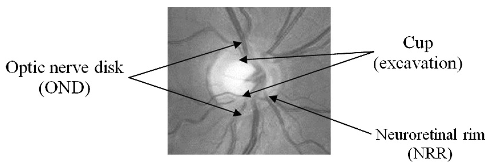
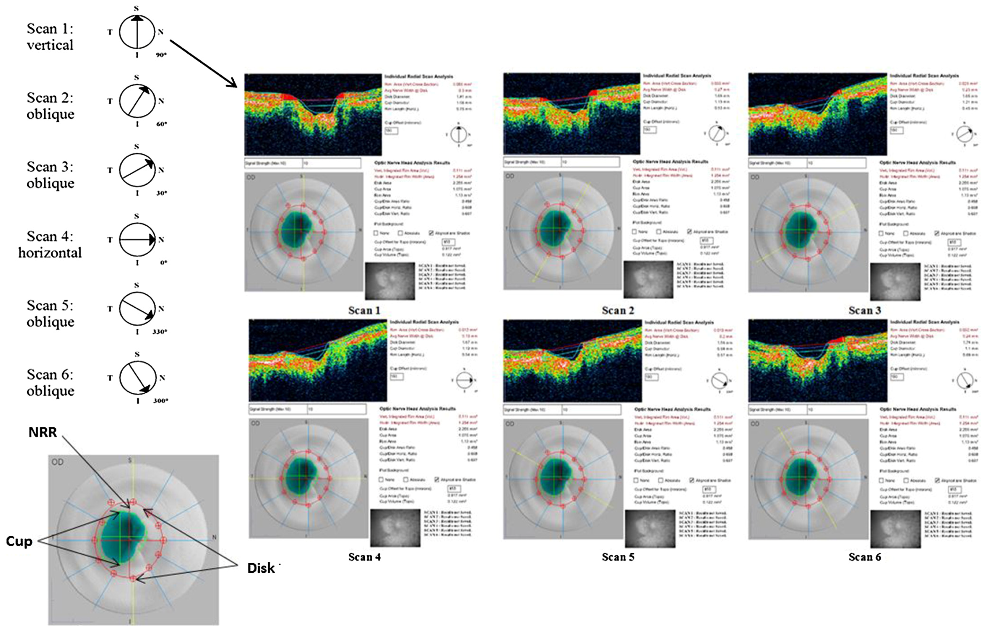
2.3. Imaging using TD-OCT and data acquisition

TD-OCT of the OD was performed with the Stratus OCT 3000 (software version 4.0, Carl Zeiss Meditec). It is a computer-assisted optical instrument that generates cross sectional two-dimensional (2D) images (scans) of the OD with axial and transverse resolutions of 10 μm and 20 μm, respectively.
The OD scans were acquired by the Fast Optic Disc protocol. This protocol was used because it is faster, there is a lower influence of eye movements on image quality, and the process is more automated compared with the Optic Disc protocol. The Fast Optic Disc protocol consists of a series of six equally spaced 4-mm radial line scans through a common central axis. With each scan pass, the Stratus OCT captures 128 longitudinal (axial) range samples (A-scans). Each A-scan consists of 1024 data points over 2 mm of depth. The Stratus OCT integrates 131 072 data points to construct a cross-sectional 2D image (scan) of OD anatomy. It displays the six scans in real time using a false color scale that represents the degree of light backscattering from tissues at different depths in the retina (Fig. 2).
The OD analysis was carried out by the Optic Nerve Head protocol. The analysis automatically calculates quantitative OD parameters for each of the six radial line scans and the output enables to interactively assess and measure them using each scan individually and a composite of all the scans. The outputs are displayed as the OD individual scans and the results of composite image measurements (Fig. 2).

2.4. Data grouping

Obtained data of the control and the POAG groups were divided into 4 age groups, i.e., (1) younger than 60 years, (2) 60–65 years, (3) 66–70 years, and (4) older than 70 years; into male and female groups; into different OD size groups, i.e., (1) the small disc area size was less than 1.5 mm2, (2) the medium disc size was between 1.5 and 2 mm2 and (3) the large disc area was considered to be larger than 2 mm2. It was considered that the range of (1) the average normal ocular AL was from 22.5 to 24.5 mm, (2) the short AL was less than 22.5 mm, and (3) the long AL was longer than 24.5 mm [21].
The spherical equivalent of refraction in diopter (D) ranged from −18.75 D to 12.37 D in the primary data of the study. We excluded eyes with spherical equivalent of refraction from −18.75 D to −6.125 D (which accounted for 0.9% of the total eyes of the study) and from 5.0 D to 12.37 D (which accounted for 2.7% of the total eyes of the study) from data analysis for this study.
We did not exclude eyes with prior intraocular or refractive surgeries. The cataract surgery accounted for 5% (2.8% in control and 2.2% in POAG groups), intraocular foreign body elimination (1 eye in the control group) as well as refractive surgery (2 eyes in the POAG group) for 0.1% of the total eyes of the study.

2.5. Statistical data analysis
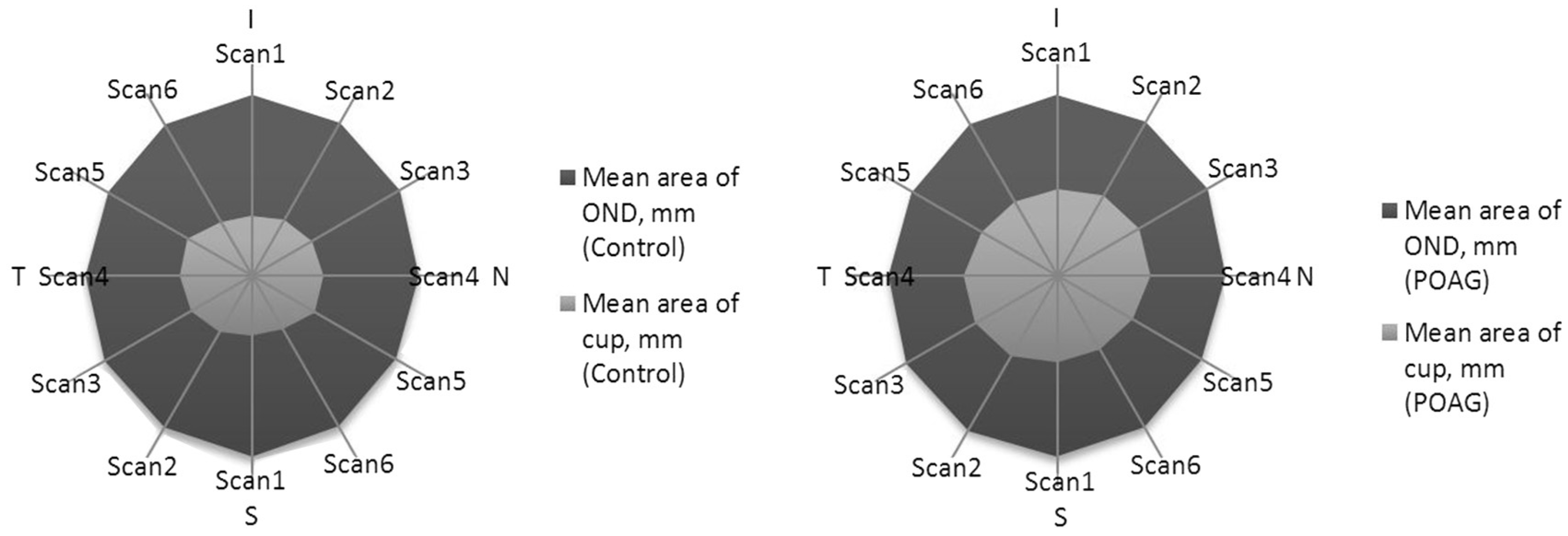
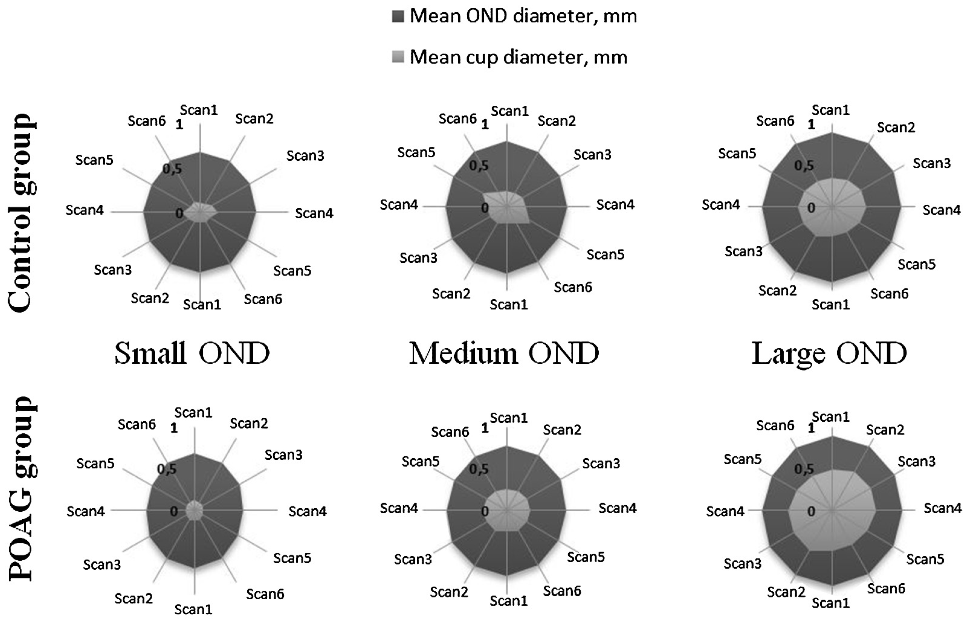
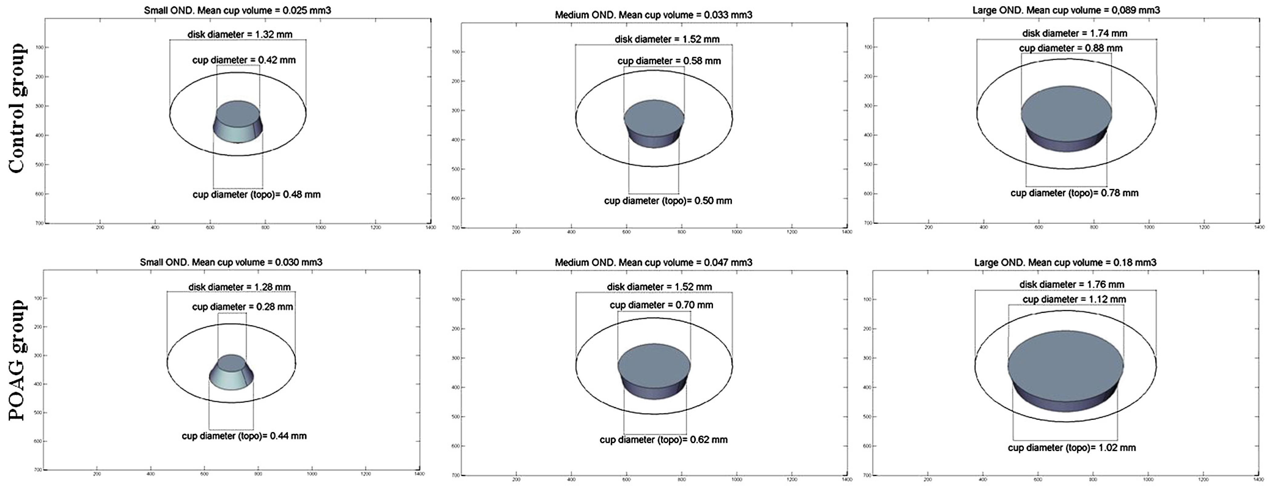
Data were expressed as mean values and standard deviations. Continuous variable normality assumption was verified using the Kolmogorov–Smirnov test. A comparison between and within the control and the POAG group were done with the parametric Student t and nonparametric Mann–Whitney–Wilcoxon tests based on distribution pattern of the data. An analysis of variance (ANOVA) was used to find whether or not the means of several groups were equal. Differences in categorical variables were determined by post hoc test analysis. The linear relation of the variables was evaluated using the correlation coefficient. According to the distribution of variables, we used Pearson or Spearman correlation coefficient. All statistical analysis was performed using the Statistical Package for Social Sciences (SPSS Inc., IL, version 18 for Windows). Statistical significance was defined as a P value less than 0.05. The results were presented as 2D models and as three-dimensional (3D) structures, using computer graphics. The OD and the cup diameters and areas were taken from the results of individual and composite scans measurements and expressed as the mean diameters and areas.

3. Results

Totally, 697 subjects were enrolled in the study. The control group was formed by healthy participants (71.6% of all the subjects). The case group included POAG patients −28.4% of all the subjects (Table 1). Stage I glaucoma was confirmed by two Humphrey SITA Standard (24-2) tests and was found in 82.3% of all POAG subjects.
The influence of age, gender, size of the OD and the ocular AL on morphometric parameters of the OD were analyzed. The mean OD area and the mean optic cup area were significantly larger in the POAG group compared to the control group. The mean NRR area was significantly larger in the control group (Table 2).
The mean OD areas were not significantly different within the control and the POAG group and between these groups when comparing the results among the four different age groups.
There was no significant difference of the NRR and the mean cup areas between the different age groups in the control group, but these values were significantly different between the age groups in the POAG group. The mean NRR area in the group of less than 60 years was larger than in the groups of 66–70 and older than 70 years. The mean cup area in the group of less than 60 years was smaller than in the other age groups (Table 2).
The mean OD area of the males was significantly larger than the females in the control group, but smaller in the POAG group. There was no significant difference of the mean OD area of the males between the control and the POAG groups. The mean OD area of the females was significantly larger in the POAG group. There was no significant difference of the NRR and the mean cup areas within the genders in the control and the POAG groups (Table 2). The NRR area size of female was significant larger in the stage I POAG group compared to the stage II.
The mean cup area was significantly smaller within the small area discs than in the medium and the large area discs, both in the control and the POAG groups. The cup area size of the large OD was significant smaller in the stage I POAG group compared to the stage II.
The mean NRR area was significantly smaller in the small area discs only in the control group. But NRR area size of the large OD was significant larger in the stage I POAG group compared to the stage II.
There were a significant linear correlation between the mean OD area and the mean cup area (Spearman r = 0.6), also between the mean OD area and the mean NRR area (Spearman r = 0.7). According to this correlation, linear equations were derived to illustrate the associations between the cup as well as NRR areas with the OD’s area size of the control group:
Cup area = −0:07 + 0:24 ∗ OD area
NRR area = 0:94 + 0:255 ∗ OD area
The mean cup area and the mean NRR area of the small discs had no significant difference between the control and the POAG groups (Table 2). Therefore it is difficult to diagnose glaucoma when the disc area is small, i.e., smaller than 1.5 mm2.
There was no significant difference of the mean cup area between the different ranges of the ocular AL. But the longer was the AL, the significantly smaller were OD and mean NRR areas in the control and POAG groups (except for mean OD area between short and medium ocular AL in POAG group) (Table 2). It was interesting to notice that mean OD area was significantly larger in POAG group when there was the medium AL. It was found significant larger OD area size in cases of long ocular AL of the stage I POAG group compared to the II stage.
In general, in our study, the longer was the ocular AL, the smaller were OD and mean NRR areas.
The shape of the OD based on OCT scans was studied: length variations of six different scans of the OD and the cup in the control and the POAG groups were analyzed. The statistical analysis of these scans described the shapes of the OD and the cup.
There was no significant difference (P > 0.05) of the OD diameters (scans 1–6) between the control and the POAG groups. All six cup diameters were significantly smaller in the control group than in the POAG group (Fig. 3).
There was no significant relationship between the diameters (scans 1–6) of the cup and different age in both groups (ANOVA, P > 0.05) (Table 3). But, there was a significant difference in the cup diameter between the control and the POAG groups (Table 3). In the POAG group, the contour of the cup was in average longer than in the control group and had a tendency to progress as the age increased (Table 3).
The OD diameters (scan 1–6) of the males and the females were not statistically significantly different between the study groups (Table 3). The cup diameters (scan 1–6) between the male and the female genders were almost the same in the control and the POAG groups (ANOVA, P > 0.05) (Table 3).
The horizontal (scan 4) diameter of the small OD was significantly longer in the control than in the POAG group. All the other diameters (scan 1–6) of the small, medium and large OD had no significant difference between the study groups. The shape of the medium and large OD did not differ significantly between the study groups (Fig. 4).
There were no significant differences in the cup diameter (scan 1–6) between the control and the POAG groups when the OD area was small. So there was no difference of the cup form of the small OD between the control and the POAG groups. But all the cup diameters (scan 1–6) of the medium and the large discs were significantly different between the control and the POAG groups, except the oblique (scan 5) diameter in the medium disc group (Fig. 4).
When comparing the cup diameters (scan 1–6) between the small, the medium and the large OD in each group, they were significantly different, except the oblique (scan 5) diameter in the control group. According to the post hoc test, the cup diameters (scan 1–6) of the small OD were not significantly shorter than the cup diameters of medium discs, but significantly shorter than the cup diameters of large discs in both study groups. Also post hoc test showed that the cup diameters of medium discs were significantly shorter than the cup diameters of large discs in both study groups, except the oblique (scan 5) cup diameter of the medium and the large discs in the control group. So as the area of OD increased, diameters of the cup also significantly increased in both study groups (Fig. 4).
The structural 3D cup illustration of the small, the medium and the large OD is presented in (Fig. 5).

4. Discussion

The evaluation of the OD size in clinical work is an essential part to diagnose and manage glaucoma disease. It enhances assessment of the features of the OD, such as the NRR and the cup areas that are necessary for accurate glaucoma diagnosis and monitoring [22]. It was reported that risk of glaucoma was independent of the OD size in white patients [22]; in our study the OD mean area, based on TD-OCT, was significantly larger in the POAG group. Limitation of the results can be explained: it is known from the literature that Stratus OCT OD parameters, which are based on locating the termination of the retinal pigment epithelium, have been reported as sensitive parameters in discriminating between glaucomatous and healthy eyes [23]. OCT cross-sectional definition can overestimate the disc size due to the inclusion of peripapillary atrophy and diffuse the RNFL loss in patients with diffuse glaucomatous damage [24,25]. According to the data of this study, the mean area of the cup was significantly larger in the POAG group, so the mean NRR area was significantly larger in the control group.
The age did not appear to be associated with the OD size in humans [22]. Likewise in our study, the age did not influence the OD mean area in both study groups also the NRR and the cup mean areas in the control group. But NRR mean area significantly decreased and the cup mean area significantly increased with the age in the POAG group. The physiological decline of the NRR and increase in the cup areas are parallel to the loss of optic nerve axons in glaucoma. It still remains difficult to discriminate physiological from pathological change [9]. It was found out that in healthy eyes the NRR area in average annually decreases by 0.28–0.39 percent, and in the case of glaucomatous changes of the OD by 0.95 percent and is significantly smaller than in the healthy group [26,27].
The influence of the gender on the disc size is debatable [9]. We found out that the mean OD area of the male gender was larger in the control group while the mean OD area of the female gender was larger in the POAG group and between the study groups. So the role of a gender as a risk factor for glaucoma is considerable, but in the Blue Mountains Eye Study was reported a higher prevalence of glaucoma in women [22], as in our study.
There was a significant linear correlation relating the NRR and the cup areas to the OD areas of the subjects in the control group of this study. Jonas et al. [28] also found out that the OD area was significantly and positively correlated with the size of the optic cup area and the NRR area in the Vellore Eye Study. The mean cup area and the mean NRR area of the small discs had no significant difference between the control and the POAG groups in our study. It was found out that it is difficult to diagnose glaucoma when the disc area is small, i.e., less than 1.5 mm2. In the morphological diagnosis of glaucoma this feature has to be taken into account. Early or moderately advanced glaucomatous OD damage may erroneously be overlooked in the small OD with relatively small optic cups, if one does not take into account that the small optic discs normally have no optic cup [28].
The interface of the OD parameters with the ocular AL is not clearly defined in the literature [8], [28]. In this study, it was evaluated that the mean cup area did not depend on the ocular AL, but the longer was the ocular AL, the significantly smaller was mean OD area and mean NRR area in the control and the POAG group.
According to this study data, the OD had the vertically oval shape in the control and the POAG groups. The result of this study correlated with the Vellore Eye Study where Jonas et al., identified that the shape of the OD was slightly vertically oval with the vertical disc diameter being about 6% longer than the horizontal disc diameter in the control group [28]. We found out that the OD had the irregular shape, i.e., it was vertically-oblique in the control and the POAG groups based on TD-OCT. We also found out that the cup had the horizontally oval shape in the control group and the vertically oval shape in the POAG. The cup shape was irregular based on TD-OCT data, i.e., it was horizontally-oblique in the control group and had more vertically-oblique shape in the POAG group.
The diameters (scans 1–6 of OCT) and the shape of the OD and the cup had no significant relation with the age groups and gender in both research groups. According to our study data we consider that these risk factors i.e., age and gender are not significant in diagnosing and monitoring glaucoma.
The structural 2D visualization demonstrated that the OD shape is more correctly oval of the small disc in the control group and more vertically oval of the small disc in the POAG group. The shape of the OD of the medium and the large discs did not significantly differ between the study groups (Fig. 4). Based on our study we assume that the small OD shape can be one of the indicators of glaucoma.
There was no significant difference in the cup diameter and shape between the control and the POAG groups when the OD area was small. So glaucoma diagnostic becomes complicated when the OD area is small.
We have obtained estimated OD morphometric parameters, i.e., size and shape, due to age, gender, size of the disc and ocular axial length, in the case–control study. The advantage of the present study was a large-scale research and the significant results which may serve as a reference for making clinical decision in differential glaucoma diagnostics. This study was limited at a time by using TD-OCT and as newer technologies like spectral-domain or swept-source OCT are currently available. Further studies are necessary to improve more detailed three-dimensional evaluations of the OD.

5. Conclusions

Based on TD-OCT, the mean OD, cup and NRR areas differed significantly between the control and POAG groups. The shape of the OD was vertically-oblique in the both study groups without significant difference. All cup diameters were significantly smaller in the control than in the POAG group and the cup had horizontally-oblique shape in the control group and vertically-oblique shape in the POAG group.
The mean OD area and shape in both study groups, the mean cup area and shape and the mean NRR area in the control group were independent of age. However, the mean NRR area significantly decreased and the mean cup area significantly increased with the age in the POAG group.
The mean OD area of the male was significantly larger in the control group. While the mean OD area and shape, i.e., the contour, of the women were significantly larger in the POAG group. The mean NRR area, cup area and shape were independent of the gender.
The structural 2D OD shape of the small OD was more regular round in the control group and more vertical round in the POAG group. The shape of the medium and the large OD did not differ between the study groups. But the mean cup area and the shape, the mean NRR area of the small OD had no significant difference between the control and the POAG groups.
The mean cup area did not depend on the ocular AL, but the longer was the ocular AL, the significantly smaller were the mean OD area and diameters and the mean NRR area in the study groups.
Data of the study has shown that based on morphometrical parameters obtained by TD-OCT it is possible to discriminate healthy and glaucomatous ODs, but further studies are needed including different stages of glaucoma and different technologies for OD morphometrics. OCT image analysis allows defining parameters that are specific for normal and case groups, and in this way it can be possible to facilitate early prediction of the disease.

Author’ Contributions

D.B. has made contributions to acquisition, analysis, interpretation of data and drafted the manuscript. A.K.Z. has made contribution to analysis, interpretation of data and drafted the manuscript. L.K. has made contributions to acquisition of data. V.B. has made contributions to acquisition of data, has been involved in the design of the study. I.J. participated in the design of the study, has been involved in revising the manuscript. A.P. participated in the design of the study. All the authors read and approved the final manuscript.

Conflicts of Interest

The authors declare that they have no conflict of interest.

R E F E R E N C E S

  1. Sanfilippo, PG; Cardini, A; Hewitt, AW; Crowston, JG; Mackey, DA. Optic disc morphology – rethinking shape. Prog Retin Eye Res 2009, 28, 227–48. [Google Scholar] [CrossRef] [PubMed]
  2. Leung, CK; Chan, W-M; Hui, Y-L; Yung, W-H; Woo, J; Tsang, M-K; et al. Analysis of retinal nerve fiber layer and optic nerve head in glaucoma with different reference plane offsets, using optical coherence tomography. Invest Ophthalmol Vis Sci 2005, 46, 891–9. [Google Scholar] [CrossRef] [PubMed]
  3. Arvind, H; George, R; Raju, P; Ve, RS; Mani, B; Kannan, P; et al. Neural rim characteristics of healthy south Indians: the Chennai glaucoma study. Invest Ophthalmol Vis Sci 2004, 49, 3457–46. [Google Scholar] [CrossRef] [PubMed]
  4. Bourne, RR. The optic nerve head in glaucoma. Community Eye Health 2006, 19, 44–5. [Google Scholar] [PubMed]
  5. Weinreb, RN; Khaw, PT. Primary open-angle glaucoma. Lancet 2004, 363, 1711–20. [Google Scholar] [CrossRef]
  6. Kwon, YH; Fingert, JH; Kuehn, MH; Alward, WLM. Primary open-angle glaucoma. New Engl J Med 2009, 360, 1113–24. [Google Scholar] [CrossRef] [PubMed]
  7. Bourne, RR; Foster, PJ; Bunce, C; Peto, T; Hitchings, RA; Khaw, PT; et al. The morphology of the optic nerve head in the Singaporean Chinese population (the TanjongPagar study): part 1 – optic nerve head morphology. Br J Ophthalmol 2009, 92, 303–9. [Google Scholar] [CrossRef] [PubMed]
  8. Bourne, RR; Foster, PJ; Bunce, C; Peto, T; Hitchings, RA; Khaw, PT; et al. The morphology of the optic nerve head in the Singaporean Chinese population (the Tanjong Pagar study): part 2 – biometric and systemic associations. Br J Ophthalmol 2008, 92, 310–4. [Google Scholar] [CrossRef] [PubMed]
  9. Garway-Heath, D; Wollstein, G; Hitchings, R. Aging changes of the optic nerve head in relation to open angle glaucoma. Br J Ophthalmol 1997, 81, 840–5. [Google Scholar] [CrossRef] [PubMed]
  10. Hellström, A; Svensson, E. Optic disc size and retinal vessel characteristics in healthy children. Acta Ophthalmol Scand 1998, 76, 260–7. [Google Scholar]
  11. Wang, Y; Xu, L; Zhang, L; Yang, H; Ma, Y; Jonas, JB. Optic disc size in a population based study in northern China: the Beijing eye study. Br J Ophthalmol 2006, 90, 353–6. [Google Scholar] [CrossRef] [PubMed]
  12. Garway-Heath, DF; Hitchings, RA. Quantitative evaluation of the optic nerve head in early glaucoma. Br J Ophthalmol 1998, 82, 352–61. [Google Scholar] [CrossRef] [PubMed]
  13. Moya, F; Brigatti, L; Caprioli, J. Effect of aging on optic nerve appearance: a longitudinal study. Br J Ophthalmol 1999, 83, 567–72. [Google Scholar] [CrossRef] [PubMed]
  14. Garway-Heath, D; Ruben, S; Viswanathan, A. Vertical cup/disc ratio in relation to optic disc size: its value in the assessment of the glaucoma suspect. Br J Ophthalmol 1998, 82, 1118–24. [Google Scholar] [CrossRef] [PubMed]
  15. Samarawickrama, C; Pai, A; Tariq, Y; Healey, PR; Wong, TY; Mitchell, P. Characteristics and appearance of the normal optic nerve head in 6-year-old children. Br J Ophthalmol 2012, 96, 68–72. [Google Scholar] [CrossRef] [PubMed]
  16. Galloway, RL. The process and development of image-guided procedures. Annu Rev Biomed Eng 2001, 13, 83–108. [Google Scholar] [CrossRef] [PubMed]
  17. Fujimoto, JG; Pitris, C; Boppart, SA; Brezinski, ME. Optical coherence tomography: an emerging technology for biomedical imaging and optical biopsy. Neoplasia 2000, 2, 9–25. [Google Scholar] [CrossRef] [PubMed]
  18. Buteikiene, D; Paunksnis, A; Barzdziukas, V; Zaliuniene, D; Balciunienė, JV; Jegelevicius, D. Correlations between digital planimetry and optical coherence tomography, confocal scanning laser ophthalmoscopy in assessment of optic disc parameters. Medicina (Kaunas) 2012, 48, 150–8. [Google Scholar] [CrossRef] [PubMed]
  19. Mills, RP; Budenz, DL; Lee, PP; Noecker, JR; Siegartel, LR; et al. Categorizing the stage of glaucoma from pre-diagnosis to end-stage disease. Am J Ophthalmol 2006, 1, 24–30. [Google Scholar] [CrossRef] [PubMed]
  20. Oduntan, OA; Mashige, KP; Raliavhegwa-Makhado, MA. Comparison of two methods of log MAR visual acuity data scoring for statistical analysis. S Afr Optom 2009, 68, 155–63. [Google Scholar]
  21. Oliveira, C; Harizman, N; Girkin, CA; Xie, A; Tello, C; Liebmann, JM; et al. Axial length and optic disc size in normal eyes. Br J Ophthalmol 2007, 91(1), 37–9. [Google Scholar] [CrossRef] [PubMed]
  22. Hoffmann, EM; Zangwill, LM; Crowston, JG; Weinreb, RN. Optic disk size and glaucoma. Surv Ophthalmol 2007, 52, 32–49. [Google Scholar] [PubMed]
  23. Manassakorn, A; Ishikawa, H; Kim, JS; Wollstein, G; Bilonick, RA; Kagemann, L; et al. Comparison of optic disc margin identified by color disc photography and high-speed ultrahigh-resolution optical coherence tomography. Arch Ophthalmol 2008, 126, 58–64. [Google Scholar] [CrossRef] [PubMed]
  24. Kim, JS; Manassakorn, A; Wollstein, G; Bilonick, RA; Kagemann, L; Sung, KR; et al. Comparison of optic disc margin identified by planimetry and high speed ultrahigh-resolution optical coherence tomography (Spectral OCT, SOCT). ARVO’06. Association for Research in Vision and Ophthalmology, 2006; poster 3642-B243. [Google Scholar]
  25. Choong, YF; Rakebrandt, F; North, RV; Morgan, JE. Acutance, an objective measure of retinal nerve fibre image clarity. Br J Ophthalmol 2003, 87, 322–6. [Google Scholar] [CrossRef] [PubMed]
  26. Laemmer, R; Schroeder, S; Martus, P; Viestenz, A; Mardin. Quantification of neuroretinal rim loss using digital planimetry in long-term follow-up of normals and patients with ocular hypertension. J Glaucoma 2007, 16, 430–6. [Google Scholar] [CrossRef] [PubMed]
  27. Mardin, CY; Horn, F; Viestenz, A; mmer R, J&#xFC; nemann, A. Healthy optic discs with large cups – a diagnostic challenge in glaucoma. Klin Monbl Augenheilkd 2006, 223, 3080–14. [Google Scholar]
  28. Jonas, JB; Thomas, R; George, R; Berenshtein, E; Muliyil, J. Optic disc morphology in south India: the Vellore eye study. Br J Ophthalmol 2003, 87, 189–96. [Google Scholar] [CrossRef] [PubMed]
Fig. 1. An image of the optic disc.
Medicina 53 00242 g001
Fig. 2. Six equally spaced radial line scans of the OD of the right eye. The outputs of the six OD scans with results of individual scan measurements and composite schematic two-dimensional image of the OD, the NRR and the optic cup (constructed from all six scans) acquired by the Fast Optic Disc protocol and analyzed by Optic Nerve Head protocol.
Medicina 53 00242 g002
Fig. 3. Visualization of the mean OD area and the mean cup area and their shapes of the control (a) and POAG (b) groups.
Medicina 53 00242 g003
Fig. 4. OD and the cup shape according to the disc area size (scale in mm).
Medicina 53 00242 g004
Fig. 5. Structural 3D form of the cup of the small, the medium and the large OD in both study groups.
Medicina 53 00242 g005
Table 1. Characteristics of the study population.
CharacteristicGroupP
ControlPOAG
N = 499N = 198
(n* = 998)(n* = 394)
Age, years 65.3 ± 8.565.1 ± 9.00.8
Male/female, %38.3/61.726.8/73.2
BCVA by Log MAR 0.15 ± 0.210.19 ± 0.210.004
Spherical equivalent of refraction, diopter1.03 ± 2.240.52 ± 2.290.001
Eye axial length, mm23.14 ± 1.2423.2 ± 1.110.4
Values are mean ± standard deviation unless otherwise indicated. BCVA by Log MAR, best corrected visual acuity by logarithm of the minimum angle of resolution; N, number of subjects; n*, number of eyes.
Table 2. The influence of age, gender, size of disc and ocular AL on morphometric parameters of OD.
ParameterControl groupP1POAG groupP2P
Optic disc area size, mm22.14 ± 0.39 2.19 ± 0.43 0.02
Age0.3
<60 years2.10 ± 0.40.22.15 ± 0.410.3*,**
60–65 years2.15 ± 0.352.21 ± 0.520.2***0.3
66–70 years2.12 ± 0.372.21 ± 0.430.9****,******0.1
>70 years2.16 ± 0.422.21 ± 0.41 0.2
Male2.17 ± 0.380.032.11 ± 0.400.020.2
Female2.11 ± 0.402.22 ± 0.440.02<0.001
Ocular AL
Short2.20 ± 0.400.04*2.16 ± 0.360.06*0.4
Medium2.14 ± 0.37<0.001**2.26 ± 0.440.008**<0.001
Long1.86 ± 0.43<0.001***1.97 ± 0.49<0.001***0.2
NRR area size, mm21.60 ± 0.38 1.33 ± 0.41 <0.001
Age
<60 years1.58 ± 0.310.41.43 ± 0.370.05*<0.001
60–65 years1.64 ± 0.531.31 ± 0.440.04** 0.02*** 0.9****,******<0.001
66–70 years1.59 ± 0.331.30 ± 0.48 <0.001
>70 years1.6 ± 0.361.3 ± 0.37 <0.001
Male 1.60 ± 0.360.81.29 ± 0.430.2<0.001
Female1.60 ± 0.401.35 ± 0.400.02<0.001
OD
Small 1.18 ± 0.24 1.14 ± 0.270.1–0.70.7
Medium1.48 ± 0.26<0.001*,**,***1.33 ± 0.31<0.001
Large1.69 ± 0.36 1.35 ± 0.45<0.001
Ocular AL
Short 1.71 ± 0.390.001*,**,***1.44 ± 0.400.01*<0.001
Medium1.59 ± 0.361.32 ± 0.42<0.001**<0.001
Long1.34 ± 0.41.16 ± 0.360.01***0.01
Cup area size, mm20.55 ± 0.41 0.86 ± 0.58 <0.001
Age
<60 years0.52 ± 0.370.30.72 ± 0.480.01*, 0.03**<0.001
60–65 years0.54 ± 0.410.93 ± 0.650.01***<0.001
66–70 years0.52 ± 0.380.91 ± 0.640.8***,****<0.001
>70 years0.58 ± 0.460.91 ± 0.550.9******<0.001
Male 0.58 ± 0.410.10.82 ± 0.530.4<0.001
Female0.53 ± 0.410.87 ± 0.60<0.001
OD
Small 0.21 ± 0.180.05*0.16 ± 0.250.03*0.4
Medium0.33 ± 0.26<0.001**,***0.46 ± 0.30<0.001**,***<0.001
Large0.69 ± 0.43 1.1 ± 0.55 <0.001
Ocular AL
Short 0.52 ± 0.430.1* 0.9** 0.5***0.72 ± 0.510.001* 0.4** 0.1***<0.001
Medium0.56 ± 0.400.94 ± 0.6<0.001
Long0.53 ± 0.470.81 ± 0.560.003
Values are mean ± standard deviation.
P, between the control and the POAG groups; P1, within the control group; P2, within the POAG group. Multiple comparisons between different age groups: *between <60 and 60–65, **between <60 and 66–70, ***between <60 and >70, ****between 60–65 and 66–70, *****between 60–65 and >70, ******between 66–70 and >70; between different disc area size groups: *between small and medium, **between small and large, ***between medium and large; between different ocular ALs: *between <22.5 mm and 22.5–24.5 mm, **between <22.5 mm and >24.5 mm, ***between 22.5–24.5 mm and >24.5 mm.
Table 3. The change of the mean diameters of the cup in different age groups and dependency of the mean diameters of the OD and the cup in genders in the control and the POAG groups.
ScansControl groupP1POAG groupP2P
Scan 1
Cup diameter by age
<60 years0.58 ± 0.380.40.76 ± 0.460.6<0.001
60–65 years0.60 ± 0.43 0.86 ± 0.50 <0.001
66–70 years0.52 ± 0.44 0.82 ± 0.56 <0.001
>70 years0.57 ± 0.46 0.84 ± 0.50 <0.001
OD diameter, mm
Male1.71 ± 0.180.951.66 ± 0.17<0.0010.024
Female1.71 ± 0.20 1.74 ± 0.20 0.02
Cup diameter, mm
Male0.58 ± 0.440.720.77 ± 0.470.23<0.001
Female0.57 ± 0.42 0.84 ± 0.51 <0.001
Scan 2
Cup diameter by age, mm
<60 years0.60 ± 0.430.50.78 ± 0.480.1<0.001
60–65 years0.63 ± 0.45 0.89 ± 0.52 <0.001
66–70 years0.56 ± 0.46 0.87 ± 0.57 <0.001
>70 years0.62 ± 0.49 0.96 ± 0.50 <0.001
OD diameter
Male1.68 ± 0.180.501.65 ± 0.180.070.15
Female1.67 ± 0.19 1.71 ± 0.20 0.005
Cup diameter
Male0.62 ± 0.480.720.84 ± 0.490.37<0.001
Female0.61 ± 0.45 0.89 ± 0.53 <0.001
Scan 3
Cup diameter by age, mm
<60 years0.67 ± 0.420.80.82 ± 0.470.20.02
60–65 years0.68 ± 0.46 0.97 ± 0.48 <0.001
66–70 years0.64 ± 0.43 0.86 ± 0.56 0.02
>70 years0.66 ± 0.48 0.94 ± 0.48 <0.001
OD diameter, mm
Male1.64 ± 0.190.031.61 ± 0.250.340.28
Female1.61 ± 0.19 1.67 ± 0.20 <0.001
Cup diameter, mm
Male0.67 ± 0.460.460.87 ± 0.480.49<0.001
Female0.65 ± 0.44 0.91 ± 0.50 <0.001
Scan 4
Cup diameter by age, mm
<60 years0.69 ± 0.390.90.80 ± 0.420.10.01
60–65 years0.67 ± 0.42 0.95 ± 0.48 <0.001
66–70 years0.68 ± 0.42 0.87 ± 0.53 0.05
>70 years0.68 ± 0.45 0.93 ± 0.46 <0.001
OD diameter, mm
Male1.59 ± 0.190.051.58 ± 0.220.290.51
Female1.57 ± 0.19 1.60 ± 0.22 0.013
Cup diameter, mm
Male0.70 ± 0.430.240.87 ± 0.450.64<0.001
Female0.67 ± 0.42 0.90 ± 0.48 <0.001
Scan 5
Cup diameter by age, mm
<60 years0.65 ± 0.370.60.76 ± 0.410.20.02
60–65 years0.65 ± 0.39 0.88 ± 0.48 <0.001
66–70 years0.60 ± 0.40 0.86 ± 0.49 <0.001
>70 years0.82 ± 3.35 0.85 ± 0.42 0.91
OD diameter, mm
Male1.59 ± 0.180.041.57 ± 0.190.190.30
Female1.57 ± 0.18 1.60 ± 0.20 0.013
Cup diameter, mm
Male0.83 ± 3.250.130.81 ± 0.400.590.96
Female0.62 ± 0.39 0.84 ± 0.46 <0.001
Scan 6
Cup diameter by age, mm
<60 years0.60 ± 0.370.40.73 ± 0.440.30.08
60–65 years0.61 ± 0.40 0.84 ± 0.52 0.001
66–70 years0.54 ± 0.40 0.81 ± 0.49 <0.001
>70 years0.59 ± 0.42 0.84 ± 0.45 <0.001
OD diameter, mm
Male1.66 ± 0.180.361.61 ± 0.190.0030.10
Female1.65 ± 0.19 1.68 ± 0.21 0.045
Cup diameter, mm
Male0.60 ± 0.400.450.76 ± 0.440.220.001
Female0.58 ± 0.40 0.82 ± 0.48 <0.001
Values are mean ± standard deviation.
P, between the control and POAG groups; P1, within the control group; P2, within the POAG group (ANOVA).

