Next Article in Journal
Pathologic fracture of the thoracic spine in a male master ultra-marathoner due to the combination of a vertebral hemangioma and osteopenia
Previous Article in Journal
Ethnic variation in self-rated health–mortality association: Results from a 17-year follow-up study in Estonia
 
 
Medicina is published by MDPI from Volume 54 Issue 1 (2018). Previous articles were published by another publisher in Open Access under a CC-BY (or CC-BY-NC-ND) licence, and they are hosted by MDPI on mdpi.com as a courtesy and upon agreement with Lithuanian Medical Association, Lithuanian University of Health Sciences, and Vilnius University.
Article

Interaction of ethidium and tetraphenylphosphonium cations with Salmonella enterica cells

by
Valeryia Mikalayeva
1,2,
Sandra Sakalauskaitė
1 and
Rimantas Daugelavičius
1,*
1
Department of Biochemistry, Faculty of Natural Sciences, Vytautas Magnus University, Kaunas, Lithuania
2
Institute of Cardiology, Medical Academy, Lithuanian University of Health Sciences, Kaunas, Lithuania
*
Author to whom correspondence should be addressed.
Medicina 2017, 53(2), 122-130; https://doi.org/10.1016/j.medici.2017.04.001
Submission received: 19 December 2016 / Revised: 1 March 2017 / Accepted: 4 April 2017 / Published: 17 April 2017

Abstract

:
Background and objective: One of the main causes of bacterial resistance to antimicrobials is multidrug resistance induced by the increased efficiency of the efflux pumps. In this study we analyzed how the conditions of assay affect the efflux of indicator substrates ethidium (Et+) and tetraphenylphosphonium (TPP+) in Salmonella enterica ser. Typhimurium cells. Impact of the outer membrane permeability barrier, composition and temperature of the medium on accumulation of the indicator compounds also was analyzed. Materials and methods: The fluorescence of Et+ and Nile Red was measured using 96-well plates and a plate reader. In parallel to traditional studies of fluorescence we applied a constructed selective electrode to follow the accumulation of Et+ in S. enterica cells. Simultaneously with monitoring of Et+ concentration in the cell incubation medium, electrochemical measurements of TPP+ accumulation were performed. Furthermore, Et+ and TPP+ were used within the same sample as agents competing for the interaction with the efflux pumps. An inhibitor phenylalanyl-arginyl-β-naphtylamide (PAβN) was applied to evaluate the input of RND-family pumps in the total efflux of these indicator compounds. Results: S. enterica cells with the intact outer membrane (OM) bound very low amounts of Et+ or TPP+. Cells with the permeabilized OM accumulate considerably higher amounts of the indicator compounds at pH 8.0, but only Et+ was considerably accumulated at pH 6.5. At conditions of electrochemical monitoring accumulation of Et+ by the permeabilized cells at 37 °C was considerably faster than at 23 °C, but at the higher temperature most of the cell-accumulated Et+ was extruded back to the medium. The fluorescence of Et+ in suspension of cells incubated in 400 mmol/L Tris buffer was about twice higher compared to 100 mmol/L one. The inhibitory action of TPP+ on Et+ efflux was evident only in 400 mmol/L Tris although PAβN effectively increased Et+ fluorescence at both buffer concentrations. Conclusions: Results of our experiments indicate that ionic strength of the incubation medium influence the selectivity, the medium temperature and the assay conditions impact the kinetics of efflux. The lower accumulated amount and the weaker fluorescence of Et+ registered in slightly acidic medium indicate that ΔΨ plays a role in the accumulation of this indicator cation. The bound amount of Et+ to the de-energized or permeabilized cells considerably varies depending on the conditions and methods of de-energization or permeabilization of cells. Tris/EDTA permeabilization of the cells does not inhibit the efflux.

1. Introduction

Extrusion of drugs from cells by efflux pumps (EPs) is one of the main reasons for bacterial resistance to antibiotics [1,2]. EPs are able to eject different lipophilic and amphiphilic compounds, such as dyes, detergents, lipids, quorum sensing signal molecules. EPs are also associated with virulence [3,4,5] and biofilm formation [6]. Therefore, information about the efflux is vital for the effective usage of available antibacterial drugs and the discovery of new ones.
Lipophilic cations, such as ethidium (Et+) and tetraphenylphosphonium (TPP+), are well known EP substrates [7,8,9,10,11]. Membrane voltage (transmembrane difference of electrical potential, ΔΨ, negative inside) drives the accumulation of these cations in bacterial cytosol. The specific feature of Et+ is its affinity to DNA. Intercalation into the double helix increases fluorescence of this indicator compound [12]. Intracellular components bind low amount of the TPP+ [13] and, therefore, this compound can be used as an indicator of ΔΨ in bacteria and mitochondria.
Fluorimetric assay of cell-accumulated Et+ is one of the most popular method to study EP activity in bacteria [14,15,16,17]. The fluorescence is related to the amount of Et+ bound to bacterial DNA and the latter depends on the intracellular concentration of this indicator. Measurements of Et+ fluorescence are very convenient for studying the competition between efflux substrates [18] because it is possible to use several compounds in the same sample. However, a gradual decrease in Et+ fluorescence during the monitoring period was observed in experiments with Pseudomonas aeruginosa cells although the cell-bound Et+ is not destructed [14]. Beside this, Martins and colleagues [15] have reported about variations in the energy-dependence of Et+ efflux from Enterobacter aerogenes cells in media of different pH.
In this study we analyzed how the assay conditions affect the efflux of Et+ from Salmonella enterica ser. Typhimurium cells. To follow the accumulation of Et+ in cells, in addition to traditional measurements of fluorescence we applied potentiometric monitoring of the concentration of this indicator by the constructed selective electrode. In parallel to Et+ measurements accumulation of TPP+ in the cells was also assayed. Beside this, Et+ and TPP+ were used simultaneously within the same sample as agents competing for the interaction with EPs and the efflux inhibitor phenylalanyl-arginyl-β-naphtylamide (PAβN) was applied to evaluate the input of RND-family pumps. Results of our experiments indicate that ionic strength of the incubation medium influence the selectivity, the medium temperature and the assay conditions impact the kinetics of efflux.

2. Materials and methods

2.1. Bacteria cultivation and preparation for experiments

Salmonella enterica ser. Typhimurium strain SL1344 wild type cells were obtained from Prof. Séamus Fanning (Institute of Food and Health, University College Dublin, Ireland). Overnight culture of cells was grown in Luria-Bertani broth, containing 0.5% NaCl (Sigma–Aldrich, Munich, Germany), diluted 1:50 in fresh medium, and the incubation was continued until the OD600 reached 1.0. The cells were collected by centrifugation at 4 °C for 10 min at 3000 g (HeraeusTM MegafugeTM 16R, Thermo Scientific, Germany). The pelleted cells were re-suspended in 100 or 400 mmol/L Tris-hydro-xyaminomethane (Tris)/HCl (Roth, Karlsruhe, Germany), pH 8.0, to obtain ~4 × 10¹⁰ cells/mL. To permeabilize the outer membrane (OM), the cells were at 37 °C 10 min incubated in 100 mmol/L Tris/HCl containing 10 mmol/L ethylene diamine tetra-acetic acid (EDTA; Sharlau, Barcelona, Spain), pH 8.0, then pelleted and re-suspended as described above. Concentrated cell suspensions were kept on ice until used, but not longer than 4 h. Heat treatment of the cells was performed incubating 1 mL of the concentrated suspension in a 1.5-mL Eppendorf tube for 10 min in a boiling water bath.

2.2. Fluorescence measurements

Stock solutions of ethidium (Et+) bromide (Acros Organics, New Jersey, USA) or Nile red (NR, Sigma–Aldrich, Munich, Germany) were added to the corresponding concentrations into testtubes containing Tris buffer solution with 0.1% glucose (Sharlau, Barcelona, Spain). After mixing, stock solutions of phenylalanyl-arginyl-β-naphtylamide (PAβN) hydrochloride (Sigma–Aldrich, Munich, Germany) and/or tetraphenylphosphonium (TPP+) chloride (Fluka, St. Gallen, Switzerland) were added. Then the concentrated cell suspension was added to obtain OD600 of 1.0, the samples were mixed and within 2 min transferred into a 96-well flat-bottom black plate, 120 μL per well (n = 3). Relative intensity of the fluorescence (excitation 535 nm or 485 nm, emission 612 nm or 535 nm for Et+ or NR, respectively) was monitored in “TECAN GENios ProTM” (Männedorf, Switzerland) plate reader, thermostating the plate at 23 °C or 37 °C. The plate was shaken 5 s before each registration point. The representative curves from 3 independent experiments are presented.

2.3. Potentiometric measurements

TPP+ and Et+ concentrations in the incubation media were potentiometric monitored using selective electrodes as described [10,19]. The Et+ sensor was prepared in accordance with a well-known procedure [20] by dissolving poly(vinyl chloride) (PVC, average Mw ~80,000; Sigma–Aldrich), plasticizer dioctyl phthalate (Sigma–Aldrich), and tetraphenylborate sodium salt (Sigma–Aldrich) in freshly distilled tetrahydrofuran (Sigma–Aldrich) while stirring. The membrane components were taken in following proportions (by weight): tetraphenylborate sodium salt, 1%; plasticizer, 66%; and PVC, 33%. After tetrahydrofuran evaporation at room temperature in fume hood, the formed membrane was fixed to the sensor body, soaked overnight in 1 mmol/L Et+ bromide solution and washed with deionized water before the experiments. The sensors were stored dry at room temperature. While assembling Et+-selective electrode the sensor was filled with 0.1 mmol/L Et+ bromide in 100 mmol/L NaCl solution and connected to an internal Ag/AgCl half-cell electrode.
The thermostated magnetically stirred glass vessels were filled with 5 mL of 100 or 400 mmol/L Tris/HCl, pH 8.0 or 6.5, containing 0.1% glucose. After calibration of the electrodes the concentrated cell suspension was added to obtain an OD600 of 1. We used the electrode potential-amplifying system with an ultralow-input bias current operational amplifier AD549JH (Analog Devices, Norwood, MA, USA). The data acquisition system PowerLab 8/35 (ADInstruments, Oxford, UK) was used to connect of the amplifying system to a computer. The agar salt bridges were used for indirect connection of the Ag/AgCl reference electrodes (Thermo Inc.; Orion model 9001) and the cell suspension in vessels. The measurements were performed simultaneously in 2–4 reaction vessels. The representative sets of curves from 3 independent series of measurements are presented.

3. Results

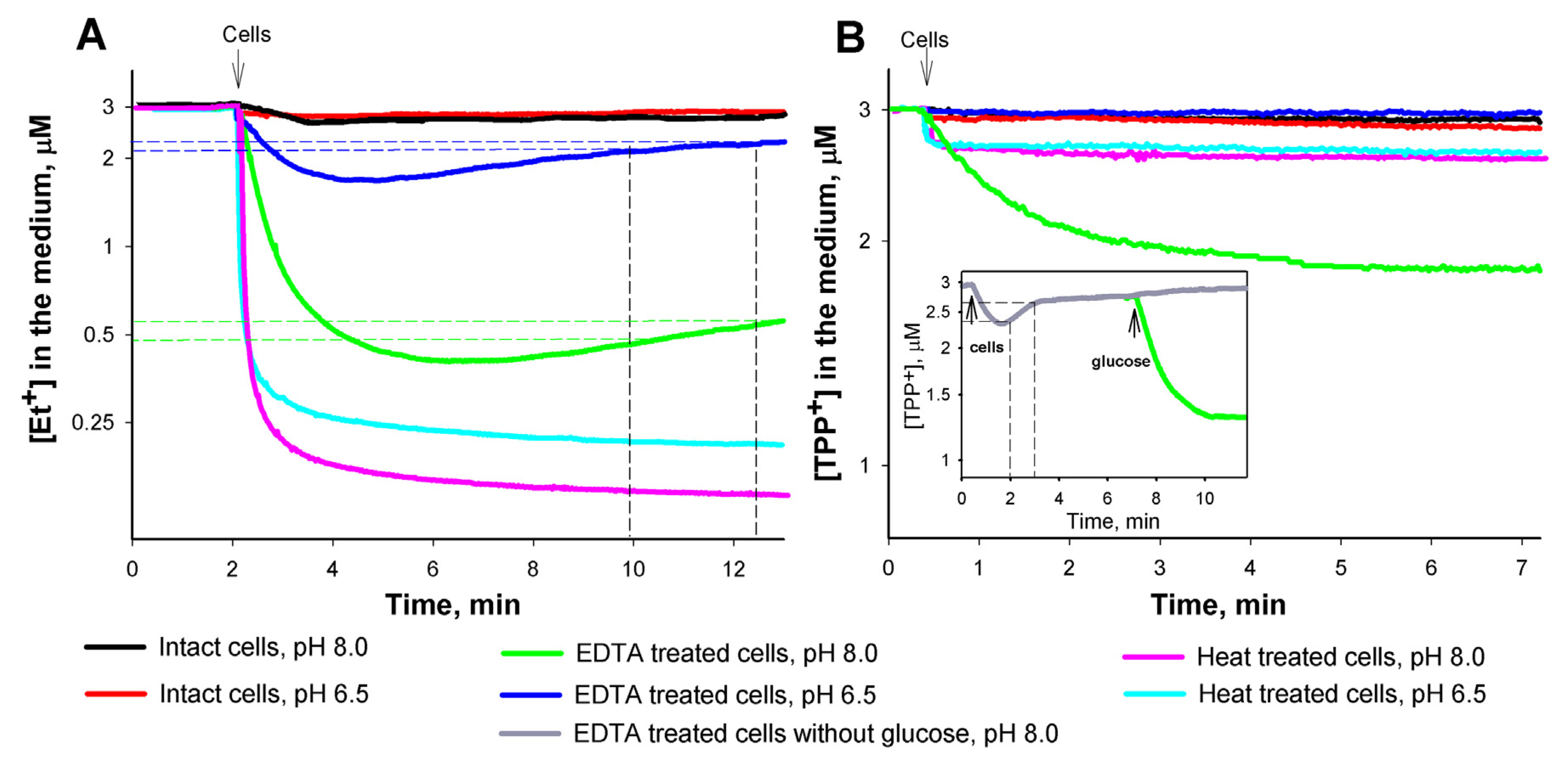
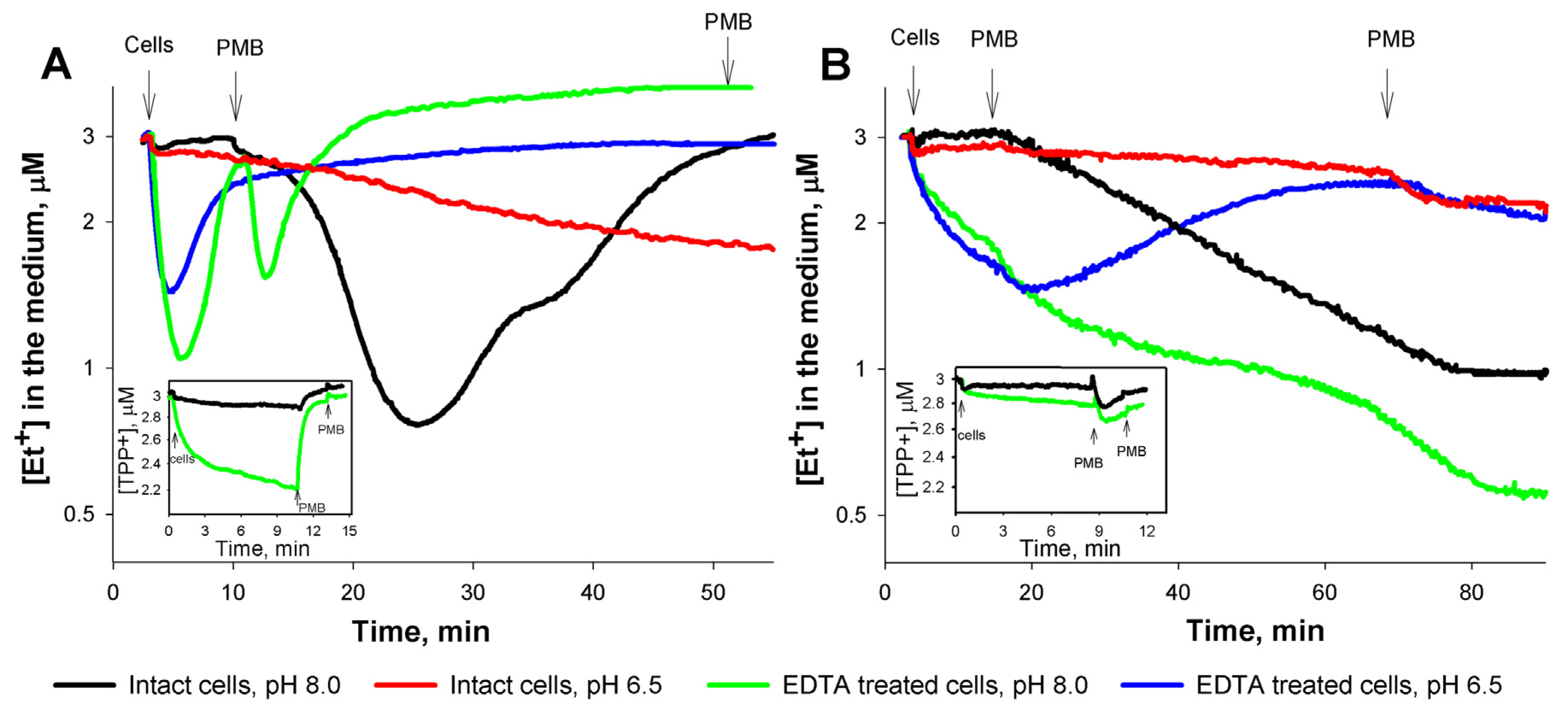
3.1. Role of the medium pH and the outer membrane (OM) permeability in the interaction of Et+ with S. enterica cells

We used 96-well plates and a plate reader to monitor Et+ fluorescence. This method allows to perform the efflux studies under conditions of controlled temperature and in media of selected composition. Also, possibility to use simultaneously several EP substrates and/or inhibitors is the advantage of this method.
Considerable difference of Et+ fluorescence was observed between suspensions of cells with the intact OM and the Tris/EDTA-permeabilized ones. In case of the permeabilized cells the initial level of Et+ fluorescence was high, especially at pH 8.0, but during the incubation at both pH it gradually decreased to the similar levels (Fig. 1B and D). In suspensions of cells with the intact OM an intensive Et+ fluorescence was observed only in the presence of RND-family efflux pump inhibitor PAβN: higher concentrations of this compound caused stronger Et+ fluorescence. At 500 μmol/L of PAβN the maximal intensity of Et+ fluorescence at pH 6.5 was less than 50% of the level registered at pH 8.0, although at low concentrations or in the absence of the inhibitor intensities of the fluorescence were similar. Permeabilization of the cell envelope using polycationic antibiotic Polymyxin B (PMB) stimulated Et+ fluorescence except when 500 μmol/L PAβN was present in pH 8.0 medium.
Availability of several methods to assess the interaction between indicator compounds and cells provides additional information on the efflux. Intensity of the fluorescence (Fig. 1) reflects DNA-bound amount of Et+. To determine amount of this indicator taken in by the cells from incubation medium we constructed selective electrodes and performed a potentiometric monitoring of the extracellular Et+ concentration.
Changes of Et+ and TPP+ concentrations in the thermostated suspensions of S. enterica cells were monitored by selective electrodes under the conditions of intense magnetic stirring. Cells with the intact OM did not bind considerable amounts of Et+ and TPP+ (Fig. 2). Tris/EDTA-permeabilized cells accumulated high amounts of the indicator compounds at pH 8.0, but only Et+ was considerably bound at pH 6.5. However, a release of the accumulated Et+ started in a few minutes (Fig. 2A). Such increase in the extracellular Et+ concentration, observed in media at both pH, is in agreement with the decrease of Et+ fluorescence registered in plate wells (Fig. 1), but proceeded much faster.
The release of the accumulated Et+ might be explained by the activation of efflux after adaptation of the cells in a warm and aerated medium. Based on changes in the extracellular concentrations, it is possible to calculate the rates of Et+ release from cells. Tris/EDTA-permeabilized cells released 0.15 ± 0.04 and 0.33 ± 0.03 nmol/min of Et+ at pH 8.0 and 6.5, respectively. During the release period the real amount of extruded Et+ should be higher because of the re-entry of this indicator back to cells. It is possible to calculate rates of the initial Et+ entry taking the same intervals of extracellular Et+ concentration as used for evaluation of the release. During the initial Et+ binding the Tris/EDTA-treated cells accumulated 0.22 ± 0.03 and 0.5 ± 0.04 nmol/min of this cation at pH 8.0 and 6.5, respectively. If rate of the re-entry during efflux is the same as rate of the initial Et+ accumulation, the total efficiency of efflux would be from 0.37 nmol/min (pH 8.0) to 0.83 nmol/min (pH 6.5). In 5 mL of the suspension at concentration of 8.3 × 10⁸ cells/mL (OD600 of 1.0) there were ~4.15 × 10⁹ cells. In such case the efflux rate was 0.9–2.0 × 10⁻¹⁹ mol/cell/min or ~900–2,000 molecules of Et+/cell/s.
In contrast to Et+, accumulation of the TPP+ at these conditions was one-stage irreversible process. Similar to Et+ reversible accumulation of TPP+ by Tris/EDTA-permeabilized cells was observed only in the absence of glucose (Fig. 2B insert). Supplement of the medium with glucose induced an additional accumulation of TPP+. The rate of TPP+ release from the cells, following the initial accumulation in the absence of glucose, was 1.45 ± 0.08 nmol/min. Taking into account the reentry of the extruded TPP+, the rate of efflux was 3.87 ± 0.1 nmol/min, corresponding to 9,300 molecules of TPP+ per cell/s. This rate is ~5 to 10 times higher than the maximal rate of Et+ efflux (see above).
Because heat treatment eliminates ion gradients and the energy-dependent processes, such cells could be considered for the role of “zero” control in the efflux studies. Addition of the heat-treated cells to the medium induced very fast and strong decrease of the extracellular concentration of Et+, but not TPP+. The amount of Et+ bound to the heated cells at pH 8.0 (14.1 nmol) was slightly, but reproducibly, higher compared to pH 6.5 conditions (13.7 nmol). Very fast binding of Et+ to the cells indicates the abolishment of barriers on the way of this compound to DNA.

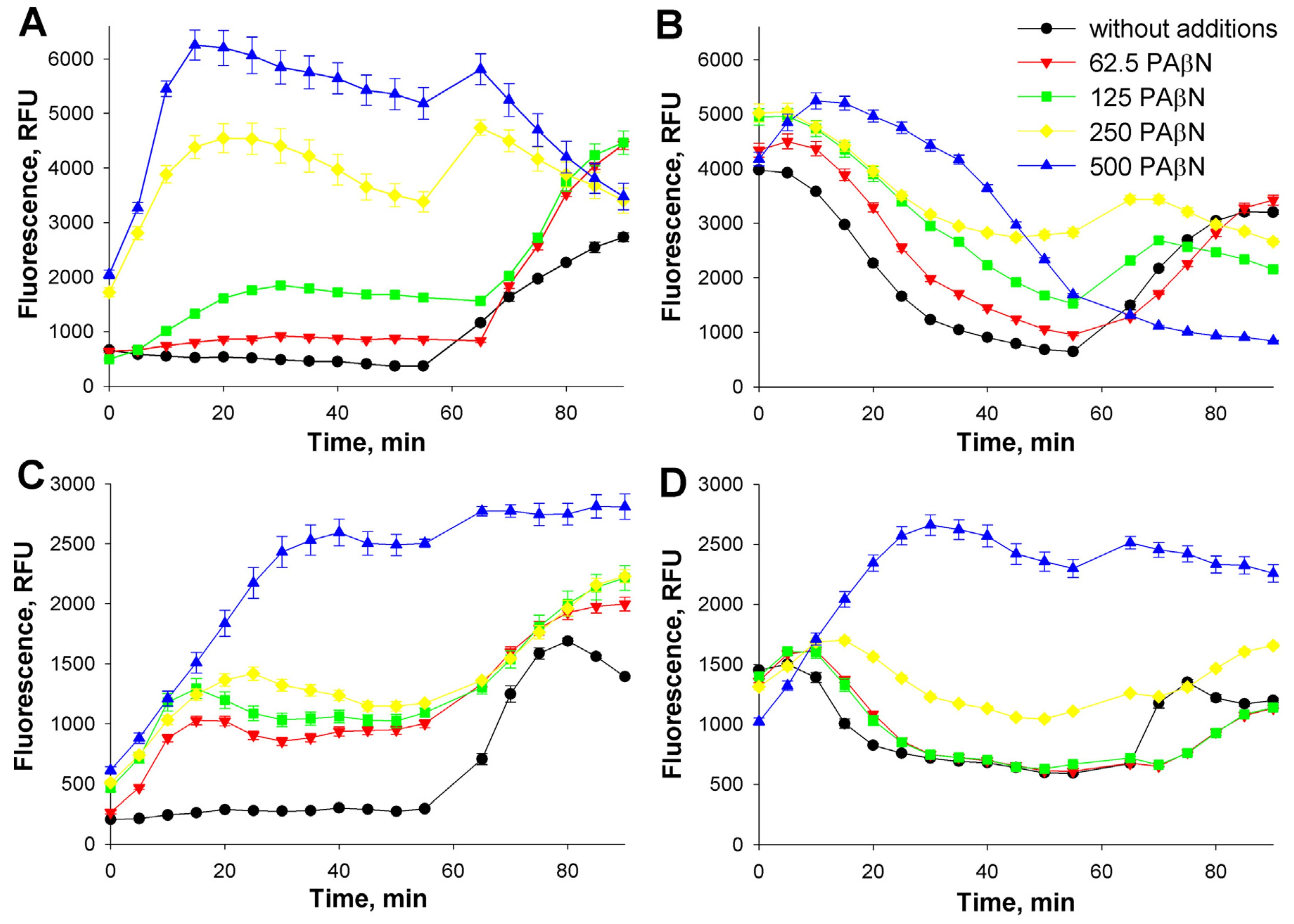
3.2. Influence of the incubation buffer concentration on the efflux

To determine the influence of incubation buffer concentration on Et+ efflux in S. enterica, the experiments were performed in 100 mmol/L and 400 mmol/L Tris/HCl buffers, pH 8.0 (Fig. 3).
In addition, the combined effects of PAβN, as the RND pump inhibitor, and TPP+, as an alternative EP substrate (competitive inhibitor of Et+ efflux), were investigated to determine possible interactions of these compounds during Et+ extrusion. In the absence of inhibitors, the maximal intensity of Et+ fluorescence was observed after 20–25 min of incubation. The fluorescence of Et+ in suspension of cells incubated in 400 mmol/L buffer was about twice higher compared to 100 mmol/L one. In the case of 100 mmol/L buffer there was no difference in the fluorescence between suspensions containing 50 μmol/L TPP+ and without this EP substrate. However, in 400 mmol/L buffer the presence of 50 μmol/L TPP+ considerably increased the fluorescence of Et+. In 100 mmol/L buffer a similar increase in Et+ fluorescence was achieved only when the extracellular TPP+ concentration was 150 μmol/L (data not shown).
In 400 mmol/L buffer (Fig. 3B) the simultaneous presence of both compounds, TPP+ and PAβN, caused higher Et+ fluorescence compared to the situations when these two Et+ efflux inhibitors were applied separately. These results indicate that more Et+ interacts with DNA when PAβN blocks RND-family EPs and TPP+ interferes with the Et+ extrusion by other efflux systems. However, in 100 mmol/L buffer the inhibitory action of TPP+ on Et+ efflux was very weak and this alternative substrate was even interfering with the inhibitory action of PAβN.

3.3. Influence of the incubation temperature on the interaction of indicator compounds with S. enterica cells

We explored the fluorescence of Et+ at two temperatures, 23 °C and 37 °C, in the same 400 mmol/L Tris/HCl buffer, pH 8.0 (Fig. 4). The levels of fluorescence during first 20 min of incubation were very close at both temperatures but at 23 °C the fluorescence was gradually increasing and after 60 min it was twice higher than at 37 °C (Fig. 4). In the presence of TPP+ at both temperatures Et+ fluorescence was increasing significantly faster and the intensity was following the TPP+ concentration. However, the effect of PAβN on the fluorescence was more distinct at 23 °C. At this temperature combination of TPP+ and PAβN increased the fluorescence more efficiently than each of these compounds separately. At both temperatures Et+ fluorescence increased after the permeabilization of cells using PMB. However, a sharp decrease of Et+ fluorescence was observed at 37 °C starting ~20th min after PMB addition (Fig. 4A). Such events cannot be explained by the more intense Et+ efflux.
Both cations, TPP+ and Et+, are accumulated in the cytosol because of the ΔΨ. For comparison, a neutral EP substrate, lipophilic stain Nile Red, was chosen. Measurements of NR fluorescence in 400 mmol/L Tris/HCl buffer indicated that this compound slowly accumulates in membranes. NR fluorescence in the presence of TPP was lower, but PAβN alone or in combination with TPP+ strengthened it (Fig. 4A insert). Permeabilization of the cell envelope by PMB induced rapid and stable increase of NR fluorescence.
Analogous experiments were performed by potentiometric monitoring of Et+ and TPP+ concentrations in the medium (Fig. 5). At both temperatures, 23 °C and 37 °C, only the permeabilized cells accumulated considerable amounts of Et+ and the initial uptake of this indicator at 37 °C was considerably faster than at 23 °C. However, after 10 min of incubation at 37 °C (but not at 23 °C) most of the cell-accumulated Et+ was extruded back to the medium. The total rate of Et+ extrusion (including the re-entry, see calculations for Fig. 2) was 5.42 ± 0.1 nmol/min at pH 6.5 and 6.00 ± 0.1 nmol/min at pH 8,0. This corresponds to extrusion of 13,000–14,400 of Et+ molecules/cell/s.
The extent and the kinetics of the PMB-induced Et+ binding were dependent on the OM permeability and the medium pH. In case of cells with the intact OM, at 37 °C PMB induced a slow accumulation of Et+, reversible at pH 8.0 but irreversible at pH 6.5. At 23 °C the efficiency of PMB was considerably lower, especially at pH 6.5. In the case of the permeabilized cells PMB induced temporary accumulation of Et+ at pH 8.0, but did not induce any additional accumulation of Et+ at pH 6.5. At 23 °C in experiments with the permeabilized cells, PMB induced a release of the accumulated Et+ at pH 6.5, but an additional accumulation of this cation at pH 8.0. Experiments with TPP+ (inserts Fig. 5) showed that PMB was able to induce an additional accumulation of TPP+ by the cells at 23 °C, but the PM depolarizing effect of PMB was clearly visible at 37 °C.
It should be mentioned that in 400 mmol/L Tris/HCl buffer the cell-accumulated amount of Et+ was considerably lower compared to the 100 mmol/L one, especially in experiments with Tris/EDTA treated cells (compare Fig. 2A and Fig. 5A). Supplement of 100 mmol/L Tris/HCl buffer, pH 8.0, with 600 mmol/L sucrose did not considerably change the amount of cell-accumulated Et+ or TPP+ (not shown). These results indicate that the increase of ionic strength, but not the osmotic pressure of incubation medium affects accumulation of the indicator cations.

4. Discussion

Measurements of Et+ fluorescence in cell suspensions is one of the most popular methods of efflux analysis in bacteria. Here we compared the accumulation of EP substrate Et+ in S. enterica cells by methods of fluorescence spectroscopy and potentiometry. Results of our experiments indicated that the quantitative measurements of the efflux following the changes in Et+ fluorescence are rather complicated. The intensity of fluorescence depends on the amount of Et+ bound to bacterial DNA and the latter is related to the intracellular concentration of this indicator. In addition to efflux, ΔΨ and the OM permeability also affect the intracellular accumulation of this indicator. Therefore, it is important to monitor continuously the interaction of this indicator compounds with the cells. For quantitative studies it is important to have control data on the state of “zero efflux”. Efflux of the indicator compounds could be switched off using the efflux pump inhibitors (e.g. PAβN), blocking the energy supply of the efflux by membrane-active compounds (e.g. PMB) or inhibitors of energy metabolism (uncouplers of oxidative phosphorylation). However, the bound amount of Et+ to the de-energized or permeabilized cells considerably varies depending on the conditions and methods of de-energization or permeabilization of cells. Beside this, the changes in the composition of the cytosol of the de-energized and permeabilized cells can affect the binding of Et+ to DNA. It is known that PMB [21], as well as Mg2+ [22] bind to the DNA decreasing Et+ fluorescence.
In parallel with Et+ accumulation measurements, we potentiometrically monitored the interaction with cells of another EP substrate TPP+. This compound is lipophilic and cationic, as Et+, but has a low affinity to intracellular components [13] and, therefore, can be used for measurements of ΔΨ in bacteria and mitochondria [23]. Interpreting results of TPP+ accumulation is more straightforward because of the low intracellular binding of this compound, but the studies using this indicator become complicated at conditions of low ΔΨ, for example, when the medium pH is 6.5 and lower.
EPs of RND family, clinically the most important in S. enterica [2], obtain energy from the proton-motive force generated as a result of cellular metabolism [1]. By selecting pH of the medium it is possible to change the inputs of the transmembrane difference of electrical potential (membrane voltage, ΔΨ) and the transmembrane difference of pH (pH gradient) without effect on the magnitude of proton-motive force [23]. However, Martins and colleagues [15] have reported about variations in the energy-dependence of Et+ efflux from Enterobacter aerogenes cells in media of different pH. Beside this, it has been shown [24,25] that E. coli cells more efficiently extrude Et+ in lower pH media.
Our experiments demonstrated that Tris/EDTA treated S. enterica cells at pH 6.5 accumulate very low amount of TPP+ indicating a low ΔΨ. The lower accumulated amount and the weaker fluorescence of Et+ registered during the first minutes of incubation of Tris/EDTA treated cells at pH 6.5 compared to pH 8.0 (Fig. 1, Fig. 2 and Fig. 5) indicate that low ΔΨ at slightly acidic medium also plays a role in the accumulation of this cation. However, at both pH values Et+ fluorescence increased with the increase of PAβN concentration in the medium indicating the active Et+ efflux. The heat-treatment could be used for determination of the maximum capacity of cells to bind Et+. The heat-treated cells at both pH values accumulated the maximal amounts of Et+ and PMB induced neither additional accumulation of Et+, nor the release of this indicator to the medium. On the other hand, lower cell envelope-permeabilizing efficiencies of PMB at acidic pH and/or in case of metabolically non-active cells should also be mentioned.
As efflux indicators Et+ and TPP+ were used rather low (1.2– 3 μmol/L) concentration in our experiments. Accumulation of high amounts of cations inside the cytosol can lead to depolarization of the PM and inhibition of the ΔΨ-dependent processes, such as ATP synthesis or efflux of lipophilic compounds. Inhibition of Et+ efflux at high concentrations of TPP+ and/or PAβN can be caused not only by the direct interaction of these compounds with EPs, but also by the depolarization of PM. In addition to this, the saturation of DNA with Et+ at high intracellular concentrations of this indicator could lead to disappearance of the correlation between the intensity of fluorescence and the intracellular Et+ concentration.
We also monitored the ability of TPP+ and PAβN to inhibit the efflux of Nile Red, a non-charged lipophilic indicator accumulating in the hydrophobic phase of membranes [26,27]. The increased NR fluorescence in the presence of PAβN and PMB can be explained by the inhibition of NR efflux, but attenuation of the permeability barrier to NR because of a high affinity of PAβN and PMB to lipopolysaccharide-containing outer surface of the OM cannot be excluded. The decrease of NR fluorescence in the presence of TPP+ could be explained by the effect of this lipophilic cation on the of membrane affinity to NR. The targets of the antibiotics usually are in the waterfilled compartments of cells, such as cytosol and periplasm. Therefore, distribution of water-soluble indicator compounds, TPP+ and Et+, between the cells and the medium should better reflect the role of efflux in accumulation of the antibiotics in bacteria.
Alongside the EPs, the OM permeability barrier is another important factor preventing the accumulation of lipophilic compounds in S. enterica cells. To explore the role of OM barrier in accumulation of the indicators inside the cells, we used Tris/EDTA-permeabilized cells in parallel to the intact ones. Tris/EDTA treatment of cells before the monitoring allowed the immediate accumulation of TPP+ and Et+ ions inside the cells. However, in 10 min after the addition of cell a decrease of Et+ fluorescence and an increase of the extracellular Et+ concentration were observed, indicating that Tris/EDTA permeabilization of the cells does not inhibit the efflux.
PMB is a very effective permeabilizer of bacterial membranes, at low concentrations increasing the permeability only of the OM [28]. This antibiotic is not an EP substrate [29] and it is convenient to use it as a permeabilizing agent. However, at 50 mg/L and higher concentrations of PMB the initial additional accumulation of Et+ is followed by the release of this indicator. The decrease in fluorescence and the increase in extracellular Et+ concentration after prolonged action of PMB could be explained by the interaction of PMB with DNA and the replacement of Et+-DNA complex by PMB-DNA one. Analogous effect of thioridazine on Et+ fluorescence was described by Spengler and colleagues [30]. Our results are in agreement with the described ones, showing that PMB [31] binds to the DNA decreasing Et+ fluorescence.
It is known that the fluidity of biological membranes decreases with the decrease of temperature. Temperature-induced deformations of the cell membrane can act as allosteric regulators of protein function [32] and this can affect the efficiency of efflux. Temperature of the incubation medium in a complex way affects accumulation of lipophilic indicators in S. enterica cells (Fig. 4 and Fig. 5). At room temperature the rate of TPP+ accumulation inside the cells is considerably reduced as compared to 37 °C temperature ([19,33] see also Fig. 5 inserts). During potentiometric measurements only Tris/EDTA treated cells accumulated Et+ at 23 °C and a considerable binding of this indicator to the intact cells started only after the addition of PMB (Fig. 5). The initial levels of Et+ fluorescence were very close at both temperatures, but during incubation at 23 °C the fluorescence was gradually increasing, especially in the presence of TPP+ and PAβN. The higher efficiency of the inhibitors at 23 °C can be explained by lower rate of Et+ entry into the cells (Fig. 5). Our data are in agreement with the protocols using room temperature for the loading of cells with Et+ or other lipophilic indicators to study the efflux at 37 °C [34]. However, Et+ accumulation stimulating effects of PAβN and TPP+ at 23 °C clearly indicate that the efflux is still active at this temperature.
Results of our previous experiments with Pseudomonas aeruginosa cells [10] showed that efflux of TPP+ is strongly dependent on the composition and the concentration of incubation buffer. However, it is not generally agreed what the composition of the incubation buffer should be during assays of the efflux activity. In our experiments the actions of PAβN and TPP+ on Et+ efflux were the incubation medium dependent. In 400 mmol/L Tris/HCl buffer the mixture of TPP+ and PAβN better prevented Et+ efflux than each of these inhibitors used separately. However, 100 mmol/L buffer the inhibitory action of TPP+ on Et+ efflux was very weak and this alternative EP substrate was even interfering with the action of PAβN. There are data that PAβN also can be a substrate of RND pumps [35,36] and, therefore, a competitive inhibitor of Et+ and TPP+ efflux. The results by Kinana and collaborators [37] suggest that this inhibitor, even when it is itself pumped out, can reduce the pumping of other compounds interfering with their binding to other parts of the binding pocket of AcrB pump. It is known [38,39] that under certain conditions PAβN not only inhibits the efflux but also permeabilizes the OM to lipophilic compounds. Beside this, activation of the energetic metabolism not only increases the efficiency of efflux but also has an impact on the amount of cations bound to the surface of Gramnegative bacteria [40]. All these facts also should be kept in mind when interpreting results of the efflux experiments.

5. Conclusions

Results of our experiments indicate that ionic strength of the incubation medium influence the selectivity, the medium temperature and the assay conditions impact the kinetics of efflux. The lower accumulated amount and the weaker fluorescence of Et+ registered at slightly acidic medium indicate that ΔΨ plays a role in the accumulation of this indicator cation. The bound amount of Et+ to the de-energized or permeabilized cells considerably varies depending on the conditions and methods of de-energization or permeabilization of cells. Tris/EDTA permeabilization of the cells does not inhibit the efflux.

Author Contributions

V.M. and R.D. conceived and designed the project. V.M. and S.S. performed the experiments and acquired the data. V.M. and R.D. wrote the paper. All authors have read and approved the final version submitted for publication.

Conflicts of Interest

All authors state that they have no any conflict of interest.

Acknowledgments

This work was supported by Research Council of Lithuania, funding grant No. MIP-040/2015.

References

  1. Nikaido, H; Pagès, JM. Broad specificity efflux pumps and their role in multidrug resistance of Gram-negative bacteria. FEMS Microbiol Rev 2012, 36, 340–63. [Google Scholar] [CrossRef] [PubMed]
  2. Li, X; Plésiat, P; Nikaido, H. The challenge of efflux-mediated antibiotic resistance in Gram-negative bacteria. Clin Microbiol Rev 2015, 28, 337–418. [Google Scholar] [CrossRef] [PubMed]
  3. Blair, J; Smith, HE; Ricci, V; Lawler, AJ; Thompson, LJ; Piddock, LJV. Expression of homologous RND efflux pump genes is dependent upon AcrB expression: implications for efflux and virulence inhibitor design. J Antimicrob Chemother 2015, 70, 424–31. [Google Scholar] [CrossRef] [PubMed]
  4. Dhanani, A; Block, G; Dewar, K; Forgetta, V; Topp, E; Beiko, RG; et al. Genomic comparison of non-typhoidal Salmonella enterica serovars Typhimurium, Enteritidis, Heidelberg, Hadar and Kentucky isolates from broiler chickens. PLOS ONE 2015, 10, e0128773. [Google Scholar] [CrossRef] [PubMed]
  5. Lin, Y; Huang, YW; Chen, SJ; Chang, CW; Yang, TC. The SmeYZ efflux pump of Stenotrophomonas maltophilia contributes to drug resistance, virulence-related characteristics, and virulence in mice. Antimicrob Agents Chemother 2015, 59, 4067–73. [Google Scholar] [CrossRef] [PubMed]
  6. Baugh, S; Phillips, CR; Ekanayaka, AS; Piddock, LJV; Webber, MA. Inhibition of multidrug efflux as a strategy to prevent biofilm formation. J Antimicrob Chemother 2014, 69, 673–81. [Google Scholar] [CrossRef] [PubMed]
  7. Midgley, M. The phosphonium ion efflux system of Escherichia coli: relationship to the ethidium efflux system and energetic studies. J Gen Microbiol 1986, 132, 3187–93. [Google Scholar] [CrossRef] [PubMed]
  8. Gros, P; Talbot, F; Tang-Wai, D; Bibi, E; Kaback, HR. Lipophilic cations: a group of model substrates for the multidrug-resistance transporter. Biochemistry 1992, 31, 1992–8. [Google Scholar] [CrossRef] [PubMed]
  9. Paixão, L; Rodrigues, L; Couto, I; Martins, M; Fernandes, P; de Carvalho, CC; et al. Fluorometric determination of ethidium bromide efflux kinetics in Escherichia coli. J Biol Eng 2009, 3, 18. [Google Scholar]
  10. Daugelavicius, R; Buivydas, A; Sencilo, A; Bamford, DH. Assessment of the activity of RND-type multidrug efflux pumps in Pseudomonas aeruginosa using tetraphenylphosphonium ions. Int J Antimicrob Agents 2010, 36, 234–8. [Google Scholar] [CrossRef] [PubMed]
  11. Ye, C; Wang, Z; Lu, W; Zhong, M; Chai, Q; Wei, Y. Correlation between AcrB trimer association affinity and efflux activity. Biochemistry 2014, 53, 3738–46. [Google Scholar] [CrossRef] [PubMed]
  12. Severini, A; Morgan, AR. An assay for proteinases and their inhibitors based on DNA/ethidium bromide fluorescence. Anal Biochem 1991, 193, 83–9. [Google Scholar] [CrossRef]
  13. Hockings, P; Rogers, PJ. The measurement of transmembrane electrical potential with lipophilic cations. Biochim Biophys Acta 1996, 1282, 101–6. [Google Scholar] [CrossRef]
  14. Babayan, A; Nikaido, H. In Pseudomonas aeruginosa ethidium bromide does not induce its own degradation or the assembly of pumps involved in its efflux. Biochem Biophys Res Commun 2004, 324, 1065–8. [Google Scholar] [CrossRef] [PubMed]
  15. Martins, A; Spengler, G; Martins, M; Rodrigues, L; Viveiros, M; Davin-Regli, A; et al. Physiological characterisation of the efflux pump system of antibiotic-susceptible and multidrug-resistant Enterobacter aerogenes. Int J Antimicrob Agents 2010, 36, 313–8. [Google Scholar] [CrossRef] [PubMed]
  16. Baylay, A; Piddock, LJ. Clinically relevant fluoroquinolone resistance due to constitutive overexpression of the PatAB ABC transporter in Streptococcus pneumoniae is conferred by disruption of a transcriptional attenuator. J Antimicrob Chemother 2015, 70, 670–9. [Google Scholar] [CrossRef] [PubMed]
  17. Blair, J; Piddock, LJ. How to measure export via bacterial multidrug resistance efflux pumps. MBio 2016. pii:e00840-00816. [Google Scholar] [CrossRef] [PubMed]
  18. Cerca, P; Martins, A; Cuoto, I; Viveiros, M; Amaral, L. Competition between substrates of the efflux pump system of Salmonella enteritidis. In Vivo 2011, 25, 597–602. [Google Scholar] [PubMed]
  19. Sutkuvienė, S; Mikalayeva, V; Pavan, S; Berti, F; Daugelavicius, R. Evaluation of the efficiency of synthesized efflux pump inhibitors on Salmonella enterica ser. typhimurium cells. Chem Biol Drug Des 2013, 82, 438–45. [Google Scholar]
  20. Cammann, K. Working with ion-selective electrodes; Springer-Verlag: Berlin, 1979. [Google Scholar]
  21. Li, W; Nordenskiold, L; Mu, Y. Sequence-specific Mg2+–DNA interactions: a molecular dynamics simulation study. J Phys Chem B 2011, 115, 14713–20. [Google Scholar] [CrossRef] [PubMed]
  22. Hayashi, M; Harada, Y. Direct observation of the reversible unwinding of a single DNA molecule caused by the intercalation of ethidium bromide. Nucleic Acids Res 2007, 35, e125. [Google Scholar]
  23. Nicholls, D; Ferguson, SJ. Bioenergetics, 4th ed.; Academic Press: San Diego, 2013. [Google Scholar]
  24. Martins, A; Spengler, G; Rodrigues, L; Viveiros, M; Ramos, J; Martins, M; et al. pH modulation of efflux pump activity of multi-drug resistant Escherichia coli: protection during its passage and eventual colonization of the colon. PLoS One 2009, 4, e6656. [Google Scholar]
  25. Amaral, L; Fanning, S; Pagès, JM. Efflux pumps of gram-negative bacteria: genetic responses to stress and the modulation of their activity by pH, inhibitors, and phenothiazines. Adv Enzymol Relat Areas Mol Biol 2011, 61–108. [Google Scholar]
  26. Bohnert, J; Karamian, B; Nikaido, H. Optimized Nile Red efflux assay of AcrAB-TolC multidrug efflux system shows competition between substrates. Antimicrob Agents Chemother 2010, 54, 3770–5. [Google Scholar]
  27. Iyer, R; Erwin, AL. Direct measurement of efflux in Pseudomonas aeruginosa using an environment-sensitive fluorescent dye. Res Microbiol 2015, 166, 516–24. [Google Scholar]
  28. Daugelavičius, R; Bakiene, E; Bamford, DH. Stages of polymyxin B interaction with the Escherichia coli cell envelope. Antimicrob Agents Chemother 2000, 44, 2969–78. [Google Scholar]
  29. Masuda, N; Sakagawa, E; Ohya, S; Gotoh, N; Tsujimoto, H; Nishino, T. Substrate specificities of MexAB-OprM, MexCD-OprJ, and MexXY-oprM efflux pumps in Pseudomonas aeruginosa. Antimicrob Agents Chemother 2000, 44, 3322–7. [Google Scholar]
  30. Spengler, G; Rodrigues, L; Martins, A; Martins, M; McCusker, M; Cerca, P; et al. Genetic response of Salmonella enterica serotype Enteritidis to thioridazine rendering the organism resistant to the agent. Int J Antimicrob Agents 2012, 39, 16–21. [Google Scholar]
  31. Kong, L; Liu, Z; Hu, X; Liu, S. Interaction of polymyxin B with ds-DNA, and determination of DNA or polymyxin B via resonance Rayleigh scattering and resonance non-linear scattering spectra. Microchim Acta 2011, 173, 207–13. [Google Scholar]
  32. de Mendoza, D. Temperature sensing by membranes. Annu Rev Microbiol 2014, 68, 101–16. [Google Scholar] [CrossRef] [PubMed]
  33. Katsu, T; Yoshimura, S; Tsuchiya, T; Fujita, Y. Temperature dependence of action of polymyxin B on Escherichia coli. J Biochem 1984, 95, 1645–53. [Google Scholar]
  34. Bailey, A; Paulsen, IT; Piddock, LJ. RamA confers multidrug resistance in Salmonella enterica via increased expression of acrB, which is inhibited by chlorpromazine. Antimicrob Agents Chemother 2008, 52, 3604–11. [Google Scholar]
  35. Lomovskaya, O; Warren, MS; Lee, A; Galazzo, J; Fronko, R; Lee, M; et al. Identification and characterization of inhibitors of multidrug resistance efflux pumps in Pseudomonas aeruginosa: novel agents for combination therapy. Antimicrob Agents Chemother 2001, 45, 105–16. [Google Scholar]
  36. Iino, R; Nishino, K; Noji, H; Yamaguchi, A; Matsumoto, Y. A microfluidic device for simple and rapid evaluation of multidrug efflux pump inhibitors. Front Microbiol 2010, 3, 40. [Google Scholar]
  37. Kinana, A; Vargiu, AV; May, T; Nikaido, H. Aminoacyl b-naphthylamides as substrates and modulators of AcrB multidrug efflux pump. Proc Natl Acad Sci U S A 2016, 113, 1405–10. [Google Scholar]
  38. Lamers, R; Cavallari, JF; Burrows, LL. The efflux inhibitor phenylalanine–arginine beta naphthylamide (PAβN) permeabilizes the outer membrane of gram-negative bacteria. PLOS ONE 2013, 8, e60666. [Google Scholar]
  39. Misra, R; Morrison, KD; Cho, HJ; Khuu, T. Importance of realtime assays to distinguish multidrug efflux pumpinhibiting and outer membrane-destabilizing activities in Escherichia coli. J Bacteriol 2015, 197, 2479–88. [Google Scholar]
  40. Hunter, R; Phoenix, VR; Saxena, A; Beveridge, TJ. Impact of growth environment and physiological state on metal immobilization by Pseudomonas aeruginosa PAO1. Can J Microbiol 2010, 56, 527–38. [Google Scholar]
Fig. 1. Effects of the medium pH and the cell OM permeability on Et+ fluorescence in suspensions of S. enterica. Experiments were performed using 96-well plates at 37 °C. The cells were incubated in 100 mmol/L Tris/HCl buffer, pH 8.0 (A and B) or 6.5 (C and D), containing 0.1% glucose, 1.2 μmol/L Et+ and PAβN at concentrations (μmol/L) indicated in the figure. Tris/EDTA-treated cells were studied in B and D. After 60 min of monitoring polymyxin B (PMB) was added into the wells to concentration of 50 mg/L.
Medicina 53 00122 g001
Fig. 2. Influence of medium pH on the accumulation of Et+ (A) and TPP+ (B) by S. enterica cells. The measurements were performed at 37 °C in 5 mL of 100 mmol/L Tris/HCl buffer, containing 0.1% of glucose. In the insert (B): the measurements were performed in 100 mmol/L Tris/HCl buffer without glucose, pH 8.0, the green line indicates the cells after addition of 0.1% of glucose.
Medicina 53 00122 g002
Fig. 3. Influence of Tris buffer concentration and the efflux inhibitors on the fluorescence of Et+ in S. enterica cell suspensions. Cells were incubated at 37 °C in 100 mmol/L (A) or 400 mmol/L (B) Tris/HCl buffer, pH 8.0, containing 0.1% glucose and 1.2 μmol/L Et+. TPP+ was added to the final concentration of 50 μmol/L, PAβN was added to concentrations (μmol/L) indicated in the figure.
Medicina 53 00122 g003
Fig. 4. Influence of incubation temperature and efflux inhibitors on the fluorescence of Et+ and Nile Red (NR, insert of A) in S. enterica cell suspensions. Measurements were performed in 400 mmol/L Tris/HCL buffer, pH 8.0, containing 0.1% glucose at 37 °C (A) or 23 °C (B). After 60 min of monitoring PMB was added to the wells. The final concentration of Et+, PAβN and PMB were 1.2 μmol/L, 62.5 μmol/L and 50 mg/L, respectively. TPP+ was added to concentrations (μmol/L) indicated in the figure.
Medicina 53 00122 g004
Fig. 5. Interaction of Et+ and TPP+ (inserts) with S. enterica cells at 37 °C (A) and 23 °C (B). Measurements were performed in 400 mmol/L Tris/HCl buffer, pH 8.0, containing 0.1% glucose. PMB was added to the final concentration of 50 mg/L (after the first addition) and 100 mg/L (after the second addition).
Medicina 53 00122 g005

