

p62 Is a Potential Biomarker for Risk of Malignant Transformation of Oral Potentially Malignant Disorders (OPMDs)
 , ,
, ,
Abstract
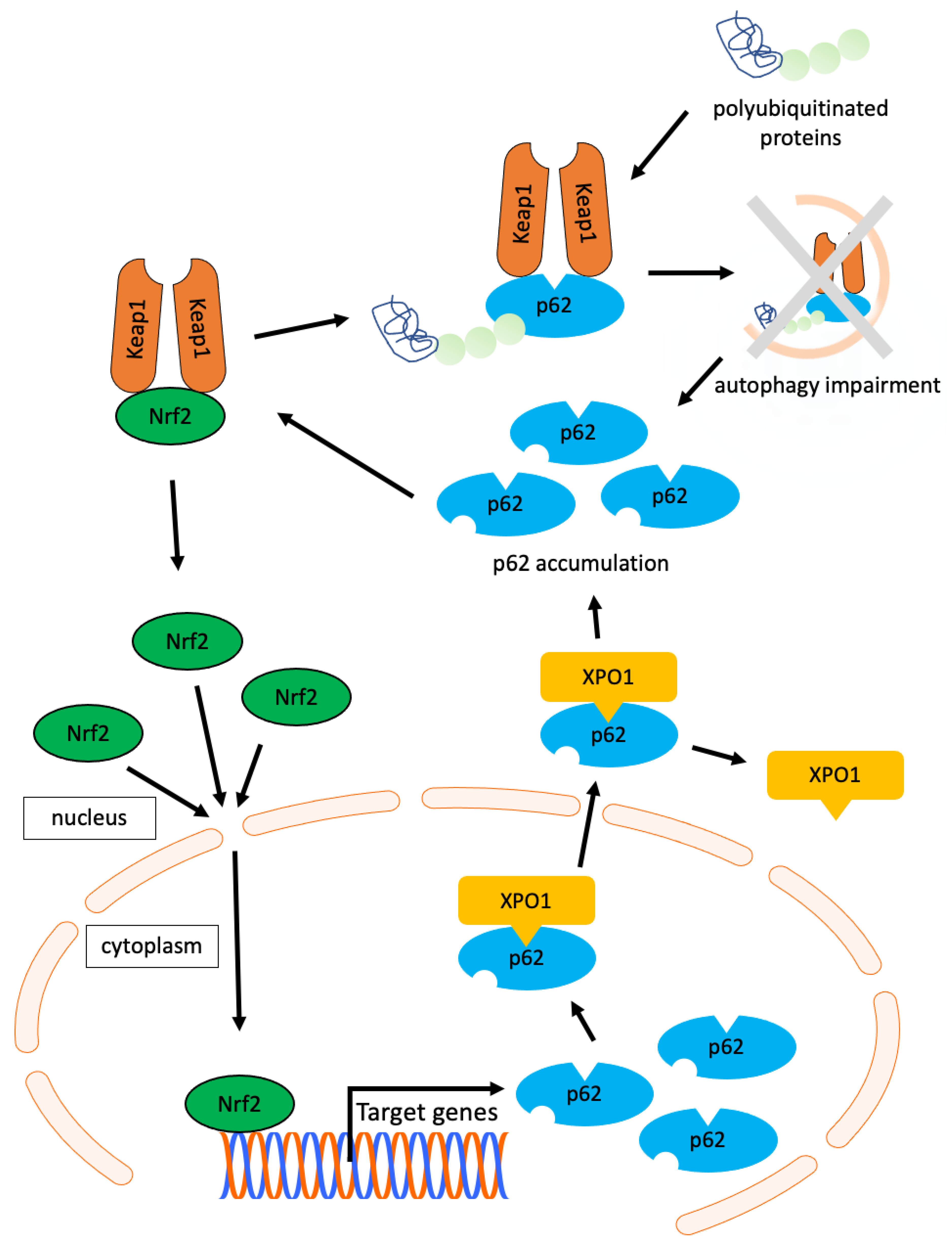
:1. Introduction
2. Materials and Methods
2.1. Samples
2.2. Immunohistochemistry
2.3. Evaluation of p62, XPO1, p53, and ki67
2.4. Statistical Analysis
3. Results
3.1. Correlation of Each Protein Expression in OPMDs
3.2. The Association between Malignant Transformation of OPMDs and Clinical Characteristics or the Expression of Each Protein
4. Discussion
5. Conclusions
Author Contributions
Funding
Institutional Review Board Statement
Informed Consent Statement
Data Availability Statement
Conflicts of Interest
References
- Reibel, J.; Gale, N.; Hille, J.; Hunt, J.L.; Lingen, M.; Muller, S.; Sloan, P.; Tilakaratne, W.M.; Westra, W.H.; Williams, M.D. Oral potentially malignant disorders and oral epithelial dysplasia. In WHO Classification of Head and Neck Tumors, 4th ed.; El-Naggar, A.K., Chan, J.K.C., Grandis, J.R., Takata, T., Slootweg, P., Eds.; iaRC: Lyon, France, 2017; pp. 112–115. [Google Scholar]
- Iocca, O.; Sollecito, T.P.; Alawi, F.; Weinstein, G.S.; Newman, J.G.; De Virgilio, A.; Di Maio, P.; Spriano, G.; Pardiñas López, S.; Shanti, R.M. Potentially malignant disorders of the oral cavity and oral dysplasia: A systematic review and meta-analysis of malignant transformation rate by subtype. Head Neck 2020, 42, 539–555. [Google Scholar] [CrossRef]
- Chaturvedi, A.K.; Udaltsova, N.; Engels, E.A.; Katzel, J.A.; Yanik, E.L.; Katki, H.A.; Lingen, M.W.; Silverberg, M.J. Oral leukoplakia and risk of progression to oral cancer: A population-based cohort study. J. Natl. Cancer Inst. 2020, 112, 1047–1054. [Google Scholar] [CrossRef]
- Gupta, S.; Jawanda, M.K.; Madhushankari, G.S. Current challenges and the diagnostic pitfalls in the grading of epithelial dysplasia in oral potentially malignant disorders: A review. J. Oral Biol. Craniofac. Res. 2020, 10, 788–799. [Google Scholar] [CrossRef]
- Kumari, P.; Debta, P.; Dixit, A. Oral potentially malignant disorders: Etiology, pathogenesis, and transformation into oral cancer. Front. Pharmacol. 2022, 13, 825266. [Google Scholar] [CrossRef]
- Dionne, K.R.; Warnakulasuriya, S.; Zain, R.B.; Cheong, S.C. Potentially malignant disorders of the oral cavity: Current practice and future directions in the clinic and laboratory. Int. J. Cancer 2015, 136, 503–515. [Google Scholar] [CrossRef] [PubMed]
- Kuribayashi, Y.; Tsushima, F.; Sato, M.; Morita, K.; Omura, K. Recurrence patterns of oral leukoplakia after curative surgical resection: Important factors that predict the risk of recurrence and malignancy. J. Oral Pathol. Med. 2012, 41, 682–688. [Google Scholar] [CrossRef] [PubMed]
- Faruk, M.O.; Ichimura, Y.; Komatsu, M. Selective autophagy. Cancer Sci. 2021, 112, 3972–3978. [Google Scholar] [CrossRef]
- Mizushima, N.; Komatsu, M. Autophagy: Renovation of cells and tissues. Cell 2011, 147, 728–741. [Google Scholar] [CrossRef] [PubMed]
- Lamark, T.; Svenning, S.; Johansen, T. Regulation of selective autophagy: The p62/SQSTM1 paradigm. Essays Biochem. 2017, 61, 609–624. [Google Scholar] [CrossRef] [PubMed]
- Sánchez-Martín, P.; Saito, T.; Komatsu, M. p62/SQSTM1: ‘Jack of all trades’ in health and cancer. FEBS J. 2019, 286, 8–23. [Google Scholar] [CrossRef]
- Cuyler, J.; Murthy, P.; Spada, N.G.; McGuire, T.F.; Lotze, M.T.; Xie, X.Q. Sequestsome-1/p62-targeted small molecules for pancreatic cancer therapy. Drug Discov. Today 2022, 27, 362–370. [Google Scholar] [CrossRef]
- Klionsky, D.J.; Petroni, G.; Amaravadi, R.K.; Baehrecke, E.H.; Ballabio, A.; Boya, P.; Bravo-San Pedro, J.M.; Cadwell, K.; Cecconi, F.; Choi, A.M.K.; et al. Autophagy in major human diseases. EMBO J. 2021, 40, e108863. [Google Scholar] [CrossRef]
- Yin, X.; Hu, L.; Feng, X.; Wang, H.; Zhang, C.; Wang, S. Simultaneous activation of impaired autophagy and the mammalian target of rapamycin pathway in oral squamous cell carcinoma. J. Oral Pathol. Med. 2019, 48, 705–711. [Google Scholar] [CrossRef] [PubMed]
- Wang, X.; Wu, W.K.K.; Gao, J.; Li, Z.; Dong, B.; Lin, X.; Li, Y.; Gong, J.; Qi, C.; Peng, Z.; et al. Autophagy inhibition enhances PD-L1 expression in gastric cancer. J. Exp. Clin. Cancer Res. 2019, 38, 140. [Google Scholar] [CrossRef]
- Thompson, H.G.; Harris, J.W.; Wold, B.J.; Lin, F.; Brody, J.P. p62 overexpression in breast tumors and regulation by prostate-derived Ets factor in breast cancer cells. Oncogene 2003, 22, 2322–2333. [Google Scholar] [CrossRef] [PubMed]
- Terabe, T.; Uchida, F.; Nagai, H.; Omori, S.; Ishibashi-Kanno, N.; Hasegawa, S.; Yamagata, K.; Gosho, M.; Yanagawa, T.; Bukawa, H. Expression of autophagy-related markers at the surgical margin of oral squamous cell carcinoma correlates with poor prognosis and tumor recurrence. Hum. Pathol. 2018, 73, 156–163. [Google Scholar] [CrossRef] [PubMed]
- Yoshida, T.; Terabe, T.; Nagai, H.; Uchida, F.; Hasegawa, S.; Nagao, T.; Miyabe, S.; Ishibashi-Kanno, N.; Yamagata, K.; Warabi, E.; et al. Association between p62 expression and clinicopathological characteristics in oral leukoplakia. Clin. Exp. Dent. Res. 2019, 5, 389–397. [Google Scholar] [CrossRef] [PubMed]
- Liu, J.L.; Chen, F.F.; Lung, J.; Lo, C.H.; Lee, F.H.; Lu, Y.C.; Hung, C.H. Prognostic significance of p62/SQSTM1 subcellular localization and LC3B in oral squamous cell carcinoma. Br. J. Cancer 2014, 111, 944–954. [Google Scholar] [CrossRef]
- Leemans, C.R.; Braakhuis, B.J.; Brakenhoff, R.H. The molecular biology of head and neck cancer. Nat. Rev. Cancer 2011, 11, 9–22. [Google Scholar] [CrossRef]
- Li, X.; He, S.; Ma, B. Autophagy and autophagy-related proteins in cancer. Mol. Cancer 2020, 19, 12. [Google Scholar] [CrossRef]
- Mathew, R.; Karp, C.M.; Beaudoin, B.; Vuong, N.; Chen, G.; Chen, H.Y.; Bray, K.; Reddy, A.; Bhanot, G.; Gelinas, C.; et al. Autophagy suppresses tumorigenesis through elimination of p62. Cell 2009, 137, 1062–1075. [Google Scholar] [CrossRef] [PubMed]
- Wang, Y.; Le, W.D. Autophagy and Ubiquitin-Proteasome System. Adv. Exp. Med. Biol. 2019, 1206, 527–550. [Google Scholar] [CrossRef] [PubMed]
- Komatsu, M.; Waguri, S.; Koike, M.; Sou, Y.S.; Ueno, T.; Hara, T.; Mizushima, N.; Iwata, J.; Ezaki, J.; Murata, S.; et al. Homeostatic levels of p62 control cytoplasmic inclusion body formation in autophagy-deficient mice. Cell 2007, 131, 1149–1163. [Google Scholar] [CrossRef] [PubMed]
- Pankiv, S.; Lamark, T.; Bruun, J.A.; Øvervatn, A.; Bjørkøy, G.; Johansen, T. Nucleocytoplasmic shuttling of p62/SQSTM1 and its role in recruitment of nuclear polyubiquitinated proteins to promyelocytic leukemia bodies. J. Biol. Chem. 2010, 285, 5941–5953. [Google Scholar] [CrossRef]
- Scholzen, T.; Gerdes, J. The Ki-67 protein: From the known and the unknown. J. Cell Physiol. 2000, 182, 311–322. [Google Scholar] [CrossRef]
- Yang, C.; Zhang, J.; Ding, M.; Xu, K.; Li, L.; Mao, L.; Zheng, J. Ki67 targeted strategies for cancer therapy. Clin. Transl. Oncol. 2018, 20, 570–575. [Google Scholar] [CrossRef]
- Torres-Rendon, A.; Roy, S.; Craig, G.T.; Speight, P.M. Expression of Mcm2, geminin and Ki67 in normal oral mucosa, oral epithelial dysplasias and their corresponding squamous-cell carcinomas. Br. J. Cancer 2009, 100, 1128–1134. [Google Scholar] [CrossRef]
- Inoue, D.; Suzuki, T.; Mitsuishi, Y.; Miki, Y.; Suzuki, S.; Sugawara, S.; Watanabe, M.; Sakurada, A.; Endo, C.; Uruno, A.; et al. Accumulation of p62/SQSTM1 is associated with poor prognosis in patients with lung adenocarcinoma. Cancer Sci. 2012, 103, 760–766. [Google Scholar] [CrossRef]
- Kitamura, H.; Torigoe, T.; Asanuma, H.; Hisasue, S.I.; Suzuki, K.; Tsukamoto, T.; Satoh, M.; Sato, N. Cytosolic overexpression of p62 sequestosome 1 in neoplastic prostate tissue. Histopathology 2006, 48, 157–161. [Google Scholar] [CrossRef]
- Iwadate, R.; Inoue, J.; Tsuda, H.; Takano, M.; Furuya, K.; Hirasawa, A.; Inazawa, J. High expression of p62 protein is associated with poor prognosis and aggressive phenotypes in endometrial cancer. Am. J. Pathol. 2015, 185, 2523–2533. [Google Scholar] [CrossRef]
- Azizian, N.G.; Li, Y. XPO1-dependent nuclear export as a target for cancer therapy. J. Hematol. Oncol. 2020, 13, 61. [Google Scholar] [CrossRef] [PubMed]
- Fu, S.C.; Huang, H.C.; Horton, P.; Juan, H.F. ValidNESs: A database of validated leucine-rich nuclear export signals. Nucleic Acids Res. 2013, 41, D338–D343. [Google Scholar] [CrossRef] [PubMed]
- Gruffaz, M.; Yuan, H.; Meng, W.; Liu, H.; Bae, S.; Kim, J.S.; Lu, C.; Huang, Y.; Gao, S.J. CRISPR-Cas9 Screening of Kaposi’s Sarcoma-Associated Herpesvirus-Transformed Cells Identifies XPO1 as a Vulnerable Target of Cancer Cells. mBio 2019, 10, e00866-19. [Google Scholar] [CrossRef] [PubMed]
- Jiang, T.; Harder, B.; Rojo de la Vega, M.; Wong, P.K.; Chapman, E.; Zhang, D.D. p62 links autophagy and Nrf2 signaling. Free Radic. Biol. Med. 2015, 88, 199–204. [Google Scholar] [CrossRef] [PubMed]
- Duffy, M.J.; Synnott, N.C.; Crown, J. Mutant p53 as a target for cancer treatment. Eur. J. Cancer 2017, 83, 258–265. [Google Scholar] [CrossRef] [PubMed]
- Lindemann, A.; Takahashi, H.; Patel, A.A.; Osman, A.A.; Myers, J.N. Targeting the DNA Damage Response in OSCC with TP53 Mutations. J. Dent. Res. 2018, 97, 635–644. [Google Scholar] [CrossRef]
- Deshmukh, A.V.; Gupta, A.; Chaudhari, A.G.; Gangane, N.M. Correlation of p53 expression with Clinical Presentation and Prognosis of Oral Squamous Cell Carcinoma Patients: A Pilot Study. Indian J. Otolaryngol. Head Neck Surg. 2022, 74, 1836–1840. [Google Scholar] [CrossRef]
- Aladhraei, M.; Kassem Al-Thobhani, A.; Poungvarin, N.; Suwannalert, P. Association of XPO1 Overexpression with NF-κB and Ki67 in Colorectal Cancer. Asian Pac. J. Cancer Prev. 2019, 20, 3747–3754. [Google Scholar] [CrossRef]


| Characteristics | Cases (%) | |
|---|---|---|
| Sex | Male | 47 (67.1) |
| Female | 23 (32.9) | |
| Age | 65> | 37 (52.9) |
| 65≤ | 33 (47.1) | |
| Drinking | Yes | 51 (72.9) |
| No | 19 (27.1) | |
| Smoking | Yes | 43 (61.4) |
| No | 27 (38.6) | |
| Location | Tongue | 24 (34.3) |
| Others | 46 (65.7) | |
| Disorder | Leukoplakia | 50 (71.4) |
| OLP | 20 (28.6) | |
| Epithelial dysplasia | Positive | 53 (75.7) |
| Negative | 17 (24.3) | |
| Developed Cancer | Yes | 6 (8.6) |
| No | 64 (91.4) |
| Characteristics | Developed Cancer Cases (%) | No Cancer Cases (%) | p | |
|---|---|---|---|---|
| Sex | Male | 3 (6.4) | 44 (93.6) | 0.39 |
| Female | 3 (13.0) | 20 (87.0) | ||
| Age | 65> | 1 (2.7) | 36 (97.3) | 0.09 |
| 65≤ | 5 (15.2) | 28 (84.8) | ||
| Drinking | Yes | 4 (7.8) | 47 (92.2) | 0.66 |
| No | 2 (10.5) | 17 (89.5) | ||
| Smoking | Yes | 3 (7.0) | 40 (93.0) | 0.67 |
| No | 3 (11.1) | 24 (88.9) | ||
| Location | Tongue | 3 (12.5) | 21 (87.5) | 0.41 |
| Others | 3 (7.0) | 43 (91.5) | ||
| Disorders | Leukoplakia | 4 (8.0) | 46 (92.0) | 1.00 |
| OLP | 2 (10.0) | 18 (90.0) | ||
| Epithelial dysplasia | Positive | 2 (13.3) | 13 (86.7) | 0.60 |
| Negative | 4 (7.3) | 51 (92.7) |
| Parameter | Developed Cancer Expression (%) | No Cancer Expression (%) | p |
|---|---|---|---|
| p62 in nucleus | 12.9 ± 5.9 | 29.7 ± 19.0 | 0.03 * |
| p62 in cytoplasm | 45.4 ± 12.5 | 29.8 ± 15.5 | 0.03 * |
| p62 aggregation | 6.5 ± 3.6 | 2.1 ± 1.9 | <0.01 * |
| XPO1 | 41.6 ± 22.9 | 38.9 ± 25.0 | 0.78 |
| p53 | 14.2 ± 8.5 | 11.9 ± 9.4 | 0.44 |
| Ki67 | 20.3 ± 6.5 | 18.0 ± 9.7 | 0.20 |
Disclaimer/Publisher’s Note: The statements, opinions and data contained in all publications are solely those of the individual author(s) and contributor(s) and not of MDPI and/or the editor(s). MDPI and/or the editor(s) disclaim responsibility for any injury to people or property resulting from any ideas, methods, instructions or products referred to in the content. |
© 2023 by the authors. Licensee MDPI, Basel, Switzerland. This article is an open access article distributed under the terms and conditions of the Creative Commons Attribution (CC BY) license (https://creativecommons.org/licenses/by/4.0/).
Share and Cite
Takasaki, R.; Uchida, F.; Takaoka, S.; Ishii, R.; Fukuzawa, S.; Warabi, E.; Ishibashi-Kanno, N.; Yamagata, K.; Bukawa, H.; Yanagawa, T. p62 Is a Potential Biomarker for Risk of Malignant Transformation of Oral Potentially Malignant Disorders (OPMDs). Curr. Issues Mol. Biol. 2023, 45, 7630-7641. https://doi.org/10.3390/cimb45090480
Takasaki R, Uchida F, Takaoka S, Ishii R, Fukuzawa S, Warabi E, Ishibashi-Kanno N, Yamagata K, Bukawa H, Yanagawa T. p62 Is a Potential Biomarker for Risk of Malignant Transformation of Oral Potentially Malignant Disorders (OPMDs). Current Issues in Molecular Biology. 2023; 45(9):7630-7641. https://doi.org/10.3390/cimb45090480
Chicago/Turabian StyleTakasaki, Ryo, Fumihiko Uchida, Shohei Takaoka, Ryota Ishii, Satoshi Fukuzawa, Eiji Warabi, Naomi Ishibashi-Kanno, Kenji Yamagata, Hiroki Bukawa, and Toru Yanagawa. 2023. "p62 Is a Potential Biomarker for Risk of Malignant Transformation of Oral Potentially Malignant Disorders (OPMDs)" Current Issues in Molecular Biology 45, no. 9: 7630-7641. https://doi.org/10.3390/cimb45090480