Article

PADI4 Haplotypes Contribute to mRNA Expression, the Enzymatic Activity of Peptidyl Arginine Deaminase and Rheumatoid Arthritis Risk in Patients from Western Mexico

by
Mónica Guadalupe Matuz-Flores
1,
Jesús Alfredo Rosas-Rodríguez
2,
Orlando Tortoledo-Ortiz
3,
Salvador Muñoz-Barrios
4,
Gloria Esther Martínez-Bonilla
5,
Jorge Hernández-Bello
1,
Christian Johana Baños-Hernández
1,
Cesar Pacheco-Tena
6,
Gabriela Athziri Sánchez-Zuno
1,
Beatriz Panduro-Espinoza
1 and
José Francisco Muñoz-Valle
1,*
1
Instituto de Investigación en Ciencias Biomédicas, Centro Universitario de Ciencias de la Salud, Universidad de Guadalajara, Guadalajara 44340, Jalisco, Mexico
2
Departamento de Ciencias Químico Biológicas y Agropecuarias, Universidad de Sonora Unidad Regional Sur, Navojoa 85880, Sonora, Mexico
3
Centro de Investigación en Alimentación y Desarrollo A.C., Coordinación de Nutrición, Lab. de Cromatografía, Hermosillo 83304, Sonora, Mexico
4
Unidad Académica de Ciencias Naturales, Universidad Autónoma de Guerrero, Chilpancingo 39086, Guerrero, Mexico
5
Servicio de Reumatología, Hospital Civil de Guadalajara Fray Antonio Alcalde, Guadalajara 44280, Jalisco, México
6
Facultad de Medicina y Ciencias Biomédicas, Universidad Autónoma de Chihuahua, Chihuahua C.P. 31109, Chih., Mexico
*
Author to whom correspondence should be addressed.
Curr. Issues Mol. Biol. 2022, 44(9), 4268-4281; https://doi.org/10.3390/cimb44090293
Submission received: 5 August 2022 / Revised: 27 August 2022 / Accepted: 3 September 2022 / Published: 16 September 2022
(This article belongs to the Collection Feature Papers in Current Issues in Molecular Biology)

Abstract

:
Citrullination is catalyzed by the peptidyl arginine deiminase 4 (PAD4) enzyme, encoded by the PADI4 gene. Increased PAD4 activity promotes the onset and progression of rheumatoid arthritis (RA). This study aimed to evaluate the association of PADI4 haplotypes with RA risk, mRNA expression, and the PAD4 activity in patients with RA from Mexico. Methodology: 100 RA patients and 100 control subjects (CS) were included. Genotyping was performed by PCR-RFLP method, PADI4 mRNA expression was quantified by real-time PCR, the contribution of PADI4 alleles (PADI4_89 G>A, PADI4_90 T>C, and PADI4_92 G>C) to mRNA expression by the ASTQ method, and PAD4 activity by HPLC. Also, the anti-CCP and anti-PADI4 antibodies were quantified by ELISA. Results: The three PADI4 polymorphisms were associated with RA susceptibility (OR = 1.72, p = 0.005; OR = 1.62; p = 0.014; OR = 1.69; p = 0.009; respectively). The 89G, 90T, and 92G alleles have a higher relative contribution to PADI4 mRNA expression from RA patients than 89A, 90C, and 92C alleles in RA patients. Moreover, the GTG/GTG haplotype was associated with RA susceptibility (OR = 2.86; p = 0.024). The GTG haplotype was associated with higher PADI4 mRNA expression (p = 0.04) and higher PAD4 enzymatic activity (p = 0.007) in RA patients. Conclusions: The evaluated polymorphisms contribute to PADI4 mRNA expression and the enzymatic activity of PAD4 in leukocytes. Therefore, the GTG haplotype is a genetic risk factor for RA in western Mexico, and is associated with increased PADI4 mRNA expression and higher PAD4 activity in these patients.

1. Introduction

Rheumatoid arthritis (RA) is an autoimmune, inflammatory, chronic, and systemic disease characterized by dysregulation of cytokine production and the presence of antibodies against citrullinated proteins (ACPA) [1].
One of the most relevant molecules in the development of this disease is the enzyme peptidyl arginine deaminase type IV (PAD4). The PAD4 enzyme catalyzes the conversion of arginine to citrulline residues. In humans, five isoforms of PAD have been described, PAD1, PAD2, PAD3, PAD4, and PAD6, distributed in many cells and tissues. Among these, PAD4 is expressed primarily on neutrophils, eosinophils, and monocytes [2,3].
High activity of PAD4 has been associated with the appearance and progression of RA because it promotes neutrophils recruited to the site of inflammation and the formation of neoantigens, and the hypercitrullination of vimentin, fibrinogen, type II collagen, and α-enolase [4,5,6], which are the primary targets of the autoantibodies in RA.
Also, PAD participates in biological processes such as the formation of extracellular neutrophil traps (NETs), which contain nuclear material, granular enzymes, and cytoplasmic proteins that can be a source of autoantigens during NETosis in autoimmune diseases [7].
The PAD4 enzyme is encoded by the PADI4 gene, which has three single nucleotide polymorphisms (SNPs) that generate amino acid substitutions (PADI4_89 G>A [rs11203366], PADI4_90 T>C [rs11203367], and PADI4_92 G>C [rs874881]): S55G, A82V, and A112G, respectively. These polymorphisms also generate a conformational change in the enzyme [8]. The conformational change caused by these SNPs can affect the interaction of the enzyme with the substrates [9].
The 3 PADI4 SNPs are in strong linkage disequilibrium, and form haplotypes. The GTG haplotype has been associated with RA, higher ACPA levels, and mRNA expression in several populations such as Japanese [8,10], Korean [11], North American [12], and western and southern Mexico [13,14]. In addition, the activity of the PAD enzyme on synovial fluid has been shown, and it correlates with ACPA positivity, disease activity, and inflammatory mediator concentrations [15,16]. However, the effects of the GTG haplotype on the activity of the PAD4 enzyme in polymorphonuclear cells (PMN) and peripheral blood mononuclear cells (PBMC) of RA patients have been poorly explored. Therefore, this study aimed to identify the relationship between PADI4 gene haplotypes with RA risk, mRNA expression in PBMC, and PAD4 activity in the leukocytes (PMN and PBMC) of Mexican patients.

2. Materials and Methods

2.1. RA Patients and Control Subjects

One hundred RA patients from western Mexico were included, classified according to the ACR/EULAR 2010. They were recruited from the rheumatology service at the Hospital Civil “Fray Antonio Alcalde”, Guadalajara, Jalisco. Their clinical activity and functional disabilities were evaluated by the DAS28 score (disease activity score) [17], and a visual analog pain scale HAQ-DI (Health Assessment Questionnaire Disability Index), respectively [18,19]. Moreover, one hundred CS classified by self-report were included, which were recruited from the general population. Only Mexican Mestizo unrelated subjects with ancestry from western Mexico (Jalisco, Nayarit, Colima, and Michoacan states) since three previous generations were enrolled in the study, to prevent population heterogeneity. The ethics committee of the University of Guadalajara approved this study (CI-05718), which was conducted following the ethical principles established in the Declaration of Helsinki (64th General Assembly, Fortaleza, Brazil, October 2013) and the Official Mexican Standard NOM-087-ECOL-SSA1-2002, Environmental protection, Environmental health, Hazardous biological-infectious waste—Classification and handling specifications. The subjects who participated in this study each signed an informed consent letter.

2.2. Laboratory Assessment and Quantification of Qutoantibodies

Peripheral blood samples were obtained from all subjects included in the study. Serum was isolated using standard protocols, aliquoted, and stored at −80 °C until use. The clinical parameters, C-reactive protein (CRP) and rheumatoid factor (RF), were determined by turbidimetry (BS120 Chemestry Analyzer, 2, Shenzhen, China) using an assay based on latex microparticles (COD31029; BioSystems, Barcelona, Spain), and with the RF latex assay (COD31030; BioSystems, Barcelona, Spain), respectively, according the manufacturer’s instructions. Individuals with RF values > 30 IU/mL were considered positive. The determination of ESR was carried out with the Wintrobe method. The quantification of antibodies against cyclic citrullinated peptides (anti-CCP) was carried out using an enzyme-linked immunosorbent assay (ELISA) (Axis-Shield Diagnostics, Scotland, UK); serum values > 5 U/mL were considered positive. An ELISA based on the determination of any anti-PADI4 immunoglobulin isotype (IgG, IgM, and IgA) was performed according to the manufacturer’s recommendations (Cayman Chemical Co, Ann Arbor, MI, USA) for the serum anti-PADI4 antibodies quantification.

2.3. PADI4 SNPs Genotyping

Three exonic PADI4 SNPs were genotyped using the PCR-RFLP method. The sets of primers were those reported by Guzman-Guzman et al. [11]. Since the PADI4 89G>A and 90T>C SNPs are in proximate sequences, a single PCR reaction was performed to amplify both polymorphic fragments. Reactions were carried out in a final volume of 25 µL containing 0.12 µM of each primer, 100 ng of gDNA, 2.5 U of Taq DNA polymerase (InvitrogenTM life technologies, Carlsbad, CA, USA), 1X enzyme supplied buffer, 4.5 mM MgCl2, 1.4 M Betaine (SIGMA Life Science, Misuri, USA), and 0.1 mM of dNTPs (InvitrogenTM life technologies, CA, USA). The cycling conditions were the following: initial denaturation at 94 °C for 3 min (min); followed by 30 cycles of 30 s (s) at 94 °C, 30 s at 62 °C, and 30 s at 72 °C; each with an ending extension step of 1 m at 72 °C. The resultant 221 bp fragment was afterward digested in separate reactions with the BtgI enzyme or with the MscI enzyme (New England, BioLabs, Inc., Watertown, MA, USA), to identify the PADI4 89G>A and 90T>C polymorphisms, respectively. The digested products were then electrophoresed on a 6% polyacrylamide gel (29 acrylamide: 1 bisacrylamide) and stained with 2% AgNO3. In the PADI4 89G>A polymorphism, the G allele was identified by two fragments (144 and 77 bp), and the A allele was recognized as a 221 bp fragment; whereas in the PADI4 90T>C polymorphism, the T allele was represented by two fragments (165 and 56 bp), a single 221 bp fragment represented the C allele. For the PADI4 92G>C genotyping, PCR was performed in a final volume of 15 µL containing 0.2 µM of each primer, 100 ng of gDNA, 2.5 U Taq DNA polymerase (Invitrogen™ life technologies, USA), 1X enzyme supplied buffer, 2.5 mM MgCl2, and 0.2 mM dNTPs (InvitrogenTM life technologies, USA), under the following conditions: initial denaturation at 94 °C for 3 min followed by 35 cycles of 30 s at 94 °C, 30 s at 56 °C, and 30 s at 72 °C; then, a final extension of 1 min at 72 °C. The PCR product resulted in a 106 bp fragment subsequently digested with the MspI restriction endonuclease (New England BioLabs, Inc.). The products were then electrophoresed on a 6% polyacrylamide gel and stained with AgNO3. The G allele was represented as two fragments (59 and 47 bp), while the C allele resulted in a single fragment of 106 bp. A 50 bp molecular weight standard (Invitrogen™ life technologies, CA, USA) and the corresponding positive/negative controls for each PADI4 SNP were loaded in all polyacrylamide gels.

2.4. RNA Isolation and cDNA Synthesis

Total RNA was extracted immediately from 5 mL of EDTA—treated peripheral blood of RA patients and controls. Total leucocytes were isolated using 5% dextran solution (Sigma-Aldrich, St. Louis, MO, USA), and the RNA was obtained using TRIzol reagent (InvitrogenTM life technologies, CA, USA), according to the manufacturer’s instructions. The RNA was analyzed on a NanoDrop 2000 spectrophotometer (Thermo Scientific, MA, USA) by measuring the optical density (OD) at 260 nm for the quantity and the OD260/280 ratio for the quality. Before cDNA synthesis, RNA samples were treated with DNAse I (InvitrogenTM Life Technologies, CA, USA). 1 µg of total RNA was subjected to reverse transcription using oligo(dT) and M-MLV reverse transcriptase, as indicated by the manufacturer (Promega, Madison, WI, USA).

2.5. Quantification of PADI4 Allelic Contribution to the mRNA Expression of PADI4

In the samples identified as heterozygous for the polymorphisms 89G>A, 90T>C, and 92G>C of the PADI4 gene, the allelic contribution to mRNA expression of PADI4 was determined by the allele-specific transcript quantification (ASTQ) method reported by Kaijzel et al. [14]. It allowed discriminating the mRNA expression of two alleles through quantifying fragments digested by restriction enzymes and the ratio of bands representing the allelic expression that reflects the relationship between the respective precursor transcripts. From 100 ng of cDNA, we proceeded to PCR amplification and subsequent digestion with the respective restriction enzymes (Btg I, for 89G>A; Msc I for 90T>C; and Msp I for 92G>C), using 10 U for each experiment. The densitometry of the bands corresponding to the products digested with the respective restriction enzymes was performed using ImageJ software (National Institutes of Health, New York, NY, USA) from 3% agarose gels (Vivantis Technologies, Ehsan, Malaysia) stained with GelRed 1X (Biotium Inc, Fremont, CA, USA). The intensity of the band was automatically determined in RUA.

2.6. Quantitative Real-Time PCR for PADI4 Expression Quantitative

The quantification of PADI4 mRNA was conducted by real-time PCR using TaqMan® probes (Applied Biosystems, Waltham, MA, USA) as a fluorescent system for both the interest gene PADI4 (ID Hs00202612 m1) and the reference gene glyceraldehyde 3-phosphate dehydrogenase (GAPDH) (ID Hs03929097 g1). All samples were analyzed in duplicate using the conditions indicated in the Gene Expression Assay protocol in a QuantStudio 5 Real-Time PCR system (Thermo Fisher Scientific, Waltham, MA, USA). As indicated previously [20], the validation of similar reaction efficiencies in both genes was done by running serial dilution curves with four cDNA points. The 2−ΔΔCq and 2−ΔCq methods were used to analyze the PADI4 expression in the GTG haplotype carriers in relation to the ACC haplotype carriers (calibrator group).

2.7. Isolation of Leukocytes

Leukocytes [including peripheral blood mononuclear cells (PBMC) and polymorphonuclear cells (PMN)] were obtained from RA patients (n = 8) and CS (n = 8) carrying the GTG or ACC haplotypes. The cells were separated by density gradient (Histopaque 1.119 and 1.077 Sigma-Aldrich, Misuri, USA) and were washed with phosphate buffer saline (PBS) (pH 7.2), and centrifuged at 4 °C for 10 min at 1800 revolutions per minute (rpm); then, the cell button was added in 1 mL of RPMI-1640 with 10% fetal calf serum. The number of viable cells was determined by the exclusion technique with 3% trypan blue staining; the total number of cells/mL was quantified by direct counting using a Neubauer chamber. The cells were subsequently lysed by sonication.

2.8. PAD4 Enzymatic Activity Analysis

The enzymatic activity of PAD4 was evaluated by the technique reported by Chikuma et al. [21], based on the conversion of the peptide Dansyl Glycyl -l- Citrulline (Dns-Gly-Cit) liberated enzymatically from the substrate, Dansyl Glycyl Arginine (Dns-Gly-Arg) (GenScript Biotech, Piscataway, NJ, USA). The generation of the Dns-Gly-Cit peptide was evaluated by HPLC and fluorescence detection. The assay medium consisted of 40 mM Tris-HCl buffer (pH 7.6), 2 mM CaCl2, 1 mM Dithiothreitol, adding the substrate Dns-Gly-Arg 20 mM. After the addition of 300 µg of protein of PBMC and PMN lysates from RA patients and CS, it was incubated at 37 °C and the reaction was stopped at different reaction times (0.5, 1, 2 and 4, and 6 h), incubating at 100 °C for 10 min. The sample was centrifuged at 4 °C for 10 min, and 20 µL was injected in triplicate for analysis by HPLC. For the analysis of PAD activity in µmol/mL/µg protein, the area of each peak corresponding to the Dns-Gly-Cit peptide at the different incubation times was integrated. The Dns-Gly-Cit was enzymatically prepared from Dns-Gly-Arg using recombinant PAD (Abcam ab170409, Cambridge, UK), and the corresponding area for each incubation time was used for the standard curve.

2.9. Statistical Analysis

Statistical analysis was performed by STATA v 9.2 and GraphPad Prism v 8.0 software. For the statistical analysis, p < 0.05 was considered significant. Variable distribution was determined using the Shapiro–Wilk normality test. The data were expressed as means and standard deviations (SD) for descriptive statistics. For the inferential statistics for two groups, a t-test was performed, and the ANOVA test was used for more than two groups. Variables non-normally distributed were presented in median and interquartile ranges (25–75) or median and percentiles 5–95. For the inferential statistics for two groups, the Mann–Whitney U test was performed and the Kruskal–Wallis test for more than two groups.

3. Results

3.1. Clinical and Demographic Characteristics of RA Patients

The clinical and demographic characteristics of RA patients and CS are shown in Table 1. We observed that most patients were female (87% women and 13% men). In patients with RA, increased levels of acute-phase reactants were observed, including ESR (39 mm/h) and hsCRP (24.7 mg/L). Regarding antibodies, the mean of RF was 89.3 U/mL [54% were positive for RF] and 100.2 U/mL for anti-CCP antibodies [72.7% were positive for anti-CCP antibodies]. In addition, high concentrations of anti-PADI4 antibodies (2026.9 U/mL) were identified by a quantitative method.

3.2. Genotype and Haplotype Frequencies of the PADI4 Gene in RA Patients and CS

The 3 SNPs of PADI4 were in Hardy–Weinberg equilibrium in the CS group (PADI4_89 G>A, p = 0.279; PADI4_90 T>C, p = 0.305; PADI4_92G>C), p = 1.00). The frequency distribution in RA patients and CS is shown in Table 2. We found significant differences in the frequencies of genotypes and alleles between RA patients and CS for each of the three polymorphisms analyzed. In addition, we performed haplotype analysis of the PADI4 gene in 40 CS and 38 RA patients who were identified as homozygous for each haplotype. We observed a higher frequency of the GTG haplotype in RA patients than in CS (57.9% vs. 32.5%), while the ACC haplotype was more frequent in CS than in patients (67.5% vs. 42.1%). After performing the haplotype association analysis, a strong association of the GTG haplotype with the risk of developing RA (OR = 2.86; p = 0.024) was found (Table 3).

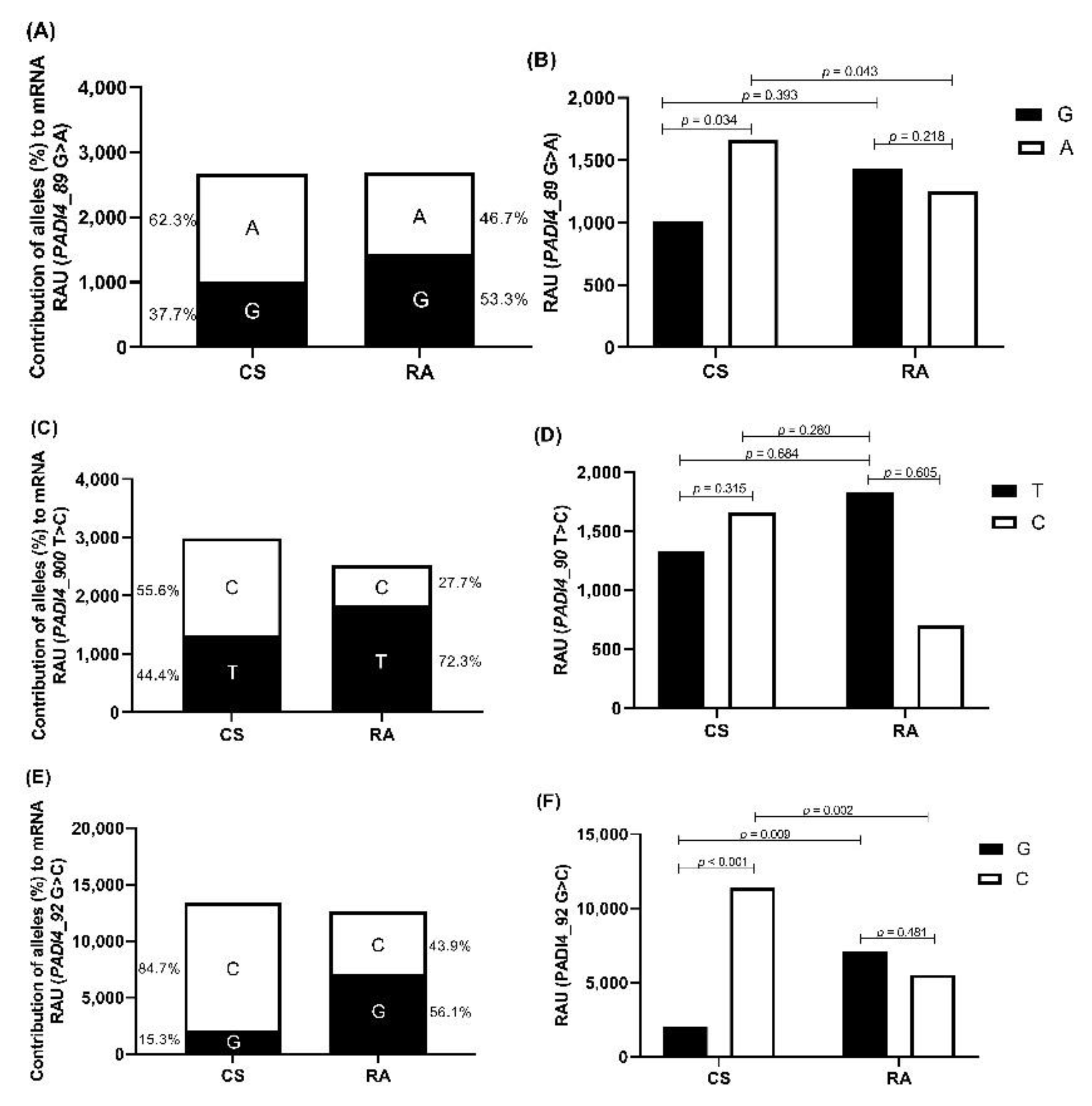
3.3. Allele-Specific Quantification of PADI4 mRNA Expression

Differences in the capacity of each PADI4 allele to contribute to the mRNA expression in RA patients and CS was detected. A higher expression of mRNA carrying the 89A allele was observed in CS (62.3% 89A allele vs. 46.7% 89G allele, ratio 1.33), while in RA patients, a higher expression of the mRNA carrying the 89G allele was observed (53.3% 89G allele vs. 37.7% 89A allele, ratio 1.41) (Figure 1A). CS expressed a higher quantity of mRNA carrying the 89>A allele than RA patients (1662.80 vs. 1255.03 RAU, p = 0.043; Figure 1B).
Regarding the 90T>C polymorphism, CS expresses more mRNA carrying the 90C allele than the 90T allele (55.6% vs. 44.4%; ratio 1.25), while RA patients expressed higher mRNA with the 90T allele (72.3% vs. 27.7%; ratio 2.61) (Figure 1C). On the other hand, CS expressed higher mRNA carrying the 90C allele than RA patients (1660.59 vs. 699.56 RAU, p = 0.280; Figure 1D).
For the 92G>C polymorphism, it was observed that the CS expressed more mRNA carrying the 92C allele than the 92G allele (84.7% vs. 15.3%; ratio 5.53), while in RA patients, a higher percentage of mRNA carrying the 92G allele was observed (56.1% vs. 43.9%; ratio 1.27; Figure 1E). Furthermore, it was observed that CS expresses more the mRNA with the 92C allele than RA patients (11380.69 vs. 5536.59 RAU, p = 0.002; Figure 1F).

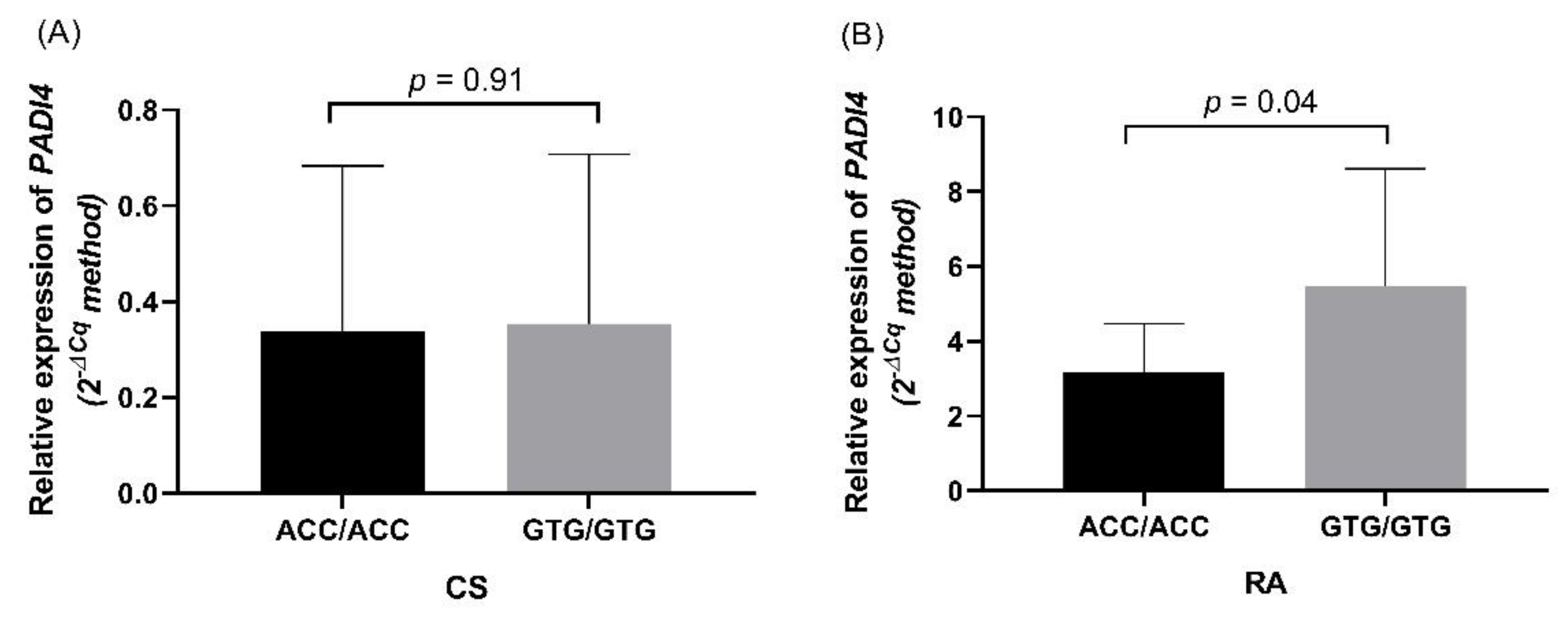
3.4. PADI4 Expression in RA Patients and CS

The PADI4 mRNA expression of 20 patients and 20 CS carrying the ACC and GTG haplotypes was quantified. The mRNA expression in CS was similar in carriers of both haplotypes (p = 0.91, Figure 2A). In RA, homozygous for the GTG haplotype had higher PADI4 mRNA expression than homozygous for the ACC haplotype (1.62 times; p = 0.04) (Figure 2B).

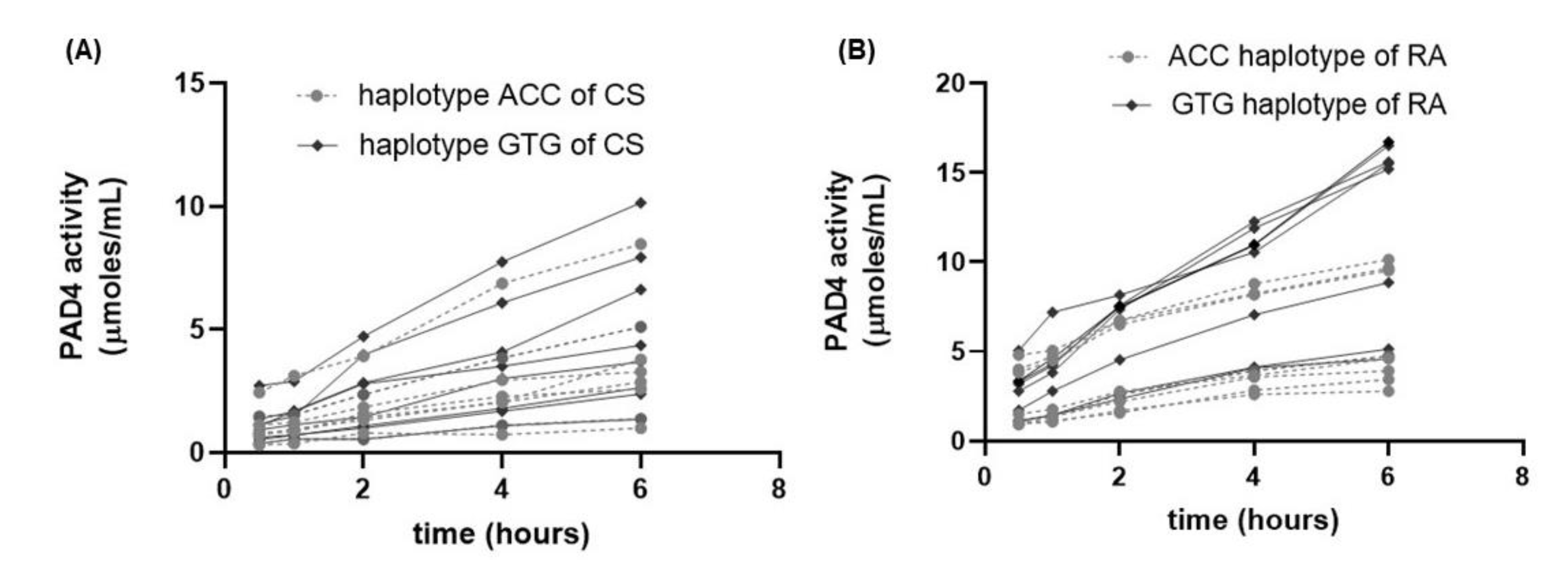
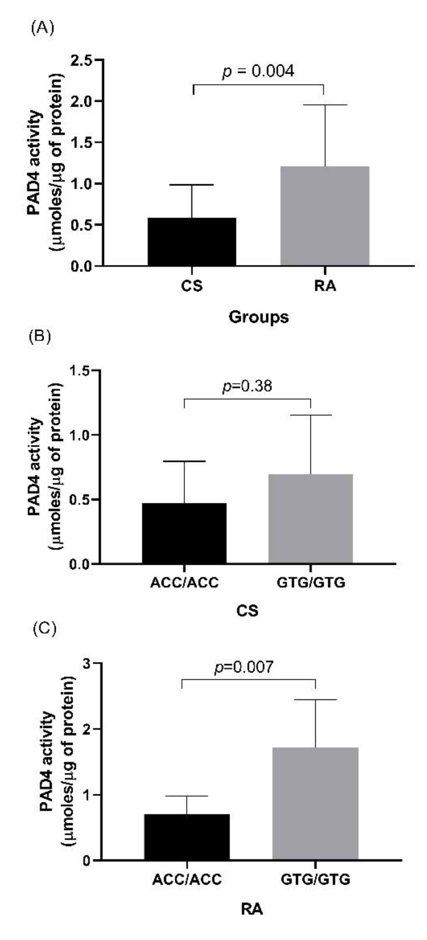
3.5. Evaluation of PAD4 Activity in CS and RA Patients

The injection of the Dns-Gly-Arg (DGA) peptide into the chromatography equipment resulted in a chromatogram with an elution peak at minute 14.4 of the peptide without citrullination due to the peptide was in the absence of the PAD enzyme, so it was used as a reference (Figure 3A).
In addition, it was observed that the DGA peptide in the presence of the recombinant enzyme presents an additional elution peak at minute 4.38, considered a citrullinated peptide (Figure 3B). An increase in the signal area corresponding to the citrullinated peptide was obtained during incubation from 30 min to 6 h (Figure 3C). In leukocytes, we compared the activity of PAD4 represented by the change in slope of the line obtained in a time of 0.5 to 6 h (Figure 4). RA patients presented a higher enzymatic activity of PAD4 compared to CS (Figure 5), and patients carrying the GTG haplotype presented more significant activity than those carrying the ACC haplotype (p = 0.007) (Figure 6).

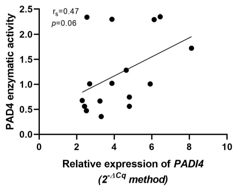
3.6. Association of Relative Expression of PADI4 Gene with PAD4 Enzymatic Activity

A positive correlation between the relative expression of the PADI4 gene and PAD4 activity was observed, but this was not statistically significant (p = 0.06, rs = 0.47) (Figure 6).

4. Discussion

RA is considered a multifactorial disease of unknown etiology with the participation of environmental and genetic factors contributing to this disease’s susceptibility, severity, and progression [22,23,24].
The PADI4 gene plays an important role in the pathophysiology of RA because the PAD4 enzyme catalyzes the change from arginine to citrulline, generating neoantigens.
This study evaluated genotypic, allelic, and haplotypic frequencies of the PADI4 gene, mRNA expression, and PAD4 activity in RA patients and CS. We observed that the 89GG, 90TT, and 92GG genotypes confer risk for RA in western Mexico. These results coincide with studies reported in Japan [8], Korea [11,25] and North America [12]. Furthermore, the genotypic frequencies for each polymorphism were very similar to those previously reported by our group of collaborators in the populations of western [14] and southern Mexico [13]. We also performed a haplotype analysis of the PADI4 gene, and an association of the GTG haplotype with the risk of developing RA was found (p = 0.024). These results agree with those previously reported by our study group in western and southern Mexican populations [13,14] as well as others, predominantly Asian [8,25,26] [8,17,18]. Therefore, our results support the finding that these SNPs and their haplotypes are genetic risk factors for RA.
It has been described that each of the two alleles present in diploid organisms contributes equally to the production of mRNA. However, different functional alleles can lead to an unequal contribution of each allele in the total mRNA production [19]. Therefore, in the present study, the contributions of the PADI4 alleles on the mRNA expression in samples from heterozygous RA and CS patients was analyzed by the ASTQ method. We found that CS expressed more mRNA carrying the non-susceptibility alleles 89A, 90C, and 92C, while the RA patients expressed more mRNA carrying the susceptibility alleles 89G, 90T, and 92G. According to the literature consulted, this is the first study evaluating the allelic contribution of PADI4 SNPs on the PADI4 mRNA expression by the ASTQ method. The results showed a differential expression of each functional PADI4 allele in RA and CS, and that each allele can contribute unequally to the expression of the PADI4 gene.
We also evaluated the gene expression of PADI4, and observed higher expression in RA patients carrying the GTG haplotypes. These results agree with a previous study carried out in this same population, where an increase in mRNA up to 3 times higher was reported in carriers of the GTG haplotype [14]. In addition, our results agree with the pioneering study of PADI4 polymorphisms by Suzuki et al. in a Japanese population [8], in which they reported that the GTG haplotype is associated with RA risk, more stable mRNA (half-lives of 11.6 in GTG carriers vs. 2.1 min in ACC carriers), an higher levels of PAD4 protein than the ACC carriers [8]. Therefore, we support the hypothesis that the mRNA of the PADI4 gene with the GTG haplotype accumulates in greater quantity than the mRNA with the ACC haplotype in RA, being able to form more PAD4 protein.
In view of the fact that we found an association of the GTG haplotype with the risk for RA and higher gene expression in carriers of these haplotypes, it was also of interest to evaluate the activity of PAD4 in leukocytes to determine the effect of these variants on the activity of the PAD4 enzyme; we observed higher PAD4 activity in patients carrying the GTG haplotype. The study by Hung et al. supports this finding. They showed that the exonic polymorphism of PADI4 increases the enzymatic activity of PAD4 in Jurkat cells [27], and that the susceptible PADI4 (GTG) haplotype for RA has a higher affinity for calcium and higher enzymatic activity than the non-susceptible haplotype. In that context, they concluded that the abnormal activation of the enzyme could be a consequence of the conformational change that the protein presents, caused by amino acid substitutions [27].
The present study suggests that the mRNA of the PADI4 gene with the GTG haplotype is more overexpressed, leading to accumulation in greater quantity than the mRNA with the ACC haplotype, which leads to a more significant formation of PAD4 protein in leukocytes from patients with RA. It could increase the production of citrullinated peptides, with an increase in the formation of anti-CCP antibodies.
Some research groups have focused on describing the activity of the PAD4 enzyme by different methods. A recent study evaluated the capacity of serum to activate PAD4 in early RA patients. That study showed that PAD4 levels were higher in RA patients than in CS, and that the serum ability to activate PAD4 is associated with ACPA and RF positivity. Also, it reported that PAD4 levels are associated with earlier disease onset, and that these levels decrease after three months of treatment with disease-modifying antirheumatic drugs (DMARDs), indicating that anti-PAD treatment could potentially be beneficial in RA [28].
In addition, PAD activity has been reported to be associated with bone loss and osteoclast activation in mouse models [29]. In a study by Damgaard et al. [15], they detected PAD4 activity in the synovial fluid of patients with RA and observed PAD activity in 4 of the 5 patient samples found soluble PAD in all five samples analyzed. Therefore, it is suggested that citrullination may occur extracellularly within inflamed joints, contributing to a local inflammatory response. It is important to note that PAD requires high calcium levels and reducing conditions to exhibit full activity, which are provided at lower levels in samples derived from CS [3].
We analyzed PAD4 activity by HPLC, which is highly sensitive and suitable for complex samples [21]; it allowed us to investigate PAD4 activity in patient samples. PAD4 has been described in peripheral blood, CD15+ granulocytes, CD68+ monocytes, CD3+ T cells, and CD20+ B cells [30]. However, analysis of samples from RA patients in peripheral blood samples and specific tissues for citrullination is essential.
One of the limitations of this study is the sample size; therefore, it is essential to develop future studies with a larger sample size that allows us to confirm the association between the presence of the GTG haplotype and increased mRNA expression or PADI4 activity. However, the strength of the present study is that we provide a pooled analysis of the association between PADI4 haplotypes with RA susceptibility, the genotypic and allelic contribution of PADI4 polymorphisms to mRNA expression, and PAD4 enzymatic activity in leukocytes (PBMC and PMN). This last is important because PAD4 is mainly expressed in neutrophils and monocytes [3,31].
In conclusion, the present work provides valuable information on the role of the PADI4 polymorphisms 89G>A, 90T>C, and 92G>C, and their haplotypes, which are associated with susceptibility to RA in our study population, and the susceptibility haplotype (GTG) is related with a greater increase in PADI4 mRNA and more significant PAD4 activity. These findings suggest the functional impact of the GTG haplotype of PADI4 in RA, with increased expression and enzymatic activity, promoting the generation of citrullinated proteins that could lead to increased production of autoantibodies.

Author Contributions

Conceptualization, J.F.M.-V.; methodology, M.G.M.-F., G.A.S.-Z., S.M.-B., O.T.-O. and J.A.R.-R.; data curation, J.F.M.-V. and M.G.M.-F.; formal analysis, J.H.-B. and C.J.B.-H.; investigation, J.A.R.-R., M.G.M.-F., G.E.M.-B., B.P.-E., C.P.-T. and O.T.-O.; resources, J.F.M.-V.; writing—original draft preparation, M.G.M.-F.; writing—review and editing, J.F.M.-V., J.A.R.-R., J.H.-B. and C.J.B.-H.; funding acquisition, J.F.M.-V. All authors have read and agreed to the published version of the manuscript.

Funding

This study was supported by funding of the National Council of Science and Technology (CONACYT Ciencia Básica, Grant No. A1-S-8774 assigned to JFMV), and Universidad de Guadalajara through Fortalecimiento de la Investigación y el Posgrado 2021.

Institutional Review Board Statement

This study was conducted according to the guidelines of the Declaration of Helsinki, and was approved by the Ethics Committee of the Universidad de Guadalajara (CI-05718).

Informed Consent Statement

Informed consent was obtained from all subjects who were involved in the study.

Data Availability Statement

The data presented in this study are not publicly available.

Conflicts of Interest

The authors declare no conflict of interest.

References

  1. Firestein, G.S.; McInnes, I.B. Immunopathogenesis of Rheumatoid Arthritis. Immunity 2017, 46, 183–196. [Google Scholar] [CrossRef] [PubMed]
  2. Nakashima, K.; Hagiwara, T.; Yamada, M. Nuclear Localization of Peptidylarginine Deiminase V and Histone Deimination in Granulocytes. J. Biol. Chem. 2002, 277, 49562–49568. [Google Scholar] [CrossRef] [PubMed]
  3. Vossenaar, E.R. Expression and Activity of Citrullinating Peptidylarginine Deiminase Enzymes in Monocytes and Macrophages. Ann. Rheum. Dis. 2004, 63, 373–381. [Google Scholar] [CrossRef] [PubMed]
  4. Burkhardt, H.; Sehnert, B.; Bockermann, R.; Engström, A.; Kalden, J.R.; Holmdahl, R. Humoral Immune Response to Citrullinated Collagen Type II Determinants in Early Rheumatoid Arthritis. Eur. J. Immunol. 2005, 35, 1643–1652. [Google Scholar] [CrossRef]
  5. Kinloch, A.; Tatzer, V.; Wait, R.; Peston, D.; Lundberg, K.; Donatien, P.; Moyes, D.; Taylor, P.C.; Venables, P.J. Identification of citrullinated α-enolase as a candidate autoantigen in rheumatoid arthritis. Arthritis Res. 2005, 7, R1421. [Google Scholar] [CrossRef]
  6. Pratesi, F.; Dioni, I.; Tommasi, C.; Alcaro, M.C.; Paolini, I.; Barbetti, F.; Boscaro, F.; Panza, F.; Puxeddu, I.; Rovero, P.; et al. Antibodies from Patients with Rheumatoid Arthritis Target Citrullinated Histone 4 Contained in Neutrophils Extracellular Traps. Ann. Rheum. Dis. 2014, 73, 1414–1422. [Google Scholar] [CrossRef]
  7. Wright, H.L.; Moots, R.J.; Edwards, S.W. The Multifactorial Role of Neutrophils in Rheumatoid Arthritis. Nat. Rev. Rheumatol. 2014, 10, 593–601. [Google Scholar] [CrossRef]
  8. Suzuki, A.; Yamada, R.; Chang, X.; Tokuhiro, S.; Sawada, T.; Suzuki, M.; Nagasaki, M.; Nakayama-Hamada, M.; Kawaida, R.; Ono, M.; et al. Functional Haplotypes of PADI4, Encoding Citrullinating Enzyme Peptidylarginine Deiminase 4, Are Associated with Rheumatoid Arthritis. Nat. Genet. 2003, 34, 395–402. [Google Scholar] [CrossRef]
  9. Slack, J.L.; Jones, L.E.; Bhatia, M.M.; Thompson, P.R. Autodeimination of Protein Arginine Deiminase 4 Alters Protein–Protein Interactions but Not Activity. Biochemistry 2011, 50, 3997–4010. [Google Scholar] [CrossRef]
  10. Ikari, K.; Kuwahara, M.; Nakamura, T.; Momohara, S.; Hara, M.; Yamanaka, H.; Tomatsu, T.; Kamatani, N. Association BetweenPADI4 and Rheumatoid Arthritis: A Replication Study. Arthritis Rheum. 2005, 52, 3054–3057. [Google Scholar] [CrossRef]
  11. Kang, C.P.; Lee, H.-S.; Ju, H.; Cho, H.; Kang, C.; Bae, S.-C. A Functional Haplotype of ThePADI4 Gene Associated with Increased Rheumatoid Arthritis Susceptibility in Koreans. Arthritis Rheum. 2006, 54, 90–96. [Google Scholar] [CrossRef] [PubMed]
  12. Plenge, R.M.; Padyukov, L.; Remmers, E.F.; Purcell, S.; Lee, A.T.; Karlson, E.W.; Wolfe, F.; Kastner, D.L.; Alfredsson, L.; Altshuler, D.; et al. Replication of Putative Candidate-Gene Associations with Rheumatoid Arthritis in >4000 Samples from North America and Sweden: Association of Susceptibility with PTPN22, CTLA4, and PADI4. Am. J. Hum. Genet. 2005, 77, 1044–1060. [Google Scholar] [CrossRef] [PubMed]
  13. Baños-Hernández, C.J.; Navarro-Zarza, J.E.; Parra-Rojas, I.; Vázquez-Villamar, M.; Ramón Padilla-Gutiérrez, J.; Valle, Y.; Reyes-Castillo, Z.; Magdalena Torres-Carrillo, N.; García-Arellano, S.; Brennan-Bourdon, L.M.; et al. PADI4 Polymorphisms and the Functional Haplotype Are Associated with Increased Rheumatoid Arthritis Susceptibility: A Replication Study in a Southern Mexican Population. Hum. Immunol. 2017, 78, 553–558. [Google Scholar] [CrossRef] [PubMed]
  14. Guzmán-Guzmán, I.P.; Reyes-Castillo, Z.; Muñoz-Barrios, S.; Ruiz-Noa, Y.; Martínez-Bonilla, G.E.; Parra-Rojas, I.; Palafox-Sánchez, C.A.; Muñoz-Valle, J.F. Polymorphisms and Functional Haplotype in PADI4: Further Evidence for Contribution on Rheumatoid Arthritis Susceptibility and Anti-Cyclic Citrullinated Peptide Antibodies in a Western Mexican Population. Immunol. Lett. 2015, 163, 214–220. [Google Scholar] [CrossRef] [PubMed]
  15. Damgaard, D.; Senolt, L.; Nielsen, M.F.; Pruijn, G.J.; Nielsen, C.H. Demonstration of Extracellular Peptidylarginine Deiminase (PAD) Activity in Synovial Fluid of Patients with Rheumatoid Arthritis Using a Novel Assay for Citrullination of Fibrinogen. Arthritis Res. 2014, 16, 498. [Google Scholar] [CrossRef]
  16. Damgaard, D.; Senolt, L.; Nielsen, C.H. Increased Levels of Peptidylarginine Deiminase 2 in Synovial Fluid from Anti-CCP-Positive Rheumatoid Arthritis Patients: Association with Disease Activity and Inflammatory Markers. Rheumatology 2016, 55, 918–927. [Google Scholar] [CrossRef] [PubMed]
  17. Prevoo, M.L.; van ’t Hof, M.A.; Kuper, H.H.; van Leeuwen, M.A.; van de Putte, L.B.; van Riel, P.L. Modified Disease Activity Scores That Include Twenty-Eight-Joint Counts. Development and Validation in a Prospective Longitudinal Study of Patients with Rheumatoid Arthritis. Arthritis Rheum. 1995, 38, 44–48. [Google Scholar] [CrossRef]
  18. Cardiel, M.H.; Abello-Banfi, M.; Ruiz-Mercado, R.; Alarcon-Segovia, D. How to Measure Health Status in Rheumatoid Arthritis in Non-English Speaking Patients: Validation of a Spanish Version of the Health Assessment Questionnaire Disability Index (Spanish HAQ-DI). Clin. Exp. Rheumatol. 1993, 11, 117–121. [Google Scholar]
  19. Kaijzel, E.; Bayley, J.-P.; van Krugten, M.; Smith, L.; van de Linde, P.; Bakker, A.; Breedveld, F.; Huizinga, T.; Verweij, C. Allele-Specific Quantification of Tumor Necrosis Factor α (TNF) Transcription and the Role of Promoter Polymorphisms in Rheumatoid Arthritis Patients and Healthy Individuals. Genes Immun. 2001, 2, 135–144. [Google Scholar] [CrossRef]
  20. Livak, K.J.; Schmittgen, T.D. Analysis of Relative Gene Expression Data Using Real-Time Quantitative PCR and the 2−ΔΔCT Method. Methods 2001, 25, 402–408. [Google Scholar] [CrossRef]
  21. Chikuma, T.; Yamada, M.; Tsuda, A.; Yamamoto, M.; Nakashima, K.; Yajima, R.; Kato, T. A Highly Sensitive High-Performance Liquid Chromatography– Fluorometric Method for the Assay of Peptidylarginine Deiminase Activity. Anal. Biochem. 2000, 285, 230–234. [Google Scholar] [CrossRef] [PubMed]
  22. Klarenbeek, N.B.; Kerstens, P.J.S.M.; Huizinga, T.W.J.; Dijkmans, B.A.C.; Allaart, C.F. Recent Advances in the Management of Rheumatoid Arthritis. BMJ 2010, 341, c6942. [Google Scholar] [CrossRef] [PubMed]
  23. Perricone, C.; Ceccarelli, F.; Valesini, G. An Overview on the Genetic of Rheumatoid Arthritis: A Never-Ending Story. Autoimmun. Rev. 2011, 10, 599–608. [Google Scholar] [CrossRef] [PubMed]
  24. Silman, A.J.; Pearson, J.E. Epidemiology and genetics of rheumatoid arthritis. Arthritis Res. 2002, 4, S265. [Google Scholar] [CrossRef]
  25. Bang, S.-Y.; Lee, K.-H.; Cho, S.-K.; Lee, H.-S.; Lee, K.W.; Bae, S.-C. Smoking Increases Rheumatoid Arthritis Susceptibility in Individuals Carrying the HLA-DRB1 Shared Epitope, Regardless of RF and ACPA. Arthritis Rheum. 2010, 62, 369–377. [Google Scholar] [CrossRef]
  26. Fan, L.Y.; Wang, W.J.; Wang, Q.; Zong, M.; Yang, L.; Zhang, H.; Sun, L.S.; Lu, T.B.; Han, J. A Functional Haplotype and Expression of the PADI4 Gene Associated with Increased Rheumatoid Arthritis Susceptibility in Chinese. Tissue Antigens 2008, 72, 469–473. [Google Scholar] [CrossRef]
  27. Hung, H.-C.; Lin, C.-Y.; Liao, Y.-F.; Hsu, P.-C.; Tsay, G.J.; Liu, G.-Y. The Functional Haplotype of Peptidylarginine Deiminase IV (S55G, A82V and A112G) Associated with Susceptibility to Rheumatoid Arthritis Dominates Apoptosis of Acute T Leukemia Jurkat Cells. Apoptosis 2007, 12, 475–487. [Google Scholar] [CrossRef]
  28. Jonsson, M.; Kantyka, T.; Falkowski, K.; Aliko, A.; Aga, A.; Lillegraven, S.; Sexton, J.; Fevang, B.; Mydel, P.; Haavardsholm, E. Peptidylarginine Deiminase 4 (PAD4) Activity in Early Rheumatoid Arthritis. Scand. J. Rheumatol. 2020, 49, 87–95. [Google Scholar] [CrossRef]
  29. Krishnamurthy, A.; Joshua, V.; Haj Hensvold, A.; Jin, T.; Sun, M.; Vivar, N.; Ytterberg, A.J.; Engström, M.; Fernandes-Cerqueira, C.; Amara, K.; et al. Identification of a Novel Chemokine-Dependent Molecular Mechanism Underlying Rheumatoid Arthritis-Associated Autoantibody-Mediated Bone Loss. Ann. Rheum. Dis. 2016, 75, 721–729. [Google Scholar] [CrossRef]
  30. Chang, X. Localization of Peptidylarginine Deiminase 4 (PADI4) and Citrullinated Protein in Synovial Tissue of Rheumatoid Arthritis. Rheumatology 2005, 44, 40–50. [Google Scholar] [CrossRef]
  31. Asaga, H.; Nakashima, K.; Senshu, T.; Ishigami, A.; Yamada, M. Immunocytochemical Localization of Peptidylarginine Deiminase in Human Eosinophils and Neutrophils. J. Leukoc. Biol. 2001, 70, 46–51. [Google Scholar] [PubMed]
Figure 1. Relative contribution of the PADI4 89G>A, 90T>C, and 92G>C polymorphisms to PADI4 mRNA expression. (A) Graphical representation of the results obtained by the ImageJ software of the allelic restriction fragments ratio indicating the relative contribution (%) of G and A alleles of the 89G>A polymorphism to PADI4 mRNA in CS and RA patients. (B) Relative area units of allelic expression (G or A) of the PADI4 89G>A polymorphism in CS and RA patients. (C) Graphical representation of the results obtained by the ImageJ software of the allelic restriction fragments ratio indicating the relative contribution (%) of T and C alleles of the 90T>C polymorphism to PADI4 mRNA in CS and RA patients. (D) Relative area units of allelic expression (T or C) of the PADI4 90T>C polymorphism in CS and RA patients. (E) Graphical representation of the results obtained by the ImageJ software of the allelic restriction fragments ratio indicating the relative contribution (%) of G and C alleles of the 92G>C polymorphism to PADI4 mRNA in CS and RA patients. (F) Relative area units of allelic expression (G or C) of the PADI4 92G>C polymorphism in CS and RA patients. RAU: relative area units. p-Values were calculated using the Mann–Whitney U test.
Cimb 44 00293 g001
Figure 2. PADI4 mRNA expression according to the ACC and GTG haplotypes of the PADI4 gene. (A) PADI4 expression according to the ACC and GTG haplotypes in CS. (B) PADI4 expression according to the ACC and GTG haplotypes in RA. P value was calculated using the Mann–Whitney U test.
Cimb 44 00293 g002
Figure 3. Fluorescent elution patterns of the reaction products generated by type PAD in PBMC and PMN lysates. (A) 20 mM Dns-Gly-Arg; (B) incubation with recombinant PAD enzyme in the presence of 20 mM Dns-Gly-Arg; (C) Incubation of 20 mM Dns-Gly-Arg with PMN and PBMC lysate from RA patients. The arrow indicates the peak corresponding to Dns-Gly-Cit.
Cimb 44 00293 g003
Figure 4. Effect of incubation time on the PAD4 activity from lysates of leukocytes from CS and RA. (A) Changes in µmoles/mL for each CS; ACC haplotype carriers are represented in the gray lines, while CS with the GTG haplotype are represented in the black lines. (B) Changes in µmoles/mL for each RA patient. RA patients carrying the ACC haplotype are represented by gray lines, while RA patients with the GTG haplotype are represented with black lines.
Cimb 44 00293 g004
Figure 5. Determination of PAD4 activity in CS and RA patients. (A) Comparison of PAD4 activity in CS and RA carrying the ACC and GTG haplotypes. (B) Comparison of PAD4 activity in CS carrying the ACC and GTG haplotypes. (C) Comparison of PAD4 activity in RA patients carrying the ACC and GTG haplotypes. The p value was calculated by the Mann–Whitney U test, comparing the value of the slope obtained for each subject.
Cimb 44 00293 g005
Figure 6. Correlation of mRNA PADI4 expression with PAD4 enzymatic activity. The p-value was calculated by the Spearman correlation (rs).
Cimb 44 00293 g006
Table 1. Clinical and demographic characteristics of RA patients.
DemographicsRA (n = 100)
Age (years) a47 ± 14
Gender
 Female % (n)
 Male % (n)
87 (87)
13 (13)
Smoking
 Smokers % (n)39 (39)
 Non-smokers % (n)61 (61)
Clinical assessment
Disease evolution (years) b5 (1–10)
Early RA ≤ 1 year of evolution % (n)29.2 (28)
Established RA > 1 year of evolution % (n)70.8 (68)
Age of disease onset a41 ± 12
DAS28 clinical activity b5.0 (3.5–5.8)
HAQ-DI functional disability b0.6 (0.2–1)
RF (U/ mL) b89.3 (28.3–300)
 Positive RF % (n)54 (54)
 Negative RF %(n)46 (46)
Anti-CCP antibodies (U/ mL) b100.2 (2.7–185.1)
 Positive anti-CCP % (n)72.7 (72)
 Negative anti-CCP % (n)27.8 (27)
Anti-PADI4 antibodies (U/ Ml) b2026.9 (188.9–3359.6)
ESR (mm/h) b39 (23–46)
hsCRP (mg/L) b24.7 (12.3–41)
a Mean ± standard deviation are shown for parametric variables; b median (25th percentile–75th percentile) for non-parametric variables; and percentages (absolute frequency) for qualitative variables. ESR, erythrocyte sedimentation rate; DAS-28, disease activity score 28; HAQ-DI, health assessment questionnaire disability index; hsCRP, high-sensitivity C-reactive protein; RA, rheumatoid arthritis; RF, rheumatoid factor. % Smoking was considered if they had a history of smoking or currently smoked.
Table 2. Genotypic and allelic frequency of PADI4 polymorphisms in RA patients and CS.
PolymorphismCS % (n = 100)RA % (n = 100)OR (IC 95%)p
PADI4_89 G>A
AA a34 (34)20 (20)1
GA52 (52)53 (53)1.73 (0.84–3.6)0.107
GG14 (14)27 (27)3.28 (1.3–8.4)0.005
Allele
A a60 (120)46.5 (93)1
G40 (80)53.5 (107)1.72 (1.14–2.62)0.007
PADI4_90 T>C
CC a33 (33)19 (19)1
TC51 (51)55 (55)1.51 (0.69–3.37)0.270
TT16 (16)26 (26)2.82 (1.12–7.14)0.014
Allele
C a58.5 (117)46.5 (93)1
T41.5 (83)53.5 (107)1.62 (1.07–2.46)0.016
PADI4_92 G>C
CC a37 (37)21 (21)1
GC48 (48)54 (54)1.98 (0.97–4.07)0.041
GG15 (15)25 (25)2.94 (1.18–7.38)0.010
Allele
C a61 (122)52 (104)1
G39 (78)48 (96)1.69 (1.11–2.57)0.009
RA, rheumatoid arthritis; CS, control subjects; OR, odds ratio; 95% CI, confidence interval. a reference category p-Values were calculated using logistic regression comparisons with the reference category.
Table 3. Distribution of the PADI4 gene haplotypes frequencies.
HaplotypeCS % (n)RA % (n)OR (IC 95%)P
GTG32.5 (13)57.9 (22)2.86 (1.03–7.99)0.024
ACC a67.5 (27)42.1 (16)1
RA, rheumatoid arthritis; CS, control subjects; OR, odds ratio; 95% CI, confidence interval. a reference category p-Values were calculated by logistic regression comparisons with the reference category.
Publisher’s Note: MDPI stays neutral with regard to jurisdictional claims in published maps and institutional affiliations.

