Article

Patients with Infections of The Central Nervous System Have Lowered Gut Microbiota Alpha Diversity

1
Department of Immunopathology of Infectious and Parasitic Diseases, Medical University of Warsaw, 02-106 Warsaw, Poland
2
Department of Adult Infectious Diseases, Medical University of Warsaw, 01-201 Warsaw, Poland
3
Department of the Medical Genetics, Medical University of Warsaw, 02-106 Warsaw, Poland
4
Department of Genetics and Clinical Immunology, National Institute of Tuberculosis and Lung Diseases, 01-138 Warsaw, Poland
*
Author to whom correspondence should be addressed.
Curr. Issues Mol. Biol. 2022, 44(7), 2903-2914; https://doi.org/10.3390/cimb44070200
Submission received: 27 May 2022 / Revised: 27 June 2022 / Accepted: 27 June 2022 / Published: 29 June 2022
(This article belongs to the Section Molecular Microbiology)

Abstract

:
There are multiple lines of evidence for the existence of communication between the central nervous system (CNS), gut, and intestinal microbiome. Despite extensive analysis conducted on various neurological disorders, the gut microbiome was not yet analyzed in neuroinfections. In the current study, we analyzed the gut microbiome in 47 consecutive patients hospitalized with neuroinfection (26 patients had viral encephalitis/meningitis; 8 patients had bacterial meningitis) and in 20 matched for age and gender health controls. Using the QIIME pipeline, 16S rRNA sequencing and classification into operational taxonomic units (OTUs) were performed on the earliest stool sample available. Bacterial taxa such as Clostridium, Anaerostipes, Lachnobacterium, Lachnospira, and Roseburia were decreased in patients with neuroinfection when compared to controls. Alpha diversity metrics showed lower within-sample diversity in patients with neuroinfections, though there were no differences in beta diversity. Furthermore, there was no significant change by short-term (1–3 days) antibiotic treatment on the gut microbiota, although alpha diversity metrics, such as Chao1 and Shannon’s index, were close to being statistically significant. The cause of differences between patients with neuroinfections and controls is unclear and could be due to inflammation accompanying the disease; however, the effect of diet modification and/or hospitalization cannot be excluded.

1. Introduction

Disruptions in the microbiota–gut–brain axis and gut dysbiosis are common in Parkinson’s disease (PD) [1,2], Alzheimer’s disease (AD) [3], and multiple sclerosis (MS) [4,5], and there is some evidence from clinical studies and experimental animal models that they could play some role in depression [6], autism spectrum disorder (ASD) [7], insomnia [8], and disturbance in the circadian rhythm [9]. The principal link between the gut bacterial community and its either protective or harmful effect on CNS function is ascribed to brain microglia playing both neuro- and immune functions [10,11].
Despite extensive microbiome research in various neurological disorders, the gut microbiome was so far not analyzed in infectious encephalitis/meningitis. However, a large study on autoimmune encephalitis did not find any significant differences in gut microbiome between anti-NMDA receptor (NMDAR)-positive patients and healthy controls in both alpha (within sample diversity) and beta diversity (comparison of diversity between ecosystems); [12].
Neuroinfections remain a considerable cause of mortality and morbidity worldwide [13]. However, because of a large number of potential pathogens, their typical transient presence and low copy number in the cerebrospinal fluid (CSF), as well as difficulties in pathogen culture and delayed serological response, the causative agent remains unidentified in 63–85% of patients [14,15,16,17,18].
In the current study, we analyzed the gut microbiome in 47 consecutive patients with a clinical diagnosis of neuroinfection (26 patients had viral encephalitis/meningitis and 8 patients had bacterial meningitis) and in 20 controls. The goal of the study was to determine whether patients with neuroinfection have any gut microbiota changes when compared to healthy subjects and whether the etiology (viral and bacterial) is reflected by differences in the gut microbiome.

2. Materials and Methods

2.1. Patients

The study group comprised 47 consecutive adult (≥18 years old) patients (24 women, 23 men) with a diagnosis of neuroinfection who were admitted to The Warsaw Hospital for Infectious Diseases between May and December 2019. Diagnosis of encephalitis was based on neurological manifestations, including a decreased level of consciousness and/or focal neurological signs and at least one abnormality of the CSF (white blood cell count ≥ 4 cells/mm3 or protein level ≥ 40 mg/dL). Meningitis was diagnosed when meningeal signs were coupled with the above CSF abnormalities; when blood cell count ≥ 300 cells/mm3 with a predominance of polymorphonuclear cells and/or protein level ≥ 200 mg/dL, bacterial meningitis was assumed. None of the patients had a history of any other medical condition or recent infections which could have had an effect on microbial composition.
The most commonly observed symptoms and signs are listed in Table 1. The chief complaint was headache (78.72%), followed by fever (65.96%), and focal neurological signs, which were observed in 40.43% of patients (Table 1). The average total cell count in CSF was 190 cells/mm3 with a dominance of lymphocytes (63%), and a mean concentration of protein was 1.51 g/L (Table 1).
Twenty-three (48.94%) patients were treated with antibiotics prior to the collection of the stool sample, but none earlier than 3 days before hospital admission. Ceftriaxone was the most commonly used antibiotic either alone (in 3 patients) or in combination with Vancomycin (2 patients); the remaining patients were treated with Vancomycin, Cloxacillin, Meropenem, Penicillin, Clarithromycin, Ampicillin, and Azithromycin. Additionally, some patients were treated with Doxycycline, Phenoxymethylpenicillin, Clindamycin, Amoxicillin, and Clavulanic acid. Antiviral treatment with Aciclovir was administered in 15 patients, and 7 patients received antiepileptic drugs.
Based on CSF analysis, viral encephalitis/meningitis was diagnosed in 26 patients, but a specific pathogen was identified in only 9 patients: 7 patients had tick-borne encephalitis virus (TBEV) and 2 patients had varicella-zoster virus (VZV) infection. The causative agent was undetermined in 17 cases (36%). Bacterial meningitis was diagnosed in eight patients, with the offending pathogen identified in 5 patients (Neisseria meningitidis in 2 cases, Haemophilus influenzae, Streptococcus pneumoniae, and Staphylococcus epidermidis in one case each). In 13 patients (27.66%), the distinction between viral and bacterial neuroinfection could not be made.
The control group consisted of twenty healthy subjects matched for age and sex. Controls did not receive any antibiotics within the last 6 months. Demographical, clinical, and laboratory data of patients and controls are shown in Table 1.

2.2. Stool Samples

Fecal samples (approx. 0.5 g) were collected from patients during their first defecation after hospital admission (within 24 h after admission and up to 5 days after first symptoms) into standard sterile stool collection tubes, which were immediately frozen and stored at −80 °C until processing. Control subjects collected stool at home into identical tubes, which were frozen within 2 h and stored at −80 °C.

2.3. Pathogen Identification

Herpes simplex virus 1 (HSV-1), herpes simplex virus 2(HSV-2), VZV, enteroviruses (EV), and human parechovirus (HPeV) were detected in CSF by the Bosphore Viral Meningitis Panel Kit (Anatolia Geneworks, Istanbul, Turkey) combined with CFX96 Real-Time System (Bio-Rad Laboratories Inc., Hercules, CA, USA). Additionally, all CSF samples were tested using in-house assays, which were previously successfully used for the identification of viral genomic DNA/RNA in CSF [16]. In short, RNA was extracted from 200 μL of CSF by Trizol LS (Thermo Fisher Scientific, Waltham, MA, USA), whereas DNA was isolated from 200 μL of CSF by NucleoSpin Plasma XS kit (Macherey Nagel, Düren, Germany) and eluted in 20 μL of water. RNA was reversely transcribed (RT) using random primers and M-MLV Reverse Transcriptase (20U). The following viruses were tested for: HSV-1/2, VZV, Epstein-Barr virus (EBV), cytomegalovirus (CMV), human herpesvirus type 6 (HHV-6), human adenoviruses (HAdVs), and EVs (Coxsackie A9, A16, B2, B3, B4, B5; ECHO 5, 6, 9, 11, 18, 30; and EV 71). Anti-TBEV IgM antibodies were detected in blood and CSF by Anti-TBE Virus ELISA (Euroimmun, Lübeck, Germany).
Patients with suspected neuroborreliosis were tested using ELISA test Borrelia IgM Rekombinant and Borrelia IgG Recombinant (Biomedica, Vienna, Austria). Positive results were confirmed by western blot test recomLine Borrelia IgM and recomLine Borrelia IgG (Mikrogen Diagnostik, Neuried, Germany). All CSF samples underwent standard bacterial cultures.

2.4. 16S rRNA Library Preparation and Sequencing

Bacterial DNA was extracted from 180–220 mg of stool using Nucleospin DNA Stool Kit (Macherey-Nagel, Düren, Germany) following the manufacturer’s protocol and suspended in 100 μL of elution buffer. DNA was measured using Qubit High Sensitivity Kit (ThermoFisher, Waltham, MA, USA). Next, V3 and V4 regions of the 16S rRNA gene were amplified (550 bp product) using 12 ng of bacterial DNA, V3-V4 primers [19], and 0.5U KAPA HiFi HotStart ReadyMix PCR Kit (Roche Molecular Diagnostics, Basel, Switzerland). PCR products were purified using a 0.8 ratio of AMPure XP beads (Beckman Coulter Life Sciences, Brea, CA, USA) and their quality was determined using Bioanalyzer and DNA 1000 kit (Agilent Technologies, Santa Clara, CA, USA). Sequencing libraries were dual-indexed using Nextera XT Index kit (Illumina, San Diego, CA, USA) and purified using a 1.1 ratio of AMPure XP beads (Beckman Coulter Life Sciences, Brea, CA, USA). All samples were normalized and sequenced (300 nt, paired-end reads) using MiSeq Reagent Kit v3 (600-cycle) on Illumina MiSeq platform.

2.5. Bioinformatics and Statistics
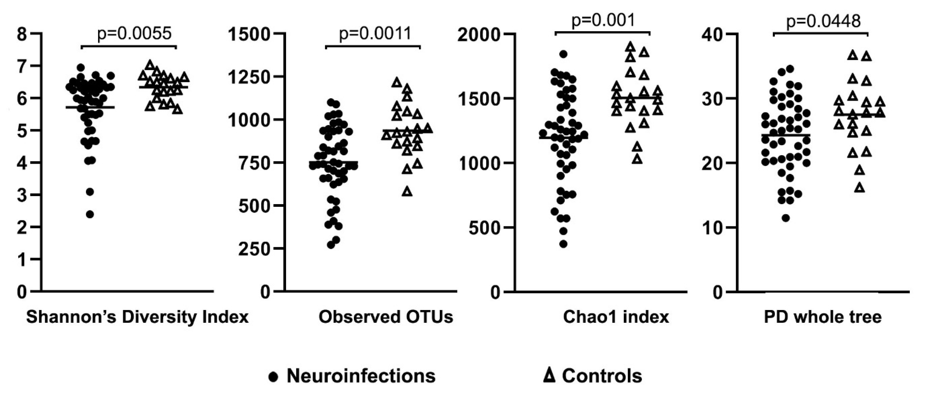
Quality control of raw reads was performed using FastQC software [20]. Reads were trimmed using trimmomatic [21] and, based on their size, selection was performed using BBTools [22]. Reads were further analyzed with QIIME version 1.9.1 [23]. In short, forward and reverse reads were merged using the fastq-join command [24]. Operational taxonomic units (OTUs) were determined by an open-reference OTU picking process in which reads were clustered against reference sequences (threshold value of 97% sequence similarity), and any unmapped reads were subsequently clustered de novo. Chimera detection was carried out using ChimeraSlayer [25]. Normalization was performed with the use of metagenomeSeq’s CSS (cumulative sum scaling) transformation [26].
Statistical analysis was performed on alpha and beta diversity metrics. Principal Coordinates Analysis (PCoA) plots (unweighted and weighted UniFrac data) for beta diversity were generated using QIIME data in PhyloToAST [27]. Abundance plots were prepared using the phyloseq package in R [28]. The Mann–Whitney U test was used for alpha diversity statistics, whereas taxonomical comparison between different groups was performed using a nonparametric t-test. Additionally, categorical variable analysis of similarities (ANOSIM) was performed.

3. Results

The number of 16S rRNA sequencing reads in patients with neuroinfections was 18,072–48,668, with a mean of 37,085 sequences per sample, whereas in controls, the number of sequences ranged from 20,863 to 53,590 (mean 40,010). Results of the open-reference OTU picking process using a normalized dataset are shown in Figure 1.
After normalization, 14 and 12 bacterial phyla were identified in neuroinfections and control patients, respectively. Representatives of Bacillota were significantly less abundant in patients than in controls (p = 0.015) and this was also true for both viral (p = 0.037) and bacterial etiology (p = 0.037). Moreover, bacterial meningitis had a significantly higher abundance of Actinomycetota (p = 0.037) compared to controls, whereas Verrucomicrobiota (p = 0.03) was more abundant in viral cases than in healthy subjects (Table 2). At the Class level, there were differences in Clostridia, Coriobacteriia, and Verrucomicrobiae (Table 2). A lower abundance of Clostridiales was observed in all patients with neuroinfection when compared to controls, whereas Coriobacteriales were significantly more prevalent in the latter group. At the Family level, there were no significant differences, with the only exception being increased ratios of Eubacteriaceae and Verrucomicrobiaceae in viral cases when compared to controls. Six bacterial genera were less abundant in the whole neuroinfection group and also in viral etiology cases when compared to controls. There were no differences in genera between bacterial meningitis and healthy subjects. Similarly, there were no statistically significant differences in gut microbial composition across any taxonomic level between viral and bacterial etiology of neuroinfection (Table 2).
Patients with neuroinfections and controls differed with respect to some alpha diversity metrics. Thus, the Shannon’s diversity index (p = 0.0055) and observed OTUs (p = 0.0011) were significantly lower in the entire group of neuroinfections compared to controls (Figure 2). Furthermore, the Shannon’s diversity index (p = 0.011), observed OTUs (p = 0.0069), Chao1 index (p = 0.004), and PD whole tree index (p = 0.0319) were significantly lower in samples with viral etiology compared to controls.
Beta diversity, defined by principal coordinate plots (PCoA) and the analysis of similarities (ANOSIM), did not differ significantly between patients with neuroinfection and controls. Similarly, there were no differences when bacterial and viral cases were compared to each other and to controls (Figure 3).
Comparisons of alpha and beta diversity metrics between patients receiving antibiotics and those who remained untreated did not reveal any significant differences (Figure 4). However, the Chao1 index and Shannon’s diversity index were borderline significant (p value was 0.0516 and 0.0689, respectively).

4. Discussion

Gut microbiota composition may affect a number of immune mechanisms, including lipopolysaccharide(LPS)-mediated modulation of inflammation [29], toll-like receptors (TLRs) expression [30], or changes in proinflammatory cytokines [31], which in turn could modify the course and outcome of CNS infections. Furthermore, such metabolites as occludin and claudin 5 could affect the permeability of the blood–brain barrier (BBB) and thus have an effect on the course of neuroinfection [32]. Unlike in patients suffering from chronic inflammatory maladies [33,34], changes in gut microbial composition were rarely studied in those with acute inflammatory processes. So far, alterations in the intestinal microbiome have been described in patients with acute pancreatitis [35] and acute myocardial infarction [36]. Moreover, experimental studies in animal models demonstrated a link between intestinal microbiota and the possible outcome of acute kidney injuries [37].
In our study, gut 16S rRNA profiling in patients with neuroinfection revealed significant differences in microbial composition at different taxonomic levels when compared to healthy subjects. In contrast to the negative study analyzing gut microbiota in 23 NMDAR encephalitis, we found a lower abundance of genera representing the family of Lachnospiraceae (Anaerostipes, Roseburi, Lachnobacterium, and Lachnospira) and genus Clostridium in the entire neuroinfection group, as well as in patients with viral etiology. Another 16S rRNA-based analysis of fecal microbiota in NMDAR encephalitis revealed dysbiosis in the gut microbiome represented mainly by the depletion of short-chain fatty acid (SCFA)-producing bacteria, which is similar to our study [38]. SCFAs such as butyrate, acetate, and succinate were associated with the pathophysiology of several neurological disorders, including AD, MS, PD, depression, and many others [39]. Moreover, based on a comparison between germ-free mice and those with normal gut flora, it was found that bacterial strains that produce SCFAs improve the integrity of the blood–brain barrier [40]. SCFAs also regulate microglia homeostasis, and it was reported that oral administration of acetate, propionate and butyrate is sufficient to drive microglia maturation in germ-free mice [41,42].
Alterations in specific gut microbiota were also reported in anti-leucine-rich glioma-inactivated 1 (anti-LGI1) autoimmune encephalitis patients. Similarly to our findings, genera Roseburia and Lachnospira were decreased in encephalitis patients when compared to healthy controls [43]. It should be noted that in the same study, genus Clostridium was more abundant in anti-LGI1 encephalitis patients than in controls [43]. However, due to the different pathogenesis of autoimmune encephalitis, the relevance of the above findings to our analysis remains unclear.
In our study, alpha diversity parameters reflecting the within-sample diversity of the gut microbiota were lower in patients with neuroinfection compared to controls. It is widely believed that increased diversity is associated with health, while its decrease is associated with dysbiosis and chronic diseases (e.g., inflammatory bowel diseases, hypertension, obesity) [33,44,45,46]. While treatment with antibiotics did not significantly affect beta diversity metrics, such alpha diversity metrics as Chao1 (p = 0.0516) and Shannon’s diversity index (p = 0.0689) were close to being statistically significant. It is likely that these differences would be more pronounced after slightly longer treatment. So far, there is little data on the effect of short-term antibiotic treatment on the human gut microbiome, as most studies concentrate on long-term sequelae and microbial recovery [47,48]. However, it was reported that 3 to 7 days of antibiotic intake might decrease the microbial load in the human gut [49]. In yet another study, Gu et al. showed that changes in alpha and beta diversity in the gut microbiota of mice exposed for four days to antibiotics occur four days after the end of treatment [50]. In another study on mice, model changes in intestinal microbiota were observed 1–5 days after the end of antibiotic treatment. These changes developed after 3 days of treatment and were highly dependable on the type of antibiotic—there were significant shifts in bacterial phyla when mice were receiving dicloxacillin and clindamycin, and only minor changes were observed when ciprofloxacin, cefotaxime, or cefuroxime were applied [51]. However, another study showed a rapid reduction in the abundance of gut bacteria in mice already on the second day of exposure to broad-spectrum antibiotics [52].
Whether alpha diversity downregulation is associated with such neurological diseases as MS and PD remains unclear, as concluded by a recent meta-analysis [53]. Similarly, studies on autoimmune encephalitis are inconclusive and contradictory. Thus, while in NMDAR encephalitis patients, alpha diversity was reported to be higher or similar to controls, in anti-LGI1 encephalitis, it was found to be lower [12,38,43]. Loss of microbial diversity can occur within just a few days due to various factors such as changes in nutrition (which are common during symptomatic diseases), hospitalization, and the administration of common drugs—and the degree of this loss may correlate with the overall disease severity [54,55,56].

5. Conclusions

In conclusion, in our analysis of the gut microbiome, some particular bacterial taxa (Clostridium, Anaerostipes, Lachnobacterium, Lachnospira, and Roseburia) were decreased in patients with neuroinfection compared to controls. Similarly, alpha diversity metrics showed lower within-sample diversity in these patients. The cause of these changes is unclear and could be due to the inflammatory process accompanying the disease; however, the effect of diet modification and/or hospitalization itself cannot be excluded. Short-term (1–3 day) antibiotic treatment did not affect beta diversity, but alpha diversity metrics were close to being statistically significant.

Author Contributions

Conceptualization, M.G., K.P., T.L. and M.R.; methodology, M.G., K.P., U.L. and A.P.; software, K.P.; investigation, M.P., M.M. and A.H.; data curation, U.L., A.P., M.M. and A.H.; writing—original draft preparation, M.G. and K.P.; writing—review & editing, K.P., T.L. and M.R.; visualization, M.G. and K.P.; supervision, K.P., T.L. and M.R. All authors have read and agreed to the published version of the manuscript.

Funding

This study was funded by grant 2017/25/B/NZ6/01463 from the National Science Center, Poland.

Institutional Review Board Statement

The study was approved by the Internal Review Board of the Medical University of Warsaw (approval number: KB/8/2015).

Informed Consent Statement

Informed consent was obtained from all subjects involved in the study.

Data Availability Statement

The datasets generated during the current study are available in the Sequence Read Archive (SRA accession: BioProject PRJNA836385).

Conflicts of Interest

Authors declare no conflict of interest.

References

  1. Scheperjans, F.; Aho, V.; Pereira, P.A.; Koskinen, K.; Paulin, L.; Pekkonen, E.; Haapaniemi, E.; Kaakkola, S.; Eerola-Rautio, J.; Pohja, M.; et al. Gut microbiota are related to Parkinson’s disease and clinical phenotype. Mov. Disord. 2015, 30, 350–358. [Google Scholar] [CrossRef] [PubMed]
  2. Shen, T.; Yue, Y.; He, T.; Huang, C.; Qu, B.; Lv, W.; Lai, H.Y. The Association Between the Gut Microbiota and Parkinson’s Disease, a Meta-Analysis. Front. Aging Neurosci. 2021, 13, 636545. [Google Scholar] [CrossRef] [PubMed]
  3. van Olst, L.; Roks, S.J.M.; Kamermans, A.; Verhaar, B.J.H.; van der Geest, A.M.; Muller, M.; van der Flier, W.M.; de Vries, H.E. Contribution of Gut Microbiota to Immunological Changes in Alzheimer’s Disease. Front. Immunol. 2021, 12, 683068. [Google Scholar] [CrossRef] [PubMed]
  4. Miyake, S.; Kim, S.; Suda, W.; Oshima, K.; Nakamura, M.; Matsuoka, T.; Chihara, N.; Tomita, A.; Sato, W.; Kim, S.W.; et al. Dysbiosis in the Gut Microbiota of Patients with Multiple Sclerosis, with a Striking Depletion of Species Belonging to Clostridia XIVa and IV Clusters. PLoS ONE 2015, 10, e0137429. [Google Scholar] [CrossRef] [PubMed] [Green Version]
  5. Cantoni, C.; Lin, Q.; Dorsett, Y.; Ghezzi, L.; Liu, Z.; Pan, Y.; Chen, K.; Han, Y.; Li, Z.; Xiao, H.; et al. Alterations of host-gut microbiome interactions in multiple sclerosis. EBioMedicine 2022, 76, 103798. [Google Scholar] [CrossRef]
  6. McGuinness, A.J.; Davis, J.A.; Dawson, S.L.; Loughman, A.; Collier, F.; O’Hely, M.; Simpson, C.A.; Green, J.; Marx, W.; Hair, C.; et al. A systematic review of gut microbiota composition in observational studies of major depressive disorder, bipolar disorder and schizophrenia. Mol. Psychiatry 2022, 27, 1920–1935. [Google Scholar] [CrossRef]
  7. Garcia-Gutierrez, E.; Narbad, A.; Rodriguez, J.M. Autism Spectrum Disorder Associated With Gut Microbiota at Immune, Metabolomic, and Neuroactive Level. Front. Neurosci. 2020, 14, 578666. [Google Scholar] [CrossRef]
  8. Li, Y.; Zhang, B.; Zhou, Y.; Wang, D.; Liu, X.; Li, L.; Wang, T.; Zhang, Y.; Jiang, M.; Tang, H.; et al. Gut Microbiota Changes and Their Relationship with Inflammation in Patients with Acute and Chronic Insomnia. Nat. Sci. Sleep 2020, 12, 895–905. [Google Scholar] [CrossRef]
  9. Voigt, R.M.; Forsyth, C.B.; Green, S.J.; Engen, P.A.; Keshavarzian, A. Circadian Rhythm and the Gut Microbiome. Int. Rev. Neurobiol. 2016, 131, 193–205. [Google Scholar]
  10. Wang, Y.; Wang, Z.; Wang, Y.; Li, F.; Jia, J.; Song, X.; Qin, S.; Wang, R.; Jin, F.; Kitazato, K.; et al. The Gut-Microglia Connection: Implications for Central Nervous System Diseases. Front. Immunol. 2018, 9, 2325. [Google Scholar] [CrossRef] [Green Version]
  11. Gubert, C.; Gasparotto, J.; Morais, L.M. Convergent pathways of the gut microbiota-brain axis and neurodegenerative disorders. Gastroenterol. Rep. 2022, 10, goac017. [Google Scholar] [CrossRef] [PubMed]
  12. Herken, J.; Bang, C.; Ruhlemann, M.C.; Finke, C.; Klag, J.; Franke, A.; Pruss, H. Normal gut microbiome in NMDA receptor encephalitis. Neurol. Neuroimmunol. Neuroinflamm. 2019, 6, e632. [Google Scholar] [CrossRef] [PubMed] [Green Version]
  13. Kennedy, P.G.E.; Quan, P.L.; Lipkin, W.I. Viral Encephalitis of Unknown Cause: Current Perspective and Recent Advances. Viruses 2017, 9, 138. [Google Scholar] [CrossRef] [PubMed]
  14. Potharaju, N.R. Incidence Rate of Acute Encephalitis Syndrome without Specific Treatment in India and Nepal. Indian J. Community Med. 2012, 37, 240–251. [Google Scholar] [CrossRef]
  15. Glaser, C.A.; Gilliam, S.; Schnurr, D.; Forghani, B.; Honarmand, S.; Khetsuriani, N.; Fischer, M.; Cossen, C.K.; Anderson, L.J. In search of encephalitis etiologies: Diagnostic challenges in the California Encephalitis Project, 1998–2000. Clin. Infect. Dis. 2003, 36, 731–742. [Google Scholar] [CrossRef]
  16. Perlejewski, K.; Bukowska-Osko, I.; Rydzanicz, M.; Pawelczyk, A.; Caraballo Corts, K.; Osuch, S.; Paciorek, M.; Dzieciatkowski, T.; Radkowski, M.; Laskus, T. Next-generation sequencing in the diagnosis of viral encephalitis: Sensitivity and clinical limitations. Sci. Rep. 2020, 10, 16173. [Google Scholar] [CrossRef]
  17. Glaser, C.A.; Honarmand, S.; Anderson, L.J.; Schnurr, D.P.; Forghani, B.; Cossen, C.K.; Schuster, F.L.; Christie, L.J.; Tureen, J.H. Beyond viruses: Clinical profiles and etiologies associated with encephalitis. Clin. Infect. Dis. 2006, 43, 1565–1577. [Google Scholar] [CrossRef] [Green Version]
  18. Granerod, J.; Ambrose, H.E.; Davies, N.W.S.; Clewley, J.P.; Walsh, A.L.; Morgan, D.; Cunningham, R.; Zuckerman, M.; Mutton, K.J.; Solomon, T.; et al. Causes of encephalitis and differences in their clinical presentations in England: A multicentre, population-based prospective study. Lancet Infect. Dis. 2010, 10, 835–844. [Google Scholar] [CrossRef] [Green Version]
  19. Klindworth, A.; Pruesse, E.; Schweer, T.; Peplies, J.; Quast, C.; Horn, M.; Glockner, F.O. Evaluation of general 16S ribosomal RNA gene PCR primers for classical and next-generation sequencing-based diversity studies. Nucleic Acids Res. 2013, 41, e1. [Google Scholar] [CrossRef]
  20. Andrews, S. FastQC: A Quality Control Tool for High Throughput Sequence Data. 2010. Available online: http://www.bioinformatics.babraham.ac.uk/projects/fastqc (accessed on 2 January 2022).
  21. Bolger, A.M.; Lohse, M.; Usadel, B. Trimmomatic: A flexible trimmer for Illumina sequence data. Bioinformatics 2014, 30, 2114–2120. [Google Scholar] [CrossRef] [Green Version]
  22. Bushnell, B.; Rood, J.; Singer, E. BBMerge—Accurate paired shotgun read merging via overlap. PLoS ONE 2017, 12, e0185056. [Google Scholar] [CrossRef] [PubMed]
  23. Caporaso, J.G.; Kuczynski, J.; Stombaugh, J.; Bittinger, K.; Bushman, F.D.; Costello, E.K.; Fierer, N.; Pena, A.G.; Goodrich, J.K.; Gordon, J.I.; et al. QIIME allows analysis of high-throughput community sequencing data. Nat. Methods 2010, 7, 335–336. [Google Scholar] [CrossRef] [PubMed] [Green Version]
  24. Aronesty, E. Ea-Utils: “Command-Line Tools for Processing Biological Sequencing Data”. 2011. Available online: https://github.com/ExpressionAnalysis/ea-utils (accessed on 2 January 2022).
  25. Haas, B.J.; Gevers, D.; Earl, A.M.; Feldgarden, M.; Ward, D.V.; Giannoukos, G.; Ciulla, D.; Tabbaa, D.; Highlander, S.K.; Sodergren, E.; et al. Chimeric 16S rRNA sequence formation and detection in Sanger and 454-pyrosequenced PCR amplicons. Genome Res. 2011, 21, 494–504. [Google Scholar] [CrossRef] [PubMed] [Green Version]
  26. Paulson, J.N.; Stine, O.C.; Bravo, H.C.; Pop, M. Differential abundance analysis for microbial marker-gene surveys. Nat. Methods 2013, 10, 1200–1202. [Google Scholar] [CrossRef] [Green Version]
  27. Dabdoub, S.M.; Fellows, M.L.; Paropkari, A.D.; Mason, M.R.; Huja, S.S.; Tsigarida, A.A.; Kumar, P.S. PhyloToAST: Bioinformatics tools for species-level analysis and visualization of complex microbial datasets. Sci. Rep. 2016, 6, 29123. [Google Scholar] [CrossRef] [Green Version]
  28. McMurdie, P.J.; Holmes, S. phyloseq: An R package for reproducible interactive analysis and graphics of microbiome census data. PLoS ONE 2013, 8, e61217. [Google Scholar] [CrossRef] [PubMed] [Green Version]
  29. Noori, M.S.; Courreges, M.C.; Bergmeier, S.C.; McCall, K.D.; Goetz, D.J. Modulation of LPS-induced inflammatory cytokine production by a novel glycogen synthase kinase-3 inhibitor. Eur. J. Pharmacol. 2020, 883, 173340. [Google Scholar] [CrossRef]
  30. Takeda, K.; Akira, S. Roles of Toll-like receptors in innate immune responses. Genes Cells 2001, 6, 733–742. [Google Scholar] [CrossRef]
  31. Bailey, M.T.; Dowd, S.E.; Galley, J.D.; Hufnagle, A.R.; Allen, R.G.; Lyte, M. Exposure to a social stressor alters the structure of the intestinal microbiota: Implications for stressor-induced immunomodulation. Brain Behav. Immun. 2011, 25, 397–407. [Google Scholar] [CrossRef] [Green Version]
  32. Jia, W.; Lu, R.; Martin, T.A.; Jiang, W.G. The role of claudin-5 in blood-brain barrier (BBB) and brain metastases (review). Mol. Med. Rep. 2014, 9, 779–785. [Google Scholar] [CrossRef] [Green Version]
  33. Gong, D.; Gong, X.; Wang, L.; Yu, X.; Dong, Q. Involvement of Reduced Microbial Diversity in Inflammatory Bowel Disease. Gastroenterol Res. Pract. 2016, 2016, 6951091. [Google Scholar] [CrossRef] [PubMed]
  34. Wehedy, E.; Shatat, I.F.; Al Khodor, S. The Human Microbiome in Chronic Kidney Disease: A Double-Edged Sword. Front. Med. 2021, 8, 790783. [Google Scholar] [CrossRef]
  35. Tan, C.; Ling, Z.; Huang, Y.; Cao, Y.; Liu, Q.; Cai, T.; Yuan, H.; Liu, C.; Li, Y.; Xu, K. Dysbiosis of Intestinal Microbiota Associated With Inflammation Involved in the Progression of Acute Pancreatitis. Pancreas 2015, 44, 868–875. [Google Scholar] [CrossRef]
  36. Han, Y.; Gong, Z.; Sun, G.; Xu, J.; Qi, C.; Sun, W.; Jiang, H.; Cao, P.; Ju, H. Dysbiosis of Gut Microbiota in Patients with Acute Myocardial Infarction. Front. Microbiol. 2021, 12, 680101. [Google Scholar] [CrossRef] [PubMed]
  37. Gong, J.; Noel, S.; Pluznick, J.L.; Hamad, A.R.A.; Rabb, H. Gut Microbiota-Kidney Cross-Talk in Acute Kidney Injury. Semin. Nephrol. 2019, 39, 107–116. [Google Scholar] [CrossRef] [PubMed] [Green Version]
  38. Gong, X.; Liu, X.; Li, C.; Chen, C.; Lin, J.; Li, A.; An, D.; Zhou, D.; Hong, Z. Alterations in the human gut microbiome in anti-N-methyl-D-aspartate receptor encephalitis. Ann. Clin. Transl. Neurol. 2019, 6, 1771–1781. [Google Scholar] [CrossRef]
  39. Mirzaei, R.; Bouzari, B.; Hosseini-Fard, S.R.; Mazaheri, M.; Ahmadyousefi, Y.; Abdi, M.; Jalalifar, S.; Karimitabar, Z.; Teimoori, A.; Keyvani, H.; et al. Role of microbiota-derived short-chain fatty acids in nervous system disorders. Biomed. Pharmacother. 2021, 139, 111661. [Google Scholar] [CrossRef]
  40. Braniste, V.; Al-Asmakh, M.; Kowal, C.; Anuar, F.; Abbaspour, A.; Toth, M.; Korecka, A.; Bakocevic, N.; Ng, L.G.; Kundu, P.; et al. The gut microbiota influences blood-brain barrier permeability in mice. Sci. Transl. Med. 2014, 6, 263ra158. [Google Scholar] [CrossRef] [Green Version]
  41. Erny, D.; Hrabe de Angelis, A.L.; Jaitin, D.; Wieghofer, P.; Staszewski, O.; David, E.; Keren-Shaul, H.; Mahlakoiv, T.; Jakobshagen, K.; Buch, T.; et al. Host microbiota constantly control maturation and function of microglia in the CNS. Nat. Neurosci. 2015, 18, 965–977. [Google Scholar] [CrossRef]
  42. Silva, Y.P.; Bernardi, A.; Frozza, R.L. The Role of Short-Chain Fatty Acids From Gut Microbiota in Gut-Brain Communication. Front. Endocrinol. 2020, 11, 25. [Google Scholar] [CrossRef] [Green Version]
  43. Ma, X.; Ma, L.; Wang, Z.; Liu, Y.; Long, L.; Ma, X.; Chen, H.; Chen, Z.; Lin, X.; Si, L.; et al. Clinical Features and Gut Microbial Alterations in Anti-leucine-rich Glioma-Inactivated 1 Encephalitis-A Pilot Study. Front. Neurol. 2020, 11, 585977. [Google Scholar] [CrossRef] [PubMed]
  44. Pickard, J.M.; Zeng, M.Y.; Caruso, R.; Nunez, G. Gut microbiota: Role in pathogen colonization, immune responses, and inflammatory disease. Immunol. Rev. 2017, 279, 70–89. [Google Scholar] [CrossRef] [PubMed]
  45. Jin, L.; Shi, X.; Yang, J.; Zhao, Y.; Xue, L.; Xu, L.; Cai, J. Gut microbes in cardiovascular diseases and their potential therapeutic applications. Protein Cell 2021, 12, 346–359. [Google Scholar] [CrossRef] [PubMed]
  46. Davis, C.D. The Gut Microbiome and Its Role in Obesity. Nutr. Today 2016, 51, 167–174. [Google Scholar] [CrossRef] [PubMed] [Green Version]
  47. Palleja, A.; Mikkelsen, K.H.; Forslund, S.K.; Kashani, A.; Allin, K.H.; Nielsen, T.; Hansen, T.H.; Liang, S.; Feng, Q.; Zhang, C.; et al. Recovery of gut microbiota of healthy adults following antibiotic exposure. Nat. Microbiol. 2018, 3, 1255–1265. [Google Scholar] [CrossRef]
  48. Kwon, Y.; Cho, Y.-S.; Lee, Y.-M.; Kim, S.-j.; Bae, J.; Jeong, S.-J. Changes to Gut Microbiota Following Systemic Antibiotic Administration in Infants. Antibiotics 2022, 11, 470. [Google Scholar] [CrossRef]
  49. Panda, S.; El khader, I.; Casellas, F.; Lopez Vivancos, J.; Garcia Cors, M.; Santiago, A.; Cuenca, S.; Guarner, F.; Manichanh, C. Short-term effect of antibiotics on human gut microbiota. PLoS ONE 2014, 9, e95476. [Google Scholar] [CrossRef]
  50. Gu, S.L.; Gong, Y.W.; Zhang, J.Y.; Chen, Y.B.; Wu, Z.J.; Xu, Q.M.; Fang, Y.H.; Wang, J.X.; Tang, L.L. Effect of the Short-Term Use of Fluoroquinolone and beta-Lactam Antibiotics on Mouse Gut Microbiota. Infect. Drug Resist. 2020, 13, 4547–4558. [Google Scholar] [CrossRef]
  51. Hertz, F.B.; Budding, A.E.; van der Lugt-Degen, M.; Savelkoul, P.H.; Lobner-Olesen, A.; Frimodt-Moller, N. Effects of Antibiotics on the Intestinal Microbiota of Mice. Antibiotics 2020, 9, 191. [Google Scholar] [CrossRef]
  52. Yao, J.; Carter, R.A.; Vuagniaux, G.; Barbier, M.; Rosch, J.W.; Rock, C.O. A Pathogen-Selective Antibiotic Minimizes Disturbance to the Microbiome. Antimicrob. Agents Chemother. 2016, 60, 4264–4273. [Google Scholar] [CrossRef] [Green Version]
  53. Plassais, J.; Gbikpi-Benissan, G.; Figarol, M.; Scheperjans, F.; Gorochov, G.; Derkinderen, P.; Cervino, A.C.L. Gut microbiome alpha-diversity is not a marker of Parkinson’s disease and multiple sclerosis. Brain Commun. 2021, 3, fcab113. [Google Scholar] [CrossRef] [PubMed]
  54. McDonald, D.; Ackermann, G.; Khailova, L.; Baird, C.; Heyland, D.; Kozar, R.; Lemieux, M.; Derenski, K.; King, J.; Vis-Kampen, C.; et al. Extreme Dysbiosis of the Microbiome in Critical Illness. mSphere 2016, 1, e00199-16. [Google Scholar] [CrossRef] [Green Version]
  55. Ravi, A.; Halstead, F.D.; Bamford, A.; Casey, A.; Thomson, N.M.; van Schaik, W.; Snelson, C.; Goulden, R.; Foster-Nyarko, E.; Savva, G.M.; et al. Loss of microbial diversity and pathogen domination of the gut microbiota in critically ill patients. Microb. Genom. 2019, 5, e000293. [Google Scholar] [CrossRef] [PubMed]
  56. Zaborin, A.; Smith, D.; Garfield, K.; Quensen, J.; Shakhsheer, B.; Kade, M.; Tirrell, M.; Tiedje, J.; Gilbert, J.A.; Zaborina, O.; et al. Membership and behavior of ultra-low-diversity pathogen communities present in the gut of humans during prolonged critical illness. mBio 2014, 5, e01361-14. [Google Scholar] [CrossRef] [PubMed] [Green Version]
Figure 1. Relative abundance above 1% (all Phylum taxa were visualized) at different bacterial taxonomic levels in patients with neuroinfection and controls.
Cimb 44 00200 g001
Figure 2. Various alpha diversity metrics in 47 patients with neuroinfection and 20 controls.
Cimb 44 00200 g002
Figure 3. PCoA of unweighted and weighted UniFrac distances at genus level in 47 patients with neuroinfection (divided by the etiology into bacterial, viral, and undefined groups) and 20 controls.
Cimb 44 00200 g003
Figure 4. Comparison between 47 patients with neuroinfection receiving antibiotics and not receiving antibiotics. (A) Beta diversity—PCoA of unweighted and weighted UniFrac distances at genus level. (B) Alpha diversity metrics.
Cimb 44 00200 g004
Table 1. Demographical, clinical, and laboratory data in 47 patients with neuroinfection and 20 controls.
NeuroinfectionsNumber (%)
All PatientsViralBacterialUndefined
Nº of patients4726 (55.32)8 (17.02)13 (27.66)
Male23 (48.94)13 (50)4 (50)6 (46.15)
Female24 (51.06)13 (50)4 (50)7 (53.85)
Age, Mean 43415044
Symptoms and signsNumber (%)
All patientsViralBacterialUndefined
Fever31 (65.96)19 (73.08)6 (75)6 (46.15)
Headache37 (78.72)22 (84.62)6 (75)9 (69.26)
Altered mental status11 (23.40)2 (7.69)4 (50)5 (38.46)
Loss of consciousness6 (12.76)2 (7.69)1 (50)3 (23.08)
Seizures of epilepsy4 (8.51)1 (3.85)03 (23.08)
Focal neurologic signs19 (40.43)13 (50)6 (75)0
CSF analysis (ref. values)Mean value/standard deviation
All patientsViralBacterialUndefined
Total cell count, cells/µL (≤5)190/238155/120619/37964/114
% of lymphocytes 63/30.4669/2720/650/37
Chlorides, mmol/L (>117)121.72/4.15121.61/3.22119.00/5.61123.49/3.91
Protein, g/L (0.15–0.45)1.51/1.621.09/0.892.51/1.871.77/2.23
L-Lactic acid, mmol/L (≤2.1)2.23/0.992.15/0.614.94/1.331.93/0.77
Glucose, mmol/L (2.4–4.7)2.80/0.872.97/0.351.64/0.733.28/1.02
Blood test (ref. values)Mean value/standard deviation
All patientsViralBacterialUndefined
WBC, ×103/µL (–10)10.65/4.049.05/2.7214.74/3.4210.87/4.48
RBC, ×106/µL (4.5–5.9)4.56/0.574.65/0.513.99/0.754.63/0.44
Platelet count, ×103/µL (150–450)238.48/63.80233.87/52.42204.50/58.14265.80/70.69
CRP, mg/L (<5)40.84/63.0413.33/8.55120.37/86.6619.77/18.77
TreatmentNumber (%)
All patientsViralBacterialUndefined
Antibiotics23 (48.94)9 (34.62)8 (100)6 (46.15)
Antiviral drugs (Aciclovir)15 (31.9)10 (38.46)05 (38.46)
Antiepileptic Drugs7 (14.89)2 (7.69)1 (12.50)4 (30.77)
Control group (%)
Nº of controls20
Male10 (50)
Female10 (50)
Age, Mean43
Table 2. Differences at taxonomic ranks between patients with neuroinfections and healthy controls.
TaxonomyControls (n = 20) vs.
Neuroinfections
(n = 47)
Bacterial
(n = 8)
Viral
(n = 26)
Phylump__ Bacillota0.0150.0370.037
p__ Actinomycetota 0.037
p__ Verrucomicrobiota 0.030
Classp__ Bacillota;c__Clostridia0.0260.0130.026
p__ Actinomycetota;c__Coriobacteriia 0.013
p__ Verrucomicrobiota;c__Verrucomicrobiae 0.039
Orderp__ Bacillota;c__Clostridia;o__Clostridiales0.0430.021
p__ Actinomycetota;c__Coriobacteriia; o__Coriobacteriales 0.021
Familyp__ Bacillota;c__Clostridia;o__Clostridiales;f__Eubacteriaceae 0.045
p__ Verrucomicrobiota;c__Verrucomicrobiae; o__Verrucomicrobiales;f__Verrucomicrobiaceae 0.045
Genusp__ Bacillota;c__Clostridia;o__Clostridiales;f__Clostridiaceae;g__Clostridium0.025 0.040
p__ Bacillota;c__Clostridia;o__Clostridiales;f__Lachnospiraceae;g__Anaerostipes0.025 0.040
p__ Bacillota;c__Clostridia;o__Clostridiales;f__Lachnospiraceae;g__Lachnobacterium0.025 0.040
p__ Bacillota;c__Clostridia;o__Clostridiales;f__Lachnospiraceae;g__Lachnospira0.025 0.040
p__ Bacillota;c__Clostridia;o__Clostridiales;f__Lachnospiraceae;g__Roseburia0.025 0.040
Publisher’s Note: MDPI stays neutral with regard to jurisdictional claims in published maps and institutional affiliations.

