
Rotating Magnetic Field-Assisted Reactor Enhances Mechanisms of Phage Adsorption on Bacterial Cell Surface

 , , , , , and
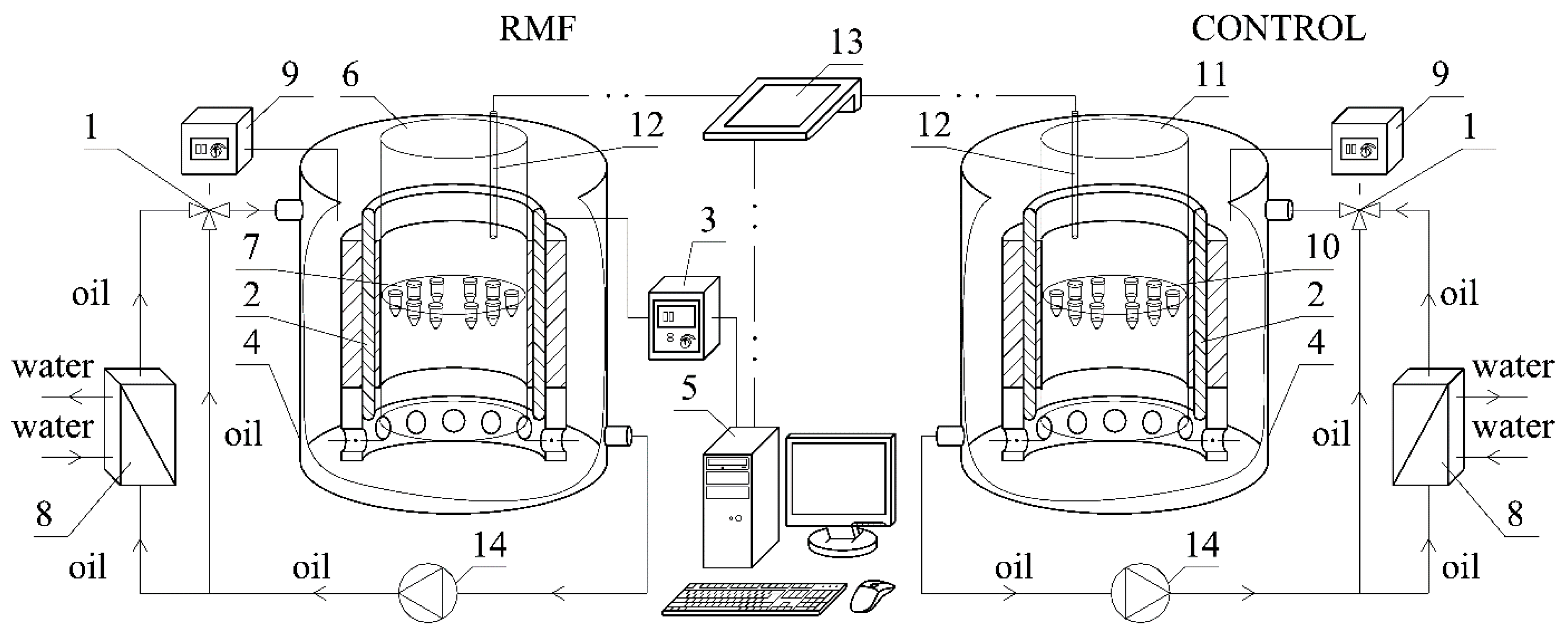
Abstract
:1. Introduction
2. Materials and Methods
2.1. Rotating Magnetic Field Assisted Reactor (RMF-AR)
2.2. Bacteria and Bacteriophage Used in Study and Culture Condition
2.3. Adsorption Rate Protocol under RMF Condition
- B—bacterial initial count, [CFU × mL−1];
- k—adsorption rate constants;
- P0—phage initial counts, [PFU × mL−1];
- P—phage count at time point, [PFU × mL−1];
- t—time, [min].
2.4. Outer Membrane Charge Assessment
2.5. Zeta Potential and Diffusion Coefficient Measurement
2.6. Statistical Analysis
3. Results
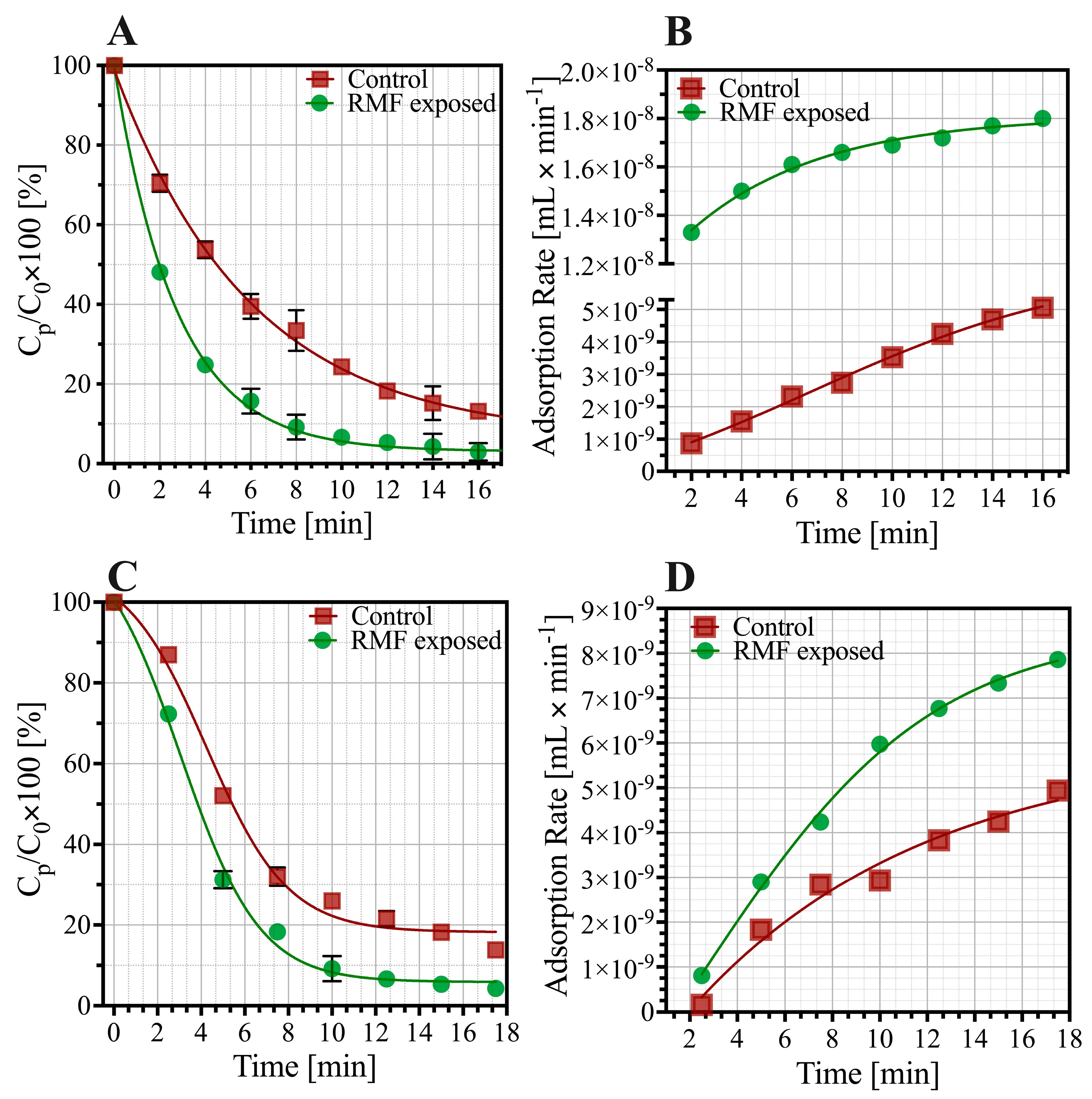
3.1. Effect of RMF-AR on the Bacteriophage Adsorption
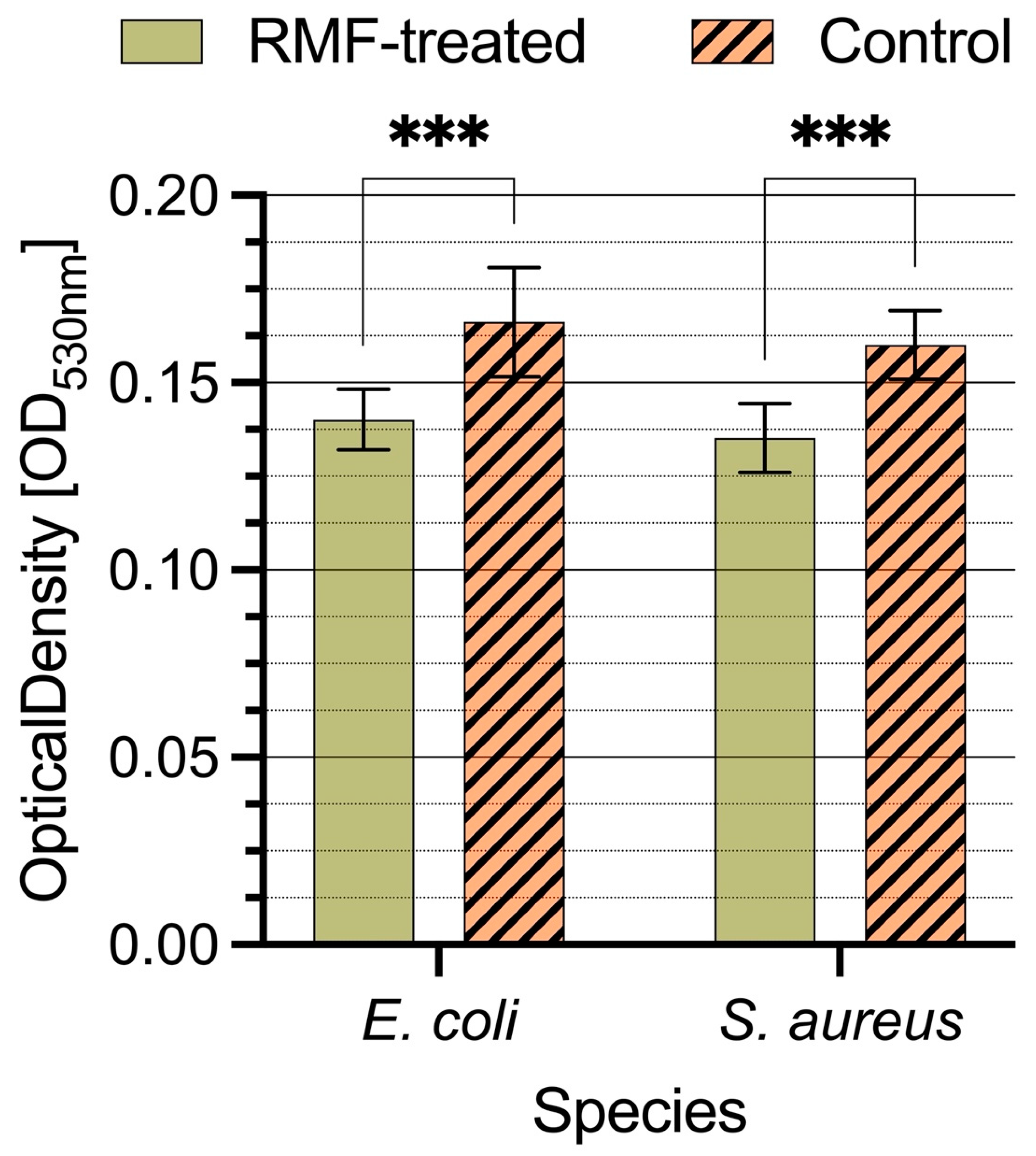
3.2. Effect of RMF-AR on Cytochrome C Binding on the Bacterial Cell Surface
3.3. Effects of RMF-AR on the Bacteria and Bacteriophage Zeta Potential
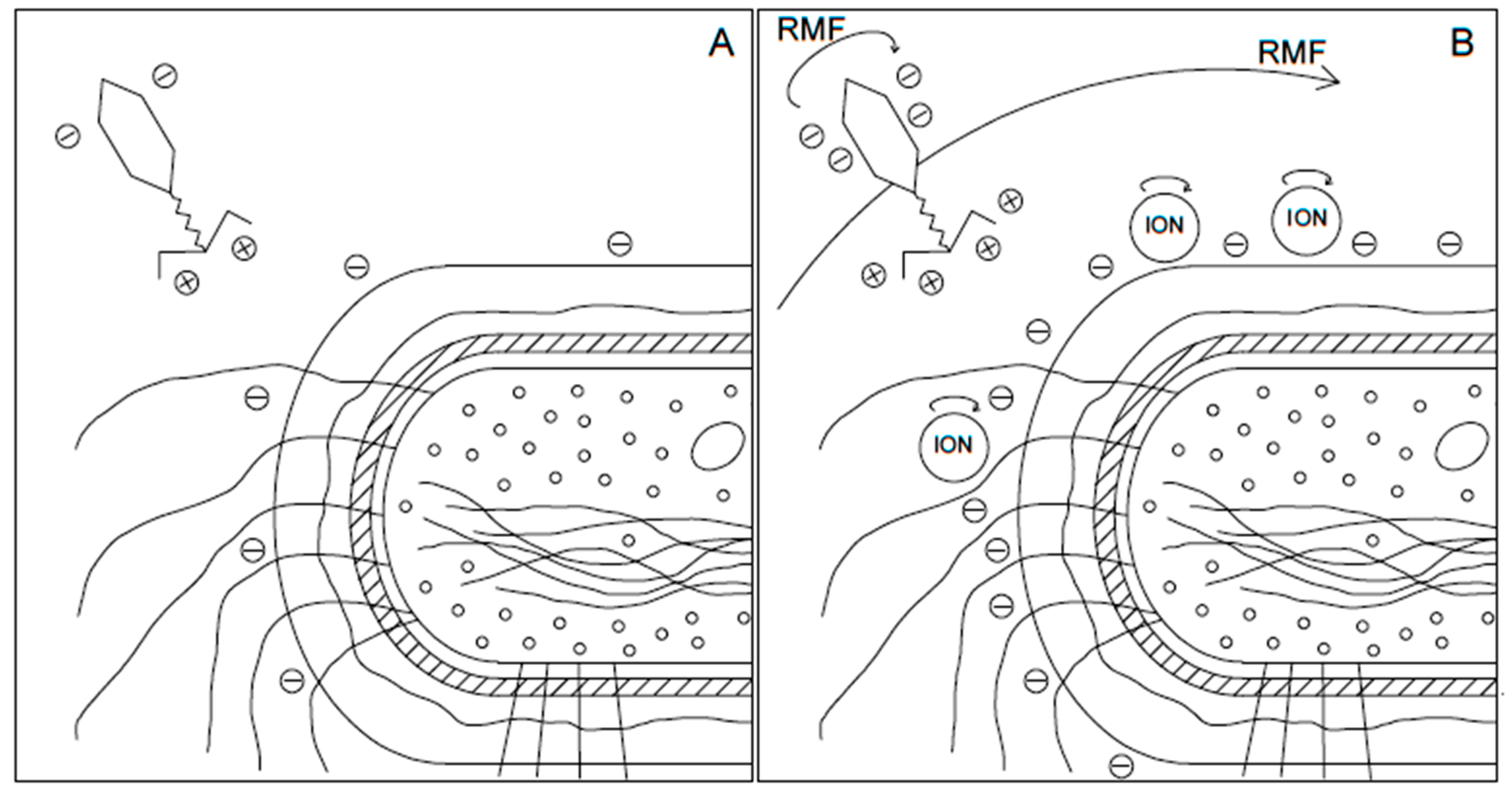
4. Discussion
5. Conclusions
Author Contributions
Funding
Conflicts of Interest
References
- Velizarov, S. Electric and Magnetic Fields in Microbial Biotechnology: Possibilities, Limitations, and Perspectives. Electromagn. Biol. Med. 1999, 18, 185–212. [Google Scholar] [CrossRef]
- Hristov, J. Magnetic field assisted fluidization—A unified approach. Part 8. Mass transfer: Magnetically assisted bioprocesses. Rev. Chem. Eng. 2010, 26, 55–128. [Google Scholar] [CrossRef]
- Ashok, A.; Doriya, K.; Rao, D.R.M.; Kumar, D.S. Design of solid state bioreactor for industrial applications: An overview to conventional bioreactors. Biocatal. Agric. Biotechnol. 2017, 9, 11–18. [Google Scholar] [CrossRef]
- Bakonyi, P.; Buitrón, G.; Valdez-Vazquez, I.; Nemestóthy, N.; Bélafi-Bakó, K. A novel gas separation integrated membrane bioreactor to evaluate the impact of self-generated biogas recycling on continuous hydrogen fermentation. Appl. Energy 2017, 190, 813–823. [Google Scholar] [CrossRef]
- Rakoczy, R. Mixing energy investigations in a liquid vessel that is mixed by using a rotating magnetic field. Chem. Eng. Process. Process Intensif. 2013, 66, 1–11. [Google Scholar] [CrossRef]
- Shanmugapriya, S.; Sarumathi, A.; Saravanan, N. Study of lipid profile changes and histopathology examination of heart under immobilization stress with static magnetic field exposure in rats. J. Environ. Biol. 2012, 2, 41–49. [Google Scholar]
- Hajiani, P.; Larachi, F. Controlling lateral nanomixing and velocity profile of dilute ferrofluid capillary flows in uniform stationary, oscillating and rotating magnetic fields. Chem. Eng. J. 2013, 223, 454–466. [Google Scholar] [CrossRef]
- Rakoczy, R. Enhancement of solid dissolution process under the influence of rotating magnetic field. Chem. Eng. Process. Process Intensif. 2010, 49, 42–50. [Google Scholar] [CrossRef]
- Rakoczy, R.; Masiuk, S. Studies of a mixing process induced by a transverse rotating magnetic field. Chem. Eng. Sci. 2011, 66, 2298–2308. [Google Scholar] [CrossRef]
- Rakoczy, R.; Konopacki, M.; Kordas, M.; Drozd, R.; Fijałkowski, K. Influence of rotating magnetic field on gas-liquid volumetric mass transfer coefficient. Chem. Process Eng. 2017, 38, 423–432. [Google Scholar] [CrossRef] [Green Version]
- Konopacka, A.; Rakoczy, R.; Konopacki, M. The effect of rotating magnetic field on bioethanol production by yeast strain modified by ferrimagnetic nanoparticles. J. Magn. Magn. Mater. 2019, 473, 176–183. [Google Scholar] [CrossRef]
- Konopacki, M.; Rakoczy, R. The analysis of rotating magnetic field as a trigger of Gram-positive and Gram-negative bacteria growth. Biochem. Eng. J. 2019, 141, 259–267. [Google Scholar] [CrossRef]
- Fijałkowski, K.; Zywicka, A.; Drozd, R.; Niemczyk, A.; Junka, A.F.; Peitler, D.; Kordas, M.; Konopacki, M.; Szymczyk, P.; Fray, M.E.; et al. Modification of bacterial cellulose through exposure to the rotating magnetic field. Carbohydr. Polym. 2015, 133, 52–60. [Google Scholar] [CrossRef]
- Nawrotek, P.; Fijałkowski, K.; Struk, M.; Kordas, M.; Rakoczy, R. Effects of 50 Hz rotating magnetic field on the viability of Escherichia coli and Staphylococcus aureus. Electromagn. Biol. Med. 2014, 33, 29–34. [Google Scholar] [CrossRef]
- Salmond, G.P.C.; Fineran, P.C. A century of the phage: Past, present and future. Nat. Rev. Microbiol. 2015, 13, 777–786. [Google Scholar] [CrossRef]
- Wojciuk, B.; Salabura, A.; Grygorcewicz, B.; Kędzierska, K.; Ciechanowski, K.; Dołęgowska, B. Urobiome: In Sickness and in Health. Microorganisms 2019, 7, 548. [Google Scholar] [CrossRef] [Green Version]
- Golkar, Z.; Bagasra, O.; Gene Pace, D. Bacteriophage therapy: A potential solution for the antibiotic resistance crisis. J. Infect. Dev. Ctries 2014, 8, 129–136. [Google Scholar] [CrossRef]
- Pei, R.; Lamas-Samanamud, G.R. Inhibition of biofilm formation by T7 bacteriophages producing quorum-quenching enzymes. Appl. Environ. Microbiol. 2014, 80, 5340–5348. [Google Scholar] [CrossRef] [Green Version]
- Szymczak, M.; Grygorcewicz, B.; Karczewska-Golec, J.; Decewicz, P.; Pankowski, J.A.; Országh-Szturo, H.; Bącal, P.; Dołęgowska, B.; Golec, P. Characterization of a Unique Bordetella bronchiseptica vB_BbrP_BB8 Bacteriophage and Its Application as an Antibacterial Agent. Int. J. Mol. Sci. 2020, 21, 1403. [Google Scholar] [CrossRef] [Green Version]
- Szot-Karpińska, K.; Golec, P.; Leśniewski, A.; Pałys, B.; Marken, F.; Niedziółka-Jönsson, J.; Wȩgrzyn, G.; Łoś, M. Modified Filamentous Bacteriophage as a Scaffold for Carbon Nanofiber. Bioconjug. Chem. 2016, 27, 2900–2910. [Google Scholar] [CrossRef]
- Monk, A.B.; Rees, C.D.; Barrow, P.; Hagens, S.; Harper, D.R. Bacteriophage applications: Where are we now? Lett. Appl. Microbiol. 2010, 51, 363–369. [Google Scholar] [CrossRef] [PubMed]
- Grygorcewicz, B.; Chajecka-Wierzchowska, W.; Augustyniak, A.; Wasak, A.; Stachurska, X.; Nawrotek, P.; Dołęgowska, B. In-milk inactivation of Escherichia coli O157:H7 by the environmental lytic bacteriophage ECPS-6. J. Food Saf. 2019, 40, e12747. [Google Scholar] [CrossRef]
- Grygorcewicz, B.; Grudziński, M.; Wasak, A.; Augustyniak, A.; Pietruszka, A.; Nawrotek, P. Bacteriophage-mediated reduction of Salmonella Enteritidis in swine slurry. Appl. Soil Ecol. 2017, 119, 179–182. [Google Scholar] [CrossRef]
- Grygorcewicz, B.; Wojciuk, B.; Roszak, M.; Łubowska, N.; Blstrokaejczak, P.; Jursa-Kulesza, J.; Rakoczy, R.; Masiuk, H.; Dogowska, B. Environmental Phage-Based Cocktail and Antibiotic Combination Effects on Acinetobacter baumannii Biofilm in a Human Urine Model. Microb. Drug Resist. 2021, 27, 25–35. [Google Scholar] [CrossRef]
- Grygorcewicz, B.; Roszak, M.; Golec, P.; Śleboda-Taront, D.; Łubowska, N.; Górska, M.; Jursa-Kulesza, J.; Rakoczy, R.; Wojciuk, B.; Dołęgowska, B. Antibiotics Act with vB_AbaP_AGC01 Phage against Acinetobacter baumannii in Human Heat-Inactivated Plasma Blood and Galleria mellonella Models. Int. J. Mol. Sci. 2020, 21, 4390. [Google Scholar] [CrossRef]
- Łubowska, N.; Grygorcewicz, B.; Kosznik-Kwaśnicka, K.; Zauszkiewicz-Pawlak, A.; Węgrzyn, A.; Dołęgowska, B.; Piechowicz, L. Characterization of the Three New Kayviruses and Their Lytic Activity Against Multidrug-Resistant Staphylococcus aureus. Microorganisms 2019, 7, 471. [Google Scholar] [CrossRef] [Green Version]
- Kropinski, A.M. Measurement of the rate of attachment of bacteriophage to cells. Methods Mol. Biol. 2009, 501, 151–155. [Google Scholar]
- Lee, C.N.; Tseng, T.T.; Lin, J.W.; Fu, Y.C.; Weng, S.F.; Tseng, Y.H. Lytic myophage Abp53 encodes several proteins similar to those encoded by host Acinetobacter baumannii and phage phiKO2. Appl. Environ. Microbiol. 2011, 77, 6755–6762. [Google Scholar] [CrossRef] [Green Version]
- Beebout, C.J.; Sominsky, L.A.; Eberly, A.R.; Van Horn, G.T.; Hadjifrangiskou, M. Cytochrome bd promotes Escherichia coli biofilm antibiotic tolerance by regulating accumulation of noxious chemicals. NPJ Biofilms Microbiomes 2021, 7, 35. [Google Scholar] [CrossRef]
- Serwer, P.; Hayes, S.J. Agarose gel electrophoresis of bacteriophages and related particles. I. Avoidance of binding to the gel and recognizing of particles with packaged DNA. Electrophoresis 1982, 3, 76–80. [Google Scholar] [CrossRef]
- Zemb, O.; Manefield, M.; Thomas, F.; Jacquet, S. Phage adsorption to bacteria in the light of the electrostatics: A case study using E. coli, T2 and flow cytometry. J. Virol. Methods 2013, 189, 283–289. [Google Scholar] [CrossRef]
- Chhibber, S.; Kaur, T.; Kaur, S. Essential role of calcium in the infection process of broad-spectrum methicillin-resistant Staphylococcus aureus bacteriophage. J. Basic Microbiol. 2014, 54, 775–780. [Google Scholar] [CrossRef]
- Shafia, F.; Thompson, T.L. Calcium ion requirement for proliferation of bacteriophage phiMU-4. J. Bacteriol. 1964, 88, 293–296. [Google Scholar] [CrossRef] [Green Version]
- Armanious, A.; Aeppli, M.; Jacak, R.; Refardt, D.; Sigstam, T.; Kohn, T.; Sander, M. Viruses at Solid-Water Interfaces: A Systematic Assessment of Interactions Driving Adsorption. Environ. Sci. Technol. 2016, 50, 732–743. [Google Scholar] [CrossRef]
- Storms, Z.J.; Sauvageau, D. Modeling tailed bacteriophage adsorption: Insight into mechanisms. Virology 2015, 485, 355–362. [Google Scholar] [CrossRef] [Green Version]
- Moldovan, R.; Chapman-McQuiston, E.; Wu, X.L. On kinetics of phage adsorption. Biophys. J. 2007, 93, 303–315. [Google Scholar] [CrossRef] [Green Version]
- Delbrück, M. Adsorption of bacteriophage under various physiological conditions of the host. J. Gen. Physiol. 1940, 23, 631–642. [Google Scholar] [CrossRef] [Green Version]
- Frymier, P.D.; Ford, R.M.; Berg, H.C.; Cummings, P.T. Three-dimensional tracking of motile bacteria near a solid planar surface. Proc. Natl. Acad. Sci. USA 1995, 92, 6195–6199. [Google Scholar] [CrossRef] [Green Version]
- Rakoczy, R.; Przybył, A.; Kordas, M.; Konopacki, M.; Drozd, R.; Fijałkowski, K. The study of influence of a rotating magnetic field on mixing efficiency. Chem. Eng. Process. 2017, 112, 1–8. [Google Scholar] [CrossRef]
- Anton-Leberre, V.; Haanappel, E.; Marsaud, N.; Trouilh, L.; Benbadis, L.; Boucherie, H.; Massou, S.; François, J.M. Exposure to high static or pulsed magnetic fields does not affect cellular processes in the yeast Saccharomyces cerevisiae. Bioelectromagnetics 2010, 31, 28–38. [Google Scholar]
- Gaafar, E.; Hanafy, M.; Tohamy, E.; Biophys, M.I.-R.J. The effect of electromagnetic field on protein molecular structure of E. coli and its pathogenesis. Rom. J. Biophys. 2008, 18, 145–169. [Google Scholar]
- Przybyl, A.; Rakoczy, R.; Konopacki, M.; Kordas, M.; Drozd, R.; Fijalkowski, K. Investigation of mixing time in liquid under influence of rotating magnetic field. Chem. Process Eng. 2017, 38, 555–565. [Google Scholar] [CrossRef] [Green Version]
- Jabłońska, J.; Augustyniak, A.; Kordas, M.; Dubrowska, K.; Sołoducha, D.; Borowski, T.; Konopacki, M.; Grygorcewicz, B.; Roszak, M.; Dołęgowska, B.; et al. Evaluation of ferrofluid-coated rotating magnetic field-assisted bioreactor for biomass production. Chem. Eng. J. 2021, 431, 133913. [Google Scholar] [CrossRef]
- Hadas, H.; Einav, M.; Fishov, I.; Zaritsky, A. Bacteriophage T4 development depends on the physiology of its host E. coli. Microbiology 1997, 143, 179–185. [Google Scholar] [CrossRef] [Green Version]
- Struk, M.; Grygorcewicz, B.; Nawrotek, P.; Augustyniak, A.; Konopacki, M.; Kordas, M.; Rakoczy, R. Enhancing effect of 50 Hz rotating magnetic field on induction of Shiga toxin-converting lambdoid prophages. Microb. Pathog. 2017, 109, 4–7. [Google Scholar] [CrossRef]


Publisher’s Note: MDPI stays neutral with regard to jurisdictional claims in published maps and institutional affiliations. |
© 2022 by the authors. Licensee MDPI, Basel, Switzerland. This article is an open access article distributed under the terms and conditions of the Creative Commons Attribution (CC BY) license (https://creativecommons.org/licenses/by/4.0/).
Share and Cite
Grygorcewicz, B.; Rakoczy, R.; Roszak, M.; Konopacki, M.; Kordas, M.; Piegat, A.; Serwin, N.; Cecerska-Heryć, E.; El Fray, M.; Dołęgowska, B. Rotating Magnetic Field-Assisted Reactor Enhances Mechanisms of Phage Adsorption on Bacterial Cell Surface. Curr. Issues Mol. Biol. 2022, 44, 1316-1325. https://doi.org/10.3390/cimb44030088
Grygorcewicz B, Rakoczy R, Roszak M, Konopacki M, Kordas M, Piegat A, Serwin N, Cecerska-Heryć E, El Fray M, Dołęgowska B. Rotating Magnetic Field-Assisted Reactor Enhances Mechanisms of Phage Adsorption on Bacterial Cell Surface. Current Issues in Molecular Biology. 2022; 44(3):1316-1325. https://doi.org/10.3390/cimb44030088
Chicago/Turabian StyleGrygorcewicz, Bartłomiej, Rafał Rakoczy, Marta Roszak, Maciej Konopacki, Marian Kordas, Agnieszka Piegat, Natalia Serwin, Elżbieta Cecerska-Heryć, Miroslawa El Fray, and Barbara Dołęgowska. 2022. "Rotating Magnetic Field-Assisted Reactor Enhances Mechanisms of Phage Adsorption on Bacterial Cell Surface" Current Issues in Molecular Biology 44, no. 3: 1316-1325. https://doi.org/10.3390/cimb44030088