
Galactose: A Versatile Vector Unveiling the Potentials in Drug Delivery, Diagnostics, and Theranostics
 , , , , , and
Abstract
:1. Introduction
2. Galactose for Drug Delivery
2.1. Galactose Conjugated to Drug Carriers
2.2. Galactose Conjugated Directly to the Parent Drug
3. Galactose for Diagnostics
4. Galactose for Theranostics
5. Conclusions
Author Contributions
Funding
Institutional Review Board Statement
Informed Consent Statement
Data Availability Statement
Conflicts of Interest
References
- Fathia, M.I.; Mubark, E.O. Review: Carbohydrates chemistry. Asian J. Sci. Technol. 2017, 7, 5038–5043. [Google Scholar]
- Mishra, S.; Upadhaya, K.; Mishra, K.B.; Shukla, A.K.; Tripathi, R.P.; Tiwari, V.K. Carbohydrate-based therapeutics: A frontier in drug discovery and development. Stud. Nat. Prod. Chem. 2016, 49, 307–361. [Google Scholar]
- Bertozzi, C.R.; Kiessling, L.L. Chemical glycobiology. Science 2001, 291, 2357–2364. [Google Scholar] [CrossRef] [PubMed]
- Fatima, M.; Karwasra, R.; Almalki, W.H.; Sahebkar, A.; Kesharwani, P. Galactose engineered nanocarriers: Hopes and hypes in cancer therapy. Eur. Polym. J. 2023, 183, 111759. [Google Scholar] [CrossRef]
- Jain, A.; Jain, A.; Parajuli, P.; Mishra, V.; Ghoshal, G.; Singh, B.; Shivhare, U.S.; Katare, O.P.; Kesharwani, P. Recent advances in galactose-engineered nanocarriers for the site-specific delivery of siRNA and anticancer drugs. Drug Discov. Today 2017, 17, 1359–6446. [Google Scholar] [CrossRef]
- Melisi, D.; Curcio, A.; Luongo, E.; Morelli, E.; Rimoli, M.G. D-Galactose as a vector for prodrug design. Curr. Top. Med. Chem. 2011, 18, 2288–2298. [Google Scholar] [CrossRef]
- Torres-Pérez, S.A.; Torres-Pérez, C.E.; Pedraza-Escalona, M.; Pérez-Tapia, S.M.; Ramón-Gallegos, E. Glycosylated Nanoparticles for Cancer-Targeted Drug Delivery. Front. Oncol. 2020, 10, 605037. [Google Scholar] [CrossRef] [PubMed]
- Ju, J.; Xu, D.; Mo, X.; Miao, J.; Xu, L.; Ge, G.; Zhu, X.; Deng, H. Multifunctional polysaccharide nanoprobes for biological imaging. Carbohydr. Polym. 2023, 317, 121048. [Google Scholar] [CrossRef]
- Deng, H.; Konopka, C.K.; Prabhu, S.; Sarkar, S.; Medina, N.G.; Fayyaz, M.; Arogundade, O.H.; Gamage, H.E.V.; Shahoei, S.H.; Nall, D.; et al. Dextran-Mimetic Quantum Dots for Multimodal Macrophage Imaging In Vivo, Ex Vivo, and In Situ. ACS Nano 2022, 16, 1999–2012. [Google Scholar] [CrossRef] [PubMed]
- D’souza, A.A.; Devarajan, P.V. Asialoglycoprotein receptor mediated hepatocyte targeting—Strategies and applications. JCR 2015, 203, 126–139. [Google Scholar] [CrossRef]
- Craparo, E.F.; Teresi, G.; Licciardi, M.; Bondí, M.L.; Cavallaro, G. Novel Composed Galactosylated Nanodevices Containing a Ribavirin Prodrug as Hepatic Cell-Targeted Carriers for HCV Treatment. JBN 2013, 9, 1107–1122. [Google Scholar] [CrossRef] [PubMed]
- Craparo, E.F.; Triolo, D.; Pitarresi, G.; Giammona, G.; Cavallaro, G. Galactosylated Micelles for a Ribavirin Prodrug Targeting to Hepatocytes. Biomacromolecules 2013, 14, 1838–1849. [Google Scholar] [CrossRef]
- Kaneko, K.; Ishihara, T. Development of liver-specific ribavirin-loaded nanoparticles with reduced cytotoxicity. Cogent. Med. 2017, 4, 1418133. [Google Scholar] [CrossRef]
- Huang, C.; Li, N.M.; Gao, P.; Yang, S.; Ning, Q.; Huang, W.; Li, Z.P.; Ye, P.J.; Xiang, L.; He, D.X.; et al. In vitro and in vivo evaluation of macromolecular prodrug GC-FUA based nanoparticle for hepatocellular carcinoma chemotherapy. Drug Deliv. 2017, 24, 459–466. [Google Scholar] [CrossRef]
- Ning, Q.; Liu, Y.F.; Ye, P.J.; Gao, P.; Li, Z.P.; Tang, S.Y.; He, D.X.; Tang, S.S.; Wei, H.; Yu, C.Y. Delivery of Liver-Specific miRNA-122 Using a Targeted Macromolecular Prodrug toward Synergistic Therapy for Hepatocellular Carcinoma. ACS Appl. Mater. Interfaces 2019, 11, 10578–10588. [Google Scholar] [CrossRef]
- Xiang, Y.; Huang, W.; Huang, C.; Long, J.; Zhou, Y.; Liu, Y.; Tang, S.; He, D.X.; Tan, X.W.; Wei, H.; et al. Facile Fabrication of Nanoparticles with Dual-Targeting Ligands for Precise Hepatocellular Carcinoma Therapy In Vitro and In Vivo. Mol. Pharm. 2020, 17, 3223–3235. [Google Scholar] [CrossRef] [PubMed]
- Wang, M.; Li, Z.; Liu, F.; Yi, Q.; Pu, C.; Li, Y.; Luo, T.; Liang, J.; Wang, J. Development of Asialoglycoprotein-Mediated Hepatocyte-Targeting Antitumor Prodrugs Triggered by Glutathione. J. Med. Chem. 2021, 64, 14793–14808. [Google Scholar] [CrossRef] [PubMed]
- Sakai, K.; Katsumi, H.; Kamano, K.; Yamauchi, K.; Hajima, A.; Morishita, M.; Sakane, T.; Yamamoto, A. Hepatic and Intrahepatic Targeting of Hydrogen Sulfide Prodrug by Bioconjugation. Biol. Pharm. Bull. 2019, 42, 273–279. [Google Scholar] [CrossRef]
- Zhang, J.; Song, H.; Ji, S.; Wang, X.; Huang, P.; Zhang, C.; Wang, W.; Kong, D. NO prodrug-conjugated, self-assembled, pH-responsive and galactose receptor targeted nanoparticles for co-delivery of nitric oxide and doxorubicin. Nanoscale 2018, 10, 4179. [Google Scholar] [CrossRef]
- Liu, X.; Shao, W.; Zheng, Y.; Yao, C.; Peng, L.; Zhang, D.; Hu, X.Y.; Wang, L. GSH-Responsive supramolecular nanoparticles constructed by β-D-galactose-modified pillar[5]arene and camptothecin prodrug for targeted anticancer drug delivery. ChemComm 2017, 53, 8596. [Google Scholar] [CrossRef]
- Kesharwani, P.; Tekade, R.K.; Gajbhiye, V.; Jain, K.; Jain, N.K. Cancer targeting potential of some ligand-anchored poly(propylene imine) dendrimers: A comparison. Nanomed. J. 2011, 7, 295–304. [Google Scholar] [CrossRef]
- Lakshminarayanan, A.; Reddy, B.U.; Raghav, N.; Ravi, V.K.; Kumar, A.; Maiti, P.K.; Sood, A.K.; Jayaraman, N.; Das, S. A galactose-functionalized dendritic siRNA-nanovector to potentiate hepatitis C inhibition in liver cells. Nanoscale 2015, 7, 16921. [Google Scholar] [CrossRef]
- Ebeid, K.; Geary, S.M.; Salem, A.K. Preparation and Characterization of a Liver Targeted, Poly(amidoamine) Based, Gene Delivery System. Methods Mol. Biol. 2022, 2455, 319–332. [Google Scholar] [PubMed]
- Sharma, R.; Porterfield, J.E.; An, H.T.; Jimenez, A.S.; Lee, S.; Kannan, S.; Sharma, A.; Kannan, R.M. Rationally Designed Galactose Dendrimer for Hepatocyte-Specific Targeting and Intracellular Drug Delivery for the Treatment of Liver Disorders. Biomacromolecules 2021, 22, 3574–3589. [Google Scholar] [CrossRef] [PubMed]
- Sodano, F.; Cristiano, C.; Rolando, B.; Marini, E.; Lazzarato, L.; Cuozzo, M.; Albrizio, S.; Russo, R.; Rimoli, M.G. Galactosylated Prodrugs: A Strategy to Improve the Profile of Nonsteroidal Anti-Inflammatory Drugs. Pharmaceuticals 2022, 15, 552. [Google Scholar] [CrossRef]
- Magliocca, S.; De Caro, C.; Lazzarato, L.; Russo, R.; Rolando, B.; Chegaev, K.; Marini, E.; Nieddu, M.; Burrai, L.; Boatto, G.; et al. Aceclofenac−Galactose Conjugate: Design, Synthesis, Characterization, and Pharmacological and Toxicological Evaluations. Mol. Pharm. 2018, 15, 3101–3110. [Google Scholar] [CrossRef] [PubMed]
- Sodano, F.; Lazzarato, L.; Rolando, B.; Spyrakis, F.; De Caro, C.; Magliocca, S.; Marabello, D.; Chegaev, K.; Gazzano, E.; Riganti, C.; et al. Paracetamol–Galactose Conjugate: A Novel Prodrug for an Old Analgesic Drug. Mol. Pharm. 2019, 16, 4181–4189. [Google Scholar] [CrossRef] [PubMed]
- Russo, R.; De Caro, C.; Avallone, B.; Magliocca, S.; Nieddu, M.; Boatto, G.; Troiano, R.; Cuomo, R.; Cirillo, C.; Avagliano, C.; et al. Ketogal: A Derivative Ketorolac Molecule with Minor Ulcerogenic and Renal Toxicity. Front. Pharmacol. 2017, 8, 757. [Google Scholar] [CrossRef] [PubMed]
- Sodano, F.; Avallone, B.; Tizzano, M.; Fogliano, C.; Rolando, B.; Gazzano, E.; Riganti, C.; Magliocca, S.; Cuozzo, M.; Albrizio, S.; et al. Ketogal Safety Profile in Human Primary Colonic Epithelial Cells and in Mice. Pharmaceuticals 2021, 14, 1149. [Google Scholar] [CrossRef]
- Di Guida, F.; Pirozzi, C.; Magliocca, S.; Santoro, A.; Lama, A.; Russo, R.; Nieddu, M.; Burrai, L.; Boatto, G.; Mollica, M.P.; et al. Galactosylated Pro–Drug of Ursodeoxycholic Acid: Design, Synthesis, Characterization, and Pharmacological Effects in a Rat Model of Estrogen-Induced Cholestasis. Mol. Pharm. 2018, 15, 21–30. [Google Scholar] [CrossRef]
- Guerrero, A.; Guiho, R.; Herranz, N.; Uren, A.; Withers, D.J.; Martínez-Barbera, J.P.; Tietze, L.F.; Gil, J. Galactose-modified duocarmycin prodrugs as senolytics. Aging Cell 2020, 19, 13133. [Google Scholar] [CrossRef]
- Tietze, L.F.; Schuster, H.J.; Krewer, B.; Schuberth, I. Synthesis and biological studies of different duocarmycin based glycosidic prodrugs for their use in the antibody-directed enzyme prodrug therapy. J. Med. Chem. 2009, 52, 537–543. [Google Scholar] [CrossRef]
- González-Gualda, E.; Pàez-Ribes, M.; Lozano-Torres, B.; Macias, D.; Wilson, J.R.; González-López, C.; Ou, H.L.; Mirón-Barroso, S.; Zhang, Z.; Lérida-Viso, A.; et al. Galacto-conjugation of Navitoclax as an efficient strategy to increase senolytic specificity and reduce platelet toxicity. Aging Cell 2020, 19, 13142. [Google Scholar] [CrossRef] [PubMed]
- Doura, T.; Takahashi, K.; Ogra, Y.; Suzuki, N. Combretastatin A4-β-Galactosyl Conjugates for Ovarian Cancer Prodrug Monotherapy. ACS Med. Chem. Lett. 2017, 8, 211–214. [Google Scholar] [CrossRef] [PubMed]
- Li, J.; Zhang, J.; Zhang, Q.; Bai, Z.; Zhao, Q.; He, D.; Wang, Z.; Chenb, Y.; Liu, B. Syntheses and anti-cancer activity of CO-releasing molecules with targeting galactose receptors. Org. Biomol. Chem. 2018, 16, 8115. [Google Scholar] [CrossRef] [PubMed]
- Mishra, N.; Yadav, N.P.; Rai, V.K.; Sinha, P.; Yadav, K.S.; Jain, S.; Arora, S. Efficient hepatic delivery of drugs: Novel strategies and their significance. Biomed. Res. Int. 2013, 2013, 382184. [Google Scholar] [CrossRef] [PubMed]
- Brochot, E.; Castelain, S.; Duverlie, G.; Capron, D.; Nguyen-Khac, E.; François, C. Ribavirin monitoring in chronic hepatitis C therapy: Anaemia versus efficacy. Antivir. Ther. 2010, 15, 687–695. [Google Scholar] [CrossRef]
- Mori, K.; Hiraoka, O.; Ikeda, M.; Ariumi, Y.; Hiramoto, A.; Wataya, Y.; Kato, N. Adenosine kinase is a key determinant for the anti-HCV activity of ribavirin. Hepatology 2013, 58, 1236–1244. [Google Scholar] [CrossRef]
- Hecker, S.J.; Erion, M.D. Prodrugs of phosphates and phosphonates. J. Med. Chem. 2008, 51, 2328–2345. [Google Scholar] [CrossRef] [PubMed]
- Nussbaumer, S.; Bonnabry, P.; Veuthey, J.L.; Fleury-Souverain, S. Analysis of anticancer drugs: A review. Talanta 2011, 85, 2265–2289. [Google Scholar] [CrossRef]
- Yu, C.Y.; Li, N.M.; Yang, S.; Ning, Q.; Huang, C.; Huang, W.; He, Z.N.; He, D.X.; Tan, X.W.; Sun, L.C. Fabrication of galactosylated chitosan–5-fluorouracil acetic acid based nanoparticles for controlled drug delivery. J. Appl. Polym. Sci. 2015, 132, 42625. [Google Scholar] [CrossRef]
- Marini, E.; Rolando, B.; Sodano, F.; Blua, F.; Concina, G.; Guglielmo, S.; Lazzarato, L.; Chegaev, K. Comparative Study of Different H2S Donors as Vasodilators and Attenuators of Superoxide-Induced Endothelial Damage. Antiox 2023, 12, 344. [Google Scholar] [CrossRef]
- Wang, J.; Li, B.; Qiu, L. Dendrimer-based drug delivery systems: History, challenges, and latest developments. J. Biol. Eng. 2022, 16, 18. [Google Scholar] [CrossRef] [PubMed]
- Zhang, F.; Trent Magruder, J.; Lin, Y.A.; Crawford, T.C.; Grimm, J.C.; Sciortino, C.M.; Wilson, M.A.; Blue, M.E.; Kannan, S.; Johnston, M.V.; et al. Generation-6 hydroxyl PAMAM dendrimers improve CNS penetration from intravenous administration in a large animal brain injury model. JCR 2017, 249, 173–182. [Google Scholar] [CrossRef] [PubMed]
- Sharma, R.; Liaw, K.; Sharma, A.; Jimenez, A.; Chang, M.; Salazar, S.; Amlani, I.; Kannan, S.; Kannan, R.M. Glycosylation of PAMAM dendrimers significantly improves tumor macrophage targeting and specificity in glioblastoma. JCR 2021, 337, 179–192. [Google Scholar] [CrossRef] [PubMed]
- De Labry Lima, A.O.; Salamanca-Fernández, E.; Del Rey, E.A.; Hoces, A.M.; Vera, M.G.; Tamayo, C.B. Safety considerations during prescription of non-steroidal anti-inflammatory drugs (NSAIDs), through a review of systematic reviews. An. Sist. Sanit. Navar. 2021, 44, 261–273. [Google Scholar]
- Bindu, S.; Mazumder, S.; Bandyopadhyay, U. Non-steroidal anti-inflammatory drugs (NSAIDs) and organ damage: A current perspective. Biochem. Pharmacol. 2020, 180, 114147. [Google Scholar] [CrossRef] [PubMed]
- Magliocca, S.; Sodano, F.; Nieddu, M.; Burrai, L.; Boatto, G.; Rimoli, M.G. New galactosylated NSAIDs prodrugs in a green context: Synthesis and stability. IJPSR 2017, 8, 1575–1581. [Google Scholar]
- Dimri, G.P.; Lee, X.; Basile, G.; Acosta, M.; Scott, G.; Roskelley, C.; Medrano, E.E.; Linskens, M.; Rubelj, I.; Pereira-Smith, O.; et al. A biomarker that identifies senescent human-cells in culture and in aging skin in-vivo. Proc. Natl. Acad. Sci. USA 1995, 92, 9363–9367. [Google Scholar] [CrossRef]
- Debacq-Chainiaux, F.; Erusalimsky, J.D.; Campisi, J.; Toussaint, O. Protocols to detect senescence-associated beta-galactosidase (SA-βgal) activity, a biomarker of senescent cells in culture and in vivo. Nat. Protoc. 2009, 4, 1798–1806. [Google Scholar] [CrossRef]
- Boger, D.L.; Johnson, D.S. CC-1065 and the duocarmycins: Unraveling the keys to a new class of naturally derived DNA alkylating agents. Proc. Natl. Acad. Sci. USA 1995, 92, 3642–3649. [Google Scholar] [CrossRef]
- Mandal, K.; Jana, N.R. Galactose Functionalized, Colloidal-Fluorescent Nanoparticle from Aggregation Induced Emission Active Molecule via Polydopamine Coating for Cancer Cell Targeting. ACS Appl. Nano Mater. 2018, 1, 3531–3540. [Google Scholar] [CrossRef]
- Ma, Y.; Chen, H.; Su, S.; Wang, T.; Zhang, C.; Fida, G.; Cui, S.; Zhao, J.; Gu, Y. Galactose as Broad Ligand for Multiple Tumor Imaging and Therapy. J. Cancer 2015, 6, 658–670. [Google Scholar] [CrossRef] [PubMed]
- Malvindi, M.A.; Di Corato, R.; Curcio, A.; Melisi, D.; Rimoli, M.G.; Tortiglione, C.; Tino, A.; George, C.; Brunetti, V.; Cingolani, R.; et al. Multiple functionalization of fluorescent nanoparticles for specific biolabeling and drug delivery of dopamine. Nanoscale 2011, 3, 5110. [Google Scholar] [CrossRef] [PubMed]
- Fu, L.; Sun, C.; Yan, L. Galactose Targeted pH-Responsive Copolymer Conjugated with Near Infrared Fluorescence Probe for Imaging of Intelligent Drug Delivery. ACS Appl. Mater. Interfaces 2015, 7, 2104–2115. [Google Scholar] [CrossRef] [PubMed]
- Lee, M.H.; Sessler, J.L.; Kim, J.S. Disulfide-Based Multifunctional Conjugates for Targeted Theranostic Drug Delivery. Acc. Chem. Res. 2015, 48, 2935–2946. [Google Scholar] [CrossRef] [PubMed]
- Quan, S.; Wang, Y.; Zhou, A.; Kumar, P.; Narain, R. Galactose-based Thermosensitive Nanogels for Targeted Drug Delivery of Iodoazomycin Arabinofuranoside (IAZA) for Theranostic Management of Hypoxic Hepatocellular Carcinoma. Biomacromolecules 2015, 16, 1978–1986. [Google Scholar] [CrossRef]
- Diaz-Dussan, D.; Peng, Y.Y.; Rashed, F.B.; Macdonald, D.; Weinfeld, M.; Kumar, P.; Narain, R. Optimized Carbohydrate-Based Nanogel Formulation to Sensitize Hypoxic Tumors. Mol. Pharm. 2023, 20, 3100–3114. [Google Scholar] [CrossRef]
- Harada, T.; Nakamura, Y.; Sato, K.; Nagaya, T.; Okuyama, S.; Ogata, F.; Choyke, P.L.; Kobayashi, H. Near-infrared photoimmunotherapy with galactosyl serum albumin in a model of diffuse peritoneal disseminated ovarian cancer. Oncotarget 2016, 7, 79408–79416. [Google Scholar] [CrossRef]
- Hu, H.; Xiao, C.; Wu, H.; Li, Y.; Zhou, Q.; Tang, Y.; Yu, C.; Yang, X.; Li, Z. Nanocolloidosomes with Selective Drug Release for Active TumorTargeted Imaging-Guided Photothermal/Chemo Combination Therapy. ACS Appl. Mater. Interfaces. 2017, 9, 42225–42238. [Google Scholar] [CrossRef]
- Asanuma, D.; Sakabe, M.; Kamiya, M.; Yamamoto, K.; Hiratake, J.; Ogawa, M.; Kosaka, N.; Choyke, P.L.; Nagano, T.; Kobayashi, H.; et al. Sensitive b-galactosidase-targeting fluorescence probe for visualizing small peritoneal metastatic tumours in vivo. Nat. Commun. 2015, 6, 6463. [Google Scholar] [CrossRef]
- Gnaim, S.; Scomparin, A.; Das, S.; Blau, R.; Satchi-Fainaro, R.; Shabat, D. Direct Real-Time Monitoring of Prodrug Activation by Chemiluminescence. Angew. Chem. 2018, 130, 9171–9175. [Google Scholar] [CrossRef]
- Schmidt, A.; Wirtz, M.; Farber, S.F.; Osl, T.; Beck, R.; Schottelius, M.; Schwaiger, M.; Wester, H.J. Effect of Carbohydration on the Theranostic Tracer PSMA I&T. ACS Omega 2018, 3, 8278–8287. [Google Scholar]
- Fallah, J.; Agrawal, S.; Gittleman, H.; Fiero, M.H.; Subramaniam, S.; John, C.; Chen, W.; Ricks, T.K.; Niu, G.; Fotenos, A.; et al. FDA Approval Summary: Lutetium Lu 177 Vipivotide Tetraxetan for Patients with Metastatic Castration-Resistant Prostate Cancer. Clin. Cancer Res. 2023, 29, 1651–1657. [Google Scholar] [CrossRef]
- Sharma, A.; Kim, E.J.; Shi, H.; Lee, J.Y.; Chung, B.G.; Kim, J.S. Development of a theranostic prodrug for colon cancer therapy by combining ligand-targeted delivery and enzyme-stimulated activation. Biomaterials 2018, 155, 145–151. [Google Scholar] [CrossRef]
- Dang, Y.; Ruan, L.; Tian, Y.; Xu, Z.; Zhang, W. Nitric Oxide Prodrug Delivery and Release Monitoring Based on a Galactose-Modified Multifunctional Nanoprobe. Anal. Chem. 2021, 93, 7625–7634. [Google Scholar] [CrossRef]
- Liu, S.; Sun, Z.; Liang, M.; Song, W.; Zhang, R.; Shi, Y.; Cui, Y.; Gao, Q. An Unrevealed Molecular Function of Corannulene Buckybowl Glycoconjugates in Selective Tumor Annihilation by Targeting the Cancer-Specific Warburg Effect. Adv. Sci. 2022, 9, 2105315. [Google Scholar] [CrossRef] [PubMed]
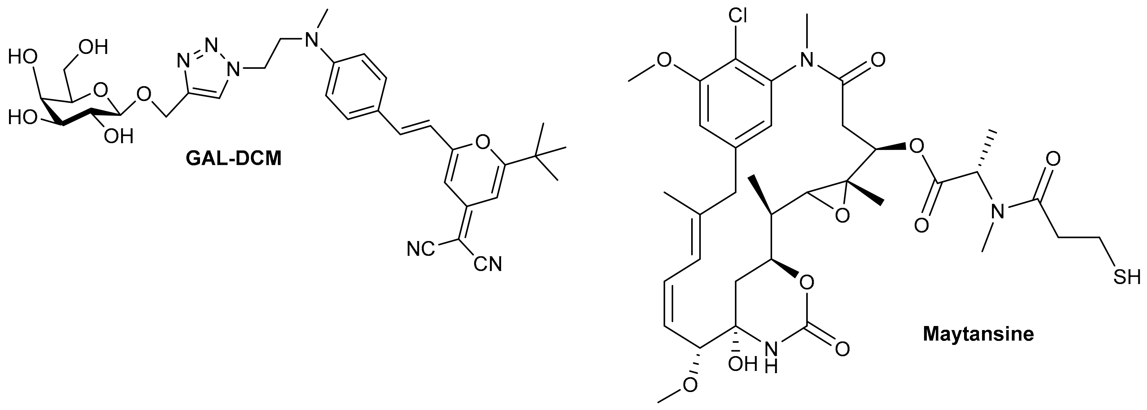
- Xie, H.N.; Chen, Y.Y.; Zhu, G.B.; Han, H.H.; Hu, X.L.; Pan, Z.Q.; Zang, Y.; Xie, D.H.; He, X.P.; Li, J.; et al. Targeted delivery of maytansine to liver cancer cells via galactose-modified supramolecular two-dimensional glycomaterial. Chem. Commun. 2022, 58, 5029. [Google Scholar] [CrossRef]
- Maiti, M.; Kikuchi, K.; Athul, K.K.; Kaur, A.; Bhuniya, S. b-Galactosidase-activated theranostic for hepatic carcinoma therapy and imaging. Chem. Commun. 2022, 58, 6413. [Google Scholar] [CrossRef] [PubMed]
- Mazumder, A.; Dwivedi, A.; Assawapanumat, W.; Saeeng, R.; Sungkarat, W.; Nasongkla, N. In vitro galactose-targeted study of RSPP050-loaded micelles against liver hepatocellular carcinoma. Pharm. Dev. Technol. 2022, 27, 379–388. [Google Scholar] [CrossRef] [PubMed]


| Drug Carrier | Active Moiety | Targeted Organ | Targeting Receptor | In Vitro Studies | In Vivo Studies | Ref. |
|---|---|---|---|---|---|---|
| PHEA-EDA-DPPE-GAL | RBV tripalmitate | Liver | ASGP-Rs | HepG2 | - | [11] |
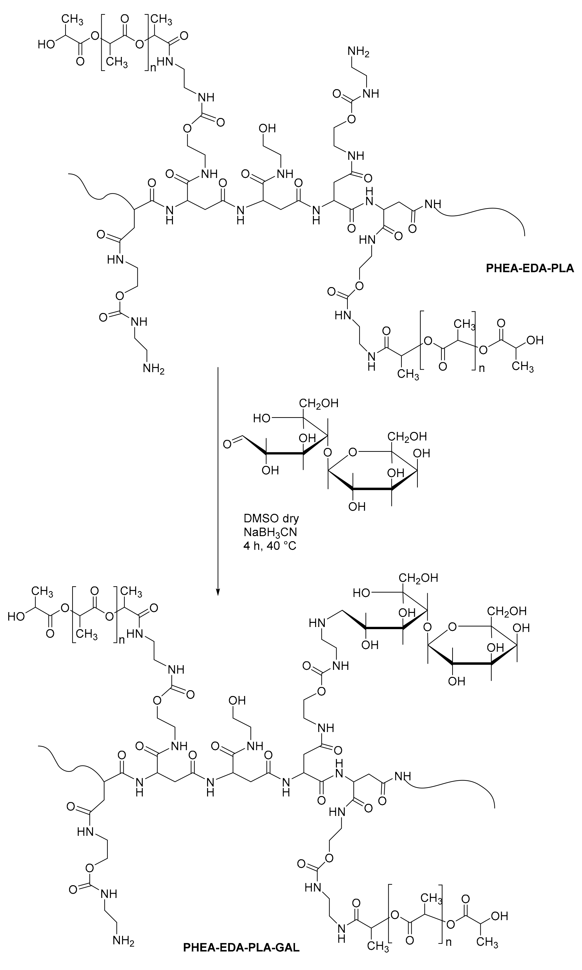
| PHEA-EDA-PLA-GAL | RBV tripalmitate | Liver | ASGP-Rs | HepG2 | - | [12] |
| AG-PLG | RMP | Liver | ASGP-Rs | HepG2 | Female ICR mice | [13] |
| GC | FUA | Liver | ASGP-Rs | HepG2 and A549 | Sprague–Dawley rats | [14] |
| GC-FUA | miRNA-122 | Liver | ASGP-Rs | HepG2 | BALB/c nude mice | [15] |
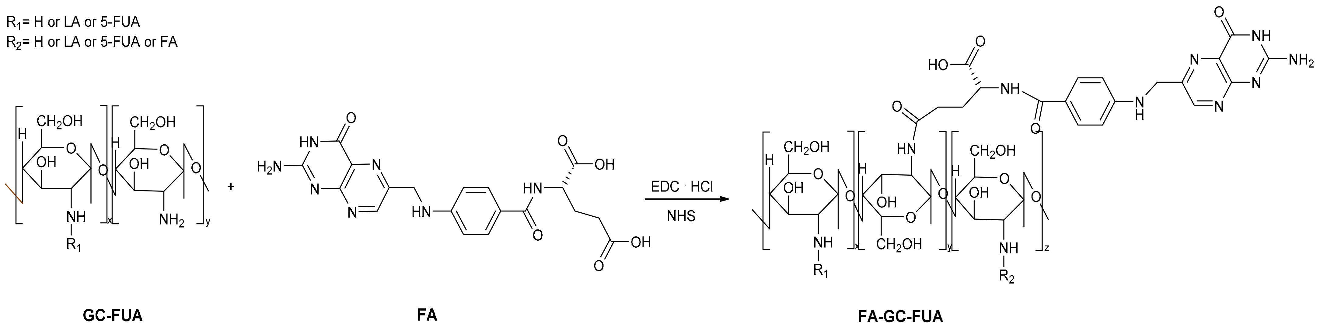
| GC-FUA | FA | Liver | ASGP-Rs and FRs | SMMC-7721 | BALB/c nude mice | [16] |
| GAL-disulfide linker sensitive to GSH | W-105 | Liver | ASGP-Rs | HepG2 | H22 tumor-bearing mice | [17] |
| PEG-Sulfo-BSA-GAL | H2S Prodrug | Liver | ASGP-Rs | HepG2 and RAW264.7 | Mice | [18] |
| P(GAL-b-DPA-b-AzMA) | Alkynyl-JSK | Liver | ASGP-Rs | HepG2 | - | [19] |
| GAL-P5 | CPT-SS-PEG | Liver | ASGP-Rs | HepG2 and HeLa | - | [20] |
| GAL-conjugated PPI dendrimers | PTX | - | - | SiHa and HeLa | - | [21] |
| DG | siRNA | Liver | ASGP-Rs | Liver cells | Mice | [22] |
| PAMAM-PEG-LA | Genetic material | - | - | - | - | [23] |
| GAL-24 | - | Liver | ASGP-Rs | HepG2 | Mice and NASH mouse model | [24] |
| Galactosylated Prodrugs | In Vitro Studies | In Vivo Studies | Ex Vivo Studies | Ref. |
|---|---|---|---|---|
| IbuGAL and OkiGAL and IndoGAL and FluGAL | Serum protein binding | Carrageenan-induced hyperalgesia Acetic Acid-induced Writing Test Ulcerogenic activity | Intestinal Permeation | [25] |
| ACEgal | Intestinal Permeability | Carrageenan-induced hyperalgesia Acetic Acid-induced Writing Test Ulcerogenic activity | Inhibition of platelet aggregation | [26] |
| PARgal | HepG2 | Post-operative Pain model Mechanical Hypergesia | - | [27] |
| Ketogal | - | Mechanical Hypergesia Thermal Hypergesia Carrageenan-induced hyperalgesia | Histological evaluation of Intestine, Stomach, Kidney, Liver | [28,29] |
| UDCAgal | Liver Permeation | EE-induced cholestasis | Analysis of bile flow | [30] |
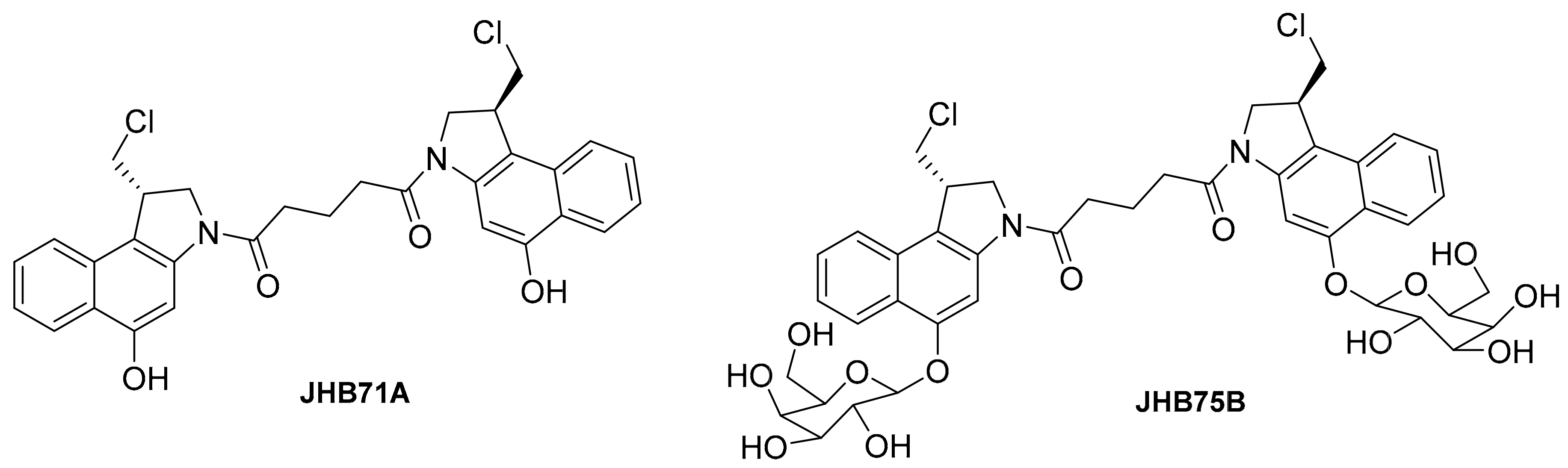
| JHB75B | Senescent cells | ACP model | - | [31,32] |
| Nav-Gal | A549 and SK-Mel-103 | Tumor xenografts and orthotopic models | - | [33] |
| CA4-βGAL-1 and CA4-βGAL-2 | Ovarian Cancer cells | - | - | [34] |
| Complex 1 | HeLa and HepG2 and MCF-7 and HT-29 | - | - | [35] |
Disclaimer/Publisher’s Note: The statements, opinions and data contained in all publications are solely those of the individual author(s) and contributor(s) and not of MDPI and/or the editor(s). MDPI and/or the editor(s) disclaim responsibility for any injury to people or property resulting from any ideas, methods, instructions or products referred to in the content. |
© 2024 by the authors. Licensee MDPI, Basel, Switzerland. This article is an open access article distributed under the terms and conditions of the Creative Commons Attribution (CC BY) license (https://creativecommons.org/licenses/by/4.0/).
Share and Cite
Battisegola, C.; Billi, C.; Molaro, M.C.; Schiano, M.E.; Nieddu, M.; Failla, M.; Marini, E.; Albrizio, S.; Sodano, F.; Rimoli, M.G. Galactose: A Versatile Vector Unveiling the Potentials in Drug Delivery, Diagnostics, and Theranostics. Pharmaceuticals 2024, 17, 308. https://doi.org/10.3390/ph17030308
Battisegola C, Billi C, Molaro MC, Schiano ME, Nieddu M, Failla M, Marini E, Albrizio S, Sodano F, Rimoli MG. Galactose: A Versatile Vector Unveiling the Potentials in Drug Delivery, Diagnostics, and Theranostics. Pharmaceuticals. 2024; 17(3):308. https://doi.org/10.3390/ph17030308
Chicago/Turabian StyleBattisegola, Chiara, Chiara Billi, Maria Cristina Molaro, Marica Erminia Schiano, Maria Nieddu, Mariacristina Failla, Elisabetta Marini, Stefania Albrizio, Federica Sodano, and Maria Grazia Rimoli. 2024. "Galactose: A Versatile Vector Unveiling the Potentials in Drug Delivery, Diagnostics, and Theranostics" Pharmaceuticals 17, no. 3: 308. https://doi.org/10.3390/ph17030308